ARTICLE
DESCRIPTION OF A NEARLY COMPLETE JUVENILE SKULL OF DIPLODOCUS
(SAUROPODA: DIPLODOCOIDEA) FROM THE LATE JURASSIC OF NORTH AMERICA
JOHN A. WHITLOCK,*,1JEFFREY A. WILSON,1and MATTHEW C. LAMANNA2
1Museum of Paleontology and Department of Geological Sciences, University of Michigan, 1109 Geddes Avenue, Ann Arbor, Michigan 48109-1079, U.S.A., jawhitl@umich.edu, wilsonja@umich.edu;
2Section of Vertebrate Paleontology, Carnegie Museum of Natural History, 4400 Forbes Avenue, Pittsburgh, Pennsylvania 15213-4080, U.S.A., lamannam@carnegiemnh.org
ABSTRACT—More than any other sauropod dinosaur group, the long-necked herbivores belonging to Diplodocoidea have been defined by their skulls. Their unique skull shape, which is extremely elongate antorbitally, with a transversely broad, square snout packed at its anterior extreme with narrow-crowned, pencil-like teeth, has served as a touchstone for describing the biology of these animals ever since the discovery of the first skull in the late 19th century. In particular, the unusual diplodocoid skull has been discussed frequently in the context of examining feeding behavior, spawning hypotheses ranging from branch stripping, propalinal shearing, and aquatic plant ‘grazing.’ Here, we describe a juvenile skull of Diplodocus (Carnegie Museum 11255) that does not share the unusually blunted snout and anteriorly sequestered teeth seen in adult specimens, suggesting that adults and juveniles may have differed greatly in their feeding behavior, an ontogenetic distinction that may be unique among sauropodomorphs.
INTRODUCTION
The skull of Diplodocus was first described by Marsh in 1884, and several additional, nearly complete skulls have been dis-covered and described since, making diplodocid cranial ele-ments some of the best-known among Sauropoda (Marsh, 1884; Hatcher, 1901; Holland, 1906, 1924; Gilmore, 1932; Janensch, 1935–36; Haas, 1963; McIntosh and Berman, 1975; Berman and McIntosh, 1978, Connely, 1997). Since its discovery, the skull of Diplodocus has played a prominent role in distinguishing diplodocids from other sauropods. Cranial characters repre-sent four of the seven features Marsh (1884) used in his ini-tial diagnosis of the Family Diplodocidae, a trend that contin-ues to this day. Wilson (2002) listed ten cranial characters as synapomorphies of Diplodocoidea (sauropods more closely re-lated to Diplodocus than to Saltasaurus), as well as seven cra-nial synapomorphies for the subgroup Diplodocidae (sauropods more closely related to Diplodocus than to Dicraeosaurus) and six for Dicraeosauridae (sauropods more closely related to
Di-craeosaurus than Diplodocus). Cranial characters made up 39.5%
of the support for the node Diplodocoidea, nearly 10% more than for Macronaria (Wilson, 2002:table 14). Likewise, cra-nial specializations contributed much of the character support for Diplodocoidea and Diplodocidae in the analysis of Up-church et al. (2004). Even at lower taxonomic levels, cranial characters are important qualifiers. Wilson (2002:appendix 4) lists 25 cranial characters as autapomorphies for the six in-cluded diplodocoid genera for which cranial material is pre-served and that were included in his analysis. Both their un-usual shape and their relative abundance—40% of diplodocoid genera are represented by some cranial material, compared to less than 33% of macronarians (Table 1)—have contributed to what has become an iconic impression of the diplodocoid skull.
*Corresponding author.
The diplodocoid skull is typically described as elongate an-torbitally, with the nares retracted to a position dorsomedial to the orbits and the jaws transversely expanded anteriorly, ter-minating in a blunt, square snout containing narrow-crowned teeth. The shape of the Diplodocus skull has been regarded as poorly suited to chewing or biting through stems (e.g., Hay, 1908; Holland, 1924), and, as a consequence, great interest has been taken in the potential uses of the skull for gathering food in other ways (Osborn, 1899; Hatcher, 1901; Holland, 1906, 1924; Hay, 1908; Tornier, 1911; Coombs, 1975; Bakker, 1986; Dodson, 1990; Fiorillo, 1991, 1995, 1998; Barrett and Upchurch, 1994; Calvo, 1994; Stevens and Parrish, 1999, 2005; Christiansen, 2000; Up-church and Barrett, 2000). The unique skull shape of Diplodocus has led researchers to propose equally unique modes of feeding, including uprooting aquatic succulents (Hatcher, 1901), scrap-ing algae from rocks (Holland, 1906), branch strippscrap-ing (Coombs, 1975; Bakker, 1986; Barrett and Upchurch, 1994), and prehen-sion of fish (Tornier, 1911) or bivalves (Sternfeld in Holland, 1924), as well as a feeding strategy employed by modern mega-herbivores: low-height cropping (Barrett and Upchurch, 1994; Stevens and Parrish, 1999; Upchurch and Barrett, 2000; Barrett and Willis, 2001; Sereno et al., 2007). Regardless of their specific functional interpretation, all studies agree that the feeding ecol-ogy of Diplodocus was clearly distinct from those employed by contemporaneous macronarian sauropods such as Camarasaurus and Brachiosaurus.
Here, we describe a juvenile skull attributable to Diplodocus that provides new insights into the life history and paleoecol-ogy of this giant herbivore. The reconstructed shape of the facial skeleton, particularly the anterior, tooth-bearing region, is trans-versely narrow and rounded anteriorly, in contrast to the square, blunt shape characteristic of adult diplodocids. This disparity in shapes between age classes implies a pattern of ontogenetic re-modeling of the facial skeleton. We interpret this pattern as an indication of resource partitioning between rapidly growing ju-veniles and adults, which were primarily invested in maintain-ing body existmaintain-ing body mass. This pattern is then compared and 442
TABLE 1. Described sauropod taxa known from cranial elements, not including taxa known solely from teeth
Category Genus Material Key reference
Non-Neosauropods Archaeodontosaurus Jaw fragment Buffetaut, 2005
Chebsaurus Skull fragments Mahammed et al., 2005
Chinshakiangosaurus Partial dentary Upchurch et al., 2007
Datousaurus Jaw fragments Dong and Tang, 1984
Gongxianosaurus Jaw fragment He et al., 1998
Lamplughsaura Partial skull Kutty et al., 2007
Lufeng taxon Partial maxilla Barrett, 1999
Mamenchisaurus Several partial skulls Ouyang and Ye, 2002
Omeisaurus Three partial skulls He et al., 1988; Tang et al., 2001
Patagosaurus Jaw fragments Bonaparte, 1979; Rauhut, 2003
Shunosaurus At least five partial skulls Chatterjee and Zheng, 2002 Tazoudasaurus Partial braincase, jaw fragments Allain and Aquesbi, 2008
Diplodocoids Amargasaurus Braincase Salgado and Bonaparte, 1991
Apatosaurus Multiple braincases, nearly complete skull Berman and McIntosh, 1978 Dicraeosaurus Two braincases, jaw fragments Janensch, 1935–36
Diplodocus Multiple adult and sub-adult skulls Holland, 1924
Limaysaurus Braincase Calvo and Salgado, 1995
Nigersaurus Nearly complete skull Sereno et al., 1999
Suuwassea Braincase, jaw fragments Harris, 2006
Tornieria Braincase, possibly jaw fragments Remes, 2006, 2009
Macronarians Abrosaurus Skull Ouyang, 1989
Ampelosaurus Partial braincase Le Loeuff, 1995
Antarctosaurus Braincase, partial dentary Huene, 1929 Atlasaurus Braincase, jaw fragments Monbaron et al., 1999 Auca Mahuevo taxon Multiple embryonic skulls Chiappe et al., 2001
Bonatitan Braincase Martinelli and Forasiepi, 2004
Bonitasaura Partial skull Apestegu´ıa, 2004
Brachiosaurus At least three partial skulls Janensch, 1935–36 Camarasaurus Multiple adult and sub-adult skulls Madsen et al., 1995
Euhelopus Nearly complete skull Wiman, 1929
Europasaurus At least 11 partial skulls Sander et al., 2006
Glen Rose taxon Partial braincase Tidwell and Carpenter, 2003
Isisaurus Partial braincase Berman and Jain, 1982
Jainosaurus Partial braincase Huene and Matley, 1933
Jobaria Partial adult skull and sub-adult braincase Sereno et al., 1999 Lirainosaurus Braincase fragment Sanz et al., 1999
?Magyarosaurus Braincase Weishampel, 1991
Malawisaurus Braincase, other dermal skull fragments Gomani, 2005
Nemegtosaurus Nearly complete skull Nowinski, 1971; Wilson, 2005
Neuquensaurus Skull fragments Huene, 1929
Neuqu ´en taxon Partial braincase Calvo and Kellner, 2006
Paluxysaurus Skull fragments Rose, 2007
Phuwiangosaurus Jaw fragments Martin et al., 1999
Quaesitosaurus Partial skull Kurzanov and Bannikov, 1983
Rapetosaurus Partial adult and sub-adult skulls Curry Rogers and Forster, 2004
R´ıo Negro taxon Partial braincase Garc´ıa et al., 2008
Saltasaurus Partial braincase Bonaparte and Powell, 1980
contrasted with the known record of cranial ontogeny in other dinosaurs.
Discovery of the Juvenile Diplodocus Skull
Several expeditions led by Earl Douglass in the early 20th cen-tury to the Carnegie Quarry in what is now Dinosaur National Monument yielded some of the most spectacular sauropod dis-coveries in North America, including several complete or nearly complete skulls (McIntosh, 1981). Most famous of these is prob-ably the complete and largely undistorted adult Diplodocus skull (Carnegie Museum [CM] 11161) collected by Douglass in 1912. Three years later, a large sub-adult skull of this genus (CM 3452) was the first—and, to date, only described—diplodocid skull found articulated with postcranial elements. Following these suc-cesses, a third, much smaller Diplodocus skull was collected by Douglass and his team in 1921, near the discovery site of CM 11161 (Fig 1; McIntosh, 1981). This skull was mentioned and fig-ured by Holland (1924:pl. 43), but until now has never been fully described.
Institutional Abbreviations—AMNH, American Museum of
Natural History, New York, U.S.A.; CM, Carnegie Museum of
Natural History, Pittsburgh, U.S.A.; CMC, Cincinnati Museum Center, Cincinnati, U.S.A.; MB, Museum f ¨ur Naturkunde der Humboldt-Universit ¨at zu Berlin, Germany; SMM, Science Mu-seum of Minnesota, St. Paul, U.S.A.; USNM, United States Na-tional Museum, Washington D.C., U.S.A.; YPM, Yale Peabody Museum, New Haven, U.S.A.; Z. PAL, Polish Academy of Sci-ence, Warsaw, Poland.
SYSTEMATIC PALEONTOLOGY SAURISCHIA Seeley, 1887 SAUROPODOMORPHA Huene, 1932
SAUROPODA Marsh, 1878 DIPLODOCOIDEA Marsh, 1884 FLAGELLICAUDATA Harris and Dodson, 2004
DIPLODOCIDAE Marsh, 1884
DIPLODOCUS Marsh, 1878
(Figs. 2–5)
CM 11255 is a diplodocid sauropod, sharing the following synapomorphies with other representatives of this clade: elon-gate prefrontal with a posterior projection approaching the parietal; squamosal-quadratojugal contact absent; paroccipital
process with rounded, tongue-like ventrolateral end; shallow quadrate fossa. CM 11255 displays a single autapomorphy of
Diplodocus: sharply defined fossa surrounding preantorbital
fen-estra. CM 11255 is distinguishable from Apatosaurus by the fol-lowing features: presence of a long quadratojugal process of the squamosal that nearly contacts the quadratojugal; basiptery-goid recess present; basipterybasiptery-goid process without marked an-teroventral flaring; closely appressed, sheet-like basal tubera with posteriorly facing concavity; paroccipital processes strongly ven-trally oriented. CM 11255 cannot be compared to the other Morrison Formation diplodocids, Barosaurus, and Supersaurus, due to their lack of recognized cranial material. CM 11255 is distinguishable from the East African diplodocid Tornieria by the presence of a basipterygoid recess and the comparatively larger contribution of the prefrontal to the margin of the orbit. It can also be tentatively distinguished by the location of the frontal-parietal suture, which is located near the anterior mar-gin of the supratemporal fenestra in Diplodocus and probably CM 11255, but located more posteriorly in Tornieria. Lastly, CM 11255 is distinguishable from the indeterminate flagellicaudatan
Suuwassea in the following characters: anteroposteriorly
com-pressed basal tubera; basisphenoid concave ventrally, not visi-ble in posterior view; basal tubera widely separated from oc-cipital condyle, spike-like, dorsoventrally narrow parasphenoid rostrum.
Few characters have been shown to consistently distinguish the skulls of different diplodocid taxa, particularly Apatosaurus and
Diplodocus (Berman and McIntosh, 1978). In the only
descrip-tion of a mostly complete skull referable to Apatosaurus (CM 11162), Berman and McIntosh (1978) noted several cranial dis-tinctions between that genus and Diplodocus, although these are primarily proportional in nature. Due to the early ontogenetic stage of CM 11255, proportional characters are potentially unreli-able. Nevertheless, the anteroventral flaring of the basipterygoid processes noted by those authors for Apatosaurus (CM 11162) is not present in CM 11255 or in any other specimen of Diplodocus, regardless of ontogenetic stage; the remainder of the characters listed by Berman and McIntosh (1978) cannot be evaluated on CM 11255 due to incomplete preservation.
Wilson (2002) distinguished between Apatosaurus and
Diplodocus on the basis of the basipterygoid recess, which is
absent in the former but present in the latter. The presence of a sharply defined fossa surrounding the preantorbital fenestra is listed as an autapomorphy of Diplodocus by Wilson (2002); this
fossa is present in CM 11255, but cannot be adequately evaluated in CM 11162; as such, its state is here considered unknown in
Apatosaurus. The basal tubera, which are globose and laterally
expanded in Apatosaurus (CM 11162, YPM 1860), are pendant and more anteroposteriorly compressed in CM 11255 and in other specimens of Diplodocus (CM 11161, CM 3452, USNM 2672).
Although there are no cranial remains known for two other Morrison diplodocids (Supersaurus, Barosaurus), the identifica-tion of two novel probable Diplodocus autapomorphies and the ease with which CM 11255 is distinguished from Apatosaurus,
Su-uwassea, and Tornieria support the current assignment of this
specimen to Diplodocus. CM 11255 is not here assigned to a species within Diplodocus: no cranial characters yet known serve to differentiate between the various species of the genus, which currently remain distinguishable solely by postcranial features. As such, the lack of associated postcranial material for CM 11255 currently prevents a specific diagnosis.
DESCRIPTION
We use traditional anatomical terminology and orientational descriptors (e.g., ‘anterior’ rather than ‘cranial’) in the descrip-tion below, following Wilson (2006).
Ontogenetic Stage
The early ontogenetic stage assigned to CM 11255 is based on the small size of the specimen and on the presence of a visible suture between the left and right parietal bones. The skull mea-sures 29.2 cm from its anterior-most point to the posterior margin of the occipital condyle, which is approximately 58% the length of the adult Diplodocus CM 11161 and approximately 66% the length of the large sub-adult CM 3452. CM 11255 is therefore the ontogenetically youngest Diplodocus skull ever fully described.
Preservation
CM 11255 was recovered in isolation, with no associated postcrania or elements belonging to other individuals (Fig 1). It was recovered from, and to some extent is still obscured by, a poorly sorted sandstone matrix. The skull is crushed later-ally, such that its left and right halves are disarticulated along the midline sutures, continuing posteriorly through the unfused parietals. Parts of the maxillae and premaxillae have been
FIGURE 1. Quarry map showing the original location of the juvenile Diplodocus skull (CM 11255) relative to other dinosaurs excavated from Carnegie Quarry at Dinosaur National Monument. Redrawn from original quarry map at Carnegie Museum of Natural History. North at top of figure; grid lines are 2 feet (∼0.61 m) apart.
damaged or destroyed, although the tooth row is well represented in these regions by in situ replacement teeth, preserved when sed-iment filled in around the highly resistant teeth after the thin bone surrounding them had been eroded away. This type of preserva-tion is not uncommon (e.g., CM 21718 [McIntosh, 1981; pers. ob-serv.], YPM 1922), although it is not typically found in direct asso-ciation with other skull elements. The skull roof and occiput are represented primarily by elements from the right side, although median elements (supraoccipital, occipital condyle, basioccipital) are largely complete. The dermal skull roof (nasal, prefrontal, frontal, and parietal) is rotated away from the midline of the fa-cial skeleton by approximately 7.5◦.
The left and right elements of the lower jaw are disarticu-lated, and the left mandible is displaced slightly medially and pos-terodorsally. As seen in ventral view, the curvature of the jaws is similar in both elements. The lower jaw of the well-preserved skull of Nemegtosaurus (Z. PAL MgD-I/9) has a similar translation of one mandible relative to the other (Wilson, 2005). No deformation of the tooth-bearing elements was noted in that specimen, although the left and right mandibles were translated relative to each other; this suggests that the curvature seen in the preserved mandibular elements of CM 11255 is likely natural and undistorted.
Dermal Roof Complex
Premaxilla—The premaxillae are elongate tooth-bearing
ele-ments that contact each other medially and the maxillae later-ally, terminating posteriorly at the external naris. Due to the lat-eral crushing, the anterior portion of the skull is deformed along the median suture between the premaxillae, creating an unusually steep angle between these bones and exaggerating their visibility in lateral view.
Four tooth positions are preserved in the right premaxilla and are presumed present in the left (Fig. 2). The anterior margins of the premaxillae are represented only by narrow, medial portions of the left and right premaxillae. Harris (2006) regarded the pre-maxillae of CM 11255 as similar in proportion and size to those of
Suuwassea; however, the premaxillae in the present specimen
ap-pear to expand laterally without the distinct ‘step’ of Suuwassea, in which the transversely wide anterior portion is clearly demar-cated by sharp lateral curvature in dorsal and ventral view. The narial fossae are not distinctly preserved.
The posterior extremes of the premaxillae are partially com-plete, preserving a portion of the anterior margin of the narial opening. There is no evidence for an elongate internarial bar, such as has been observed in some adult (USNM 2673) and sub-adult (CM 3452) Diplodocus skulls.
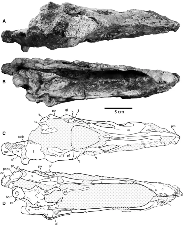
Maxilla—The maxillae are thin, sheet-like bones that are
di-vided by the antorbital fenestra into an ascending process and a posteriorly directed process that contacts the jugal and quadra-tojugal (Fig. 2). As in adult Diplodocus, the preserved portion of the maxilla-jugal suture is coarsely sinuous in lateral view. The as-cending process remains in contact with the premaxilla until they both reach the external naris. Posteriorly, the ascending process contacts the nasal and the lacrimal.
Similar to adult Diplodocus (AMNH 696, CM 11161, CM 3452, USNM 2672) and other diplodocoids (Dicraeosaurus, MB.R. 2336; Apatosaurus, CM 11162, CMC VP 7180), the external sur-faces of the maxillae in this region are ornamented by sev-eral shallow, elongate depressions, roughly corresponding to the positions and orientations of tooth families within the bone. The lateral plate of the maxilla is partially preserved posteriorly. The poor anterior preservation contributes to the convex shape of the ventral margin as preserved.
Immediately posterior to the preserved dentigerous margin of the maxilla is a large, distinct fossa that dorsally overlaps the three posterior-most replacement teeth. The fossa is pierced
posteriorly by a large, subcircular preantorbital fenestra. The sharp outline of the fossa is best preserved on the left side (Fig. 2D). In other Diplodocus skulls (CM 11161, CM 3452, USNM 2672), the subnarial foramen and anterior maxillary foramen re-side in a shallow groove situated slightly anterodorsal to the preantorbital fossa, along the contact with the premaxilla; nei-ther these foramina nor the groove are distinctly preserved in CM 11255.
Nasal—The nasal is a roughly quadrangular element with
an-teriorly projecting rami that form the right and left posterolat-eral margins of the naris. The preserved portions of these rami contact the lacrimal and maxilla anteriorly; the main body of the nasal contacts the prefrontal laterally and would have contacted the frontal posteriorly, forming the anterior-most medial element of the braincase. The main body of the nasal rises dorsally above the orbit, elevating the posterior margin of the naris, as seen in some other individuals of Diplodocus (AMNH 696, CM 11161, CM 3452, USNM 2672; Figs. 2B, 3D). A small (∼1 cm long) piece of the anterior ramus is preserved on the left side (Figs. 2D, 3C, 4C). This process forms the posterolateral margin of the naris. It is probable that the orientation of the rami and the main body of the nasal obscured the naris in lateral view and that the naris pointed strictly anterodorsally in life.
Jugal—The jugal is a V-shaped element that connects the
max-illa and quadratojugal with the lacrimal and postorbital. Its tact with the maxilla is a broadly sinuous suture, whereas its con-tacts with the remaining three elements are more linear (Figs. 2C, D). A dorsally directed process along the lacrimal/jugal contact, as illustrated by Wilson and Sereno (1998:fig 6) in their recon-struction of Diplodocus, appears to be at least incipiently present, although less substantial in this specimen. Medially, the jugal has a small contact with the palate via a lateral process of the ptery-goid.
As in adult Diplodocus, the jugal is broad, flat, and excluded from the ventral margin of the skull. It forms parts of the margins of three skull openings: the orbit, the antorbital fenestra, and the lateral temporal fenestra.
Lacrimal—The lacrimal is a bar-shaped element, oriented
ap-proximately dorsoventrally. It contacts the jugal ventrally, the nasal and prefrontal dorsally, and the maxilla along the dorsal portion of its anterior margin. The suture with the jugal is nearly linear, with a reduced version of the “stepped” contact in some adult skulls (CM 11161; Fig. 2). Ventral to its anterior contact with the maxilla, the lacrimal forms an anteroposteriorly elon-gate portion of the margin of the antorbital fenestra; the cross-sectional shape becomes more mediolaterally elongate dorsal to this fenestra. There, a strong ridge on the lateral surface of the lacrimal extends dorsally to join the laterally expanded prefrontal and form the anterior portion of the dorsal orbital margin.
The long axis of the lacrimal in CM 11255 is much more ver-tically oriented than is typical of more ontogenever-tically mature skulls, suggesting that the infraorbital and antorbital regions ex-perienced a greater degree of anteroposterior and anteroventral lengthening during growth than did neighboring regions. A con-sequence of the orientation of the lacrimal is that the tear-drop shape typical of eusauropod orbits is not as strongly expressed; although there is still an anteroventral ‘corner’ on the orbit, the shape is generally subcircular.
Prefrontal—The prefrontal meets and slightly overlaps the
lacrimal at a scarf joint. Along with the frontal, which it contacts posteriorly, the prefrontal forms most of the dorsal margin of the orbit. In lateral view, the prefrontal is anteroposteriorly broad, with only a gentle ventral concavity. In dorsal view, it is arcuate and contacts the frontal and nasal along a concave medial mar-gin. This concavity lends the posterior half of the prefrontal a ‘hooked’ shape, which has been recognized as a diplodocid fea-ture (Berman and McIntosh, 1978; Wilson, 2002). The prefrontal forms much of the lateral margin of the skull roof in dorsal view.
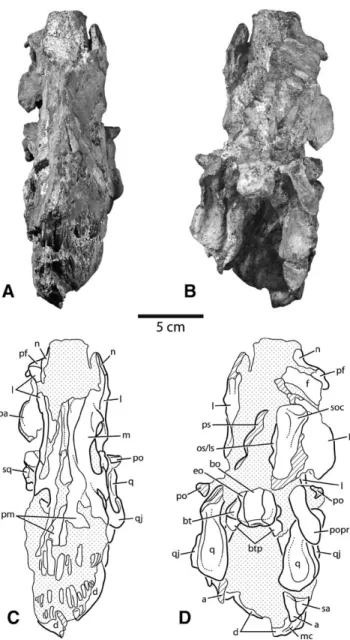
FIGURE 2. Photographs and interpretive line drawings of the juvenile skull of Diplodocus (CM 11255). A, B, right lateral view; C, D, left lateral view. Stippled areas indicate matrix; hatching indicates broken bone. Scale bar equals 5 cm. Abbreviations: a, angular; aof, antorbital fenestra; bo, basioccipital; d, dentary; eo, exoccipital-opisthotic; f, frontal; j, jugal; ltf, lateral temporal fenestra; l, lacrimal; m, maxilla; mc, Meckelian canal; n, nasal;
o, orbit; os/ls, orbitosphenoid/laterosphenoid; pa, parietal; paof, preantorbital fenestra; pf, prefrontal; pm, premaxilla; po, postorbital; pop, paroccipital
process; ps, parasphenoid; psaf, posterior surangular foramen; pt, pterygoid; q, quadrate; qj, quadratojugal; sa, surangular; soc, supraoccipital; spl, splenial; sq, squamosal.
FIGURE 3. Photographs and interpretive line drawings of the skull of Diplodocus (CM 11255). A, C, anterior view; B, D, posterior view. Stip-pled areas indicate matrix, hatching indicates broken bone. Scale bar equals 5 cm. Abbreviations: a, angular; bo, basioccipital; bt, basal tu-bera; btp, basipterygoid processes; d, dentary; eo, exoccipital-opisthotic;
f, frontal; l, lacrimal; m, maxilla; mc, Meckelian canal; n, nasal; os/ls,
or-bitosphenoid/laterosphenoid; pa, parietal; pf, prefrontal; po, postorbital;
popr, paroccipital process; pm, premaxilla; ps, parasphenoid; q, quadrate; qj, quadratojugal; sa, surangular; sq, squamosal; soc, supraoccipital.
Frontal—In lateral view, the frontal is a narrow, arcuate
element that contacts the prefrontal anteriorly and the pos-torbital laterally. In dorsal view, the frontals can be seen to contact the parietals posteriorly and the nasal anteriorly. The frontal-parietal suture is obscured and so cannot be compared to the relatively linear suture of Suuwassea (Harris, 2006) or the sin-uous sutures known in Apatosaurus (CM 11162) and Diplodocus (CM 11161). The frontal forms much of the dorsal skull roof, and contacts the orbitosphenoid ventrally.
The lateral margin of the right frontal has been eroded away, exposing the trabecular internal structure of the bone, bordered by a thin veneer of cortical bone on the dorsal and ventral faces. In other specimens of Diplodocus (CM 11161, CM 3452, USNM
2672, USNM 2673), the lateral portion of the frontal that forms the orbital margin is greatly expanded laterally, creating a broad shelf dorsal to the orbit. A notch is commonly associated with the anterior margin of this expanded margin. In Apatosaurus (CM 11162), this expansion is well developed, and increases the width of the skull in this region well beyond the lateral extent of the parietals. Based on the mediolateral position of the posterior-most preserved portion of the postorbital, CM 11255 did not have such a prominent lateral expansion.
Postorbital—The postorbital is a triradiate element, with
elon-gate processes contacting the frontal dorsally and the jugal ante-riorly. The third process is comparatively short and inserts into a groove in the squamosal. CM 11255 preserves the jugal process, which forms most of the ventral margin of the orbit. The contact with the jugal occurs along a shallowly angled, planar suture. Al-though the postorbital and lacrimal are closely situated in adult specimens, this relationship in CM 11255 appears unusually close. There is a longitudinal ridge along the lateral surface of the jugal process, expanding the element into a flat shelf dorsally and ren-dering it triangular in cross-section. This creates a narrow fossa surrounding the lateral temporal fenestra.
Squamosal—The squamosal is an arcuate element when
viewed laterally, contacting the parietal, postorbital, and quadrate, and forming a portion of the margin of both temporal fenestrae. The right side of CM 11255 preserves a small portion of the anteriorly directed process that overlies the head of the quadrate in lateral view. In CM 11255, this process is partially obscured by matrix (Fig. 2A), but is revealed by computed tomography (CT) to extend anteriorly to nearly contact the posterodorsally directed squamosal process of the quadratojugal, which contrasts with the widely spaced position of these elements in adult Diplodocus (CM 11161). In posterior view, the pre-served portion of the squamosal is almost entirely obscured by the paroccipital process, although a small portion of the lateral margin can be seen overlapping the quadrate (Fig. 3B, D).
Quadratojugal—The quadratojugal of CM 11255 is an
antero-posteriorly elongate, dorsoventrally narrow element that con-tacts and laterally overlaps the quadrate; its elongate squamosal process extends 2.3 cm along the lateral margin of the quadrate toward the squamosal. This process tapers dorsally and termi-nates in a broken surface; it can be inferred to have extended to near the midpoint of the quadrate, within a centimeter of con-tact with the squamosal. The anteroventral corner of the lateral temporal fenestra is preserved on the left side only. Much of the anterior contact with the maxilla is not preserved.
The dorsal margin of the quadratojugal is broadly sinuous. It is dorsally convex anteriorly along the contacts with the maxilla and jugal, and it is concave posteriorly where it forms the anteroven-tral corner of the lateral temporal fenestra. In dorsal and venanteroven-tral views, the quadratojugal bulges laterally as it overlaps the articu-lar head of the quadrate (Fig. 4).
Parietal—The parietal of CM 11255 contacts the frontal
an-teriorly and delimits the dorsomedial margin of the supratem-poral fenestra. The partially preserved median contact between the parietals is a highly interdigitated suture. In adult and large sub-adult Diplodocus (CM 11161, CM 3452, USNM 2672, USNM 2673), the parietals are fused. Coupled with the small size of the specimen, the presence of a patent interparietal suture in CM 11255 suggests that fusion of these elements only occurred as the animal approached maturity.
The lateral wing of the parietal arches strongly dorsolaterally, obscuring the supratemporal fenestra in posterior view—a con-dition described as characterizing Diplodocus by Berman and McIntosh (1978). The dorsolateral margin of the parietal in
Ap-atosaurus is nearly linear, unlike the state preserved in CM 11255.
In dorsal view, a nuchal fossa can be seen between the lateral wing of the parietal and the parietal-supraoccipital suture. Although the depth of this fossa is somewhat exaggerated by an
FIGURE 4. Photographs and interpretive line drawings of the juvenile skull of Diplodocus (CM 11255). A, C, dorsal view; B, D, ventral view. Stippled areas indicate matrix, hatching indicates broken bone. Scale bar equals 5 cm. Abbreviations: a, angular; bo, basioccipital; bs, basisphenoid;
bt, basal tubera; btp, basipterygoid processes; d, dentary; eo, exoccipital-opisthotic; f, frontal; l, lacrimal; m, maxilla; n, nasal; nf, nuchal fossa; os/ls,
orbitosphenoid/laterosphenoid; pa, parietal; pf, prefrontal; pm, premaxilla; po, postorbital; popr, paroccipital process; pt, pterygoid; q, quadrate; qj, quadratojugal; soc, supraoccipital; t, teeth.
area of broken bone in the center of the concavity, the nuchal fossa appears to invade quite deeply, reaching nearly to the depth of the parietal-frontal suture, and is therefore more strongly con-cave than that seen in Suuwassea (Harris, 2006) and some speci-mens of Diplodocus (CM 11161). It is quite similar to the fossae in some other Diplodocus specimens, however, including other presumed sub-adult skulls (CM 3452), as well as in Apatosaurus (CM 11162).
The supratemporal region is narrow in dorsal view. The pre-served mediolateral width of the right parietal at the greatest extent of the lateral wing is 4.1 cm, resulting in a minimum tal transverse width estimate of 8.2 cm, roughly 24% of the to-tal skull length (compared to ∼29% in adult Diplodocus and ∼46% in Apatosaurus). The proportional width of CM 11255 is more congruent with smaller Diplodocus skulls (SMM P84.15.3; Erickson and Hanks, 2001).
Palatal Complex
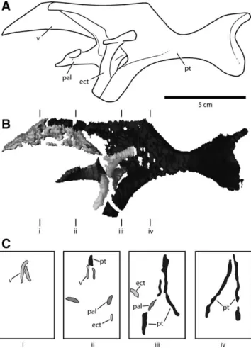
The palatal complex of CM 11255 is represented by the quadrates, pterygoids, ectopterygoids, palatines, and vomers (Fig. 5). The latter four structures are visible entirely or in large part only through CT scanning.
Quadrate—The quadrate connects the palate with the dermal
skull roof through its insertion into a recess of the squamosal posterodorsally and with the facial skeleton by contact with the quadratojugal laterally. The quadrate also contacts the
FIGURE 5. Palatal complex of juvenile Diplodocus (CM 11255), based on data obtained using computed tomography (CT) scanning. A, Recon-struction of the palate in left lateral view; B, palatal complex in lateral view; C, Cross-sections taken through B at transects i–iv. Cross-sections in anterior view. Scale bar equals 5 cm. Abbreviations: ect, ectopterygoid;
pal, palatine; pt, pterygoid; v, vomer.
paroccipital process posteriorly (Fig. 3B, D). The wing-like ptery-goid flange of the quadrate extends anteromedially, and is visible in lateral view through the lateral temporal fenestra. This flange is broad and flat, terminating in a broad, arcuate contact with ptery-goid.
In lateral view, the quadrate is distinctly concave posteriorly, unlike the relatively straight or only slightly bent condition seen in adult exemplars of Apatosaurus (CM 11162), Diplodocus (CM 11161), and Suuwassea (Harris, 2006). This curvature is slightly exaggerated by breakage at mid-shaft, but even allowing for some distortion the quadrate was much more strongly curved than those of larger Diplodocus skulls. The quadrate fossa is shallow, as in other diplodocoids, and does not continue onto other ele-ments laterally.
The articular surface of the quadrate condyle is visible in lat-eral view, with the medial aspect projecting well beyond the ven-tral margin of the quadratojugal. The articular face is roughly triangular with a peak facing posteriorly. The surface is beveled such that it faces slightly ventrolaterally.
Pterygoid—The pterygoid forms much of the posterior portion
of the palate, contacting the pterygoid wing of the quadrate pos-teriorly, the palatine and ectopterygoid laterally, and the vomer anteriorly. Anteriorly, the pterygoid bifurcates into anterodor-sally and anteroventrally directed processes. The anterodorsal process is broad and sheet-like and has a slightly concave, elon-gate dorsal margin that extends anteriorly to contact the pala-tine and vomer (Fig. 5). Much of the ventral portion of this pro-cess is indistinguishable from matrix in the CT images, suggesting that portion of the element was quite thin. The pterygoid an-teroventral process contacts the ectopterygoid to form the trans-verse pterygoid hook. In contrast to the autapomorphic condition described for adult Diplodocus by Wilson (2002), the pterygoid is situated posterolateral to the ectopterygoid when the two ele-ments are articulated (Fig. 5).
Like the other dermal skull elements of the left side, the left pterygoid is displaced dorsally, posteriorly, and slightly medially. The pterygoids are vaulted inward at an angle of approximately 37.3◦from vertical and arch dorsally as they approach their mid-line contact (Fig. 5C). The right pterygoid is more strongly in-clined dorsomedially (35◦from the vertical) than the left (2.3◦). In adult Diplodocus (CM 11161), the pterygoids are inclined at ap-proximately 30◦from the vertical (McIntosh and Berman, 1975).
Palatine—The palatine is thin and arcuate in CM 11255,
con-tacting the pterygoid posteriorly along a concave, anterodorsally inclined suture. The dorsal portion of the palatine and the con-tact with the vomer is not well preserved (see Vomer, below). The anteroventral portion of the palatine forms a dorsoventrally flattened process that contacts the maxilla along its lateral and an-terior margins. The anan-terior-most portion of this process is over-lapped by a posterior process of the maxilla that projects from the floor of the preantorbital fenestra. The ectopterygoid con-tacts the palatine along the palatine’s ventral margin, near the posterior margin of the maxillary contact.
Vomer—The vomer is trapezoidal in lateral view, contacting
the maxilla anteriorly and the pterygoid posteriorly. It is over-lapped to some degree along its posteroventral margin by the palatine, although that contact is poorly preserved. The vomers are strongly vaulted, although this is probably exaggerated by the minor lateral compression of the skull.
Ectopterygoid—The ectopterygoid is an arched, strap-like
element that articulates laterally with the maxilla and postero-medially with the pterygoid in a ventrally directed, anteroposte-riorly elongate ‘hook’ that extends to near the level of the ven-tral margin of the maxilla. As discussed above, the articulation of the ectopterygoid with the pterygoid appears to position the former anteromedial to the latter, unlike the condition described for adult Diplodocus by Wilson (2002). In both cases, the articula-tion is primarily along the posterior margin of the ectopterygoid,
and the ectopterygoid process of the pterygoid curves laterally to meet the ectopterygoid, so the variation seen here is perhaps not significant.
Braincase
The braincase is largely missing, particularly those elements surrounding the foramen magnum. As with the circumorbital and skull roof elements, the right side preserves more of these bones than does the left. The braincase is oriented at the same slight angle to the facial skeleton as is the skull roof.
Basioccipital—The basioccipital makes up the main body of
the occipital condyle, and in CM 11255 it is mostly complete. Lit-tle of the foramen magnum remains dorsal to it, including the portion of the margin that was presumably formed by this ele-ment. The condyle is D-shaped in occipital view, with a nearly flat posterodorsal margin. Including allowances for ventral rotation of the structure, the occipital condyle is oriented strongly ven-trally. The suture between the basioccipital and the exoccipital-opisthotic that forms the dorsolateral ‘shoulder’ of the condyle is plainly visible on both sides (Fig. 3B, D).
Supraoccipital—The supraoccipital is a midline element that
contacts the parietals and the exoccipital-opisthotics. In CM 11255, the supraoccipital is represented primarily by a large, tri-angular eminence formed by the confluence of two crests: a trans-verse crest that extends laterally towards the nuchal fossa, the insertion of which is obscured by a broken surface; and a promi-nent, sagittal crest that extends down the midline of the element, flaring slightly at its dorsal terminus (Fig. 3B, D). In Apatosaurus,
Suuwassea, and other crania of Diplodocus, a narrow isthmus of
the supraoccipital extends ventrally to form a small portion of the dorsal margin of the foramen magnum, although this cannot be determined in CM 11255 due to a lack of preservation.
Exoccipital-Opisthotic—The exoccipital-opisthotic forms part
of the posterior braincase, contacting the supraoccipital and ba-sioccipital medially, the parietal dorsally, and the squamosal lat-erally. Together, the right and left exoccipital opisthotics form much of the lateral margin of the foramen magnum. In CM 11255, these elements are primarily represented by the left and right paroccipital processes. The blade-like paroccipital processes are strongly angled ventrally, as in other Diplodocus specimens (CM 11161, CM 3452). This is in contrast to the more laterally directed processes seen in Suuwassea (Harris, 2006) and Apatosaurus (CM 11162). As in other diplodocid sauropods, the paroccipital pro-cess expands ventrolaterally, predominantly on its dorsal mar-gin. The exoccipital-opisthotic makes a small contribution to the ‘shoulders’ of the occipital condyle (Figs. 3, 4).
Basisphenoid—In diplodocoids, the basisphenoid serves as the
only bony connection between the braincase and palate, contact-ing the pterygoids via elongate basipterygoid processes. Poste-riorly, it meets the basioccipital at the base of the neck of the occipital condyle. Dorsally, the basisphenoid projects anteriorly as the parasphenoid rostrum, visible though the left orbit. This process is rod-like and elongate, reaching anteriorly to near the anterior margin of the orbit. It is generally similar in shape and proportion to its homologues in adult Diplodocus, and is unlike the much larger, dorsoventrally expanded parasphenoid rostrum of Suuwassea (Harris, 2006). A prominent ridge just dorsal to the parasphenoid rostrum is identified as the ventral extreme of the antotic crest.
Near its contact with the basioccipital, the basisphenoid is ex-panded into paired basal tubera that are separated from each other by a narrow sulcus. A narrow pit (the ‘basipterygoid recess’; Wilson, 2002) is present just posterior to this sulcus, as in other specimens of Diplodocus. The basal tubera of CM 11255 are diag-nostic for this genus in two ways. First, unlike the more massive, globose basal tubera seen in Apatosaurus, those of CM 11255 are flat and semi-concave posteriorly, as in other Diplodocus
skulls (CM 11161, CM 3452). Second, as in other specimens of
Diplodocus, the basal tubera of CM 11255 are pendulous,
pri-marily visible ventral to the occipital condyle in occipital view. In Apatosaurus (CM 11162, CMC VP 7180, YPM 1860), the basal tubera do not descend as far ventrally and are more lat-erally oriented, such that they are primarily visible lateral to the condyle.
Anteroventral to the basal tubera, a deep concavity separates the paired basipterygoid processes. In CM 11255, the basiptery-goid processes are thin and elongate, similar in shape to those of other Diplodocus crania. Anteroventrally, there is a condyle for articulation with the pterygoid. Unlike their counterparts in
Ap-atosaurus, the basipterygoid processes of CM 11255 do not flare
anteroventrally. The angle between the basipterygoid processes cannot be determined due to deformation of the left process and breakage of the right process.
Orbitosphenoid and Laterosphenoid—Posterodorsal to the
parasphenoid, the right orbitosphenoid and laterosphenoid are visible in medial view (Fig. 2C, D). Much of the internal surface of the orbitosphenoid has been destroyed, including the foram-ina for cranial nerves I and IV, respectively. A small foramen for the passage of cranial nerve III is preserved near the ven-tral margin of the orbitosphenoid. An ovate, anteroposteriorly oriented foramen situated slightly posterodorsal to the foramen for cranial nerve III may have accommodated the endolymphatic sac. Ventrally, the orbitosphenoid meets the parietal in a sin-uous, patent suture. Posteriorly, the orbitosphenoid abuts the supraoccipital in a straight, dorsoventrally oriented patent suture. The internal surface of the laterosphenoid is marked by a large, approximately anteroposteriorly oriented tuberosity, the dorsal margin of which forms the ventral margin of the foramen for cranial nerve V. The anteroventral corner of this tuberosity, where the openings for cranial nerves IX–XI are expected, has been destroyed.
Cranial Openings
Six cranial openings can be identified in CM 11255: the prean-torbital fenestra, the anprean-torbital fenestra, the orbit, the external naris, the supratemporal fenestra, and the lateral temporal fenes-tra. The subnarial foramen, anterior maxillary foramen, and post-temporal fenestra are not preserved.
Preantorbital Fenestra—The preantorbital fenestra is a small,
elliptical opening that pierces the maxilla in the posterodorsal corner of a sharply defined fossa that extends anteriorly and ven-trally. As in other neosauropods, the preantorbital fenestra is in-ternally connected with the antorbital fenestra by a narrow bridge of bone composed of a posteromedial projection of the maxilla and an anterior projection of the palatine, inserting on the ven-tral margin of the preantorbital fenestra. The preantorbital fen-estra may therefore represent a pneumatic continuation of the antorbital fenestra, similar to the invasions of the maxilla by the antorbital sinus in some theropods (Witmer, 1997).
Antorbital Fenestra—Located posterodorsal to the
preantor-bital fenestra, the antorpreantor-bital fenestra opens laterally and is with-out a distinct fossa surrounding it. It is bound largely by processes of the maxilla; the ascending process surrounds the fenestra on its dorsal margin, and the posterior process contributes most of the anteroventral margin. The remainder of the antorbital fenestra is enclosed by the jugal and the lacrimal; the nasal is excluded from its margin.
As in adult Diplodocus, the outline of the antorbital fenes-tra of CM 11255 is roughly teardrop-shaped, with the acute posterodorsal corner formed by the confluence of the maxilla and lacrimal. The dorsal margin is not as concave as in adult
Diplodocus (CM 11161, USNM 2672, USNM 2673), more closely
resembling the condition seen in other sub-adult Diplodocus (CM 3452).
Orbit—Five bones bound the orbit: the lacrimal, prefrontal,
frontal, postorbital, and jugal. It is a subcircular opening in CM 11255, with the sharply notched ventral margin typical of eu-sauropods weakly expressed (Wilson and Sereno, 1998). The pri-mary cause of this shape disparity is the vertical orientation of the lacrimal, creating a wider angle between that element and the postorbital process of the jugal. The expansion of this angle gives the orbit its rounded appearance. The orbit is more strongly arched dorsally, where it is bounded by the prefrontal and frontal, than it is ventrally, where it is proscribed by the postorbital. The anteroposterior length of the orbit is difficult to determine due to the loss of the frontal process of the postorbital.
External Naris—The external naris is bounded posteriorly and
posterolaterally by the nasal, anterolaterally by the maxillae, and anteriorly by the premaxillae. From the position of preserved ele-ments surrounding it, the naris faced entirely dorsally. The trans-verse breadth of the naris expands posteriorly, giving the open-ing a triangular shape in dorsal view. There is no evidence for a large internarial bar dividing the naris anteriorly, although such bars have been observed in other Diplodocus crania (CM 3452, USNM 2762).
Supratemporal Fenestra—In adult skulls of Diplodocus, the
supratemporal fenestrae are bounded by the frontals and pos-torbitals anteriorly, the parietals posteriorly, and the squamosal ventrally. However, due to the incomplete preservation of these elements in CM 11255, little is known about the condition of the supratemporal fenestra in this specimen. It was likely a mediolat-erally elongate, oval opening, largely obstructed in posterior view by the large lateral wing of the parietal. The partially preserved right supratemporal fenestra is fully visible in lateral view.
Lateral Temporal Fenestra—The lateral temporal fenestra
is poorly preserved, although an estimate of its shape can be inferred from the bones forming its margin. As in other diplodocids, it is roughly divisible into two sections: a rounded, anterior portion and a dorsoventrally compressed, anteroposteri-orly elongate posterior portion. In CM 11255, its anterior margin is approximately even with the anterior margin of the orbit in lat-eral view. This is unlike the condition in larger Diplodocus skulls (e.g., CM 11161), in which the lateral temporal fenestra extends well anterior to the orbit.
Lower Jaw
The lower jaw is similar to that described for larger specimens of Diplodocus, save only for its rounded anterior end, which dis-tinguishes it from the iconic squared shape of adults.
Dentary—As in other Diplodocus (CM 11161, USNM 2672),
the dentary of CM 11255 comprises approximately one-half the length of the mandible. The internal surfaces of both dentaries are preserved medially, and the ventral margin is visible in both elements as well. As a consequence of the lateral crushing of the skull, the dentaries are disarticulated from each other at the symphysis, and the left is displaced posteriorly, dorsally, and me-dially. In ventral view, the dentaries are similar, although the better-preserved right element is slightly more strongly curved. The orientation of the symphysis suggests that the two dentaries would have met at a sharp angle, unlike the symphysis in adult
Diplodocus, which was oriented essentially perpendicular to the
anterior rami of the dentaries. The Meckelian groove is visible on the medial surface of the right dentary, arching dorsally to the broken edge of the bone.
Surangular—The surangular forms the dorsal portion of the
posterior half of the mandible. In lateral view, it contacts the den-tary anteroventrally and the angular ventrally. Medially, it con-tacts the dentary anteriorly and the splenial and prearticular ven-trally. The surangular contacts the articular posteriorly in other exemplars of Diplodocus, but this cannot be confirmed in CM 11255. The surangular is broad and sheet-like, and there is no
ev-idence for a well-developed coronoid eminence. There is also no evidence of the anterior surangular foramen that occurs in other
Diplodocus (CM 11161, CM 3452).
The surangular and angular (along with the articular) form the retroarticular process, which protrudes farther posterior to the quadrate in CM 11255 than is seen in adult Diplodocus. In this way, it is more similar to the condition of the large sub-adult CM 3452. As in that specimen, however, the mandibular cotyle does not extend greatly posterior to the articular head of the quadrate, being instead quite rounded dorsally. This indicates that the lower jaw of CM 11255 was not capable of being signif-icantly displaced anteriorly during the bite stroke, in contrast to what has been previously proposed for adult Diplodocus (Barrett and Upchurch, 1994; Calvo, 1994; Upchurch and Barrett, 2000; Barrett and Upchurch, 2005).
Angular—The angular forms the ventral margin of the
mandible and contributes to the unusually well developed retroarticular process. In lateral view, it contacts the surangular dorsally and the dentary anteriorly. Medially, it contacts the sple-nial anterodorsally and the prearticular dorsally.
Splenial—The splenial is a triradiate bone with two closely
ap-pressed anterior processes that contact the dentary and a pos-terior process that separates the prearticular from the angular. The anteroventral process is elongate and triangular in shape. The anterodorsal process is also triangular in shape, but much broader at the base and does not extend far anteriorly. Whether the anterodorsal process is further subdivided (as in CM 11161; McIntosh and Berman, 1975) cannot be determined.
Prearticular—The prearticular is a subquadrangular element
that contacts the surangular dorsally and the angular and splenial ventrally. It arches slightly dorsally at mid-length, where it forms a portion of the ventral margin of the adductor fossa, and is very similar to the prearticular described for the adult Diplodocus CM 11161 (McIntosh and Berman, 1975).
Dentition
The teeth of CM 11255 are of the narrow-crowned type typi-cal of diplodocoids (Calvo, 1994). As in other Diplodocus, they lack marginal denticles. Seven functional maxillary tooth posi-tions are preserved on the right side, and 10–11 are estimated on the left. This is the standard Diplodocus condition (AMNH 696, CM 11161, CM 3452, USNM 2672). In the lower jaw, there are eight preserved functional teeth in the left dentary and six in the right. Based on other Diplodocus skulls, CM 11255 would have had between 10 and 11 dentary teeth. The upper teeth are larger than their lower counterparts, and are gener-ally in a better state of preservation. None show definite traces of wear.
Similar to adult teeth, those of CM 11255 are subcircular in cross-section near the apicobasal midpoint of the crown, and be-come labiolingually compressed more apically. The upper teeth are unusual for a diplodocoid, however, in having mesiodistal asymmetry. Unlike the teeth preserved in specimens of adult
Diplodocus (AMNH 696, CM 11161, USNM 2672, USNM 2673),
the apices of each tooth of CM 11255 are slightly distally inclined. The teeth are sharply pointed, a condition Holland (1924) consid-ered unusual. However, in situ teeth in other Diplodocus skulls (CM 3452, USNM 2672, and USNM 2673) are also pointed, sug-gesting that this shape is in fact typical for unworn crowns. As in those skulls (but not CM 11161), the teeth of CM 11255 are also closely appressed, occasionally contacting their mesial and/or dis-tal neighbors.
In adult diplodocoids, the dentition is restricted to the ante-rior extremity of the snout. Although much of the ventral mar-gins of the maxillae of CM 11255 are missing, a large propor-tion of the dentipropor-tion is represented by both funcpropor-tional (Fig. 2A, B) and replacement (Fig. 2C, D) teeth. The preservation of the
replacement teeth allows the reconstruction of the position of the distal-most maxillary tooth, which is located farther posteriorly than is typical for adult Diplodocus (Fig. 2). That is, the tooth row in adults ends well anterior to the preantorbital fenestra, and of-ten anterior to the subnarial foramen. Conversely, in CM 11255, the tooth row appears to end posterior to at least the subnarial foramen, and perhaps quite close to the anterior margin of the preantorbital fenestra.
The replacement teeth in each maxillary tooth family are first formed well within the bone, with the most distally located re-placement tooth forming nearly within the preantorbital fossa (2D). The most mesially positioned upper teeth (i.e., those in the premaxilla and the mesial-most positions of the maxilla) appear to travel in an arcuate path through the jaw bones as a result of the ‘stepped’ shape of the snout visible in lateral view; more distal teeth seem to form in more linear families. The erupted teeth are oriented at a slight angle to the ventral margin of the jaw and tilted slightly mesially (Fig. 2A, C), as in other sub-adult
Diplodocus (CM 3452). Visual estimates of the size and position
of the replacement teeth of CM 11255 suggest that, even as ju-veniles, Diplodocus individuals carried in excess of four or five replacement teeth per alveolus in the maxilla. In the dentary of CM 11255, replacement teeth are present within 7 mm of the pre-served ventral margin of the jaw, possibly explaining the ventrally projecting ‘chin’ of diplodocids as a reservoir for replacement teeth (Wilson and Sereno, 1998). The presence of so many teeth in such a small jaw may be evidence of rapid replacement rates, such as that observed in the rebbachisaurid diplodocoid
Niger-saurus (Sereno et al., 2007).
DISCUSSION
Reconstructing the Snout of CM 11255
Adult individuals of Diplodocus and most other diplodocoids are well known for having snouts that are broad and square in dorsoventral view, with anteriorly sequestered teeth. The youngest known juvenile Diplodocus (CM 11255), however, has a highly rounded snout and a tooth row that extends farther poste-riorly than in adults (Figs. 6, 7). Although taphonomic processes have slightly distorted the snout, there are three main lines of evi-dence that suggest that we have correctly reconstructed its shape: the preserved position of the teeth, the orientation and size of the palatal elements, and the shape of the dentary.
Evidence from Teeth—In adult Diplodocus, the
posterior-most tooth in the maxillary tooth row (tooth 10 or 11) is located very far anteriorly in the jaw, well anterior to the preantorbital fenestra. The lateral compression experienced by CM 11255 is unlikely to have displaced its tooth row posteriorly; displacement of that type would result in visible damage or deformation to the pre-preantorbital region of the skull, which is not evident in the specimen. In contrast, the most likely consequence of lateral crushing is exaggeration of the length of the skull due to folding at the premaxillary symphysis, causing the anterior ends of the premaxillae to protrude farther anteriorly than they did in life. This can be refuted as a major impactor here due to the close as-sociation of the anterior end of the preserved upper tooth row with the anterior end of the right dentary.
Evidence from the Palate—The pterygoids are elements with
multiple local angles when viewed in cross-section (Fig. 5C). In
Diplodocus and Apatosaurus, the angle between the pterygoids
at mid-height is approximately 60◦(Berman and McIntosh, 1978). The right pterygoid of CM 11255—the better preserved of the two—has an angle with the vertical of 35◦. If the right pterygoid is mirrored, the resultant median angle is 70◦, greater than that seen in adult Diplodocus. This suggests that the right pterygoid is largely undistorted, and when mirrored provides a conservative estimate of palatal width. Using the right pterygoid to reconstruct the width of the palate indicates that the width to be added is only
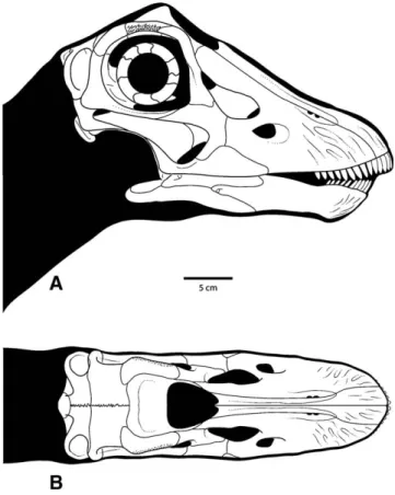
FIGURE 6. Reconstruction of CM 11255. A, lateral view; B, dorsal view. Scale bar equals 5 cm.
FIGURE 7. Transformation grids based on sutures and other landmarks showing the regions of the Diplodocus skull that underwent the greatest amount of shape change through ontogeny. Grids based on Figure 6 and a reconstruction of the adult skull of Diplodocus (Wilson and Sereno, 1998:fig. 6). A, lateral view; B, dorsal view.
11 mm, for a maximum skull width of 88 mm at the quadrates. If the entire right side of the skull, to the midline suggested by the right pterygoid, is mirrored, the maximum skull width is only 102 mm. Even using this higher estimate, the reconstructed snout is still quite round in comparison to adults (see Comparisons with Adult Diplodocus, below).
Evidence from the Dentary—As noted above, lateral
crush-ing of the skull has displaced the left mandible, disarticulatcrush-ing it from the right at the dentary symphysis. This disarticulation and subsequent displacement appears to have been the main im-pact of compression on the mandibles because their longitudinal rami appear to be relatively straight and the anterior curvature of each dentary is similar (Fig. 4B, D). These elements are gently rounded in ventral view, not squared as seen in larger Diplodocus skulls.
Lateral crushing of this extent is not typically seen in larger
Diplodocus skulls, although slight crushing is apparent in two
other specimens, one adult (USNM 2672), and a larger sub-adult (CM 3452). In both of these latter specimens, the dentaries are symmetrical and appear to retain their original morphology (squared in USNM 2672, more gently rounded in CM 3452) and are, again, disarticulated along the symphysis, with one mandible displaced. Even in a clearly distorted skull (USNM 2673), the de-formation occurs along the lateral ramus of the mandible, not at its anterolateral corner. The only observed case of deformation altering the anterior shape of the dentary is the dicraeosaurid diplodocoid Dicraeosaurus (MB.R. 2372), in which the square-ness of the dentary is exaggerated, although the ventral margin is itself undeformed. It is unlikely, then, that taphonomic deforma-tion has greatly altered the shape of the dentaries of CM 11255, and consequently these elements constitute a reasonable proxy for snout shape in this specimen.
Comparisons with Adult Diplodocus
Varricchio (1997) listed ten ways in which the crania of di-nosaurs have been shown to vary ontogenetically, two of which (increase in tooth count, shortening and deepening of the skull) are potentially related to changes in feeding behavior. Others (relative decrease in size of the orbit, relative increase in antor-bital length) are common in many amniote groups and are not directly related to feeding behavior.
As expected, the relative contributions of the orbit and brain-case to overall skull size are dramatically larger in CM 11255 rel-ative to large adult skulls (Fig. 7). Accordingly, there is a smaller contribution of the antorbital region to skull length in CM 11255, which has the effect of shortening the face. This shorter propor-tional length is accompanied by a snout that is narrower and rounder in dorsal view. The upper tooth row occupies a much larger proportion of the jaw margin and extends much farther posteriorly than in adults. This suggests a large-scale ontoge-netic remodeling of the facial skeleton involving rearrangement of the food-gathering apparatus, which has not been previously reported for any sauropodomorph dinosaur.
Implications for Facial Remodeling in Diplodocus—In
mam-mals, snout shape has been shown to serve as a proxy for feed-ing behavior (Bou ´e, 1970; Solounias et al., 1988; Dompierre and Churcher, 1996). Broad, anteriorly flat snouts belong to mam-mals that crop low-lying grasses near the ground, and narrow, pointed snouts belong to mammals that selectively browse for particular plants or plant parts. Using a modification of a metric used by Solounias et al. (1988), the snout shape of sauropods can be quantified. The premaxillary-maxillary index (PMI) is calcu-lated by superimposing a triangle over a dorsal view of the snout with the hypotenuse drawn at 26◦. The area of this triangle that is covered by snout is divided by the total area of the triangle to determine the PMI; higher numbers indicate squarer snouts. The most conservative reconstruction of CM 11255 has a PMI of
56%; this is well below that of adult Diplodocus (PMI= 84%; Whitlock, 2007). This pointed snout is also seen in other juvenile specimens of Diplodocus (CMC VP 8300; Whitlock, 2006). The ontogenetic disparity in snout shapes in this genus may be evi-dence of resource partitioning between adults and juveniles that might have had vastly different energetic needs. Fiorillo (1998) presented patterns of enamel microwear as evidence for resource partitioning between adults and juveniles of a different Morri-son sauropod, Camarasaurus. Ongoing research has shown a dif-ference in wear patterns between relatively round snouted
(Di-craeosaurus) and relatively square snouted (Apatosaurus, adult Diplodocus) diplodocoids that is consistent with selective
brows-ing versus non-specific browsbrows-ing similar to the ‘grazbrows-ing’ behavior of ruminants (Whitlock, 2007). Unfortunately, microwear has not yet been recovered from a definitive juvenile Diplodocus tooth for comparison with adult patterns.
Curry (1999) and Lehman and Woodward (2008) presented sigmoidal growth curves for the diplodocid Apatosaurus. As-suming that these curves accurately represent the pattern of growth rates through diplodocid ontogeny, juvenile Diplodocus in the exponential growth phase may have required more energy-rich foodstuffs than adults that had reached their growth plateau. Barrett (2000) suggested a similar scenario for basal sauropodomorphs involving opportunistic carnivory in juveniles. Jarman (1974) noted that small ungulates, which have compa-rably higher metabolic rates than large ungulates, have narrow snouts for selective browsing of plant parts with high digestibility and high caloric content. It is possible that a similar scenario could have occurred in an organism whose growth curve involves many orders of magnitude increase in mass. Earlier ontogenetic stages likely required easily digestible, high-calorie foods to maintain a higher metabolism, and used a narrow, pointed snout to selectively obtain them. Once full size had been reached, energetic goals may have been attained by higher-volume, less nutritious, non-specific browsing by blunt-snouted adult
Diplodocus individuals. Resource partitioning may also have
oc-curred out of necessity, easing intraspecific competition between adults and their offspring, or, as noted by Jarman (1974), because a larger skull (such as that of adult Diplodocus) is less suited for selective herbivory.
Comparisons with Other Dinosaurs
Of the numerous dinosaurian taxa that have been examined for ontogenetic cranial variation, six are of particular interest. Three related taxa, the sauropodomorphs Camarasaurus, Rapetosaurus, and Massospondylus, are examined, as well as the theropods
Al-bertosaurus and Tyrannosaurus. To elucidate the condition in
an ornithischian, the basal ceratopsian Psittacosaurus is also dis-cussed.
Camarasaurus—Camarasaurus is known from multiple skulls, including a juvenile preserving most of the facial skeleton (CM 11338). Ikejiri (2004) and Ikejiri et al. (2005) suggested that there was little remodeling of the craniofacial skeleton throughout the ontogeny of this genus. McIntosh et al. (1996) posited that a re-duction in alveolar count did occur with advancing ontogenetic stage, contrary to the typical dinosaurian condition (Varricchio, 1997; but see Carr, 1999). Fiorillo (1998) used enamel microwear patterns to suggest resource partitioning between adults and ju-veniles in browse height, but not necessarily browse type. For sauropods like Camarasaurus, whose relatively high forelimb-to-hind limb and low neck-to-torso ratios suggest a higher browse height than is posited for diplodocoids, browse height was most likely a function of body size and therefore maturity. In other words, given their smaller size, younger individuals necessar-ily browsed at lower heights than older individuals. In contrast,
Diplodocus has been interpreted as a mid- to low-height feeder
and Parrish, 1999, 2005; Upchurch and Barrett, 2000). The varia-tion in resource acquisivaria-tion may have been as much a funcvaria-tion of behavior as it is of physiology.
Rapetosaurus—The derived titanosaurian sauropod
Rapetosaurus is known from cranial elements belonging to
an adult and a juvenile, including facial bones from each (Curry Rogers and Forster, 2004). However, the reconstructed skull of this taxon is based almost entirely on the adult, because very little of the facial skeleton is preserved in the juvenile (Curry Rogers and Forster, 2004:Fig. 1). Although the reconstructed snout is somewhat rounded in dorsal view, the preserved dentary (Curry Rogers and Forster, 2004:fig. 28) approaches a square shape, more so than in the juvenile Diplodocus but less than in adults. In the absence of more complete juvenile skulls, little can be said about the ontogenetic development of the blunt snout in
Rapetosaurus.
Massospondylus—Sues et al. (2004) described four skulls attributable to Massospondylus carinatus, representing several stages of growth. M. carinatus appears to have added maxillary tooth positions with age, and there are more denticles per crown in juvenile specimens than in adults, but the general shape of the snout does not appear to have varied greatly (Sues et al., 2004). Gow (1990) noted a few ontogenetic changes in the braincase of M. carinatus, primarily the late ossification of a posterior ex-tension of the laterosphenoid separating the vena cerebralis me-dia and cranial nerve V, and increased muscle scarring on the supraoccipital. Reconstructions of three skulls of M. carinatus (Gow et al., 1990:fig. 7) suggest that the orbit became propor-tionally smaller and the antorbital region proporpropor-tionally longer with increasing size, as is typical of many other dinosaurian taxa (Varricchio, 1997).
Albertosaurus and Tyrannosaurus—The premaxillae and maxillae of Albertosaurus were subject to ontogenetic variation, particularly in the later sub-adult stages, when the snout broad-ened transversely (Carr, 1999). Additionally, the skull as a whole became more robust with age, a pattern also seen in
Tyran-nosaurus. Carr (1999) hypothesized that this variation may be
the result of variation in foraging behaviors, such that older in-dividuals were more capable of grasping and holding live prey or tearing apart large carrion; an alternate explanation proposed was that the increased robustness and broadness was a physiolog-ical response to increased skull size and bite force. Additionally, the teeth became more robust throughout ontogeny, with a cor-responding reduction in the number of alveoli. The implication is that, as in Diplodocus, tyrannosaurid theropods were capable of variation in response to differing feeding behaviors or require-ments at different ontogenetic stages.
Psittacosaurus—The basal ceratopsian Psittacosaurus is known from many individuals of varying sizes and stages of ontogeny. In P. mongoliensis, alveolar count more than doubles throughout ontogeny, eventually reaching 12 maxillary and den-tary teeth from approximately five in the youngest individuals (Sereno, 1990). In P. mongoliensis and P. xinjiangensis, the large sagittal crest is not present in young individuals and developed as the animal matured (Sereno and Chao, 1988). Additionally, Makovicky et al. (2006) demonstrated that the presence of a well-developed flange on the dentary is age related, only appearing in older sub-adults. Those authors also found that overall skull shape, however, did not significantly vary with age. Unlike in Diplodocus, it appears that the ontogenetic variation in
Psittacosaurus was not related to a substantial change in feeding
behavior, but was instead a response to increased body size and muscle development with age.
CONCLUSION
CM 11255 is the smallest recognized skull of Diplodocus. It shares several synapomorphies with adult skulls, but the pres-ence of unfused parietal bones and the small size of the specimen
(60% of the anteroposterior length of the adult skull CM 11161) indicate that it pertains to a juvenile individual. Unique to this individual are the extreme posterior position of the distal-most tooth in the maxillary tooth row and the rounded dental arcade, in contrast to the squared snout and anteriorly sequestered tooth row in adults. The larger sub-adult Diplodocus CM 3452 strongly resembles CM 11255 in both conditions, and it is hypothesized that juvenile and sub-adult individuals of Diplodocus share a fa-cial morphology that is distinct from that of adults, particularly with regard to the tooth bearing elements and the dental arcade. Similar morphologies (rounded versus blunt snouts) have been shown to be related to food gathering in mammals. The condition in Diplodocus indicates ontogenetic niche partitioning, as has been suggested for Camarasaurus and tyrannosaurid theropods. Juvenile and sub-adult Diplodocus appear to have been selective browsers, whereas square-snouted adults were likely low-height non-selective browsers, similar to what has been proposed for the diplodocoids Brachytrachelopan (Rauhut et al., 2005),
Di-craeosaurus (Upchurch and Barrett, 2000; Barrett and Upchurch,
2005), and Nigersaurus (Sereno et al., 2007). ACKNOWLEDGMENTS
We thank A. Henrici (CM), C. Mehling (AMNH), M. Brett-Surman (USNM), K. Curry Rogers (SMM), N. Klein (MB), and O. Hampe (MB) for access to specimens. A. Shaw (CM) as-sisted with additional preparation of the specimen. C. Blaine and K. Schwarz (UM Hospitals) proved CT imaging assistance. B. Miljour (UM Museum of Paleontology) greatly improved the il-lustrations. D. Brinkman (YPM) provided a photo of YPM 1922. Reviewers M. D’Emic and T. Ikejiri provided comments on pre-vious drafts of the manuscript. Editor H.-D. Sues and reviewers P. Barrett and J. Harris improved later drafts with helpful com-ments. This work was supported in part by funds from the Scott Turner Award at the University of Michigan and the Geological Society of America (8689-07) awarded to J. Whitlock.
LITERATURE CITED
Allain, R., and N. Aquesbi. 2008. Anatomy and phylogenetic relation-ships of Taz oudasaurus naimi (Dinosauria, Sauropoda) from the late Early Jurassic of Morocco. Geodiversitas 30:345–424.
Apesteguia, S. 2004. Bonitasaura salgadoi gen. et sp. nov.: a beaked sauro-pod from the Late Cretaceous of Patagonia. Naturwissenschaften 91:493–497.
Bakker, R. T. 1986. The Dinosaur Heresies. Kensington Press, New York, 481 pp.
Barrett, P. M. 1999. A sauropod dinosaur from the Lower Lufeng For-mation (Lower Jurassic) of Yunnan Province, People’s Republic of China. Journal of Vertebrate Paleontology 19:785–787.
Barrett, P. M. 2000. Prosauropod dinosaurs and iguanas: speculations on the diets of extinct reptiles; pp. 43–78 in H.-D. Sues (ed.), Evolution of Herbivory in Terrestrial Vertebrates: Perspectives from the Fossil Record. Cambridge University Press, Cambridge, U.K.
Barrett, P. M., and P. Upchurch. 1994. Feeding mechanisms of Diplodocus. Gaia 10:195–203.
Barrett, P. M., and P. Upchurch. 2005. Sauropodomorph diversity through time: paleoecological and macroevolutionary implications; pp. 125–151 in K. A. Curry Rogers and J. A. Wilson (eds.), The Sauropods: Evolution and Paleobiology. University of California Press, Berkeley, California.
Barrett, P. M., and K. J. Willis. 2001. Did dinosaurs invent flowers? Dinosaur-angiosperm coevolution revisited. Biological Reviews of the Cambridge Philosophical Society 76:411–447.
Berman, D. S., and S. L. Jain. 1982. The braincase of a small sauropod dinosaur (Reptilia: Saurischia) from the Upper Cretaceous Lameta Group, Central India, with review of Lameta Group Localities. An-nals of Carnegie Museum 51:405–422.
Berman, D. S., and J. S. McIntosh. 1978. Skull and relationships of the Upper Jurassic sauropod Apatosaurus (Reptilia, Saurischia). Bul-letin of Carnegie Museum of Natural History 8:1–35.
Bonaparte, J. F. 1979. Faunas y paleobiogeograf´ıa de los tetr ´apodos Mesozoicos de Am ´erica del Sur. Ameghiniana 16:217–238.