HAL Id: hal-03133856
https://hal.archives-ouvertes.fr/hal-03133856
Submitted on 7 Feb 2021HAL is a multi-disciplinary open access
archive for the deposit and dissemination of sci-entific research documents, whether they are pub-lished or not. The documents may come from teaching and research institutions in France or abroad, or from public or private research centers.
L’archive ouverte pluridisciplinaire HAL, est destinée au dépôt et à la diffusion de documents scientifiques de niveau recherche, publiés ou non, émanant des établissements d’enseignement et de recherche français ou étrangers, des laboratoires publics ou privés.
Distributed under a Creative Commons Attribution - NonCommercial| 4.0 International License
Immune signatures distinguish frequent from
non-frequent exacerbators among children with severe
asthma
Karine Adel-Patient, Marta Grauso, Rola Abou Taam, Blanche Guillon,
Céline Dietrich, Franois Machavoine, Nicolas Garcelon, Mélanie Briard,
Hassan Faour, Antoine Neuraz, et al.
To cite this version:
Karine Adel-Patient, Marta Grauso, Rola Abou Taam, Blanche Guillon, Céline Dietrich, et al.. Im-mune signatures distinguish frequent from non-frequent exacerbators among children with severe asthma. Allergy, Wiley, 2021, �10.1111/all.14759�. �hal-03133856�
Immune signatures distinguish frequent from non-frequent exacerbators among children with 1 severe asthma. 2 3 To the editor, 4 5 Asthma is a heterogeneous condition with multiple phenotypes.1,2 Almost 5% of children with severe 6
asthma (SA) remain symptomatic despite high doses of inhaled corticosteroids (ICS) and other 7 controllers.3 Asthma has long been regarded as a type-2 (T2) disorder, but a dominant Th1 signature, 8 with Th17 and Th2 cells in a mixed cytokine milieu, was recently described in bronchoalveolar lavages 9 (BAL) from children with SA.4 Moreover, among children with SA, cellular components such as IL-17-10
producing mucosal-associated invariant T cells may distinguish frequent (FE) from non-frequent 11
exacerbators (nFE).5 The inflammatory profiles of children with SA thus appear to be highly complex.
12
We aimed to provide preliminary results to identify immune signatures associated with clinical 13
phenotypes of SA. 14
Twenty children with SA were included (supplementary material & table 1) and their blood and BAL 15
collected. FE (n=13) were defined as children with at least three severe exacerbations (SE) in the 16
previous year and nFE (n=7) as those having one or two.5 The two groups did not differ, except for the
17
number of steroid bursts. Samples were also collected from control subjects with non-asthmatic (NA) 18
severe respiratory conditions (n=10). Preliminary analysis i) confirmed a mixed T1/T2-type cellular 19
profile in BAL (figure 1A), associated with blood eosinophilia (table 1), in children with SA relative to 20
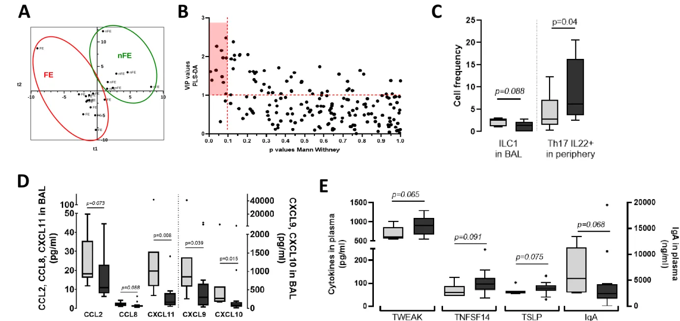
NA, and ii) showed the immune profile of FE to differ from that of nFE (T2 versus T1 phenotype; 21 respectively, figure 1B&C). We then performed a comprehensive non-targeted analysis of cytokines 22 and cells in blood and BAL from children with SA (supplementary material) and then constructed a 23 model to distinguish FE from nFE through supervised analysis (PLS-DA) (figure 2A). The model classified 24
patients with a good predictive value (R²Y=0.799), with both sensitivity and specificity of 100%. 25 Although all data were used to construct the model, we identified a set of constituents that mostly 26 supported the differences between the patient groups (supplementary table 2, VIP>1). In parallel, we 27 performed univariate comparisons of each constituent (supplementary table 2). Finally, we identified 28 11 immune constituents to be the most discriminative (PxVIP plotting, VIP>1 and p<0.1, figure 2B). A 29
higher frequency of ILC1 and Th1-associated chemokines in BAL (CCL2/8, CXCL9-11, figure 2C&D) 30
confirmed the more pronounced T1 phenotype in nFE. Conversely, the local T2 phenotype of FE was 31
associated with a higher frequency of activated Th17 (IL22+) cells in the blood (figure 2C) that tended
32
to correlate with the number of exacerbations (r=0.41, p=0.08). Previous studies found that the 33
number of circulating Th17 cells was higher in children with moderate-to-severe than mild asthma,6
that these cells may be steroid resistant,7 and that PBMC IL-17 secretion was induced by
35
dexamethasone.8 Th17 IL-22+ cells may therefore emerge after repeated systemic steroid
36
administration received to treat exacerbations, leading to a mixed T2/T17 phenotype. FE also exhibited 37
lower concentrations of total IgA and higher concentrations of TWEAK, TNFSF14, and TSLP in plasma 38
(figure 2E), and TLSP levels significantly correlated with the number of exacerbations (r=0.45, 39
p=0.045). This is consistent with previous studies showing the involvement of these individual 40
constituents in the pathogenesis and severity of asthma in children. Plasma constituents, such as 41
sTNFR1, CCL26, Pentraxin3, CXCL10, IL-32, and sIL16RB, were also highly discriminant, despite high p-42
values. All these constituents thus contributed to characterize the FE phenotype and univariate 43
analysis alone would not have allowed their identification. For example, the T2 chemokine CCL26 44
tended to be higher in FE and significantly correlated with blood eosinophil counts (r=0.432, p=0.025). 45
CCL26 is a potent chemoattractant for eosinophils and elevated concentrations of CCL26 in plasma 46
have been shown to be related to mucosal eosinophilia and the severity of various eosinophilic 47 disorders (e.g.9), which may suggest tissue eosinophilia in FE. 48 In conclusion, despite a small sample size and ICS that may affect the immune response, our study 49 shows the potential interest of high-dimensional/non-targeted multivariate analysis to identify specific 50 biological signatures of children with different clinical phenotypes of SA. Although confirmation in an 51 independent cohort is needed, our study provides new leads for delineating asthma pathogenesis and 52 identifying new set of targets for diagnosis and personalized treatment. 53 54 Authors: 55
Karine Adel-Patient1, PhD, Marta Grauso1, PhD, Rola Abou-Taam2, MD, Blanche Guillon1, MSc, Céline
56
Dietrich3, MSc, François Machavoine3, MSc, Nicolas Garcelon4,5, PhD, Mélanie Briard1, Hassan Faour4,5,
57
MSc, Antoine Neuraz4,5, MD, PhD, Christophe Delacourt2, MD, PhD, MSc, Thierry Jo Molina4,6, MD, PhD,
58
Maria Leite-de-Moraes3, PhD, and Guillaume Lezmi2,3, MD, PhD
59 60
1 Université Paris-Saclay, CEA, INRAE, Département Médicaments et Technologies pour la Santé
61
(DMTS), SPI, Laboratoire d’Immuno-Allergie Alimentaire, F-91191, Gif-sur-Yvette, France 62
2 AP-HP, Hôpital Necker-Enfants Malades, Service de Pneumologie et Allergologie Pédiatriques,
F-63
75015, Paris, France. 64
3 Université de Paris, Institut Necker Enfants Malades, Equipe Immunorégulation et
65 Immunopathologie, Inserm UMR1151, CNRS UMR8253, F-75015, Paris, France. 66 4 Université de Paris, UMRS 1138, INSERM, Sorbonne Paris-Cité, F-75006 Paris, France 67 5AP-HP, Hôpital Necker-Enfants Malades, Service Informatique Médicale, F-75015 Paris, France 68
6 AP-HP, Centre-Université de Paris, Hôpital Necker-Enfant-Malades, Service d'Anatomie et Cytologie 69 Pathologiques, F-75015 Paris, France 70 71 Corresponding authors: 72 Karine Adel-Patient: 73 DRF/Institut Joliot/DMTS/SPI/Laboratoire d’Immuno-Allergie Alimentaire 74 CEA de Saclay, Bat 136 75 91191 Gif-sur-Yvette, France 76 karine.adel-patient@cea.fr 77 78 Guillaume Lezmi: 79
Institut Necker-Enfants Malades, Laboratory of Immunoregulation and Immunopathology, CNRS 80 UMR8253 and INSERM UMR1151, Paris, France 81 guillaume.lezmi@aphp.fr 82 83 Author contributions: 84 GL, KAP, and MLM: designed the research. 85 KAP, MG, BG, CDi, TM, MB, and FM: performed the research. 86 GL, RAT, NG, HF, AN, and CD: were responsible for patient recruitment or establishing the patient 87 database. 88 KAP and GL: analyzed the data. 89 KAP, GL, and MLM: wrote the manuscript. 90 91 Abbreviations: BAL: bronchoalveolar lavage fluids, ICS: inhaled corticosteroids, NA: non-asthmatic, 92 PLS-DA: partial least squares-discriminant analysis, SA: severe asthma, FE: frequent exacerbators, nFE: 93 non-frequent exacerbators. 94
95 Acknowledgements 96 We thank Naima Cortes-Perez for her help in the experiments and all the patients involved in the study 97 and their parents. 98 99 Financial support 100 This work was supported by the INRAE-AlimH Department and grants from the Legs Poix, Chancellerie 101 des Universities, Paris, France, and ANR-18-CE14-0011-01 SevAsthma-children, Paris, France. 102 103 Conflict of interests 104 The authors have no conflict of interests to declare. 105 106 107 Figure legends 108 109 Figure 1. Complex immune profile of children with different SA phenotypes. A. Higher frequency of 110 Th1 cells (trend for Th2) in BAL from children with SA (grey bars) than in that from non-asthmatic 111 children with severe respiratory disorders (NA, white bars). B. The higher frequency of Th1 cells in BAL 112 of children with SA relative to that in NA children (white bars) was significant only in non-frequent 113
exacerbators (nFE, n=7; light grey). Conversely, the higher number of Th2 cells in BAL (B) and 114 eosinophils in blood (C) was significant only in frequent exacerbators (FE, n=13; dark grey). Data are 115 shown as box and Tukey whisker plots. P values were obtained using the Kruskal-Wallis test comparing 116 all groups together. 117 118 Figure 2. Identification of immune constituents that discriminate between SA children with frequent 119 (FE) and non-frequent (nFE) exacerbations. A. Graph of the individuals provided by PLS-DA modelling. 120 B. VIP x p values plot of all measured immune constituents and selection of the most discriminating 121 and significant (red rectangle: VIP>1 and p<0.1; because of the small number of patients in each group, 122 we tolerated p<0.1 as a cut-off). Identified discriminant immune constituents within cellular (C) or 123 soluble constituents in BAL (D) or plasma (E), represented as box and Tukey whisker plots. Exact p 124 values (Mann Whitney test) are indicated. 125 126 127
References 128 129 1. Lezmi G, de Blic J. Assessment of airway inflammation and remodeling in children with severe 130 asthma: The next challenge. Pediatric pulmonology. 2018;53(9):1171-1173. 131 2. Kuruvilla ME, Lee FE, Lee GB. Understanding Asthma Phenotypes, Endotypes, and Mechanisms 132 of Disease. Clinical reviews in allergy & immunology. 2019;56(2):219-233. 133 3. Chung KF, Wenzel S. From the authors: International European Respiratory Society/American 134 Thoracic Society guidelines on severe asthma. Eur Respir J. 2014;44(5):1378-1379. 135 4. Wisniewski JA, Muehling LM, Eccles JD, et al. TH1 signatures are present in the lower airways 136
of children with severe asthma, regardless of allergic status. J Allergy Clin Immunol. 137 2018;141(6):2048-2060 e2013. 138 5. Lezmi G, Abou-Taam R, Garcelon N, et al. Evidence for a MAIT-17-high phenotype in children 139 with severe asthma. J Allergy Clin Immunol. 2019;144(6):1714-1716 e1716. 140 6. Chien JW, Lin CY, Yang KD, Lin CH, Kao JK, Tsai YG. Increased IL-17A secreting CD4+ T cells, 141 serum IL-17 levels and exhaled nitric oxide are correlated with childhood asthma severity. Clin 142 Exp Allergy. 2013;43(9):1018-1026. 143 7. Nagakumar P, Puttur F, Gregory LG, et al. Pulmonary type-2 innate lymphoid cells in paediatric 144 severe asthma: phenotype and response to steroids. Eur Respir J. 2019;54(2). 145 8. Gupta A, Dimeloe S, Richards DF, et al. Defective IL-10 expression and in vitro steroid-induced 146 IL-17A in paediatric severe therapy-resistant asthma. Thorax. 2014;69(6):508-515. 147 9. Yamada T, Miyabe Y, Ueki S, et al. Eotaxin-3 as a Plasma Biomarker for Mucosal Eosinophil 148 Infiltration in Chronic Rhinosinusitis. Front Immunol. 2019;10:74. 149 150