HAL Id: hal-02370548
https://hal.archives-ouvertes.fr/hal-02370548
Submitted on 26 May 2020
HAL is a multi-disciplinary open access
archive for the deposit and dissemination of
sci-entific research documents, whether they are
pub-lished or not. The documents may come from
teaching and research institutions in France or
abroad, or from public or private research centers.
L’archive ouverte pluridisciplinaire HAL, est
destinée au dépôt et à la diffusion de documents
scientifiques de niveau recherche, publiés ou non,
émanant des établissements d’enseignement et de
recherche français ou étrangers, des laboratoires
publics ou privés.
Distributed under a Creative Commons Attribution| 4.0 International License
and changes in stress-responsive gene expression in male
Balb/c mice after repeated administration of a Rhodiola
rosea L. root extract
Anne-laure Dinel, Isabelle Guinobert, Céline Lucas, Claude Blondeau, Valérie
Bardot, Isabelle Ripoche, Lucile Berthomier, Véronique Pallet, Sophie Laye,
Corinne Joffre
To cite this version:
Anne-laure Dinel, Isabelle Guinobert, Céline Lucas, Claude Blondeau, Valérie Bardot, et al..
Reduc-tion of acute mild stress corticosterone response and changes in stress-responsive gene expression in
male Balb/c mice after repeated administration of a Rhodiola rosea L. root extract. Food Science &
Nutrition, Wiley, 2019, 7 (11), pp.3827-3841. �10.1002/fsn3.1249�. �hal-02370548�
Food Sci Nutr. 2019;7:3827–3841. www.foodscience-nutrition.com
|
3827 Received: 29 March 2019|
Revised: 24 July 2019|
Accepted: 27 July 2019DOI: 10.1002/fsn3.1249
O R I G I N A L R E S E A R C H
Reduction of acute mild stress corticosterone response and
changes in stress‐responsive gene expression in male Balb/c
mice after repeated administration of a Rhodiola rosea L. root
extract
Anne‐Laure Dinel
1,2,3| Isabelle Guinobert
4,5| Céline Lucas
3| Claude Blondeau
4,5|
Valérie Bardot
4,5| Isabelle Ripoche
6| Lucile Berthomier
6| Véronique Pallet
1,2|
Sophie Layé
1,2| Corinne Joffre
1,2This is an open access article under the terms of the Creative Commons Attribution License, which permits use, distribution and reproduction in any medium, provided the original work is properly cited.
© 2019 The Authors. Food Science & Nutrition published by Wiley Periodicals, Inc.
1Integrated Nutrition and Neurobiology, UMR 1286, INRA, Bordeaux, France 2Integrated Nutrition and Neurobiology, UMR 1286, Bordeaux University, Bordeaux, France 3Integrated Nutrition and Neurobiology, UMR 1286, NutriBrain Research and Technology Transfer, Bordeaux, France 4Groupe Pileje, Paris, France 5Naturopôle, Les Tiolans, Saint‐Bonnet de Rochefort, France 6CNRS, SIGMA Clermont, Clermont‐Ferrand Chemistry Institute, Clermont Auvergne University, Clermont Ferrand, France Correspondence Anne‐Laure Dinel, Integrated Nutrition and Neurobiology, UMR 1286, INRA, 33076 Bordeaux, France. Email: anne‐laure.dinel@inra.fr
Abstract
Rhodiola rosea L. (R. rosea) is an adaptogenic plant increasing body resistance to stress.
Its efficacy has been evidenced mainly in chronic stress models, data concerning its effect in acute stress and underlying mechanisms being scarce. The objective was to investigate the effect of repeated doses of a R. rosea hydroethanolic root extract (HRE) on hypothalamic pituitary adrenal response in a murine model of acute mild stress and also the mechanisms involved. Stress response was measured in Balb/c mice having received by gavage HRE (5 g/kg) or vehicle daily for 2 weeks before being submitted to an acute mild stress protocol (open‐field test then elevated plus maze). Corticosterone was measured in plasma from mandibular vein blood drawn before and 30, 60, and 90 min after initiation of the stress protocol. Mice were sac- rificed at 90 min, and the hippocampus, prefrontal cortex, and amygdala were ex-cised for high‐frequency RT‐PCR gene expression analysis. At 30 min after acute mild stress induction, corticosterone level in mice having received the HRE was lower than in control mice and comparable to that in nonstressed mice in the HRE group. HRE administration induced brain structure‐dependent changes in expression of sev-eral stress‐responsive genes implicated in neuronal structure, HPA axis activation, and circadian rhythm. In the acute mild stress model used, R. rosea HRE decreased corticosterone level and increased expression of stress‐responsive genes, especially in the hippocampus and prefrontal cortex. These findings suggest that R. rosea HRE could be of value for modulating reactivity to acute mild stress. K E Y W O R D S acute mild stress, circadian rhythm, corticosterone, nutritional supplementation, rhodiola
1 | INTRODUCTION
Stress is the physiological reaction to environmental threats or pressure and can be self‐driven or of external origin (Anghelescu, Edwards, Seifritz, & Kasper, 2018). It is manifested by a wide vari-ety of physical and psychological symptoms. If persistent and left untreated, stress can result in serious health problems including burnout, depression, post‐traumatic stress disorder, anxiety, and cardiovascular, gastrointestinal, neurological, and musculoskeletal diseases. Stress appears to be a particular problem in our modern society. Work‐related stress is experienced by all sections of society, being estimated to affect 22% of the European workforce (Milczarek & Gonzales, 2009). The World Health Organization has called stress “the health epidemic of the 21st century,” recognizing its substan- tial impact on personal life and also its social and economic conse-quences (Anghelescu et al., 2018; Subhani et al., 2018).
Stress management strategies include nonpharmacological ap-proaches, such as cognitive behavioral therapy and relaxation, but recourse to pharmacological treatment is standard if stress and its symptoms become harmful. Anxiolytics and antidepressants, as-sociated with known risks of adverse effects and dependency, are generally indicated for more severe situations. Several plants, in-cluding chamomile, melissa, and rhodiola, have been shown to be valuable for managing stress and its consequences, with fewer ad-verse effects and a lower risk of dependency (Sarris, McIntyre, & Camfield, 2013). Rhodiola rosea L. (rosenroot or golden root), man-ifesting adaptogenic properties, is among those most widely used (Anghelescu et al., 2018; Kasper & Dienel, 2017). Extracts of adap-togenic plants can normalize body functions and reinforce systems compromised by stress (Anghelescu et al., 2018). They have no spe-cific pharmacological properties and act by increasing resistance to a broad spectrum of adverse expressions of stress. Preclinical in vivo and ex vivo studies in animal models and experiments on cell lines have highlighted several biochemical and pharmacological stress‐ reducing properties of R. rosea extracts (Abidov, Crendal, Grachev, Seifulla, & Ziegenfuss, 2003; Olsson, von Scheele, & Panossian, 2009; Panossian, Hambardzumyan, Hovhanissyan, & Wikman, 2007; Panossian, Hovhannisyan, Abrahamyan, Gabrielyan, & Wikman, 2009). In clinical studies, various extracts of R. rosea were found to be effective and safe, improving mental work capacity, concen-tration, task performance, fatigue, burnout symptoms, and overall mood, besides reducing stress level and self‐reported mild anxiety (Cropley, Banks, & Boyle, 2015; Darbinyan et al., 2000; Edwards, Heufelder, & Zimmermann, 2012; Kasper & Dienel, 2017; Panossian, Wikman, Kaur, & Asea, 2009; Punja, Shamseer, Olson, & Vohra, 2014). R. rosea was approved by the European Medicines Agency Committee on Herbal Medicinal Products for the indication “tem-porary relief of symptoms of stress such as fatigue and sensation of weakness” (EMA/HPMC, 2012).
Stress response typically begins with activation of the hypothal-amus–pituitary–adrenal (HPA) axis, one of the main stress response pathways, and the production of corticosteroids (Anghelescu et al., 2018; Subhani et al., 2018). Acute or chronic stress produces
characteristic changes in the HPA axis, including an increase in cor-tisol in humans and corticosterone in rodents, as well as a reduc-tion in the sensitivity of the HPA axis to feedback down‐regulation (Anghelescu et al., 2018; Panossian, Wikman, et al., 2009). Chronic stress results in persistent elevation of cortisol or corticosterone levels, which may lead to fatigue, depression, and other symptoms (Anghelescu et al., 2018). The reduction in stress‐induced damage by R. rosea is characterized by a decrease in or the prevention of hormonal changes characteristic of stress, including cortisol or corti-costerone release, as shown in humans suffering from chronic stress following administration of the standardized R. rosea root extract SHR‐5 during 28 days (Olsson et al., 2009) and in rabbits subjected to acute stress after 7 days of SHR‐5 administration (Panossian et al., 2007). HPA axis modulation by R. rosea extracts also involves the in-hibition of stress‐induced protein kinases and nitric oxide in animals (Panossian, Wikman, et al., 2009). The HPA axis is not the only target of R. rosea. For instance, R. rosea extracts stimulated energy metab-olism in rodents via the activation of ATP synthesis in mitochondria (Abidov et al., 2003) and might protect against neurodegenerative brain diseases through antioxidative and anti‐inflammatory mecha-nisms (Lee et al., 2013; Zhang, Zhu, Jin, Yan, & Chen, 2006).
Investigations of the molecular mechanisms underlying central corticosteroid action following a stress event led to the identifica-tion of genetic pathways and, in particular, stress‐responsive genes (Hunter et al., 2016; Kohrt et al., 2016). Modification of target gene transcription, the so‐called genomic action of corticosteroids, is therefore most likely one of the main mechanisms underlying cor-ticosteroid action in the brain (Gray, Kogan, Marrocco, & McEwen, 2017). These genomic effects can occur within 15–30 min after the activation of corticosteroid receptors and may last for less than an hour or up to several days, depending on the duration of exposure to the hormone and the type of stress (Dong, Poellinger, Gustafsson, & Okret, 1988; Morsink, Joels, et al., 2006). These stress‐respon-sive genes are divided into several functional classes according to their implication in energy metabolism, signal transduction, neuronal structure, vesicle dynamics, neurotransmitter catabolism or cell ad-hesion, their encoding of neurotrophic factors and their receptors, and their involvement in the regulation of glucocorticoid signaling (Andrus et al., 2012; Datson, Morsink, Meijer, & de Kloet, 2008; Datson et al., 2012; Hunter et al., 2016). The effects of R. rosea ex-tracts on these stress‐responsive genes are unknown. Furthermore, all the data on R. rosea reported so far have been obtained following intense stress, either acute or chronic. Characterizing the effects of
R. rosea on the HPA axis and stress‐responsive gene transcription
under acute mild stress conditions would contribute to a better un-derstanding of how extracts of this adaptogenic plant act to prevent the negative effects of stress.
The purpose of this study was therefore to evaluate, in a murine model of acute mild stress, the effects on the HPA axis of repeated administration of a hydroethanolic root extract (HRE) of R. rosea, phytochemically characterized by high‐performance thin‐layer chro- matography (HPTLC) and ultra‐high‐performance liquid chromatog-raphy coupled with mass spectrometry (UHPLC‐MS). Corticosterone
secretion and stress‐responsive gene expression were determined in the prefrontal cortex (PFC), amygdala, and hippocampus, the main structures implicated in stress management.
2 | MATERIAL AND METHODS
2.1 | Preparation of the R. rosea HRE
The R. rosea HRE was obtained according to the patented process WO2001056584A1 by crushing frozen fresh roots of R. rosea and leaching with 20%–70% (v/v) ethanol. The extract was then concen-trated under reduced pressure to evaporate ethanol. The salidro-side titer was adjusted within the range of 0.7–1.4 mg/ml by adding glycerin to the concentrated extract. The batch of HRE used in this study (16H321), containing 83% glycerin, had a salidroside content of 1.02 mg/ml and a dry drug: dry genuine extract ratio of 17:1. This glycerin‐containing HRE corresponds to the standardized extract of R. rosea marketed in France under the brand name “Extrait de plante fraîche standardisé (EPS) R. rosea” (PiLeJe Laboratoire, France).
2.2 | LC/MS analysis of the R. rosea HRE
UHPLC analysis was performed on an Ultimate 3000 RSLC UHPLC system (Thermo Fisher Scientific Inc., MA, USA) coupled to a qua-ternary rapid separation pump (Ultimate autosampler) and a rapid separation diode array detector. Compounds were separated on an Uptisphere Strategy C18 column (25 × 4.6 mm; 5 μm; Interchim, Montluçon, France), maintained at 40°C. The mobile phase was a mixture of 0.1% (v/v) formic acid in water (phase A) and 0.1% (v/v) formic acid in acetonitrile (phase B). The gradient of phase A was 100% (0 min), 80% (10 min), 73% (35 min), 0% (40–50 min), and 100% (51–60 min). The flow rate was 0.8 ml/min and the injection volume 10 µl. The UHPLC system was connected to an Orbitrap mass spec- trometer (Thermo Fisher Scientific Inc., MA, USA) operating in nega-tive electrospray ionization mode. Source operating conditions were as follows: 3 kV spray voltage for negative mode; 320°C heated cap-illary temperature; 400°C auxiliary gas temperature; sheath, sweep, and auxiliary gas (nitrogen) flow rate 60, 17.5, and 3.5 arbitrary units, respectively; and collision cell voltage between 20 and 50 eV. Full scan data were obtained at a resolution of 35,000 whereas MS2 data were obtained at a resolution of 17,500. Data were processed using Xcalibur software (Thermo Fisher Scientific Inc., MA, USA).
The constituents of the R. rosea HRE were identified according to their retention times and mass spectral data and by comparison with authentic standards, if available, or otherwise with published data.
2.3 | HPTLC analysis of R. rosea HRE
Standards were diluted in methanol at a concentration of 0.5 mg/ ml for rosavin and 0.1 mg/ml for salidroside (Sigma Aldrich, Saint Louis, USA). One mL of the R. rosea HRE (without added glycerol) was diluted in 3 ml of a mixture of 50% ethanol and water (50/50: v/v). The resultant solution was shaken and centrifuged for 3 min at 6,600 g. The supernatant solution was transferred into individual vials and then analyzed by HPTLC. HPTLC analysis was performed on 200 × 100 mm silica gel 60 F 254 HPTLC glass plates (Merck, Darmstadt, Germany), using a Camag HPTLC system (Muttenz, Switzerland) equipped with an Automatic TLC Sampler (ATS 4), an Automatic Developing Chamber ADC2 with humidity con-trol, a TLC Visualizer, WinCATS software and for derivatization, a Chromatogram Immersion Device III, and a TLC Plate Heater III. Standard solutions and samples were applied as bands 8.0 mm wide, up to a 8.0 mm from the lower edge of the plate and 15 mm from the left and right edges. The space between bands was 11.3 mm, and each plate contained 16 tracks. The development distance was 70.0 mm from the lower edge of the plate. The temperature within the developing chamber was set at 21°C and the relative humidity at 37%. The mobile phase was a solution of ethyl acetate, water, formic acid, and methanol (volume ratio: 77/10/2/13). Derivatization was performed by dipping (speed: 5, time: 0) in a reagent comprising 10% sulfuric acid in methanol and heating at 100°C for 5 min. Plates were analyzed under UV at 366 nm.
2.4 | Animals and experimental design
Seven‐week‐old male Balb/c mice, a highly stress‐sensitive strain (Janvier, Le Genest‐Saint‐Isle, France), were housed under a normal 12‐hr light/dark cycle (07 hr–19 hr) with food (AO4 diet; Safe, Augy, France) and water available ad libitum in a controlled environment (22 ± 1°C, 40% of humidity). The mice were handled daily for 1 week before the start of the experiment to minimize stress reactions to manipulation. During the following 2 weeks, they received each morning a supplement comprising either R. rosea HRE (a 5 g/kg solu-tion containing 80% glycerin, i.e., 4 g/kg; test group, n = 8) or glycerin
alone (4 g/kg; control group, n = 8) administered by gavage using a V0105040 feeding probe (ECIMED, Boissy‐Saint‐Léger, France). The two groups received the same amount of glycerin. The volume of supplementation was adapted to the weight of each mouse. At the end of this period, the mice were subjected to an acute mild stress protocol and anxiety‐like behavior was evaluated. Blood was drawn from the mandibular vein before initiation of the stress protocol (at t0 min) and then at t30 min and t60 min. Mice were sacrificed at t90 min, and brain structures (hippocampus, hypothalamus, and amyg-dala) and plasma were excised and frozen at −80°C (Figure 1).
2.5 | Induction of acute mild stress
On the last day of supplement administration, half the mice in each group were subjected to acute mild stress. The stress protocol con-sisted in subjecting the mice to an open‐field (OF) test for 10 min immediately followed by an elevated plus maze (EPM) test for 5 min (see the following sections for details; Figure 1). Experiments were performed in the morning, one hour after gavage, under conditions of dim light and low noise. Both tests induce mild stress in animals by subjecting them to anxiogenic conditions (Treit, Menard, & Royan, 1993).
2.6 | Evaluation of anxiety‐like behavior
Anxiety‐like behavior was evaluated after induction of acute mild stress as previously reported by Dinel et al. (2011). Mouse behav-ior was videotaped and scored using “Smart” software (Noldus, Wageningen, Netherlands).
2.6.1 | OF test
Mice were exposed to an unfamiliar square (40 × 40 cm) OF from which escape was prevented by surrounding walls (16 cm high). The apparatus was virtually divided into 4 central squares defined as the central area (anxiogenic) and 12 squares along the walls, defined as the periphery. Each mouse was placed in the central area and al-lowed to freely explore the OF for 10 min. Parameters recorded to evaluate anxiety‐like behavior comprised the number of entries into the central area and the percentage of time spent in this area (Dinel et al., 2011).2.6.2 | EPM test
The EPM was a plus‐shaped acryl maze with two opposing open arms (30 × 8 cm) and two opposing closed arms (30 × 8 × 15 cm) connected by a central platform (8 × 8 cm), elevated 120 cm above the floor. Each mouse was placed in the center of the maze facing an open arm, a situation that is highly anxiogenic. The test was performed over a period of 5 min. The number of arm entries and the percent of time spent in open arms were calculated to evalu-ate the basal level of anxiety. An entry was scored as such only when the mouse placed all its four limbs in any particular arm. Areduction in the percentage time spent in the open arms and the number of entries into these is considered as an index of anxi-ety‐like behavior, independent of locomotor activity (Dinel et al., 2011).
2.7 | Biochemical measurements
2.7.1 | Measurement of corticosterone
Corticosterone was measured in plasma before and 30, 60, and 90 min after initiation of the stress protocol, using a DetectX corti-costerone immunoassay kit (Euromedex, Strasbourg, France) (Dinel, Joffre, et al., 2014).
2.7.2 | Assessment of RNA expression using
Fluidigm microfluidic arrays
One microgram of total RNA was obtained from each brain area as described in Dinel et al. (Dinel, Andre, et al., 2014) and was reverse‐transcribed with SuperScript III reverse transcriptase (Invitrogen, Cergy‐Pontoise, France). Diluted cDNA (1.3 µl, 5 ng/ µl) was added to DNA Binding Dye Sample Loading Reagent (Fluidigm), EvaGreen (Interchim, Montluçon, France), and Tris‐ EDTA (TE) buffer with low EDTA to constitute the Sample Mix plate. In the Assay Mix plate, 10 µl of primer pairs (100 µM) was added to the Assay Loading Reagent (Fluidigm) and TE buffer with low EDTA to a final concentration of 5 µM. After priming of the chip in the Integrated Fluidic Circuit Controller, Sample Mix (5 µl) and Assay Mix (5 µl) were loaded into the sample inlet wells. One well was filled with water as a contamination control. To verify specific target amplification and quantitative polymerase chain reaction (Q‐PCR) process efficiencies, a control sample (mouse gDNA, Thermo Fisher, Waltham, USA) was treated, preamplified, and quantified in a control assay (RNasePTaqMan probe, Thermo Fisher) using the same process in the same plate at the same time. The expected value of cycle quantification was around 13. The chip was inserted into the IFC controller, in which 6.3 nl of Sample Mix and 0.7 nl of Assay Mix were blended. Real‐time PCR was performed using the Biomark System (Fluidigm) on the GenoToul platform (Toulouse, France) with the following protocol: Thermal Mix at 50°C, 2 min; 70°C, 30 min; 25°C, 10 min, Uracil‐DNA N‐ glycosylase (UNG) at 50°C, 2 min, Hot Start at 95°C, 10 min, PCR Cycle of 35 cycles at 95°C, 15 s; 60°C, 60 s and Melting curves (from 60°C to 95°C). Results were analyzed using the Fluidigm Real‐Time PCR Analysis software v.4.1.3. (San Francisco, USA) to control specific amplification for each primer. Then, the raw data of the qPCR were analyzed using GenEx software (MultiD analy-ses AB, Freising, Germany) in order to choose the best reference gene for normalizing mRNA expression and to measure the rela- tive expression of each of the 93 genes analyzed in the group re-ceiving the HRE and the control group. GAPDH was found to be the best reference gene in this experiment and was therefore used for normalization of gene expression.
2.8 | Statistical analysis
2.8.1 | Bivariate statistical analysis
All data were expressed as the mean value ± SEM (standard error of the mean). A p‐value of 0.05 was considered as significant. Data were analyzed using a one‐way ANOVA (one factor: supplementation) or a two‐way ANOVA with supplementation (HRE, control), and stress (stress; no stress) as between factors followed by a Bonferroni post hoc analysis when interaction was significant (GraphPad software, La Jolla, US). Heatmaps were obtained using the Permut Matrix pro-gram (Caraux & Pinloche, 2005).
2.8.2 | Principal component analysis (PCA)
PCA was used to assess the gene expression pattern under stress conditions in the group receiving R. rosea HRE and the control group. The PCA is a dimension reduction technique that clusters data into principal components (PC) maximizing the variance of the data con-sidered. These PCs are uncorrelated linear combinations of the initial variables which can be interpreted as a pattern. PCA generates factor loadings which reflect the correlation of each variable with the PC and attributes a PC score for each individual. We selected the number of components using the Cattell criterion. Statistical analyses were performed using the XLSTAT program (Addinsoft, Paris, France).
3 | RESULTS
3.1 | Phytochemical profile of R. rosea HRE
HPTLC analysis showed that R. rosea HRE contains salidroside and rosavin (Supplementary data, Fig. S1A). UHPLC‐MS analysis
confirmed the presence of these two compounds (peaks 7 and 15) (Fig. S1B and Table S1). Three monoterpene glycosides corre-sponding to rhodiolosides E, B (or C) and rosiridin (peaks 13 and 24) and several phenylpropane derivatives, including rosarin and rosin, were identified (peaks 15–16 and 18). Five flavonoids were also de- tected: herbacetin, kaempferol, rhodamine, rhodopsin, and kaemp-ferol‐7‐O‐rhamnoside (peaks 22, 25, 21, 19, and 23, respectively).
3.2 | R. rosea HRE did not impact behavior in acute
mild stress protocol
As expected, we did not observed any significant effect of the diet (glycerin or R. rosea HRE) on time spent in open arm in the EPM (Figure 2a) or on time spent in center area in the OF (Figure 2b).
3.3 | R. rosea HRE modulated corticosterone
secretion consecutive to acute mild stress
Corticosterone was measured in plasma prepared from blood sam-ples drawn before the induction of acute mild stress and 30, 60, and 90 min after the start of the stress protocol. At t0, mice having re-ceived R. rosea HRE exhibited a significantly higher plasma corticos-terone level (110.8 ng/ml) than mice given the control supplement (glycerin alone, 31.31 ng/ml) (t = 2.789, p < .01; Figure 3a). A t30, t60, and t90, R. rosea HRE induced a decrease in corticos-terone secretion compared with the control (F (1,24) = 8.352, p < .01, Figure 3b; F (1,25) = 6.165, p < .05, Figure 3c; and F (1,26) = 5.954, p < .05, Figure 3d, respectively). At t30, we also observed a stress effect (F (1,24) = 6.391, p < .05, Figure 3b) and a stress × supplemen-tation interaction (F (1,24) = 4.544, p < .01) indicating that 30 min after the induction of acute mild stress, administration of R. rosea HRE restored corticosterone secretion to the basal level. F I G U R E 2 Anxiety‐like behavior of adult mice subjected to acute mild stress having received a R. rosea HRE or glycerin (control) supplement for 2 weeks by daily gavage. (a) Time (in seconds) spent in the open arms of the elevated plus maze. (b) Time (in seconds) spent in the center area of the open‐field. Data are presented as means ± SEM (n = 8 per group). HRE, hydroethanolic root extract
3.4 | R. rosea HRE modulated stress‐responsive
gene expression in a structure‐dependent manner
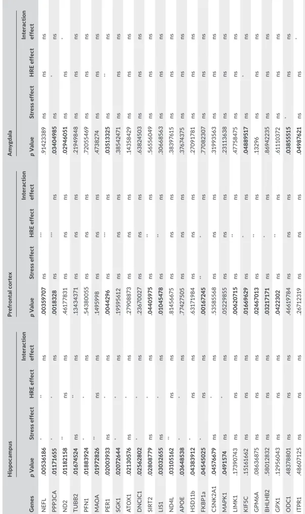
The expression of 93 genes implicated in stress reactivity was ana-lyzed. Administration of R. rosea HRE modulated the pool of stress‐ responsive genes described by Datson et al. (2008, 2012), Andrus et al. (2012), and Kohrt et al. (2016). The genes modulated differed between the hippocampus, PFC, and amygdala and could be classi-fied by function. All significant genes and results are presented in Tables 1 and 2. In the hippocampus, 13 genes were significantly overexpressed after repeated administration of R. rosea HRE. These genes were implicated in signal transduction (CSNK2A1, F (1,22) = 4.694,p < .05; MAPK1, F (1,22) = 5.248, p < .05; SGK1, F (1,22) = 6.591, p < .05), neuronal structure (NEFL, F (1,22) = 8.870, p < .01; TUBB2, F (1,22) = 8.077, p < .01; PPP3CA, F (1,22) = 4.396, p < .05; PFN1, F (1,22) = 4.892, p < .05), oxidative stress (ATOX1, F (1,22) = 7.753, p < .05; APOE, F (1,22) = 4.450, p < .05, SIRT2, F (1,22) = 7.711, p < .05) and regulation of the HPA axis (LIS1, F (1,22) = 5.623, p < .05; DNCIC1, F (1,22) = 4.493, p < .05). PER1 expression, implicated in circadian rhythm, was also increased after HRE administration (F (1,22) = 7.774, p < .05). Stress affected the expression of 11 genes including NEFL, F (1,22) = 7.624, p < .05; PPP3CA, F (1,22) = 7.701, p < .05; PFN1 F (1,22) = 7.359, p < .05; SGK1, F (1,22) = 5.088, p < .05; DNCIC1, F (1,22) = 6.041, p < .05 and APOE, F (1,22) = 4.866, p < .05) that were also regulated by R. rosea HRE. The mitochondrial genes ND2 (F (1,22) = 12.17, p < .01) and ND4L (F (1,22) = 10.17, p < .01) were also upregulated by stress along with MAOA (F (1,22) = 10.68, p < .01), HSD11b (F (1,22) = 7.636, p < .05), and FKBP1a (F (1,22) = 6.701, p < .05), the expression of which is classically induced by chronic or acute stress.
In the PFC, acute mild stress affected only FKBP1a (F (1, 16) = 16.10, p < .01). R. rosea HRE also increased the expression of genes implicated in neuronal structure (NEFL, F (1,16) = 16.14,
p < .001; PP3CA, F (1,16) = 19.07, p < .001; LIMK1, F (1,16) = 14.98, p < .01; GPM6A, F (1,16) = 8.791, p < .01), oxidative stress (SIRT2, F (1,16) = 9.914, p < .01; and GPX1, F (1,16) = 8.822, p < .01), HPA axis regulation (LIS1, F (1,16) = 12.96, p < .01; KIF5C, F (1,15) = 6.141, p < .05; FKBP1a, F (1,16) = 7.889, p < .05; BHLHB2, F (1,16) = 7.892, p < .05), and circadian rhythm (PER1, F (1,16) = 16.90, p < .001).
The amygdala was less responsive than the hippocampus and PFC to R. rosea HRE, only six genes being modulated by this supplement and/or stress. As in the other structures, PPP3CA, KIF5C, and PER1 were overexpressed following R. rosea HRE administration (F (1,18) = 8.174, p < .05; F (1,19) = 5.581, p < .05 and F (1,19) = 10.06, p < .01, respectively). Acute mild stress in-duced an increase in OD1 expression (F (1,19) = 5.575, p < .05). Interestingly, ND2 and ITPR1 expressions were similarly increased F I G U R E 3 Corticosterone secretion in adult mice having received a R. rosea HRE or glycerin (control) supplement for 2 weeks by daily gavage before the induction of acute mild stress (a) and at t30 (b), t60 (c), and t90 min (d) after initiation of the stress protocol. Glycerin versus HRE: *p < .05, **p < .01; glycerin stress versus HRE stress: $$, p < .01. HRE, hydroethanolic root extract
TA B L E 1 Stress‐responsive genes studied by high‐frequency RT‐qPCR in the prefrontal cortex, hippocampus, and amygdala
Symbol Name Category Sequence (5′−3′) References
TUBB2‐F Tubulin, beta 2A class IIA Neuronal structure TCGGCGCTAAGTTTTGGGAG Datson et al., EJP
2008
TUBB2‐R TGCAAGTCACTGTCGCCATG
NEFL‐F Neurofilament, light
polypeptide
Neuronal structure TGCAGACATTAGCGCCATGC Datson et al., EJP
2008
NEFL‐R TCTCGCTCTTCGTGCTTCTCAG
GPM6A‐F Glycoprotein m6a Neuronal structure ACTGCTGGAGACACACTGGATG Datson et al., EJP
2008
GPM6A‐R AAGAAAGCAGCCGCAATGCC
LIMK1‐F LIM domain containing,
protein kinase Neuronal structure TCCGAGCACATCACCAAAGG Datson et al., EJP 2008
LIMK1‐R AGGCGAGGCAGATGAAACAC
PPP3CA‐F Protein phosphatase 3,
catalytic subunit, alpha isoform
Neuronal structure CTGGTCGCTGCCATTTGTTG Datson et al., EJP
2008
PPP3CA‐R ATCGTCGGAGCAGATGTTGAG
PFN1 F1 Profilin 1 Neuronal structure ATCGTAGGCTACAAGGACTCGC Datson et al., EJP
2008
PFN1 R2 AACCTCAGCTGGCGTAATGC
DNCIC1‐F Dynein cytoplasmic 1
intermediate chain 1
Glucocorticoid signaling AACTTCGTGGTTGGCAGTGAG Datson et al., EJP
2008
DNCIC1‐R ACCGATGCCTGCTTTGCTTC
LIS1‐F Platelet‐activating factor
acetylhydrolase, iso-form 1b, subunit 1
Glucocorticoid signaling GATGTGGGAAGTGCAAACTGG Datson et al., EJP
2008
LIS1‐R CTGATTTGGCCGCACCATAC
KIF5C‐F Kinesin family member
5C
Glucocorticoid signaling ATGTAAAGGGGTGCACCGAGAG Datson et al., EJP
2008
KIF5C‐R ACGTGTCGGTTTGCTTTGCC
FKBP1a‐F FK506‐binding protein
1a
Glucocorticoid signaling TCCTCTCGGGACAGAAACAAGC Datson et al., EJP
2008
FKBP1a‐R AGTTTGGCTCTCTGACCCACAC
ODC1‐F Ornithine decarboxylase,
structural 1
Glucocorticoid signaling TCGCCAGAGCACATCCAAAG Datson et al.,
Hippocampus 2012
ODC1‐R TTTTGCCCGTTCCAAGAGAAG
BHLHB2‐F Basic helix‐loop‐helix
family, member e40
Glucocorticoid signaling AACGGAGCGAAGACAGCAAG Datson et al.,
Hippocampus 2012
BHLHB2‐R ATCCTTCAGCTGGGCAATGC
CSNK1A1‐F Casein kinase 1, alpha 1 Glucocorticoid signaling CGTCGGTGGAAAATACAAACTGG Datson et al.,
Hippocampus 2012
CSNK1A1‐R TCTCGTACAGCAACTGGGGATG
SGK1‐F Serum/glucocorticoid‐
regulated kinase 1 Glucocorticoid signaling CGTCAAAGCCGAGGCTGCTCGAAGC Arteaga et al., PNAS 2008
SGK1‐R GGTTTGGCGTGAGGGTTGGAGGAC
ITPR1‐F
Inositol 1,4,5‐trisphos-phate receptor 1
Glucocorticoid signaling ATCGGCCACCAGTTCCAAAG Mahfouz et al., PNAS
2016
ITPR1‐R AGCCAAGTAATGCCCTGTAGCC
HSD11b1‐F Hydroxysteroid 11‐beta
dehydrogenase 1
Glucocorticoid signaling GGAAGGTCTCCAGAAGGTAGTGTC This study
HSD11b1‐R GAGGCTGCTCCGAGTTCAAG
SGK1‐F serum/glucocorticoid‐
regulated kinase 1
Glucocorticoid signaling CGTCAAAGCCGAGGCTGCTCGAAGC Arteaga et al., PNAS
2008
SGK1‐R GGTTTGGCGTGAGGGTTGGAGGAC
MAPK1‐F
Mitogen‐activated pro-tein kinase 1
Glucocorticoid signaling AGCTAACGTTCTGCACCGTG Datson et al., EJP
2008
MAPK1‐R TGATCTGGATCTGCAACACGGG
PER1‐F Period circadian clock 1 Circadian rythm TGTCCTGCTGCGTTGCAAAC This study
PER1‐R TTGAGACCTGAACCTGCAGAGG
MAOA‐F Monoamine oxidase A Mood regulation TGAGGTATCTGCCCTGTGGTTC Datson et al., EJP
2008
MAOA‐R CCCCAAGGAGGACCATTATCTG
SIRT2‐F Sirtuin 2 Mood regulation TCCACTGGCCTCTATGCAAACC This study
SIRT2‐R TTGGCAAGGGCAAAGAAGGG
APOE‐F Apolipoprotein E Lipid metabolism TGCGAAGATGAAGGCTCTGTG This study
APOE‐R GGTTGGTTGCTTTGCCACTC
by HRE administration under stress conditions (stress × supple-mentation interaction F (1,19) = 4.399, p < .05; F (1,18) = 6.837,
p < .05, respectively).
PCA of all genes studied in the hippocampus (Figure 4a), PFC (Figure 4b), and amygdala (Figure 4c) was performed to identify those contributing most to the observed differences between the treatment groups. Remarkably, PCA analysis showed clear separa-tion of the variables: the first component (“F1”) explained 33.46%, 41.18%, and 26.46% of total variance in the hippocampus, PFC, and amygdala, respectively. Pattern 1 revealed that the genes studied were mostly upregulated in the hippocampus and PFC whereas their regulation was more heterogeneous in the amygdala. The second component (“F2”) explained 13.62%, 13.90%, and 17.60% of total variance in the hippocampus, PFC, and amygdala, respectively. This component could reveal a gene classification by functionality.
Phylogenetic analysis based on Pearson's correlation was per- formed for the three brain structures studied (Figure 5). The heat-map generated demonstrated that gene regulation depends on the group considered (HRE‐supplemented or control), especially as re-gards the PFC. However, we did not observe any real gene clusters.
4 | DISCUSSION
The objective of this study was to evaluate the effect on the HPA axis of chronic administration of a R. rosea HRE in a murine acute mild stress model by measuring corticosterone secretion and assess-ing cerebral expression of stress‐responsive genes.4.1 | R. rosea HRE decreased stress‐induced
corticosterone secretion
In the acute mild stress model used in this study, Balb/c mice were consecutively subjected to an OF and an EPM test. We chose to use Balb/c mice as studies have shown this strain to be highly stress‐sen-sitive compared with other strains (Moloney, Dinan, & Cryan, 2015). Both tests used in this study induce stress in animals by placing them in anxiogenic environments: an open place in the OF test and open arms in the EPM test (Treit et al., 1993). The basal level of corticosterone was higher in mice receiving R. rosea HRE than in control mice receiving a supplement containing glycerin alone. This difference might be explained by the organolep-tic characteristics and higher viscosity of the HRE compared with glycerin alone, which could have created additional stress during ad-ministration of these supplements (Hoggatt, Hoggatt, Honerlaw, & Pelus, 2010). Even if the percentage of increase was important, the level of corticosterone in mice having received the R. Rosea HRE was far below levels obtained after a stress, even in low reactive mice (Mattos et al., 2013). Moreover, we did not observe any behavioral difference in anxiety‐like tests between glycerin‐ and R. Rosea HRE‐ treated mice.Thirty minutes after acute mild stress induction, control mice presented, as expected, an increase in corticosterone secretion, whereas mice receiving R. rosea HRE did not. At t60 and t90, the percentage corticosterone increase was comparable between stress‐free and stressed mice. We hypothesize that the effect of experimentally induced acute mild stress was masked by that of gavage. We nevertheless observed that at both times, mice having received R. rosea HRE presented a lower percentage increase in cor-ticosterone as compared to the control group. This result implies that administration of R. rosea HRE resulted in better regulation of stress homeostasis, characterized by more effective control of corti-costerone increase that probably led to more efficient restoration of corticosterone level to the basal value.
At the intracellular level, high corticosteroid levels impact the balance between trophic and atrophic factors within neurons (Liu et al., 2017). For instance, glucocorticoids have been shown to in- hibit cell proliferation in the dentate gyrus by reducing the prolif-eration of granule cell precursors (Gould & Tanapat, 1999; Saaltink & Vreugdenhil, 2014). Moreover, chronic stress results in persistent inhibition of granule cell production and changes in the structure of the dentate gyrus, raising the possibility that stress alters hippo-campal function through this mechanism (Gould & Tanapat, 1999). By preventing the substantial increase in corticosterone level, R.
rosea extracts could prevent this negative impact of corticosteroids.
Our results confirm those of previous studies demonstrating the impact of R. rosea extracts on inhibition of the HPA axis, as illus-trated notably by the serum level of corticosteroids in rats (Cifani
Symbol Name Category Sequence (5′−3′) References
ND2‐F NADH dehydrogenase 2,
mitochondrial
Mitochondria TTCATAGGGGCATGAGGAGGAC Hunter et al., PNAS
2016
ND2‐R GTGAGGGATGGGTTGTAAGGAAG
ND4L‐F NADH dehydrogenase
4L, mitochondrial
Mitochondria CCATACCAATCCCCATCACCA Hunter et al., PNAS
2016
ND4L‐R GGACGTAATCTGTTCCGTACGTGT
ATOX1‐F Antioxidant 1 copper
chaperone
Stress oxydant ACGAGTTCTCCGTGGACATGAC This study
ATOX1‐R TGCAGACCTTCTTGTTGGGC
GPX1‐F Glutathione peroxidase 1 Stress oxydant TCGGACACCAGAATGGCAAG This study
GPX1‐R AGGAAGGTAAAGAGCGGGTGAG
Abbreviation: CORT, dosage of corticosterone. TA B L E 1 (Continued)
T A B LE 2 Ex pr es si on o f s tr es s‐ re sp on si ve g en es in th e hi pp oc am pu s, p re fr on ta l c or te x, a nd a my gd al a of a du lt m ic e ha vi ng re ce iv ed a R . r os ea H RE o r g ly ce rin (c on tr ol ) s up pl em en t b y da ily gav ag e fo r 2 w ee ks G ene s H ipp oc am pu s Pr efr on ta l c or te x A m yg da la p V alue St res s e ff ec t H RE e ff ec t In te ra cti on ef fe ct p V alue St res s e ff ec t H RE e ff ec t In te ra cti on ef fe ct p V alue St res s e ff ec t H RE e ff ec t In te ra cti on ef fe ct N EF L .0 05 36 186 * ** ns .0 03 59 70 7 ns *** .9 14 23 38 9 ns ns ns PP P3 C A .0 11 71 65 5 * * ns .0 018 32 8 ns *** ns .0 340 49 85 ns * ns N D2 .0 118 21 58 ** ns ns .4 61 77 83 1 ns ns ns .0 29 46 051 ns ns * TU B B2 .0 16 74 52 4 ns ** ns .1 343 43 71 ns ns ns .2 19 49 84 8 ns ns ns PF N 1 .0 18 83 924 * * ns .5 43 80 05 5 ns ns ns .7 20 55 46 9 ns ns ns M AO A .0 19 72 82 6 ** ns ns .14 95 99 8 ns ns ns .4 73 82 74 ns ns ns PE R1 .0 20 03 93 3 ns * ns .0 04 42 96 ns *** ns .0 351 33 25 ns ** ns SG K1 .0 207 26 44 * * ns .1 95 95 61 2 ns ns ns .3 85 42 47 1 ns ns ns ATO X1 .0 213 057 6 ns * ns .2 79 08 87 3 ns ns ns .14 35 84 29 ns ns ns D N C IC1 .025 62 802 * * ns .2 36 70 02 7 ns ns ns .6 38 24 50 3 ns ns ns SI RT 2 .0 2808 77 9 ns * ns .0 44 05 975 ns ** ns .56 556 04 9 ns ns ns LI S1 .03 03 26 55 ns * ns .0 10 45 47 8 ns ** ns .3 06 68 563 ns ns ns N D 4L .0 31 051 62 ** ns ns .8 14 56 67 5 ns ns ns .3 83 976 15 ns ns ns A PO E .0 36 485 38 * * ns .7 74 27 50 5 ns ns ns .3 76 74 37 5 ns ns ns H SD 11 b .0 43 83 91 2 * ns ns .6 31 71 98 4 ns ns ns .2 70 91 78 1 ns ns ns FK B P1a .0 45 45 02 5 * ns ns .0 01 67 24 5 ** * ns .7 70 82 307 ns ns ns C SN K 2A1 .0 45 766 79 ns * ns .53 58 55 68 ns ns ns .3 19 93 56 3 ns ns ns M AP K 1 .0 49 15 74 ns * ns .0 52 29 85 5 ns ns ns .2 311 36 38 ns ns ns LIMK 1 .1 73 90 74 3 ns ns ns .0 06 20 71 5 ns ** ns .47 75 847 5 ns ns ns K IF 5C .1 516 16 62 ns ns ns .0 16 696 29 ns * ns .0 48 89 51 7 ns * ns G PM 6A .0 86 36 875 ns ns ns .0 24 67 01 3 ns ** ns .1 32 96 ns ns ns B H LH B2 .5 801 28 32 ns ns ns .0 32 17 17 1 ns * ns .8 6942 23 5 ns ns ns G PX1 .1 29 56 04 3 ns ns ns .0 42 23 02 ns ** ns .6 111 03 72 ns ns ns O D C1 .4 83 78 80 1 ns ns ns .4 661 97 84 ns ns ns .0 38 555 15 * ns ns IT PR1 .4 86 071 25 ns ns ns .2 67 12 31 9 ns ns ns .0 49 87 621 ns ns * A bb re vi at io n: H RE , h yd ro et ha no lic ro ot e xt ra ct . *** p < .0 01 ; ** p < .0 1; *p < .0 5, n s, n ot s ig ni fic an t.
et al., 2010; Xia, Li, Wang, Wang, & Wang, 2016). The antistress properties of R. rosea extracts have been attributed to their inter-ference with both the HPA axis and the sympathoadrenal system (Panossian, Hovhannisyan, et al., 2009; Panossian & Wagner, 2005; Panossian, Wikman, et al., 2009; Panossian, Wikman, & Wagner, 1999). However, all these results were obtained in animals subjected to intense acute or chronic stress. In this study, we demonstrated for the first time that a specific R. rosea extract affects HPA axis reactivity even under conditions of mild stress of short duration. The dampening of corticosterone secretion could be due to a decrease in stress reactivity amplitude or to better control of the glucocorticoid pathway.
4.2 | R. rosea HRE upregulated the expression of
functional stress‐responsive genes
One of the main mechanisms of action of corticosteroids in the brain is their genomic effect, resulting in modification of target gene tran-scription. Corticosteroid‐mediated transcriptional changes within the brain have been studied by means of large‐scale gene expression profiling (Datson et al., 2008, 2012; Hunter et al., 2016; Kohrt et al., 2016). The resulting gene expression profile showed a highly dy-namic transcriptional response to glucocorticoid receptor activation throughout a specific time window, shifting from exclusively down‐ regulation of genes 1 hr after glucocorticoid receptor activation to both up‐ and down‐regulation after 3 hr (Morsink, Steenbergen, et al., 2006). We investigated the impact of R. rosea HRE, 1h30 after the induction of acute mild stress, on the expression of stress‐respon-sive genes (Datson et al., 2008, 2012; Hunter et al., 2016) in the PFC and amygdala, structures involved in the regulation of stress, as well as in the hippocampus, a medial temporal lobe structure implicated in the formation of stable memories and highly susceptible to stress (Kim & Diamond, 2002). Interestingly, most genes modulated in the PFC, amygdala, and hippocampus by R. rosea HRE belong to four main functional groups of genes implicated in the functioning of neuronal structures, glu-cocorticoid signaling, circadian rhythm, and mood regulation, respectively.
Supplementation with R. rosea HRE upregulated genes coding for structural components of the cytoskeleton, such as beta‐tubu- lin (TUBB2) and neurofilament light polypeptide (NEFL), genes me-diating neurite outgrowth, including glycoprotein M6A (GPM6A) (Alfonso, Fernandez, Cooper, Flugge, & Frasch, 2005), as well as genes specifically involved in the dynamics of the actin cytoskeleton of neurons, calcineurin subunit A (PPP3CA), and profilin 1 (PFN1). Genes affecting the actin cytoskeleton were modulated by the HRE in all three brain structures studied, but acute mild stress affected their expression only in the hippocampus. The actin cytoskeleton is involved in the morphology of dendritic spines, and changes in actin cytoskeletal configurations have been postulated to influence long‐term potentiation, affecting synaptic transmission (Meng et al., 2002; Smart & Halpain, 2000). Under stress, these mechanisms are dysregulated and the connectivity between the various brain structures is impaired (Christoffel, Golden, & Russo, 2011). Several studies have demonstrated that stress induces adverse changes in the morphology and strength of hippocampal excitatory synapses, inducing a generalized atrophy of dendrites and spines in the PFC (Goldwater et al., 2009; Sandi et al., 2003; Stewart et al., 2005; Wellman, 2001). By upregulating genes implicated in neuronal struc-ture genes, R. rosea HRE might prevent adverse changes in synaptic plasticity and consequently functional disorders, such as those ob-served in pathological behaviors or depression. F I G U R E 4 Graphic representation, defined by the first two principal components (F1 and F2), of the Principal Component Analysis (PCA) of gene expression measured by RT‐PCR in the hippocampus (a), prefrontal cortex (b), and amygdala (c) of adult mice having received a R. rosea HRE or glycerin (control) supplement by daily gavage for 2 weeks before the induction of acute mild stress. HRE, hydroethanolic root extract F I G U R E 5 Phylogenic relationship based on Pearson's correlation in the hippocampus (a), prefrontal cortex (b), and amygdala (c) of adult mice having received a R. rosea HRE or glycerin (control) supplement for 2 weeks by daily gavage before the induction of acute mild stress. The genes highlighted were modulated by stress, HRE supplementation, or interaction. HRE, hydroethanolic root extract
R. rosea HRE also had an impact on the glucocorticoid
signal-ing pathway. Glucocorticoids have been shown to modulate motor activity and axonal transport by regulating transcription levels of dynein cytoplasmic 1 intermediate chain 1 accessory subunit poly-peptide (DNCIC1), lissencephaly 1 protein (LIS1), and 5c (KIF5C), a member of the kinesin family (Datson, van der Perk, de Kloet, & Vreugdenhil, 2001; Jimenez‐Mateos, Wandosell, Reiner, Avila, & Gonzalez‐Billault, 2005; Kanai et al., 2000; Morsink, Steenbergen, et al., 2006). In our model, R. rosea HRE upregulated the expression of DNCIC1, LIS1, and KIF5C in both the PFC and the hippocampus. KIF5C expression was also upregulated in the amygdala, after HRE supplementation. Acute mild stress affected DNCIC1 expression only in the hippocampus. This modulation of gene expression could act as a primer of the glucocorticoid signaling system. In particular, by upregulating these genes, R. rosea HRE could modify glucocorti-coid receptor trafficking (Harrell et al., 2004), thereby modulating glucocorticoid receptor translocation and consequently glucocorti-coid receptor signaling. Our results showed that R. rosea HRE modu-lated glucocorticoid receptor signaling by changing the expression of genes affecting receptor levels and receptor binding affinity prefer- entially in the PFC and hippocampus. Moreover, FKBP1a, a glucocor-ticoid receptor cochaperone affecting the binding affinity of ligands to glucocorticoid receptors (Kovacs, Cohen, & Yao, 2005; Kovacs, Murphy, et al., 2005; Riggs et al., 2004; Sakisaka, Meerlo, Matteson, Plutner, & Balch, 2002; Wochnik et al., 2005) was upregulated by acute mild stress in the PFC and hippocampus but its expression was also affected by R. rosea HRE in the PFC. In our model, R. rosea HRE also induced in the hippocampus an upregulation of CSNK2A1 and MAPK1 expression, two genes involved in glucocorticoid sig-nal transduction. Previous studies showed that acute administration of glucocorticoids downregulates CSNK2A1 (Datson et al., 2001; Morsink, Steenbergen, et al., 2006), but down‐regulation of this gene was not observed under our 90 min postacute mild stress con-ditions. This increase in hippocampal CSNK2A1 expression under basal and stress conditions in mice receiving R. rosea HRE could act as a primer of the system, thwarting the impact of acute mild stress and preventing the negative impact of glucocorticoids. R. rosea HRE could impact circadian rhythm by modulating PER1. Acute exposure to stressors has been shown to increase PER1 ex-pression in hypothalamic nuclei while suppressing PER1 levels in the central nucleus of the amygdala (Al‐Safadi et al., 2014). In this study, we did not observe any impact of acute mild stress on PER1 expres-sion, but R. rosea HRE upregulated PER1 in all three brain structures examined. R. rosea HRE is therefore likely to have an impact on circa-dian rhythm. It is important to bear in mind that modulation of PER1 expression could affect the circadian expression of corticosterone itself. Tanaka et al. recently demonstrated that hypertensive rats presented adverse changes in PER1 expression and that this abnor-mal adrenal circadian clock may affect steroid hormone secretion by the adrenal gland (Tanaka et al., 2019). Nevertheless, we observed this modulation of PER1 at 90 min after stress induction and a sup-plementary analysis would be necessary to establish a 24 hr time course of gene expression.
Finally, R. rosea HRE modulated the expression of SIRT2, a gene im-plicated in mood regulation. Adverse changes in SIRT2 expression have been reported in mood disorders, with a decrease in SIRT2 expression consecutive to a chronic stress. Treatment with the antidepressant fluoxetine reversed the stress‐induced changes in SIRT2 (Liu et al., 2015). By upregulating SIRT2 expression in the hippocampus and PFC, R. rosea HRE could act like an antidepressant. Previous research has demonstrated that salidroside, one of the active substances of R. rosea HRE, prevented the development of depression‐like behavior as effec-tively as fluoxetine (Zhu et al., 2015). The antidepressant effect of R. rosea extracts might be mediated by their impact on SIRT2 expression. Other genes were regulated by R. rosea HRE but their modulation depended more strongly on the brain structure considered. In the amygdala, the R. rosea HRE and acute mild stress interaction damped the expression of ND2, a mitochondrial membrane respiratory chain gene, suggesting an essential role of mitochondrial activity as an adaptive response to stress, as previously proposed (Vishnyakova et al., 2016). In the PFC, BHLHB2, a gene implicated in neurotrophic factor activity and neuronal excitability, was upregulated by R. rosea HRE, suggesting improved communication between neurons.
To conclude, in the model of acute mild stress used, R. rosea HRE decreased corticosterone levels and increased the expression of stress‐responsive genes, especially in the hippocampus and PFC. Most of the genes affected are implicated in neuronal structure and could impact synaptic transmission and plasticity as well as the glucocorticoid signaling regulation pathway. This upregulation by R.
rosea HRE is associated with damping of corticosterone secretion
and a faster return to the basal profile. This result could be explained by a greater efficacy of HPA axis feedback with a more appropri-ate adaptation of the animals receiving R. rosea HRE to a new envi-ronment. Moreover, R. rosea extracts might modulate the circadian rhythm and potentially biological processes driven by the circadian clock. Complementary studies would be needed to reinforce these preliminary data. Mapping of the signaling pathways and transcrip-tion factors involved, both in cell cultures and in animal models, could help to decipher the impact of HRE extracts under stress conditions. The new data presented here nevertheless suggest that R. rosea HRE could be of value in modulating reactivity to acute mild stress. CONFLIC T OF INTEREST
This work was funded by Groupe PiLeJe. Financial support was provided to Sigma Clermont for the performance of the chromato-graphic analyses and to INRA/Nutribrain for the conduct of in vivo experiments (service provision). The specific roles of the authors including Isabelle Guinobert, Claude Blondeau, and Valérie Bardot from Groupe PiLeJe are articulated in the “author contributions” section.
ETHICAL STATEMENTS
Animal husbandry and experimental procedures were in accordance with the EU Directive 2010/63/EU for animal experiments and were
approved by the national ethical committee for the care and use of animals (approval ID A13169).
ORCID
Anne‐Laure Dinel https://orcid.org/0000‐0002‐8668‐9413
REFERENCES
Abidov, M., Crendal, F., Grachev, S., Seifulla, R., & Ziegenfuss, T. (2003). Effect of extracts from Rhodiola rosea and Rhodiola crenulata (Crassulaceae) roots on ATP content in mitochondria of skeletal mus-cles. Bulletin of Experimental Biology and Medicine, 136(6), 585–587. https ://doi.org/10.1023/B:BEBM.00000 20211.24779.15
Alfonso, J., Fernandez, M. E., Cooper, B., Flugge, G., & Frasch, A. C. (2005). The stress‐regulated protein M6a is a key modulator for neu-rite outgrowth and filopodium/spine formation. Proceedings of the
National Academy of Sciences of the United States of America, 102(47),
17196–17201. https ://doi.org/10.1073/pnas.05042 62102
Al‐Safadi, S., Al‐Safadi, A., Branchaud, M., Rutherford, S., Dayanandan, A., Robinson, B., & Amir, S. (2014). Stress‐induced changes in the expression of the clock protein PERIOD1 in the rat limbic forebrain and hypothalamus: Role of stress type, time of day, and predict-ability. PLoS ONE, 9(10), e111166. https ://doi.org/10.1371/journ al.pone.0111166 Andrus, B. M., Blizinsky, K., Vedell, P. T., Dennis, K., Shukla, P. K., Schaffer, D. J., … Redei, E. E. (2012). Gene expression patterns in the hippo-campus and amygdala of endogenous depression and chronic stress models. Molecular Psychiatry, 17(1), 49–61. https ://doi.org/10.1038/ mp.2010.119
Anghelescu, I. G., Edwards, D., Seifritz, E., & Kasper, S. (2018). Stress management and the role of Rhodiola rosea: A review. International
Journal of Psychiatry in Clinical Practice, 22(4), 242–252. https ://doi.
org/10.1080/13651 501.2017.1417442
Arteaga, M. F., Coric, T., Straub, C., & Canessa, C. M. (2008). A brain‐spe-cific SGK1 splice isoform regulates expression of ASIC1 in neurons.
Proceedings of the National Academy of Sciences of the United States of America, 105(11), 4459–4464. https ://doi.org/10.1073/pnas.15203
76113
Caraux, G., & Pinloche, S. (2005). PermutMatrix: A graphical environ-ment to arrange gene expression profiles in optimal linear order.
Bioinformatics, 21(7), 1280–1281. https ://doi.org/10.1093/bioin
forma tics/bti141
Christoffel, D. J., Golden, S. A., & Russo, S. J. (2011). Structural and synap-tic plasticity in stress‐related disorders. Reviews in the Neurosciences,
22(5), 535–549. https ://doi.org/10.1515/RNS.2011.044
Cifani, C., Micioni Di B., M. V., Vitale, G., Ruggieri, V., Ciccocioppo, R., & Massi, M. (2010). Effect of salidroside, active principle of Rhodiola
rosea extract, on binge eating. Physiology & Behavior, 101(5), 555–
562. https ://doi.org/10.1016/j.physb eh.2010.09.006
Cropley, M., Banks, A. P., & Boyle, J. (2015). The effects of Rhodiola rosea L. extract on anxiety, stress, cognition and other mood symptoms.
Phytotherapy Research, 29(12), 1934–1939. https ://doi.org/10.1002/
ptr.5486
Darbinyan, V., Kteyan, A., Panossian, A., Gabrielian, E., Wikman, G., & Wagner, H. (2000). Rhodiola rosea in stress induced fatigue—A dou-ble blind cross‐over study of a standardized extract SHR‐5 with a repeated low‐dose regimen on the mental performance of healthy physicians during night duty. Phytomedicine, 7(5), 365–371. https :// doi.org/10.1016/S0944‐7113(00)80055‐0
Datson, N. A., Morsink, M. C., Meijer, O. C., & de Kloet, E. R. (2008). Central corticosteroid actions: Search for gene targets. European
Journal of Pharmacology, 583(2–3), 272–289. https ://doi.
org/10.1016/j.ejphar.2007.11.070
Datson, N. A., Speksnijder, N., Mayer, J. L., Steenbergen, P. J., Korobko, O., Goeman, J., … Lucassen, P. J. (2012). The transcriptional response to chronic stress and glucocorticoid receptor blockade in the hip-pocampal dentate gyrus. Hippocampus, 22(2), 359–371. https ://doi. org/10.1002/hipo.20905
Datson, N. A., van der Perk, J., de Kloet, E. R., & Vreugdenhil, E. (2001). Identification of corticosteroid‐responsive genes in rat hippocampus using serial analysis of gene expression.
European Journal of Neuroscience, 14(4), 675–689. https ://doi.
org/10.1046/j.0953‐816x.2001.01685.x
Dinel, A. L., Andre, C., Aubert, A., Ferreira, G., Laye, S., & Castanon, N. (2011). Cognitive and emotional alterations are related to hippocam-pal inflammation in a mouse model of metabolic syndrome. PLoS
ONE, 6(9), e24325. https ://doi.org/10.1371/journ al.pone.0024325 Dinel, A. L., Andre, C., Aubert, A., Ferreira, G., Laye, S., & Castanon, N. (2014). Lipopolysaccharide‐induced brain activation of the indoleam-ine 2,3‐dioxygenase and depressive‐like behavior are impaired in a mouse model of metabolic syndrome. Psychoneuroendocrinology, 40, 48–59. https ://doi.org/10.1016/j.psyne uen.2013.10.014
Dinel, A. L., Joffre, C., Trifilieff, P., Aubert, A., Foury, A., Le Ruyet, P., & Laye, S. (2014). Inflammation early in life is a vulnerability factor for emotional behavior at adolescence and for lipopolysaccharide‐ induced spatial memory and neurogenesis alteration at adulthood.
Journal of Neuroinflammation, 11, 155. https ://doi.org/10.1186/
s12974‐014‐0155‐x
Dong, Y., Poellinger, L., Gustafsson, J. A., & Okret, S. (1988). Regulation of glucocorticoid receptor expression: Evidence for transcriptional and posttranslational mechanisms. Molecular Endocrinology, 2(12), 1256–1264. https ://doi.org/10.1210/mend‐2‐12‐1256
Edwards, D., Heufelder, A., & Zimmermann, A. (2012). Therapeutic ef-fects and safety of Rhodiola rosea extract WS(R) 1375 in subjects with life‐stress symptoms–results of an open‐label study. Phytotherapy
Research, 26(8), 1220–1225. https ://doi.org/10.1002/ptr.3712
EMA/HPMC. (2012). Community herbal monograph on Rhodiola rosea L., rhizoma et radix. EMA/HMPC/232091/2011. Committee on Herbal Medicinal Products (HMPC).
Goldwater, D. S., Pavlides, C., Hunter, R. G., Bloss, E. B., Hof, P. R., McEwen, B. S., & Morrison, J. H. (2009). Structural and functional alterations to rat medial prefrontal cortex following chronic restraint stress and recovery. Neuroscience, 164(2), 798–808. https ://doi. org/10.1016/j.neuro scien ce.2009.08.053
Gould, E., & Tanapat, P. (1999). Stress and hippocampal neurogenesis.
Biological Psychiatry, 46(11), 1472–1479. https ://doi.org/10.1016/
S0006‐3223(99)00247‐4
Gray, J. D., Kogan, J. F., Marrocco, J., & McEwen, B. S. (2017). Genomic and epigenomic mechanisms of glucocorticoids in the brain. Nature
Reviews Endocrinology, 13(11), 661–673. https ://doi.org/10.1038/
nrendo.2017.97
Harrell, J. M., Murphy, P. J., Morishima, Y., Chen, H., Mansfield, J. F., Galigniana, M. D., & Pratt, W. B. (2004). Evidence for glucocorticoid receptor transport on microtubules by dynein. Journal of Biological
Chemistry, 279(52), 54647–54654. https ://doi.org/10.1074/jbc.
M4068 63200
Hoggatt, A. F., Hoggatt, J., Honerlaw, M., & Pelus, L. M. (2010). A spoon- ful of sugar helps the medicine go down: A novel technique to im-prove oral gavage in mice. Journal of the American Association for
Laboratory Animal Science, 49(3), 329–334.
Hunter, R. G., Seligsohn, M., Rubin, T. G., Griffiths, B. B., Ozdemir, Y., Pfaff, D. W., … McEwen, B. S. (2016). Stress and corticosteroids regulate rat hippocampal mitochondrial DNA gene expression via the glucocorticoid receptor. Proceedings of the National Academy of
Sciences of the United States of America, 113(32), 9099–9104. https ://
Jimenez‐Mateos, E. M., Wandosell, F., Reiner, O., Avila, J., & Gonzalez‐ Billault, C. (2005). Binding of microtubule‐associated protein 1B to LIS1 affects the interaction between dynein and LIS1. The
Biochemical Journal, 389(Pt 2), 333–341. https ://doi.org/10.1042/ BJ200 50244 Kanai, Y., Okada, Y., Tanaka, Y., Harada, A., Terada, S., & Hirokawa, N. (2000). KIF5C, a novel neuronal kinesin enriched in motor neurons. Journal of Neuroscience, 20(17), 6374–6384. https ://doi.org/10.1523/ JNEUR OSCI.20‐17‐06374.2000 Kasper, S., & Dienel, A. (2017). Multicenter, open‐label, exploratory clin-ical trial with Rhodiola rosea extract in patients suffering from burn-out symptoms. Neuropsychiatric Disease and Treatment, 13, 889–898. https ://doi.org/10.2147/NDT.S120113
Kim, J. J., & Diamond, D. M. (2002). The stressed hippocampus, synap-tic plasticity and lost memories. Nature Reviews Neuroscience, 3(6), 453–462. https ://doi.org/10.1038/nrn849
Kohrt, B. A., Worthman, C. M., Adhikari, R. P., Luitel, N. P., Arevalo, J. M. G., Ma, J., … Cole, S. W. (2016). Psychological resilience and the gene regulatory impact of posttraumatic stress in Nepali child soldiers.
Proceedings of the National Academy of Sciences of the United States of America, 113(29), 8156–8161. https ://doi.org/10.1073/pnas.16013
01113
Kovacs, J. J., Cohen, T. J., & Yao, T. P. (2005). Chaperoning steroid hor-mone signaling via reversible acetylation. Nuclear Receptor Signaling,
3, e004. https ://doi.org/10.1621/nrs.03004
Kovacs, J. J., Murphy, P. J. M., Gaillard, S., Zhao, X., Wu, J.‐T., Nicchitta, C. V., … Yao, T.‐P. (2005). HDAC6 regulates Hsp90 acetylation and chap-erone‐dependent activation of glucocorticoid receptor. Molecular
Cell, 18(5), 601–607. https ://doi.org/10.1016/j.molcel.2005.04.021
Lee, Y., Jung, J. C., Jang, S., Kim, J., Ali, Z., Khan, I. A., & Oh, S. (2013). Anti‐ Inflammatory and Neuroprotective Effects of Constituents Isolated from Rhodiola rosea. Evidence‐Based Complementary and Alternative
Medicine: Ecam, 2013, 514049. https ://doi.org/10.1155/2013/514049
Liu, R., Dang, W., Du, Y., Zhou, Q., Jiao, K., & Liu, Z. (2015). SIRT2 is in-volved in the modulation of depressive behaviors. Scientific Reports,
5, 8415. https ://doi.org/10.1038/srep0 8415
Liu, W., Ge, T., Leng, Y., Pan, Z., Fan, J., Yang, W., & Cui, R. (2017). The role of neural plasticity in depression: From hippocampus to prefrontal cortex. Neural Plasticity, 2017, 6871089. https ://doi. org/10.1155/2017/6871089
Mahfouz, A., Lelieveldt, B. P., Grefhorst, A., van Weert, L. T., Mol, I. M., Sips, H. C., … Meijer, O. C. (2016). Genome‐wide coexpression of ste-roid receptors in the mouse brain: Identifying signaling pathways and functionally coordinated regions. Proceedings of the National Academy
of Sciences of the United States of America, 113(10), 2738–2743. https
://doi.org/10.1073/pnas.15203 76113
Mattos, G. E., Heinzmann, J. M., Norkowski, S., Helbling, J. C., Minni, A. M., Moisan, M. P., & Touma, C. (2013). Corticosteroid‐binding glob-ulin contributes to the neuroendocrine phenotype of mice selected for extremes in stress reactivity. Journal of Endocrinology, 219(3), 217–229. https ://doi.org/10.1530/JOE‐13‐0255
Meng, Y., Zhang, Y. U., Tregoubov, V., Janus, C., Cruz, L., Jackson, M., … Jia, Z. (2002). Abnormal spine morphology and enhanced LTP in LIMK‐1 knockout mice. Neuron, 35(1), 121–133. https ://doi.org/10.1016/ S0896‐6273(02)00758‐4
Milczarek, M., & Gonzales, E. R. (2009). OSH in figure: Stress at work—facts
and figures. Luxembourg: European Agency for Safety and Health at
Work.
Moloney, R. D., Dinan, T. G., & Cryan, J. F. (2015). Strain‐dependent vari-ations in visceral sensitivity: Relationship to stress, anxiety and spinal glutamate transporter expression. Genes, Brain, and Behavior, 14(4), 319–329. https ://doi.org/10.1111/gbb.12216
Morsink, M. C., Joels, M., Sarabdjitsingh, R. A., Meijer, O. C., De Kloet, E. R., & Datson, N. A. (2006). The dynamic pattern of glucocorti-coid receptor‐mediated transcriptional responses in neuronal PC12
cells. Journal of Neurochemistry, 99(4), 1282–1298. https ://doi. org/10.1111/j.1471‐4159.2006.04187.x
Morsink, M. C., Steenbergen, P. J., Vos, J. B., Karst, H., Joels, M., De Kloet, E. R., & Datson, N. A. (2006). Acute activation of hippocampal glucocorticoid receptors results in different waves of gene expres-sion throughout time. Journal of Neuroendocrinology, 18(4), 239–252. https ://doi.org/10.1111/j.1365‐2826.2006.01413.x
Olsson, E. M., von Scheele, B., & Panossian, A. G. (2009). A randomised, double‐blind, placebo‐controlled, parallel‐group study of the stan-dardised extract shr‐5 of the roots of Rhodiola rosea in the treatment of subjects with stress‐related fatigue. Planta Medica, 75(2), 105–112. https ://doi.org/10.1055/s‐0028‐1088346
Panossian, A., Hambardzumyan, M., Hovhanissyan, A., & Wikman, G. (2007). The adaptogens rhodiola and schizandra modify the response to immobilization stress in rabbits by suppressing the increase of phosphorylated stress‐activated protein kinase, nitric oxide and cor-tisol. Drug Target Insights, 2, 39–54. https ://doi.org/10.1177/11773 92807 00200011
Panossian, A., Hovhannisyan, A., Abrahamyan, H., Gabrielyan, E., & Wikman, G. (2009). Pharmacokinetic and pharmacodynamic study of interaction of Rhodiola rosea SHR‐5 extract with warfarin and the-ophylline in rats. Phytotherapy Research, 23(3), 351–357. https ://doi. org/10.1002/ptr.2631
Panossian, A., & Wagner, H. (2005). Stimulating effect of adaptogens: An overview with particular reference to their efficacy following single dose administration. Phytotherapy Research, 19(10), 819–838. https ://doi.org/10.1002/ptr.1751
Panossian, A., Wikman, G., Kaur, P., & Asea, A. (2009). Adaptogens exert a stress‐protective effect by modulation of expression of mo-lecular chaperones. Phytomedicine, 16(6–7), 617–622. https ://doi. org/10.1016/j.phymed.2008.12.003
Panossian, A., Wikman, G., & Wagner, H. (1999). Plant adaptogens. III. Earlier and more recent aspects and concepts on their mode of action. Phytomedicine, 6(4), 287–300. https ://doi.org/10.1016/ S0944‐7113(99)80023‐3
Punja, S., Shamseer, L., Olson, K., & Vohra, S. (2014). Rhodiola rosea for mental and physical fatigue in nursing students: A randomized con-trolled trial. PLoS ONE, 9(9), e108416. https ://doi.org/10.1371/journ al.pone.0108416
Riggs, D. L., Cox, M. B., Cheung‐Flynn, J., Prapapanich, V., Carrigan, P. E., & Smith, D. F. (2004). Functional specificity of co‐chaperone interac-tions with Hsp90 client proteins. Critical Reviews in Biochemistry and
Molecular Biology, 39(5–6), 279–295. https ://doi.org/10.1080/10409
23049 0892513
Saaltink, D. J., & Vreugdenhil, E. (2014). Stress, glucocorticoid receptors, and adult neurogenesis: A balance between excitation and inhibi-tion? Cellular and Molecular Life Sciences, 71(13), 2499–2515. https :// doi.org/10.1007/s00018‐014‐1568‐5 Sakisaka, T., Meerlo, T., Matteson, J., Plutner, H., & Balch, W. E. (2002). Rab‐alphaGDI activity is regulated by a Hsp90 chaperone complex. EMBO Journal, 21(22), 6125–6135. Sandi, C., Davies, H. A., Cordero, M. I., Rodriguez, J. J., Popov, V. I., & Stewart, M. G. (2003). Rapid reversal of stress induced loss of syn-apses in CA3 of rat hippocampus following water maze training.
European Journal of Neuroscience, 17(11), 2447–2456. https ://doi.
org/10.1046/j.1460‐9568.2003.02675.x Sarris, J., McIntyre, E., & Camfield, D. A. (2013). Plant‐based medicines for anxiety disorders, part 2: A review of clinical studies with sup-porting preclinical evidence. CNS Drugs, 27(4), 301–319. https ://doi. org/10.1007/s40263‐013‐0059‐9 Smart, F. M., & Halpain, S. (2000). Regulation of dendritic spine stabil-ity. Hippocampus, 10(5), 542–554. https ://doi.org/10.1002/1098‐ 1063(2000)10:5<542:AID‐HIPO4 >3.0.CO;2‐7
Stewart, M. G., Davies, H. A., Sandi, C., Kraev, I. V., Rogachevsky, V. V., Peddie, C. J., … Popov, V. I. (2005). Stress suppresses and learning