HAL Id: tel-03122768
https://tel.archives-ouvertes.fr/tel-03122768
Submitted on 27 Jan 2021
HAL is a multi-disciplinary open access archive for the deposit and dissemination of sci-entific research documents, whether they are pub-lished or not. The documents may come from teaching and research institutions in France or abroad, or from public or private research centers.
L’archive ouverte pluridisciplinaire HAL, est destinée au dépôt et à la diffusion de documents scientifiques de niveau recherche, publiés ou non, émanant des établissements d’enseignement et de recherche français ou étrangers, des laboratoires publics ou privés.
Impact of Vascular PPAR beta/delta Expression on
Tumor Progression and Metastasis Formation
Siyue Du
To cite this version:
Siyue Du. Impact of Vascular PPAR beta/delta Expression on Tumor Progression and Metastasis Formation. Molecular biology. Université Côte d’Azur, 2020. English. �NNT : 2020COAZ6015�. �tel-03122768�
Impact de l’expression vasculaire PPAR
beta/delta sur la progression des tumeurs
et la formation des métastases
Siyue DU
Institut de Biologie Valrose (iBV), Faculté de Médecine, Equipe
Kay-Dietrich Wagner
Présentée en vue de l’obtention
du grade de docteur en Sciences de la Vie et de la Santé – Mention Interactions
moléculaire et cellulaires d’Université Côte d’Azur Dirigée par : Nicole WAGNER
Co-encadrée par : Kay-Dietrich WAGNER Soutenue le : 24 septembre 2020
Devant le jury, composé de : Ez-Zoubir AMRI, DR, iBV, Nice David BISHOP-BAILEY, DR, UK Andreas BIKFALVI, PR, Bordeaux Nathalie PIERROT, DR, Belgique Nicole WAGNER, DR, iBV, Nice Kay-Dietrich WAGNER, DR, iBV, Nice
THÈSE DE DOCTORAT
Impact de l’expression vasculaire PPAR beta/delta sur
la progression des tumeurs et la formation de métastases
Impact of vascular PPAR beta/delta expression on tumor
progression and metastasis formation
Composition du Jury:
Président du jury
Ez-Zoubir AMRI, DR, Université Cote d’Azur, iBV, Nice
Rapporteurs
David Bishop-Bailey, DR, North Cornwall Research Institute, UK
Andreas Bikfalvi, PR,
Université de Bordeaux, LAMC-ACML, Bordeaux
Examinatrice
Nathalie Pierrot, DR, Université Catholique de Louvain, Belgique
Directeurs de thèse
Nicole WAGNER, DR, Université Cote d’Azur, iBV, Nice
Impact de l’expression vasculaire PPAR beta/delta sur
la progression des tumeurs et la formation de métastases
RÉSUMÉ
Les récepteurs activés par les proliférateurs de peroxysomes (PPARs) sont des récepteurs nucléaires et fonctionnent comme des facteurs de transcription impliqués dans certaines maladies chroniques telles que le trouble du métabolisme lipidique et le cancer, qui incluent PPAR alpha, PPAR beta/delta, PPAR gamma. Parmi eux, PPAR beta/delta a montré des effets explicitement proangiogéniques sur l'angiogenèse physiologique et pathologique. L'angiogenèse est connue pour être une caractéristique du cancer. Il a été suggéré que PPAR beta/delta était impliqué dans la régulation du commutateur angiogénique dans la progression tumorale. Cependant, jusqu'à présent, on ne sait toujours pas dans quelle mesure l'expression de PPAR beta/delta dans l'endothélium tumoral influence la progression tumorale et la formation de métastases. Ainsi, nous avons abordé cette question en utilisant des souris transgéniques avec une surexpression PPAR beta/delta spécifique de l'endothélium conditionnel inductible. Nous avons également induit des tumeurs syngéniques suite à une surexpression spécifique PPAR beta/delta dans les cellules endothéliales. Nous avons observé une néovascularisation tumorale plus élevée et une augmentation de la croissance tumorale et de la formation de métastases dans les tumeurs d'animaux avec une surexpression PPAR beta/delta spécifique vasculaire. Afin d'identifier des cibles moléculaires potentielles en aval de PPAR beta /delta dans l'endothélium tumoral, nous avons trié les cellules endothéliales des tumeurs et effectué le séquençage de l’ARN. Nous avons identifié le facteur de croissance dérivé des plaquettes B (PDGFB), le récepteur beta du facteur de croissance dérivé des plaquettes (PDGFR beta) et le récepteur tyrosine kinase KIT (c-KIT) en tant que nouvelles molécules dépendantes de PPAR beta/delta par l’essai de ChIP. Ainsi, nous avons démontré que l'activation de PPAR beta/delta, quelle que soit son action sur différents types de cellules cancéreuses, favorise la progression tumorale et la
MOTS CLEFS : PPAR beta/delta; angiogenèse tumorale; progression tumorale; formation de métastases; cellules endothéliales; facteurs proangiogéniques
Impact of vascular PPAR beta/delta expression on
tumor progression and metastasis formation
SUMMARY
Peroxisome proliferator- activated receptors (PPARs) are nuclear receptors and function as transcriptional factors involved in some chronic diseases such as lipid metabolism disorder and cancer, which include PPAR alpha, PPAR beta/delta, PPAR gamma. Among them, PPAR beta/delta has exhibited explicitly proangiogenic effects on physiological and pathological angiogenesis. Angiogenesis has been known as a hallmark of cancer. PPAR beta/delta has been suggested to be involved in the regulation of the angiogenic switch in tumor progression. However, until now, it is still unclear to what extent the expression of PPAR beta/delta in tumor endothelium influences tumor progression and metastasis formation. Thus, we addressed this question using transgenic mice with conditional inducible vascular-endothelium specific PPAR beta/delta overexpression. We also induced syngenic tumors following specific PPAR beta/delta overexpression in endothelial cells. We observed higher tumor neovascularization and enhanced tumor growth and metastasis formation in tumors of animals with vascular-specific PPAR beta/delta overexpression. In order to identify potential downstream molecular targets of PPAR beta/delta in tumor endothelium, we sorted endothelial cells from the tumors and performed RNA sequencing. We identified platelet- derived growth factor B (PDGFB), platelet- derived growth factor receptor beta (PDGFR beta) and the receptor tyrosine kinase KIT (c-KIT) as new PPAR beta/delta dependent molecules by ChIP assay. Thus, we demonstrated that PPAR beta/delta activation, regardless of its action on different cancer cell types, promotes tumor progression and metastasis formation through enhancement of tumor angiogenesis.
KEY WORDS:PPAR beta/delta; tumor angiogenesis; tumor progression; metastasis formation; endothelial cell; proangiogenic factors.
REMERCIEMENTS
How time flies! Three years passed, just like yesterday! This is really a precious time for me!
I first want to thank my supervisor, Dr Nicole Wagner, who gives me the chance to experience and helps me to initiate the doctoral journey in Nice, a beautiful city in France. Dr Nicole Wagner also provides many favorable advices on my thesis correction. I especially should thank my co-supervisor, Dr Kay Wagner, who gives me many experimental helps and much time to discuss about my
project. Dr Kay Wagner’s strong professional background knowledge and
experimental techniques benefit me a lot! This help also made me quickly adapt to the laboratory environment. Under the patient guidance, I always feel encouraged, want to think more and try my best to finish my project. I believe, each doctoral student suffers some pain or trouble more or less during the process for becoming a real PhD. Benefit from the key guidance, I persist. To this point, I think I am a lucky one. I will remember the guidance throughout my life no matter what field I would be in the future.
I also feel lucky to meet such an interesting project about tumor angiogenesis. Before my coming, some partial established results from in vivo already exist. This therefore makes me much easier to start and continue. Under of the guidance of my supervisors, my experimental research has been running on the right track. I also feel very happy to experience my PhD journey in France where I can learn my favorite French under the favorable language atmosphere. I am really grateful to my supervisors for giving me the opportunity to be here.
I also want to thank all of the colleagues who work in the 11st floor where there exists a better academic atmosphere.
I want also to thank my mother, exclusively as my ‘’heart master’’, always encourages and enlightens me. Each communication between us and her support often let me free from worse sentiments and feel better for my work and life.
I should particularly thank my motherland China and china scholarship council (CSC) which provide me the financial support in the past three years make me concentrated on my studying.
I finally want to pose a famous quote from Albert Einstein, also attached on the door of our lab, “Life is like riding a bicycle. To keep your balance, you have to keep moving”. So, in the future, I still need to continuously seek for the balance in my life.
Abbreviations
13S-HODE 13S-hydroxyoctadecadienoic acid 15S-HETE 15S- hydroxyeicosatetraenoic acid ACACA acetyl-CoA carboxylase alpha ADAM a disintegrin and metalloproteinase ADAMTS a disintegrin and metalloproteinase with thrombospondin motifs
Akt protein kinase B
AKT1 AKT serine/ threonine kinase-1 AML acute myeloid leukemia ANG angiopoietins
ANGPTL4 angiopoietin-like protein 4 AP-1 activator protein-1
APC adenomatous polyposis coli BAK Bcl-2 homologous antagonist/killer BCL-2 B- cell lymphoma 2
BCL-6 B- cell lymphoma 6 bFGF basic fibroblast growth factor BM basement membrane
CAF carcinoma- associated fibroblasts CAM cell adhesion molecule CCL2 chemokine C-C motif ligand 2 CCL20 chemokine C-C motif ligand 20 CCL5 C-C motif chemokine ligand 5 CCR5 C-C motif chemokine receptor 5 CD36 fatty acid translocase
CDH5/CD144 VE-cadherin/ cadherin-5 CDK1 cyclin-dependent kinase 1 CDK9 cyclin dependent kinase 9
Cdkn1c cyclin-dependent kinase inhibitor 1c ChIP Chromatin Immunoprecipitation assay
mast/stem cell growth factor receptor CLIC4 calcium intracellular channel protein 4 CML chronic myeloid leukemia
c-Myc MYC proto-oncogene, bHLH transcription factor CNV choroidal neo-vascular
COX2 Cyclooxygenase 2
CRBP1 cellular retinol binding protein 1 CRC colorectal cancer
CreERT2 Cre fused to mutated hormone-binding domains of the estrogen receptor CSC cancer stem cell
CSF-1R colony-stimulating factor 1 receptor CTC circulating tumor cell
CTNNB1 catenin beta-1
CXCL1 chemokine C-X-C motif ligand 1 CXCL8 chemokine C-X-C motif ligand 8 CXCL12 chemokine C-X-C motif ligand 12 CXCR2 C-X-C motif receptor 2 CXCR4 C-X-C motif receptor 4 EC endothelial cells ECM extracellular matrix
EDG2 endothelial differentiation gene 2 EGF epidermal growth factor EGFR epidermal growth factor receptor EIF4G1 eukaryotic translation initiation factor 4 gamma 1
EMT epithelial-mesenchymal transition EPC endothelial progenitor cell EPO erythropoietin
ER estrogen receptor
ERK extracellular signal-regulated kinase ES cell embryonic stem cell
FABP5 fatty acid binding protein 5 FAK focal adhesion kinase FATP fatty acid transport protein FLT3 Fms-like tyrosine kinase receptor-3 FoxO1 forkhead box protein O1 GIST gastrointestinal stromal tumor Glut1 glucose transporter-1 GSK-3beta glycogen synthase kinase HCC hepatocellular carcinoma HDAC histone deacetylase HIF hypoxia inducible factor-1 HSC hematopoietic stem cell HSCs hepatic stellate cells
HUVEC human umbilical vein endothelial cells ICAM intercellular adhesion molecule IGF-1 insulin- like growth factor 1 ILs Interleukins
JAK Janus kinase
LEC lymphatic endothelial cell LLC1 lung carcinoma cell
MAP microtubule-associated protein MAPK mitogen activated protein kinase MCAM/ CD146 melanoma cell adhesion molecule MET mesenchymal-epithelial transition MMP matrix metalloproteinases MMP9 matrix metalloproteinase-9 mTOR mammalian target of rapamycin NCoR nuclear receptor corepressor NFATc3 nuclear factor activated T cells c3 NF-kB nuclear factor kappa B
NPC nasopharyngeal carcinoma NSCLC non-small-cell lung carcinoma Ntrk2 TrkB neurotrophin receptor gene
PC pericyte
PCOS polycystic ovary syndrome PDGF platelet derived growth factor PDGFR platelet derived growth factor receptor Pdgfrb D849V/+ gain-of-function mutation (D849V)
of the PDGFR beta
PDK4 pyruvate dehydrogenase kinase 4 PECAM/CD31 platelet endothelial cell adhesion molecule PGC-1 peroxisome proliferator-activated receptor gamma coactivator 1 PGE2 prostaglandin E2 PGI2 prostacyclin PI3K phosphoinositide-3-kinase PKC protein kinase C PLC phospholipase C
PPAR peroxisome proliferator activated receptor PPAR beta KO BMT transplanted with bone marrow from PPAR beta KO mice
PPRE peroxisome proliferator response element PTEN phosphatase and tensin homolog Puma p53 upregulated modulator of apoptosis Ras a small GTPase
RTK receptor tyrosine kinase RXR retinoid X receptor SCF stem cell factor
SFK SRC family kinase pathway Sirt1 Sirtuin-1
SLC1A5 solute carrier family 1 member 5 SMAD Sma and Mad proteins from Caenorhabditis elegans and Drosophila SMRT silencing mediator for retinoid and
thyroid hormone receptor Sox18 SRY-related HMG-box 18
SRC Proto-oncogene tyrosine-protein kinase SRC-2 steroid receptor coactivator-2 STAT3 Signal transducer and activator of transcription 3
T2DM type 2 diabetes mellitus TAM tumor-associated macrophage TEAD1 transcriptional enhancer factor TEF-1/ TEA domain family member 1 TF transcription factor
TFBS transcription factor binding sites TGF beta transforming growth factor beta Thbs1/TSP1 thrombospondin-1
Tie2 TEK tyrosine kinase/ angiopoietin receptor TIMP tissue inhibitors of metalloproteinase TKI tyrosine kinase inhibitors
TME tumor microenvironment TNF alpha tumor necrosis factor alpha TRF2 telomeric repeat binding factor 2 VCAM vascular cell adhesion molecule VEGF vascular endothelial growth factor VEGFR vascular endothelial growth factor receptor VGPC villin-expressing gastric progenitor cell VSMC vascular smooth muscle cell Wt1 Wilms’ tumor suppressor 1 YAP1 yes-associated protein 1
ω3-PUFA Omega-3 long-chain polyunsaturated fatty acids
TABLE OF CONTENTS
TITRE + IDENTIFICATION du JURY………..i
RESUME ……….ii
MOTSCLEFS ……….iii
SUMMARY ………..iv
KEY WORDS ………iv
REMERCIEMENTS (NOTE OF THANKS) ……….v
ABBREVATIONS ………..vii
Table of Contents ..……...1
1. INTRODUCTION ………3
Chapter I: Angiogenesis ………3
1.1. Normal Angiogenesis and Tumor Angiogenesis ………..3
1.1.1. Molecular basis of angiogenesis ……….4
1.1.2. Tumor angiogenesis ……….6
1.2. Molecular mechanisms in tumor angiogenesis ………9
1.2.1. Vascular endothelial growth factor and its receptors
(VEGF/VEGFR) ………..9
1.2.2. Platelet- derived growth factor B and its receptor Platelet-
derived growth factor beta (PDGFB/PDGFRB) ……….12
1.2.3. Basic fibroblast growth factor (bFGF) ………..19
1.2.4. Cell Adhesion Molecules (CAMs) ……….20
1.2.5. Matrix Metalloproteinases (MMPs) ………..33
1.2.6. Extracellular Matrix and Basement Membrane (ECM and BM)..39
1.2.7. Other key signal molecules ……….42
1.3. Role of c-KIT in Tumor Progression ………49
Chapter II: Peroxisome Proliferator Activated Receptors ……….59
2.2. Role of PPAR beta/delta in Cancer ………...69
2.3. Molecular mechanisms of transcriptional regulation by PPAR
beta/delta and PPAR beta/delta target genes ……….76
2.4. PPAR beta/delta agonists and antagonists ……….88
2.4.1. Agonists ………89
2.4.2. Antagonists and Inverse Agonists ……….91
3. Aims to study PPAR beta/delta in tumor angiogenesis …………..93
3.1.1. Objective 1: Is PPAR beta/delta involved in tumor
angiogenesis and progression? ………93
3.1.2. Objective 2: What is the specific effect of vascular PPAR
beta/delta for tumor angiogenesis and progression? ……….94
3.1.3. Objective 3: What are the molecular mechanisms of
vascular PPAR beta/delta expression on tumor angiogenesis?.95
2. Paper ……….97
3. Discussion and Conclusion ………149
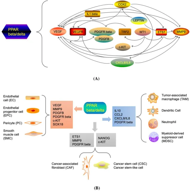
3.1. Cross-talk between PPAR beta/delta and signal molecules involved in
angiogenesis ………153
3.2. Role of PPAR beta/delta activation on the hallmarks of cancer ….157
3.3. Potential involvement of PPAR beta/delta in the tumor micro-
environment ………..161
1. Introduction
Chapter I
Angiogenesis
1.1. Normal Angiogenesis and Tumor Angiogenesis
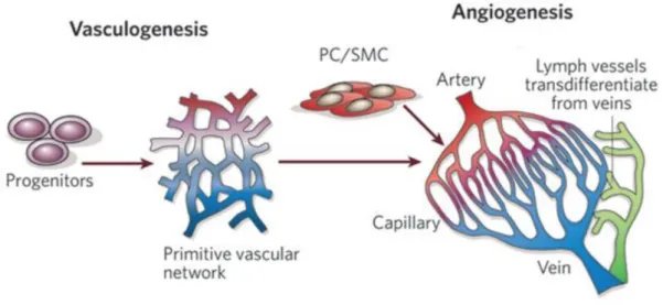
Angiogenesis is the physiological process through which new capillary networks can be formed from the pre-existing vasculature (Carmeliet, 2005; Carmeliet et al., 2011). This process is different from vasculogenesis which denotes de novo blood vessel formation (mostly during embryogenesis) in which endothelial progenitor cells migrate to sites of vascularization, differentiate into endothelial cells, and coalesce into the initial vascular plexus (Risau et al., 1995; Carmeliet et al., 2003). Angiogenesis refers to the development of new blood vessels from pre-existing capillaries.
Vasculogenesis, is the process of blood vessel formation in the embryo, occurring by a
de novo production of endothelial cells. Angiogenesis, is the formation of new capillary
networks from the pre-existing vasculature. PC: Pericyte; SMC: Smooth muscle cell
(Image modified from Carmeliet, 2005).
1.1.1. Molecular basis of angiogenesis
Apart from the interaction of pro-angiogenic factors and anti-angiogenic factors, angiogenesis is a cellular and molecular biological process involved with multiple mechanisms and multiple molecules including extracellular matrix (ECM) components, cell adhesion molecules (CAMs), matrix metalloproteinases (MMPs) and basement membrane (BM) components.
Angiogenesis occurs throughout life in both health and disease and relies on migration, proliferation, and differentiation of endothelial cells (ECs), which line the inside wall of blood vessels. During this process, the first step is the degradation of the vascular basement membrane, then vascular endothelial cells migrate into the surrounding matrix and proliferate and form solid sprouts connecting neighboring vessels.
Under the influence of various pro-angiogenic signals, such as growth factors and hypoxia, a fraction of previously quiescent vascular ECs can rapidly switch to an angiogenic state, and differentiate into specialized tip cells with a pro-migratory phenotype and vessel sprouting is initiated, whereas the following ECs, named as stalk cells, proliferate to elongate the branch, contribute to the structural integrity of nascent vessels, and form a vascular lumen. In this process, upon the stimuli of pro-angiogenic factors, such as VEGF (vascular endothelial
growth factor), ECs lose their adherence junctions, then matrix
which helps tip cells to extend and migrate around to form a long columnar filopodia, and guide the new sprout toward an angiogenic stimulus (De Bock et al., 2013; Potente et al., 2011). Upon formation of the new vessel, this undergoes stabilization and maturation, a process mainly mediated by intercellular adhesion and mural cell coverage including pericyte cells (PC) and vascular smooth muscle cells (SMC).
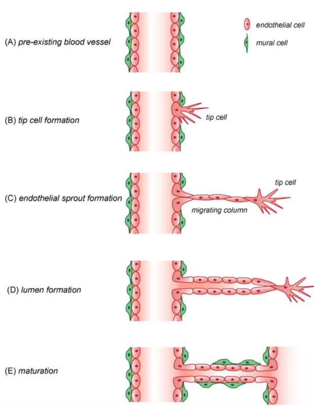
Figure 2. A schematic representation for the angiogenic process is shown. (A) Angiogenesis starts from pre-existing vessels. (B) Previously quiescent ECs switch to tip cells with migration ability. (C) Tip cells form columnar filopodia which probe the surrounding environment, which is followed by the migrating column formed by
proliferating ECs, the so-called stalk cells. (D) ECs sprouting and vascular lumen formation within stalk cells coalesce. (E) The newly forming vessel undergoes a process of stabilization and maturation that is mainly mediated by intercellular adhesion and mural cell coverage including PCs and SMCs (Image from Francavilla et al., 2009).
1.1.2. Tumor angiogenesis
Angiogenesis is a physiological and vital process in growth and development, in which pro-angiogenic factors and anti-angiogenic factors coordinate. Once the balance in the expression levels between pro-angiogenic and anti-angiogenic signal molecules is broken, causing angiogenesis in pathological conditions, such as diabetic retinopathy and tumor growth. For example, once the imbalance between pro-angiogenic and anti-angiogenic signaling molecules gradually comes to a point at which angiogenesis is triggered by tumor cells, then the so-called “angiogenic switch” of tumors is turned on (Bergers & Benjamin, 2003).
Angiogenesis is also a fundamental step by which most of benign tumors transit to malignant ones. When the growth and development of solid tumors
exceed 1 mm3, they need to develop rapidly a new vascular network to supply
nutrients, oxygen and immune cells, and also to remove waste products (McDougall et al., 2006). In this case, angiogenesis becomes an essential condition (Folkman, 1971; Figg & Folkman, 2008). Due to the abnormal structure and function of the vessels in the tumor tissue, and due to the fact that the vascular wall structure is imperfect in tumors, the microvasculature is prone to leakage. Thus, the tumor cells do not need to undergo a complicated invasion process and can directly penetrate into the blood vessels or lymphatic vessels to circulate through the intravascular system and then proliferate at another distant site, which represents the origin of metastasis formation (Folkman,1971). As a
result of the intensive angiogenesis, most malignant tumors grow rapidly and have the ability to spread to adjacent or distant organs, which makes it life threatening.
Normal vasculature tissue comprises organized ECs forming a monolayer and mural cells. However, unlike normal vasculature, tumor blood vessels are structurally and functionally highly abnormal. Probably due to the high levels of pro-angiogenic factors secreted by tumor and stromal cells, such as VEGF, tumor vasculature is immature and unstable. ECs lining tumor vessels are disorganized and hypermigrative, and are seperated by weak, loose and irregular inter-endothelial junctions. The structural abnormalities lead to tumor blood vessels hyperpermeability, vascular leakage, poor tumor perfusion and increased hypoxia. Moreover, hypoxia is a major driver of tumor neovascularization. More invasive and aggressive cancer cells are selected under the hypoxic microenvironment and can escape from the tumor-killing action of immune cells. In addition, mural cells also demonstrate structural abnormalities, as they are loosely associated with ECs or detached from ECs, or even absent. In such conditions, the tumor vessel abnormalities further facilitate invasive tumor cells to metastasize in distant organs. Furthermore, the abnormal tumor perfusion impedes the delivery of chemotherapeutic drugs to tumor itself, thus largely reducing the efficacy of anti-tumor therapies (Jain, 2005; De Bock et al., 2011; Carmeliet et al., 2011; Goel et al., 2011).
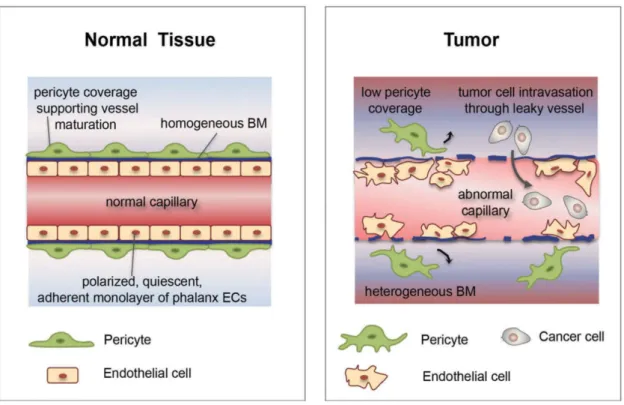
Figure 3. Tumor vessels are structurally and functionally abnormal.
Normal vasculature tissue comprises organizaed layers of ECs and pericytes. ECs line the inside wall of blood vessels, which are referred to as phalanx cells. Tumor vasculature is heterogeneous in size and shape, which is highly irregular and excessively branched. ECs lining tumor vessels are disorganized and are separated by wide and loose junctions. In addition, tumor vessels are covered by fewer pericytes which are detached from endothelial cells. This leads to tumor vascular leakage and increased permeability, therefore further facilitating invasive cancer cells to metastasize in distant organs (Image from Cantelmo et al., 2017).
1.2. Molecular Mechanisms in Tumor Angiogenesis
1.2.1. Vascular endothelial growth factor and its receptos (VEGF/VEGFR)
Pro-angiogenic factors include protein growth factors and steroids secreted by vascular endothelial cells, tumor and stromal cells. A common feature of these factors is the ability to initiate and promote angiogenesis. Vascular endothelial growth factor (VEGF), is the most important and most studied of all the pro-angiogenic factors which have been discovered and identified so far. The VEGF glycoproteins family comprises 6 members, VEGF-A, VEGF-B, VEGF-C, VEGF-D, VEGF-E and placenta growth factor (PIGF-1 and PIGF-2).
There also exist multiple isoforms of VEGF in tissues because of alternative pre-mRNA splicing of 8 exons. However, differential selection of terminal exon splice site determines the different fate of VEGFxxx isoforms, leading to 2 families of isoforms, the pro-angiogenic VEGF isoforms including VEGF121, VEGF165, VEGF189 and VEGF206, and the antiangiogenic isoforms such as VEGF121b, VEGF165b and VEGF189b. Among them, VEGF165 is in most angiogenic states, and is the most physiologically relevant and most frequently expressed isoform in tissues, with characteristics in between highly diffusible VEGF121 and ECM-bound VEGF189 (Harper et al., 2008; Ladomery et al., 2007).
VEGFs play different biological functions in embryonic development, physiological and pathological conditions, by binding to 3 different tyrosine kinases receptor (VEGFRs) including VEGFR1 (Flt1), VEGFR2 (Flk1/KDR) and VEGFR3 (Flt4). VEGFA plays the most important role during vasculogenesis and angiogenesis and stimulates directly ECs proliferation and migration (Nagy et al., 2007). During the angiogenic process, endothelial cells consist of tip cells in
leading stage and stalk cells in vascular proliferation. The direction of migration of tip cells and of microtube formation are determined based on the VEGF concentration gradient. VEGF gradients mainly regulate the formation of tip cells filopodia and proliferation of stalk cells in microtubule branches (Gerhardt et al., 2003; Hellstrom et al., 2007). In addition to, VEGFA can also upregulate the expression of matrix metalloproteases (MMPs) involved in the degradation of extracellular matrix (ECM) and in ECs migration (Bergers et al., 2000).
Activation of VEGF/VEGFR signaling is essential for vascular homeostasis, canonical VEGF signaling through binding of VEGFR1/VEGFR2 to regulate cell survival and guide cell proliferation and cell migration (Lee et al., 2007).
VEGFR2 as the dominant signal directly involved in pathological
neovascularization, plays the main role in promoting vascular permeability and ECs mitogenesis, which has therefore become an important target for drug therapy. Even though VEGF/VEGFR2 signaling is an important promoter of ECs function, it is also shown that VEGF/VEGFR2 plays a negative role in vessel maturation by inhibiting vascular smooth muscle cell (VSMC) function, and disrupting pericyte coverage (Greenberg et al., 2008).
In some conditions, VEGFR1 functions as an antagonistic receptor, which can prevent VEGFA binding to VEGFR2 (Park et al., 1994). But, VEGFR1 plays important role in haematopoiesis, which can recruit monocytes (Barleon et al., 1996) and other bone marrow-derived cells into the tumor microenvironment (TME) to promote tumor angiogenesis. VEGFR-3 is an important regulator of lymphangiogenesis. Lymphangiogenesis is a similar biological process as angiogenesis through which the formation of new lymphatic vessels occurs from existing lymphatic ECs. A study displayed that in human cancers, a strong
correlation between the levels of VEGFC/VEGFD expression and lymph node metastasis exists (Cao, 2005; Tobler & Detmar, 2006).
VEGF signaling also may participate in tumor vascularization by recruiting endothelial progenitor cells (EPCs) to sites of neovascularization in adults. Overexpression of VEGF has been linked to an aggressive phenotype in numerous cancers, such as breast cancer, which further contributes to tumor progression and poor prognosis in many carcinomas (Konecny et al., 2004; Foekens et al., 2001; Nakopoulou et al., 2002; Banerjee et al., 2007).
There exist various vascular inhibitors, including anti-VEGF antibodies (Ferrara et al., 2005), anti-VEGFR antibodies (Ellis et al., 2008), small molecule nucleic acids which specifically recognize VEGF, soluble VEGF receptors, and various small molecule inhibitors that target VEGF signaling pathway (Ferrara et al., 2005).
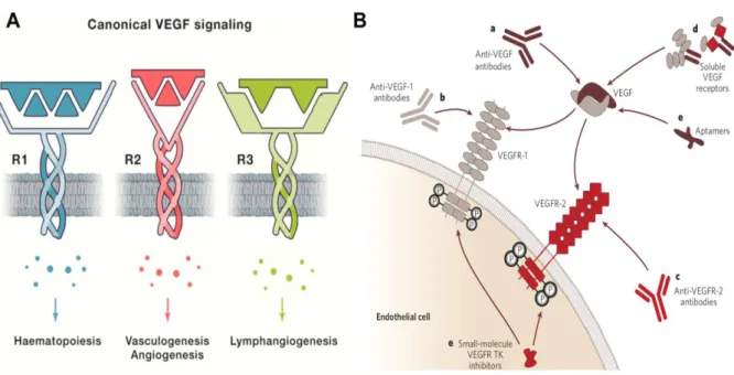
Figure 4. VEGF activation, and signaling pathways, and strategies to inhibit VEGF signaling.
A. VEGF binds to different receptors involved in various critical biological process, VEGFR2 being the dominant signaling pathway in physiological and pathological vasculogenesis and angiogenesis. B. There are various monoclonal antibodies targeting the VEGF-A (a), the VEGF receptors (b,c), and soluble VEGF receptors (d). Small molecule VEGF RTK inhibitors that inhibit ligand-dependent receptor autophosphorylation of VEGFR1 and VEGFR2, and aptamers that bind to the heparin-binding domain of VEGF165, such as pegaptanib (e) (Image A left from Apte et al.,
2019; Image B right from Ferrara et al., 2005).
1.2.2. Platelet-derived growth factor B and its receptor Platelet-derived growth factor beta (PDGFB/PDGFRB)
PDGF/PDGFR signaling pathway is essential for tumor angiogenesis. The PDGF (platelet derived growth factor) family comprises 5 members (PDGF-A, -B, AB, C and D), and there exist 3 types of PDGF receptors (PDGFRA, B and -AB). Many studies have shown that PDGF/PDGFR signaling plays various roles in cell growth, survival, cell proliferation and cell migration, therefore it has been implicated in different conditions and diseases including wound healing, angiogenesis and tumor (Sato et al., 1993; Raica & Cimpean, 2010).
PDGF especially plays a critical role in the maintenance and maturation of the newly forming vascular wall by promoting pericyte recruitment, which consequently regulates vessel permeability and tumor vessel maturation in tumor angiogenesis. Reduction of up to 90% of the pericyte coverage in mice results in structural and functional abnormalities in the microvasculature, threatening embryonic and postnatal survival (Enge et al., 2002). Previous studies have also shown that pericyte recruitment in developmental
angiogenesis depends on endothelium-derived PDGFB and PDGFB receptor expression on pericytes (Lindahl et al., 1997; Hellström et al., 1999).
Additionally, studies have demonstrated long before that mice deficient for PDGFB died perinatally and displayed several renal, cardiovascular and hematological abnormalities (Levéen et al., 1994; Soriano et al., 1994). Furthermore, in another in vivo study, a classical Pdgfbret/ret mouse model also
demonstrated that in mice transplanted into PDGFB retention motif deficient in the endogenous PDGFB protein, pericytes were fewer and were partially detached from the vessel wall in tumors, resulting in increased tumor vessel diameter and haemorrhage (Abramsson et al., 2003).
Thus, a defect in pericyte state caused by genetic deletion of PDGFB and PDGFRB leads to hemorrhage in early embryos, as a consequence of defective vascular development, and the formation of microvessels with many of the typical hallmarks of tumor vessels. The expression of PDGFRB is correlated with tumor progression and metastasis, and the inhibition of PDGFB/PDGFRB axis would theoretically diminish tumor development and invasion.
Furthermore, it is noteworthy that PDGF also can regulate indirectly angiogenesis by inducing VEGF secretion and transcription (Wang et al., 1999). However, interestingly, it has been also demonstrated that in the presence of both VEGF and PDGF, VEGF-R2 can limit pro-angiogenic signals. Under conditions of PDGF-mediated angiogenesis, VEGF can inhibit the pericyte coverage of newly forming vessels, blood vessel maturation, and disrupt VSMCs function, leading to vessel destabilization. VEGF-mediated activation of VEGFR2 can suppress the PDGFRB signaling in VSMCs through the induction of the
assembly of a receptor complex consisting of PDGFRB and VEGFR2. Additionally, deletion of VEGF in tumor cells can also disrupt the PDGFRB/VEGFR2 complex formation, and then increase tumor vessel maturation (also mentioned above in VEGF part, Greenberg et al., 2008). Hence, there exists a specific antagonistic relationship between PDGF and VEGF during neoangiogenesis.
Importantly, previous studies have shown that PDGFBB can promote endothelial cell proliferation and tube formation and angiogenesis in vitro through specifically binding to PDGFR- beta, thus suggesting that PDGF may mediate amplified angiogenesis by directly activating PDGFR- beta expressed on endothelial cells (Battegay et al., 1994). In fact, it was initially reported that upregulation of PDGFR beta expression in vascular endothelial cells was associated with a malignant glioma phenotype, indicating its potential involvement in angiogenesis (Hermansson et al., 1988).
Furthermore, genetic ablation of Pdgfb in endothelial cells in vivo resulted in organ defects in development including cardiac, placental and renal abnormalities similar to those observed in complete Pdgfb null mice (Bjarnegard et al., 2004). However, interestingly, in contrast to the embryonic lethality in Pdgfb null mice, most of the endothelium- specific Pdgfb knockout mice can survive until into adulthood with persistent microvascular pathologies such as brain microhemorrhages and kidney glomerulus abnormalities, clarifying the role of endothelial- derived PDGFB expression during development and possibly providing a model to address a role of pericytes in the microvasculature of adult mice (Bjarnegard et al., 2004). These observations also suggest that mice can tolerate up to 90% pericyte loss and survive into adulthood without severe organ
dysfunction but die during embryonic stage due to the more extensive pericyte loss (Bjarnegard et al., 2004).
Moreover, the overexpression of PDGFR beta has been shown to promote PDGF- induced endothelial progenitor cells (EPCs) proliferation, migration and angiogenesis via activation of PI3K/Akt signaling pathway (Wang et al., 2012). In fact, it has been previously demonstrated that the interaction between PDGFBB and PDGFR beta can induce PDGFR phosphorylation and activate PI3K (Heldin et al., 1998). And PI3K signal cascade activation can promote various cellular process including cell survival, proliferation and angiogenesis (Cantley, 2002). PDGF receptor signal transduction has been suggested to be involved in various tumor- associated processes such as tumor angiogenesis and tumor fibroblasts recruitment and regulation (Ostman, 2004). PDGFR beta has been shown previously to promote early endothelial cell differentiation and regulate vascular or hematopoietic development (Rolny et al., 2006). Subsequently, it was shown that PDGFR beta constitutive activity promoted angiogenesis in vivo and in vitro
(Magnusson et al., 2007). Moreover, teratomas generated from Pdgfrb D849V/+ ES
cells in nude mice were more densely vascularized compared with wild-type ES cells- derived teratomas. And it was also suggested that the increased PDGFR beta kinase activity is associated with elevated expression of VEGFA and VEGFR2, directly acting on ECs and leading to enhanced vessel formation (Magnusson et al., 2007).
Another study also firstly described an interplay between angiogenic factors
FGF2 and PDGF-BB that synergistically promoted murine tumor
neovascularization and metastasis (Nissen et al., 2007). The interaction between different angiogenic factors in the tumor microenvironment (TME) may result in
the formation of disorganized and primitive vasculatures that further facilitate tumor growth and metastasis in mice (Nissen et al., 2007; Cao et al., 2008). And subsequently, it was further shown that FGF2 induced PDGFR beta expression in the newly forming blood vessels, and PDGFR beta receptor was suggested to be required for vascular stability (Zhang et al., 2009). It has also been shown that PDGFR beta can stimulate proliferation, migration and tube formation of differentiated ECs (Wyler von Ballmoos et al., 2010).
PDGF-BB was shown to induce EPO expression that promoted tumor growth and tumor angiogenesis through direct induction ECs proliferation, migration and sprouting and tube formation. PDGF-BB can directly act on stromal cells, mural cells such as pericytes or VSMCs that express its receptor PDGFR beta to activate EPO expression, leading to enhanced tumor angiogenesis (Xue et al., 2011). The telomeric protein TRF2 has been demonstrated to regulate angiogenesis via direct transactivation of PDGFR beta (EI Maï et al., 2014). This also further indicates the important role of PDGFBB and PDGFR beta in promotion of neovascularization and re-endothelialization via stimulation on EPCs (Wang et al., 2012). Ovarian PDGFB administration has been shown to patially restore follicular development and improve ovarian angiogenesis in a rat model with PCOS (polycystic ovary syndrome) (Di Pietro et al., 2016).
Most recently, a study has shown that lymphatic endothelial cells (LEC)- specific deletion of Pdgfb in vivo can prevent smooth muscle cell (SMC) recruitment to dermal collecting lymphatics, resulting in vessel dilation (Wang et al., 2017). PDGFB has been considered to be required for SMC recruitment to
large diameter vessels, but LEC- specific overexpression of Pdgfb is not sufficient to induce SMC recruitment to lymphatic capillaries (Wang et al., 2017).
Moreover, in various tumor types, PDGF receptors exert different cell type-specific effects. They are directly involved in activation of malignant cell growth and tumor progression in many tumor types. PDGFRB as a key regulator in TME and some common malignancies, such as breast cancer, whose expression in stroma indicates prognostic significance in patients with cancer. In patients with breast cancer, high stromal PDGFRB expression is associated with poor prognosis, and high levels of PDGF/PDGFRs in tumor tissue predict low response to chemotherapy treatment and shortened survival (Paulsson et al., 2009; Seymour et al., 1993).
PDGF and its receptor may also affect cancer growth and progression by directly acting on TME. And at this point, some studies have focused on the role of PDGF receptor signals in cancer-associated fibroblasts (CAFs) that are responsible for deposition and remodeling of ECM components in tumor stromal matrix (Gialeli et al., 2014). CAFs, as primary or metastatic tumor stroma, is an important component in tumor formation and progression (Shan et al., 2017). It has been shown that PDGF-activated CAFs upregulates the expression of the pro-apoptotic protein Puma, which causes Puma- dependent binding of Bak to Bcl-2 and the pro-apoptotic change of Bak, leading to enhanced cell apoptosis in
vivo (Rizvi et al., 2014).
In addition, PDGF as important growth and survival factor, has been suggested to be involved in the CAF- cancer cell crosstalk which may affect the radiation protective effects of cancer cells (Chu et al., 2014). Interestingly, Yang
macrophages (TAMs) recruitment in TME as no detectable PDGFR expression was observed in monocytes and macrophages. And PDGFR beta primarily expressed perivascular cells and stromal fibroblasts has been considered as the crucial receptor to mediate PDGF-BB-recruited TAMs, implicating a key role of receptor PDGFR beta in tumor stroma to promote metastasis (Yang et al., 2016).
Lu et al. demonstrated that PDGF-BB expression was increased
significantly in HepG2 cells under hypoxic conditions. It has been suggested that hypoxia- induced HCC-derived PDGF-BB stimulates the accumulation and proliferation of HSCs in the tumor stroma, and enhances VEGFA expression in HSCs, possibly leading to the promotion of angiogenesis in HCC (Lu et al., 2015). In fact, PDGF-BB has also been previously reported to affect the angiogenic properties of HSCs during liver fibrosis (Jia et al., 2011). Most recently, PDGFR beta has also been indicated to promote the migration and invasion of human pterygial fibroblasts via activation of the ERK/MMP-1 signaling pathway (Mai et al., 2019). Moreover, it has been further suggested that specific targeting of PDGFR beta kinase activity in the TME can inhibit tumor growth and vascularization in tumors with high PDGF-BB expression such as LLC and B16/PDGF-BB (Tsioumpekou et al., 2020).
Based on the evidence provided by many previous studies, PDGF/PDGFR has undoubtedly become a valid anti-tumor target in drug development. PDGFR inhibitors and PDGFR-targeting monoclonal antibodies have been developed in some clinical studies. However, further development will be still essential to improve response to drug treatment and resistance of PDGFR-directed cancer therapies.
In conclusion, given the crucial role of PDGFB/PDGFRB in endothelial cells, EPCs, and pericyte and SMC recruitment, PDGFB/PDGFRB has been suggested to be involved in neovascularization as key angiogenic factors.
1.2.3. Basic fibroblast growth factor (bFGF)
Basic fibroblast growth factor (bFGF/ FGF2/ FGFB) is encoded by the FGF2 gene, which belongs to the FGF proteins family (Kim,1998). bFGF, first identified in mouse pituitary extracts, can promote mouse 3T3 cell growth (Gambarini & Armelin,1982). bFGF, located in sub-endothelial ECM and BM of blood vessels, functions as angiogenic peptides and broadly regulates cell survival, cell proliferation and migration (Montesano et al., 1986). Additionally, bFGF can stimulate ECM degradation, ECs proliferation and migration in the angiogenic process (Presta et al., 2005).
bFGF can also directly regulate angiogenesis by binding to its receptor, hence promoting subsequent biological processes such as wound repair. Importantly, it has been reported that bFGF is overexpressed in many tumor types and promotes tumor progression and metastasis (Figg & Folkman, 2008). High levels of bFGF are also correlated largely with the extent of disease and clinical status, and correlate with poor prognosis in patients with cancer. And it has been shown that the treatment with anti-bFGF drugs can prevent the tumor formation. Thus, bFGF-based cancer therapy may be another desirable therapeutic approach (Nguyen et al., 1994; Hu et al., 2016).
1.2.4. Cell adhesion molecules (CAMs)
Cell adhesion molecules (CAMs) are glycoproteins on the cell surface which mediate the interactions between adjacent cells and between cells and the surrounding extracellular matrix (ECM). The adhesion interactions of cell-to-cell and cell-to-ECM play a key role in angiogenesis, and members of CAMs are strongly involved in the each step of tumor vascularization by mediating the adhesion interactions and participating in related intracellular signaling pathways involved in the maturation of newly forming vessels (Stromblad et al., 1996; Ramjaun et al., 2009).
Under the stimuli of pro-angiogenic factors, the functional state of ECs will change and some of them will acquire the specialized properties to convert into endothelial tip cells and/or stalk cells. Tip cells are the main leaders in the process of vessel sprouting, which are crucially involved in the degradation of BM, changes in adhesive properties of ECs and ECs migration (Hillen et al., 2007). Changes in the ECs adhesive capacity are mainly due to the dynamic regulation of CAMs involved in multiple angiogenic steps. In addition, ECs also need to degrade the ECM (Folkman,1971), then proliferate, arrange themselves and migrate into the perivascular space. During this initial phase, ECs need to adhere to one to another and to the ECM to construct and extend the primary neo-forming vessels.
Cell adhesion molecules include four families: integrins, cadherins, immunoglobulin superfamily members and selectins. Among them, integrins are the major members of CAMs responsible for adhesion interactions between cells and the ECM. Apart from helping cell anchor to the matrix, integrins can also connect to the cell cytoskeleton. Hence, the integrin binding indeed plays a
critical role in a variety of cellular biological process, such as cell adhesion, cell migration and cell cytoskeletal organization (Karimi et al., 2018).
Figure 5. Functional contribution of CAMs to each step of tumor angiogenesis.
(a) The angiogenic process begins with a pre-existing blood vessel in which quiescent ECs are converted into specialized tip cells. (b,c) Tip cell formation and Vascular sprout formation. The tip cells, less proliferative, extend filopodia and migrate away from its parent vessel to form a new vascular sprout. (c,d) Vascular sprout formation and tubulogenesis. Stalk cells, highly proliferative, consist in the neck length of the neo-vascular sprout, and undergo a process of vacuole formation and fusion to form the vessel lumen, namely tubulogenesis.
(e) Anastomosis. The tip cells eventually encounter the tips from other adjacent vascular sprouts, leading to the anastomosis formation where cell-to-cell adhesive processes facilitate the intercellular connection at the joining point, finally which results in a new vessel formation. (f) A vascular bed formation. Angiogenic processes occurring in parallel leads to formation of a vascular plexus. (g) Vascular remodelling. The primary vascular plexus eventually is pruned to optimize blood flow. The related CAMs and potential signal events have been implicated directly in each adhesive step during the angiogenic process (Ramjaun & Hodivala-Dilke, 2009). (Image from Ramjaun &
Hodivala-Dilke, 2009)
Integrins
Integrins play a critical role in tumor angiogenesis, tumor progression and metastasis. The adhesion interactions between ECs and ECM proteins, are mainly mediated by integrins expressed on cell surface (Weis, 2007). Integrins can bind to ECM proteins specifically such as collagen, laminin, fibronectin, and vitronectin. Integrins have been implicated in both angiogenesis and vasculogenesis. Previous studies have demonstrated that endothelial -specific knockout of various integrin α and β subunits results in embryonic lethality,
indicating the important role of integrins in angiogenesis, therefore called the “key” to switch on angiogenesis (Giancotti & Ruoslahti, 1999). The interactions with ECM proteins can promote integrin clustering and subsequent integrin-mediate intercellular signal transduction pathways. Integrin binding has become an essential step in multiple intracellular signaling pathways (Humphries et al., 2006; Hynes, 2002). Integrins have been identified as a common target in cell-specific adhesion where they promote different cellular functions. Most recently, several reviews have shown the details that integrins can activate the principal signal transduction pathways relevant in cancer initiation and tumor angiogenesis, tumor progression and metastasis, including the the Rho family GTPases, phosphoinositid-3-kinase (PI3K)/AKT, the Rat Sarcoma (RAS)-mitogen activated protein kinases (MAPKs) and the Nuclear Factor kappa B (NF-κB) pathways (Thorpe et al., 2015; Montor et al., 2018; Tang et al., 2018; Yaeger & Corcoran, 2019; Cooper & Giancotti, 2019). But there are still some previous studies that need to be elaborated as below.
Focal adhesions (FAs/ cell-matrix adhesions) are the principle structures in promoting the interaction of cells with the ECM (Petit & Thiery, 2000; Burridge et al., 1988), as the important signal transduction centers, through which integrins can transduce signals from the ECM to regulate cell survival, cell growth and invasion (Wozniak et al., 2004). Focal adhesion kinase (FAK) locates in focal adhesions structures and plays a crucial role in integrin-mediated signal transductions, which also is involved in signaling pathways by other cell surface receptors (Zhao & Guan, 2011). Cancer cell metastasis is a dynamic and multistep process in which turnover of focal adhesions is an essential and necessary step (Ridley et al., 2003; Lauffenburger & Horwitz, 1996). There exists
excessive activation of FAK in multiple human cancers (Cohen & Guan, 2005; Golubovskaya & Cance, 2007; McLean et al., 2005). Active FAK can promote cancer cell migration through several downstream signaling pathways such as Src, or PI3K mediated pathways (Zhao & Guan, 2011).
Integrins can interact with focal adhesion kinase (FAK) or non-receptor tyrosine kinase Src through the intracellular domain to regulate the Rho A signaling pathway. It can also be linked to the cytoskeleton via binding proteins such as actinin (Lo, 2006; Mitra & Schlaepfer, 2006). Integrin β1 subunit can directly activate FAK through the cytoplasmic tail domain (Schaller et al., 1995). Studies have demonstrated that endothelial specific deletion of FAK resulted in a mid-late embryonic lethal phenotype due to largely impaired blood vessel development (Shen et al., 2005). Src is one of the targets of FAK that functions during angiogenesis. The integrin-induced Src pro-angiogenic activity has been observed in ECs (Courter et al., 2005; Zaric & Ruegg, 2005). Because VEGF receptor-mediated signaling can also activate Src and induce the interaction between FAK and integrins through the phosphorylation of FAK (Eliceiri et al., 2002), which makes the signal transduction of FAK-Src even more complex.
PI3-kinase is another key target of FAK in ECs. A previous study has shown that embryonic death between E9.5 and E10.5 can be induced in mice due to the deletion of PI3-kinase (Bi et al., 1999). And It has been also demonstrated also that an endothelial-specific deletion of PI3-kinase isoform can largely reduce ECs migration and impair angiogenesis (Graupera et al., 2008). In
addition, PI3-kinase also plays a key mediator function during
lymphangiogenesis. It has been demonstrated also that there was a developmental lymphangiogenic defect caused by the breakdown of the
interaction between PI3-kinase and its activator, Ras (Gupta et al., 2007). Integrins-induced PI3-kinase activity functions through the well-established AKT-mTOR-4E-BP1 pathway and controls cap-dependent translation, which plays a key role in ECs survival and proliferation (Sudhakar et al., 2003). Forkhead box protein O1 (FOXO1), a downstream target of the PI3-kinase/AKT signaling pathway, has also been shown to be involved in angiogenesis. FOXO1, globally deleted in mice, can lead to embryonic lethality at E11 due to impairment of the vascular development (Furuyama et al., 2004). The activity of PI3-kinase induced can lead to the activation of PLC-γ/PKC cascade in addition to activation of Rac1 and NF-kB (Tzima et al., 2002; Sasamoto et al., 2005).
Another important signal transduction involved in angiogenesis is the ERK/ MAP pathway. The activation of RAF, MEK and ERK can be induced indirectly by integrin combination to fibronectin (Sudhakar et al., 2003), and it also has been reported that integrin can directly activate ERK (Roberts et al., 2003), which initiates the cascade of ERK signaling pathway. A previous study has shown that MEK1-deficient mice died at E10.5 due to extensive developmental vascular defects (Giroux et al., 1999). FAK may also play a role in associating integrin activity with growth factor-induced ERK signaling (Hood et al., 2003). Currently, it has been shown that the activation of the RAS/RAF/MEK/MAPK cascade can promote cancer cell survival, proliferation, invasion and angiogenesis by regulating gene transcription (Martinelli et al., 2017). MEK1/2 has been considered as a transducer of the growth factor receptor (such as EGFR) /RAS/ RAF/ MAPK signaling cascade, which is related to the development and progression of human cancers (Martinelli et al., 2017).
Integrins are formed by heterodimers composed of alpha-subunits and beta-subunits. The important involvement of αvβ integrins in angiogenesis was elucidated firstly in 1994 (Brooks et al., 1994). Currently, it has been observed that there exists a differential expression of integrins in cancer cells and the
healthy tissues, especially of α3β1, α4β1, αvβ5, αvβ6, αvβ8, α6β4, α5β1 and
αvβ3 (Nieberler et al., 2017). Integrins implicated in tumor angiogenesis mainly includes αvβ5, α2β1, αvβ3 and α5β1. Currently, many experimental studies have demonstrated that the inhibition of α1β1, α2β1 and α5β1 can suppress tumor angiogenesis and reduce tumor growth (Ruegg & Alghisi, 2010; Jahangiri et al., 2014).
Among of the many integrins identified, αvβ3 was the first integrin reported
to be preferentially expressed in angiogenic endothelial cells (Brooks et al., 1994), subsequently considered to be strongly involved in angiogenesis. Integrin αvβ3 was selectively expressed on tumor vessels but not on quiescent blood vessels in normal cells. Integrin αvβ3 can interact with matrix metalloproteinase-2 (MMPmetalloproteinase-2) (Brooks et al.,1996) to promote tip cells migration, which plays an important role in the initiation of tumor angiogenesis. It has been shown also that integrin αvβ3 expressed on lymphokine-activated killer cells can bind to platelet endothelial cell adhesion molecule (PECAM1/ CD31) (Piali et al.,1995), a member of the immunoglobulin superfamily that plays various roles in cell
adhesion and angiogenesis. Integrin αvβ3 as the receptor of ECM component
vitronectin, mediates the adhesion of ECs-to-ECM to promote ECs survival. It
has been shown that the binding of αvβ3 to vitronectin can protect ECs from
apoptosis through the activation of the FAK/ PI3K/ AKT signaling cascade, ERK and NF-kB signaling pathways (Scatena et al., 1998; Wang & Milner, 2006).
Apart from angiogenesis, integrin αvβ3 also can be expressed in many cell types such as tumor cells, leukocytes, fibroblasts and smooth muscle cells, and may bind to different ligands in many settings hence to mediate different biological processes.
Integrin α5β1, the fibronectin receptor, is another key player in ECs adhesion signal transduction. It has been shown that α5β1 is up-regulated during angiogenesis (Kim et al., 2002), which can activate intracellular signal transduction and protect ECs from apoptosis, and further regulate ECs proliferation and migration. Studies in mice also demonstrated that antagonists
of α5β1 can inhibit angiogenesis further leading to tumor regression (Kim et al.,
2000). Integrin α5β1 can also activate the FAK/RAF/MEK/ERK signal
transduction and the P38 signaling pathways (Sudhakar et al., 2003; Wang & Milner, 2006). The deletion of relevant elements of the P38 pathway caused developmental vascular defects in an in vivo mouse model (Mudgett et al., 2000). As well as the αvβ3, α5β1 can also regulate ECs proliferation via PI3kinase and
ERK signaling pathways (Wilson et al., 2003). α5β1 has been considered to be
involved in the vascular remodeling and maturation of newly forming vessels
(Ramjaun & Hodivala-Dilke, 2009). Additionally, integrin α1β1 and α2β1 have
been shown to be upregulated by VEGF-mediated during early phases of tumor angiogenesis (Hong et al., 2004). Their binding to both collagens and laminins, can protect ECs from apoptosis and enhance EC proliferation through growth factor induced ERK signaling (Perruzzi et al., 2003; Senger et al., 2002). Integrin αvβ5 binding to vitronectin, can promote ECs survival and proliferation also again via the growth factor-activated ERK pathway (Perruzzi et al., 2003), and it can also enhance ERK signal transduction downstream of the VEGF receptor
(Hood et al., 2003). Integrin α6β4, the laminin-5 receptor, has also been involved in angiogenesis through the activation of the ERK and NF-kB signaling pathways to promote ECs migration (Nikolopoulos et al., 2004). Finally, even if integrin-mediated cell-cell and cell-ECM adhesion play an extremely important role in tumor angiogenesis, the other CAMs also function as a necessary part.
Cadherins
The cadherin family are calcium-dependent CAMs responsible for cell-to-cell recognition and cell-to-cell-to-tissue adhesion (Yagi & Takeichi, 2000; Gumbiner, 2005). The classic cadherins, calcium-dependent homophilic adhesion
molecules, have been considered to be linked to specific junctional structures,
namely “adherens junctions” (AJs) (Niessen & Gottardi, 2008). Cadherins
anchoring to the actin cytoskeleton can stabilize the junctional structure, maintain cell morphology and control cell motility (Francavilla et al., 2009). The expression of cadherins is tissue specific. E-cadherin is mostly expressed in epithelial cells, N-cadherin in ECs, smooth muscle cells, fibroblasts and the nervous system (Gumbiner, 2005; Cavallaro et al., 2006). Vascular endothelial (VE)-cadherin, also known as cadherin-5 (CDH5/ CD144), is an endothelium-specific adhesion molecule located at adherens junctions in endothelial cells and plays an important role in regulating vascular permeability, which is also a potential target for tumor angiogenesis (Dejana et al., 2008). VE-cadherin can associate with VEGFR2 and modulate its signaling pathways. It has been shown that the absence of VE-cadherin can cause endothelial cell growth regulated by VEGFR2 in an uncontrolled way, affecting the process of vascular development (Grazia-Lampugnani et al., 2003). The interaction between VE-cadherin and the
cytoskeleton can mediate strong cell–cell adhesion (Dejana et al., 2008). Many experimental studies have suggested the potential involvement of VE-cadherin in
tumor angiogenesis, and the specific antibodies have long been shown to
suppress cancer vascularization and tumor growth (Corada et al., 2002; Liao et al., 2002).
It is important to note that E-cadherin, also known as CDH1, has a crucial role in epithelial cell behavior and cancer suppression (Van Roy & Berx, 2008). N-cadherin is commonly found in cancer cells and provides a trans-endothelial mechanism in EMT process. Loss of function of E-cadherin has been largely linked to cancer cell invasion and tumor metastasis (Beavon, 2000; Cheung et al., 2013).
Figure 6. A. Schematic representation of VE-cadherin-mediated endothelial cell junctions.
p120, p120-catenin; α, α-catenin; β, β-catenin. VE-cadherin can mediate intercellular adhesion through trans-interactions formed by its extracellular domain, its cytoplasmic domain anchoring to the actin cytoskeleton via α- and β-catenins, which leads to the stabilization of VE-cadherin at cell-cell junctions. The cytoplasmic tail of VE-cadherin binding to p120-catenin and β-catenin, β-catenin further interacts with α-catenin, which associates with the actin cytoskeleton (Image from Rho et al., 2017).
B. Signal transduction pathways involved in VEGF-mediated phosphorylation and increased vascular permeability.
VEGF stimulating VEGFR2 can induce vascular permeability through activation of non-receptor tyrosine kinase Src. T cell-specific adaptor (TSAd) binding to phosphor-Y951 domain within VEGFR2 subsequently can recruit and activate Src. Activated Src can directly phosphorylate VE-cadherin and indirectly initiate a Vav2/Rac/PAK signalling cascade to increase vascular permeability. Additionally, VEGF/VEGFR2 signal transduction can also induce activation of FAK that can further phosphorylate β-catenin. The phosphorylation of VE-cadherin and its associated catenins can cause dissociation of the VE-cadherin/catenin complex, which finally leads to the increased vascular permeability (Rho et al., 2017) (Image from Rho et al., 2017).
Immunoglobin-like cell adhesion molecules
Ig-CAM superfamily mainly includes endothelial cell specific adhesion molecule (ESAM), junctional adhesion molecule (JAM), intercellular adhesion molecule (ICAM), carcinoembryonic antigen related cell adhesion molecule (CEACAM), vascular cell adhesion molecule (VCAM), platelet endothelial cell adhesion molecule (PECAM) and melanoma cell adhesion molecule (MCAM/
CD146). Ig-CAMs have been implicated in tumor angiogenesis, however, the extent of involvement in tumor progression and metastasis still remains unclear and needs to be further elucidated.
MCAM (CD146/ MUC18) was initially identified as a marker of melanoma progression, and also shown to play a key role in melanoma progression and metastasis (Shih et al.,1994). It was also reported in the same year that MCAM was overexpressed in tumor blood vessels and its upregulation largely correlated to tumor angiogenesis. It has been also shown that expression of CD146 not only exists on melanoma cells but also on the vascular ECs of primary and metastatic melanomas, indicating a complex role of CD146 involved in tumor angiogenesis, tumor progression and metastasis (Sers et al., 1994). Later, a study in 2003 further demonstrated that MCAM may exert a critical role in angiogenesis of multiple tumor types and it had been clearly identified as a target for inhibition of tumor angiogenesis (Yan et al., 2003). MCAM has also been shown to inhibit EC proliferation, adhesion and migration, implicating its potential involvement in the initial steps of angiogenesis and a crucial role in vascular EC behaviour (Kang et al., 2006). MCAM has been implicated in several oncogenic signal transduction pathways including PI3K/AKT (Li et al., 2003), VEGF/VEGFR (Jiang et al., 2012; Jouve et al., 2015) and NF-kB (Zheng et al., 2009), which is a key driver in tumor progression and metastasis. MCAM can also promote tumor angiogenesis and metastasis by increasing the levels of VEGF, VEGFR2 and CD31 expression (Wu et al., 2011). It has been found to promote the epithelial-mesenchymal transition (EMT) process (Luo et al., 2012; Zeng et al., 2012), an initial step for invasion and metastasis of malignancies through regulating the cytoskeleton remodelling. Overexpression of MCAM has
been shown to promote EMT and to activate AKT signalling pathway, and high levels of MCAM expression are largely correlated with poor prognosis of patients with breast cancer (Liang et al., 2017).
Selectins
Selectins have been considered to promote tumor metastasis, and to contribute to tumor progression (Borsig, 2018; Läubli & Borsig, 2010). Selectins are vascular cell adhesion receptors mainly including P-selectins, L-selectins and E-selectins. P-selectin is expressed on activated platelets and ECs, L-selectin on leukocytes and E-L-selectins on activated ECs (McEver, 2002). Major carriers of selectin ligands are mucins correlated with tumor progression that have been used as diagnostic markers in cancer (Hollingsworth & Swanson, 2004), including MUC1.MUC2, MUC4, MUC16 and MUC18. Tumor cells carrying selectin ligands can enter into the blood circulation and then encounter the selectins on the leukocytes, platelets and ECs (Läubli & Borsig, 2010). It has been shown that the absence of P-selectins can weaken tumor metastasis (Borsig et al., 2001). E-selectins has also been shown to be involved in tumor cell extravasation through loosening the VE-cadherin junctions in endothelium (Hauselmann et al., 2016). More recently, a study has also demonstrated that E-selectins in the bone vascular niche can facilitate metastasis by the induction of mesenchymal-epithelial transition (MET) of breast cancer cells and the activation of Wnt signal transduction (Esposito et al., 2019). In conclusion, cell adhesion is a critical step in tumor angiogenesis, and/or directly plays an important role in tumor progression and metastasis.