HAL Id: hal-00675122
https://hal.archives-ouvertes.fr/hal-00675122
Submitted on 26 Apr 2012
HAL is a multi-disciplinary open access
archive for the deposit and dissemination of
sci-entific research documents, whether they are
pub-lished or not. The documents may come from
teaching and research institutions in France or
abroad, or from public or private research centers.
L’archive ouverte pluridisciplinaire HAL, est
destinée au dépôt et à la diffusion de documents
scientifiques de niveau recherche, publiés ou non,
émanant des établissements d’enseignement et de
recherche français ou étrangers, des laboratoires
publics ou privés.
Organization of the Photosynthetic Apparatus in
Chlamydomonas.
André Nordhues, Mark Aurel Schöttler, Ann-Katrin Unger, Stefan Geimer,
Stephanie Schönfelder, Stefan Schmollinger, Mark Rütgers, Giovanni Finazzi,
Barbara Soppa, Frederik Sommer, et al.
To cite this version:
André Nordhues, Mark Aurel Schöttler, Ann-Katrin Unger, Stefan Geimer, Stephanie Schönfelder, et
al.. Evidence for a Role of VIPP1 in the Structural Organization of the Photosynthetic Apparatus
in Chlamydomonas.. The Plant cell, American Society of Plant Biologists (ASPB), 2012, 24 (2),
pp.637-59. �10.1105/tpc.111.092692�. �hal-00675122�
Evidence for a Role of VIPP1 in the Structural Organization of
the Photosynthetic Apparatus in Chlamydomonas
W OAAndre´ Nordhues,
aMark Aurel Scho¨ttler,
aAnn-Katrin Unger,
bStefan Geimer,
bStephanie Scho¨nfelder,
a,cStefan Schmollinger,
aMark Ru¨tgers,
aGiovanni Finazzi,
dBarbara Soppa,
bFrederik Sommer,
aTimo Mu¨hlhaus,
aThomas Roach,
eAnja Krieger-Liszkay,
eHeiko Lokstein,
cJose´ Luis Crespo,
fand Michael Schroda
a,g,1aMax-Planck-Institut fu¨r Molekulare Pflanzenphysiologie, D-14476 Potsdam-Golm, Germany bZellbiologie/Elektronenmikroskopie, Universita¨t Bayreuth, 95440 Bayreuth, Germany
cInstitut fu¨r Biochemie und Biologie/Pflanzenphysiologie, Universita¨t Potsdam, D-14476 Potsdam-Golm, Germany
dLaboratoire de Physiologie Cellulaire et Ve´ge´tale, Unite´ Mixte de Recherche 5168 Centre National de la Recherche Scientifique/Commissariat a` l’Energie Atomique et aux E´nergies Alternatives/Universite´ Joseph Fourier, Commissariat a` l’Energie Atomique Grenoble, 38054 Grenoble, France
eCommissariat a` l’Energie Atomique Saclay, iBiTec-S, Centre National de la Recherche Scientifique Unite´ de Recherche Associe´e 2096, Service de Bioe´nerge´tique, Biologie Structurale et Me´canisme, 91191 Gif-sur-Yvette cedex, France
fInstituto de Bioquı´mica Vegetal y Fotosı´ntesis, Consejo Superior de Investigaciones Cientı´ficas-Universidad de Sevilla, 41092 Seville, Spain
gFachbereich Biologie, Molekulare Biotechnologie und Systembiologie, Technische Universita¨t Kaiserslautern, 67663 Kaiserslautern, Germany
The vesicle-inducing protein in plastids (VIPP1) was suggested to play a role in thylakoid membrane formation via membrane vesicles. As this functional assignment is under debate, we investigated the function of VIPP1 in Chlamydo-monas reinhardtii. Using immunofluorescence, we localized VIPP1 to distinct spots within the chloroplast. In VIPP1-RNA interference/artificial microRNA cells, we consistently observed aberrant, prolamellar body-like structures at the origin of multiple thylakoid membrane layers, which appear to coincide with the immunofluorescent VIPP1 spots and suggest a defect in thylakoid membrane biogenesis. Accordingly, using quantitative shotgun proteomics, we found that unstressed vipp1 mutant cells accumulate 14 to 20% less photosystems, cytochrome b6f complex, and ATP synthase but 30% more light-harvesting complex II than control cells, while complex assembly, thylakoid membrane ultrastructure, and bulk lipid composition appeared unaltered. Photosystems in vipp1 mutants are sensitive to high light, which coincides with a lowered midpoint potential of the QA/QA2 redox couple and increased thermosensitivity of photosystem II (PSII), suggesting structural defects in PSII. Moreover, swollen thylakoids, despite reduced membrane energization, in vipp1 mutants grown on ammonium suggest defects in the supermolecular organization of thylakoid membrane complexes. Overall, our data suggest a role of VIPP1 in the biogenesis/assembly of thylakoid membrane core complexes, most likely by supplying structural lipids.
INTRODUCTION
The thylakoids of chloroplasts represent an internal membrane system that is detached from the inner envelope membrane. As thylakoid lipids are not synthesized at the thylakoid membranes but rather at the chloroplast inner and outer envelope mem-branes and the endoplasmic reticulum, a transport system must exist that enables a flow of lipid components from these biogenic membranes to the thylakoids (Benning, 2008, 2009). Ultrastruc-tural studies suggested lipid transport via vesicles that bud off
from the inner envelope and fuse with the thylakoids (Carde et al., 1982). The M30 protein was identified in pea (Pisum sativum) as a plastid protein that is associated with both envelope and thyla-koid membranes and therefore was suggested as a candidate protein for lipid transfer from envelope to thylakoids (Li et al., 1994). M30 was renamed vesicle-inducing protein in plastids1 (VIPP1) based on the phenotype observed in the Arabidopsis
thaliana hcf155 mutant, which expresses M30/VIPP1 to
;
20% of wild-type levels: hcf155 plants have much less and distorted thylakoids and reduced amounts of photosystem I (PSI), photo-system II (PSII), light-harvesting complex B, cytochrome b6fcomplex, and ATP synthase compared with wild-type plants. Moreover, hcf155 plants lack vesicles originating from the inner chloroplast envelope (Kroll et al., 2001; Aseeva et al., 2007). These observations led to the proposal that VIPP1 is essential for the formation of thylakoid membranes via vesicle traffic, a conclusion that was supported by the almost complete lack of
1Address correspondence to schroda@biologie.uni-kl.de.
The author responsible for distribution of materials integral to the findings presented in this article in accordance with the policy described in the Instructions for Authors (www.plantcell.org) is: Michael Schroda (schroda@biologie.uni-kl.de).
WOnline version contains Web-only data.
OAOpen Access articles can be viewed online without a subscription.
thylakoids in a cyanobacterial vipp1 mutant strain (Westphal et al., 2001). However, in a similarly constructed cyanobacterial
vipp1 mutant strain, Fuhrmann et al. (2009a) only found reduced,
less well arranged thylakoid layers and reduced amounts of (trimeric) PSI. Because VIPP1 is an essential protein, the disrup-tion of the VIPP1 gene generated merodiploid cells that still accumulated >25% of wild-type VIPP1 levels. Hence, Gao and Xu (2009) generated a cyanobacterial strain expressing VIPP1 under control of the copper-responsive petE promoter and under copper-depleted conditions observed that depletion of VIPP1 correlated first with a loss of photosynthetic activity (in particular of PSII) before thylakoid membranes were depleted. Therefore, Gao and Xu questioned the role of VIPP1 in thylakoid formation. The picture becomes even more confusing when looking at the proposed function for the closest homolog of VIPP1 in prokary-otes, the phage shock protein A (PspA) (Joly et al., 2010). The phage shock response is induced by agents that potentially affect the integrity of the plasma membrane and normally lead to a loss of the proton motive force. Examples for inducing agents are filamentous phage infection, severe heat shock, depletion of the protein membrane insertase YidC, or blockage of the twin-Arg (TAT) or Sec translocons (Brissette et al., 1990; Kleerebezem and Tommassen, 1993; van der Laan et al., 2003; DeLisa et al., 2004). PspA in its oligomeric form was shown to suppress proton leakage from damaged membranes by directly interacting with membrane lipids phosphatidylserine and phosphatidylglycerol (PG; Kleerebezem et al., 1996; Kobayashi et al., 2007). Func-tional conservation between PspA and VIPP1 is suggested by the findings that both proteins improved protein export via the twin-Arg translocon pathway (DeLisa et al., 2004), and both proteins assemble into rotationally symmetric rings of >1 MD (Aseeva et al., 2004; Hankamer et al., 2004; Liu et al., 2007; Standar et al., 2008; Fuhrmann et al., 2009b). Still, some specificity for PspA and VIPP1 function must exist because both are present in cyanobacteria, but cyanobacterial PspA cannot substitute for the function of cyanobacterial VIPP1 (Westphal et al., 2001).
Several opinions exist not only regarding the function of VIPP1 but also concerning its localization. In chloroplasts of higher plants and algae, VIPP1 was localized to thylakoids and the inner envelope (Li et al., 1994; Kroll et al., 2001; Liu et al., 2005), but a localization only to inner envelopes was proposed by Aseeva et al. (2004). Moreover, VIPP1 was also found in stromal fractions (Li et al., 1994; Liu et al., 2005). In cyanobacteria, VIPP1 was initially reported to be localized exclusively to the plasma mem-brane (Westphal et al., 2001). However, a dual localization of VIPP1 to plasma membrane and thylakoids was reported (Srivastava et al., 2005), and, more recently, VIPP1 was also detected in the cytoplasm (Srivastava et al., 2006; Fuhrmann et al., 2009b). Eventually, these data suggest that VIPP1 is in an equilibrium between membrane-bound and soluble forms, as appears to be the case for bacterial PspA (Brissette et al., 1990; Kleerebezem and Tommassen, 1993).
VIPP1 activity appears to be tightly linked to plastidic chap-erones, especially the HSP70 and HSP90 systems (Nordhues et al., 2010). VIPP1 interacts with a specialized J-domain protein termed CDJ2, which delivers VIPP1 to chloroplast HSP70B (Liu et al., 2005). HSP70B and CDJ2 catalyze the assembly and disassembly of VIPP1 oligomers (Liu et al., 2007). The interaction
of VIPP1 with chloroplast HSP70 was confirmed in higher plants (Aseeva et al., 2007), and VIPP1 was found to interact also with chloroplast HSP90, which might support HSP70 during VIPP1 oligomer assembly/disassembly (Heide et al., 2009). To eluci-date why the chloroplast HSP90/HSP70 systems catalyze the assembly/disassembly of VIPP1 oligomers, it is essential to understand the function of VIPP1. However, as outlined above, the functional analyses done so far in higher plants and cyano-bacteria did not yet provide definitive conclusions on VIPP1 function. We therefore decided to perform a functional analysis of VIPP1 in the unicellular green alga Chlamydomonas reinhardtii, in which the interaction between VIPP1 and the chaperones was discovered. While doing so, we found that, unlike higher plants and cyanobacteria, volvocean algae contain two VIPP paralogs. In C. reinhardtii, these two paralogs share 50% identical and 65% similar residues and were termed VIPP1 and VIPP2 (S. Schmollinger and M. Schroda, unpublished results). Although the presence of two VIPP paralogs complicated the analysis, our results strongly suggest that these VIPPs are redundantly in-volved in the biogenesis/assembly of thylakoid membrane pro-tein complexes at distinct sites within the chloroplast and not in vesicle-mediated thylakoid membrane formation as proposed earlier.
RESULTS
C. reinhardtii VIPP1 Is Efficiently Downregulated by RNA Interference
To investigate the function of VIPP1 in C. reinhardtii, we first used an RNA interference (RNAi) strategy to reduce VIPP1 function. To do this, we made a construct harboring a piece of genomic, intron-containing VIPP1 DNA in sense orientation followed by a complementary piece of cDNA in antisense orientation (see Supplemental Figure 1 online). Expression of the RNAi construct is driven by the strong HSP70A-RBCS2 fusion promoter (Schroda et al., 2000). The ARG7 gene present on the same vector was used as selectable marker for the transformation of the Arg-auxotrophic cw15-325 recipient strain. This construct silenced the VIPP1 gene efficiently, as shown by the reduction of VIPP1 protein to <5% of wild-type levels in
;
10% of the Arg-prototrophic transformants (Figure 1A; see Supplemental Figure 4 online). However, as wild-type VIPP1 levels recovered within 1 to 6 months in VIPP1-RNAi strains the RNAi construct itself appears to be silenced, as observed earlier with other inverted repeat constructs (Yamasaki et al., 2008). Therefore, during the course of this work, we regularly had to generate fresh VIPP1-RNAi strains. Eventually, >30 independent lines were characterized. Control strains were generated by transforming the cw15-325 strain with a plasmid containing the ARG7 gene alone.VIPP1-RNAi/amiRNA Strains Are Sensitive to High Light Despite the strong reduction of VIPP1 levels in VIPP1-RNAi strains (Figure 1A), we observed no obvious phenotypes in cells grown at low light intensities in Tris-acetate-phosphate (TAP)-NH4medium: Cells grew normally under mixotrophic conditions
(Figure 1B), had fully developed thylakoid membranes (see Figures 3C and 10A), and seemed to assemble thylakoid mem-brane protein complexes like control cells (see Supplemental Figure 2 online). The composition of bulk thylakoid membrane lipids in VIPP1-RNAi strains was indistinguishable from control cells (see Supplemental Figure 3 online), and we could not
observe consistent changes in the number of thylakoids per granum stack (as judged from the analysis of electron micro-graphs from 50 cells each of two control and five VIPP1-RNAi lines). This observation was unexpected, as Arabidopsis vipp1 mutants expressing VIPP1 at
;
20% of wild-type levels have an albinotic phenotype and cannot grow photoautotrophically dueFigure 1. Photobleaching of High Light–Exposed VIPP1-RNAi Strains.
(A) VIPP1 protein levels in VIPP1-RNAi strains are strongly reduced. Whole-cell proteins from transformants generated with VIPP1-RNAi construct pMS439 corresponding to 2mg chlorophyll were separated on a 14% SDS-polyacrylamide gel and analyzed by immunoblotting. CF1b served as loading control.
(B) High light sensitivity of VIPP1-RNAi strains. Ten microliters containing 107cells were spotted onto TAP-NH
4agar plates and photographed directly
or after a 4-d incubation at the indicated light intensities.
(C) Bleaching of VIPP1-RNAi strains. A control strain and VIPP1-RNAi strain #4 grown in TAP-NH4medium were exposed to a light intensity of;1000
mE m 2s 1for 10 h.
(D) The chlorophyll content rapidly declines in high light–exposed VIPP1-RNAi strains. A control strain (n = 3) and five VIPP1-RNAi strains (#3, #26, #44, #49, and #93) were treated as in (A), and the chlorophyll content was determined. Chlorophyll contents are given relative to the concentrations determined prior to the shift to high light, which were set to 1. Error bars representSE. r.u., relative units.
(E) PSII and PSI in VIPP1-RNAi strains are very sensitive to high light. Equal cell densities of a control strain and VIPP1-RNAi strains #3, #14, and #93 were subjected to high light as in (A), and PSII and PSI activities were measured by determining the DCMU-sensitive and -insensitive fractions of the electrochromic shift signal.
(F) Subunits of PSII and PSI are rapidly degraded in VIPP1-RNAi strains exposed to high light intensities. Whole-cell proteins from high light–exposed control and VIPP1-RNAi strain #27 grown in TAP-NH4medium were separated on 14% SDS-polyacrylamide gels and analyzed by immunoblotting.
to a degraded thylakoid membrane system (Kroll et al., 2001; Aseeva et al., 2007).
In previous work, we observed that the C. reinhardtii VIPP1 gene was strongly induced when dark-grown cells were shifted into the light (Liu et al., 2005). In C. reinhardtii, the thylakoid membranes are fully developed also in the dark; this suggested to us that VIPP1 may be of particular importance in the light. We therefore exposed control and VIPP1-RNAi strains to increasing light intensities (up to 500mE m22s21) and observed that VIPP1-RNAi strains containing very low levels of VIPP1 failed to grow and bleached at higher light intensities. By contrast, no effect of high light was observed with the control strain and with a VIPP1-RNAi strain expressing VIPP1 to
;
50% of wild-type levels (#18; Figure 1B).To substantiate this finding, we exposed mixotrophically grown control and VIPP1-RNAi strains to high light intensities (
;
1000 mE m22 s21) (Figure 1C). Within 10 h, we observed severe bleaching of VIPP1-RNAi strains. On average, the chlo-rophyll content declined to;
40% of starting levels, while in the control strain, it increased by;
60% during high light treatment as a consequence of growth (Figure 1D).Assessment of PSII and PSI activities by measurements of the electrochromic shift revealed a fast decline of PSII activity in high light–treated VIPP1-RNAi strains, with activities dropping to
;
15 to 35% of the initial values already after 90 min and to;
10% after 7.5 h at high light (Figure 1E). In the control, PSII activity dropped only to;
50% of the initial value. Also, PSI activities declined in high light–treated VIPP1-RNAi strains but generally not as fast as PSII activities. The 7.5-h high light treatment resulted only in an;
10% drop of PSI activity in the control, but the loss of PSI activity in the VIPP1-RNAi strains ranged between;
40 and;
90% and correlated with the residual VIPP1 levels present in these strains (Figure 1E; see Supplemental Figure 4 online).Protein gel blot analyses revealed that the D1, CP43, and OEE33 subunits of PSII were reduced by >75% after 3 h at high light (Figure 1F). Interestingly, light-harvesting complex II (LHCII) levels in VIPP1-RNAi cells grown under low light conditions appeared to be higher than in control cells, and it took 7 h at high light until LHCII levels were reduced by >50%. A reduction of PSI core subunits PsaA and PsaD in VIPP1-RNAi cells by more than half was observed only after 7 h at high light and therefore was not as fast as the loss of the PSII core subunits, which is in line with the spectroscopic data. Levels of CF1b and cytochrome f, subunits of the ATP synthase and cytochrome b6f complex,
respectively, were barely reduced in high light–exposed VIPP1-RNAi strains. No decline in any of the investigated subunits was observed in high light–treated control cells.
VIPP1 and VIPP2 were expressed constitutively in control cells grown at low light intensities, and levels of both proteins in-creased strongly during exposure to high light (Figure 1F). While high light treatment led to an increase in levels of plastidic chaperones HSP70B and HSP90C and of the LHCSR3 protein in control cells, none of these proteins increased in high light– treated VIPP1-RNAi strains. As the genes coding for these three proteins are nuclear encoded, this finding suggests that VIPP1-RNAi strains might be defective in a retrograde high light signal-ing pathway. The existence of more than one high light signalsignal-ing
pathway is implied by the observation that VIPP1-RNAi strains are still capable of inducing VIPP2 expression in response to high light exposure.
Interestingly, already under low light conditions, VIPP1-RNAi cells expressed VIPP2 at much higher levels than control cells, and they increased VIPP2 expression further during exposure to high light intensities (Figure 1F). Apparently, VIPP1-RNAi cells try to compensate for the lack of VIPP1 by expressing additional VIPP2, but VIPP2 cannot completely substitute VIPP1. This may have two reasons, which are not mutually exclusive: First, VIPP2 may be functionally redundant with VIPP1, but its expression levels may be too low to completely compensate for the VIPP loss in VIPP1-RNAi strains. Second, VIPP2 overexpression in
VIPP1-RNAi strains may replenish the total VIPP pool, but VIPP2
function may be distinct from that of VIPP1 and, therefore, VIPP2 cannot substitute VIPP1. To distinguish between these possibil-ities, we needed to get an estimate of the relative cellular expression levels of VIPP1 and VIPP2. For this, we separated total cell proteins next to dilution series of purified, recombinant VIPP1 and VIPP2 and immunodetected VIPP1 and VIPP2 with specific antisera. Exposure times were chosen such that signals from recombinant VIPP1 and VIPP2 were about equal, thus allowing for a direct comparison of VIPP1 and VIPP2 signals from total cell proteins (Figure 2). This procedure allowed us to estimate that the total VIPP pool size in VIPP1-RNAi cells of the cw15-325 background was
;
25% of that in control cells, with a VIPP1:VIPP2 ratio of;
1:4.Although these data support the first scenario, we could not exclude the second one as the two VIPPs are expressed at strongly different ratios in VIPP1-RNAi cells compared with control cells. We therefore decided to repress VIPP1 in a strain background (cw15-302) that we knew to be unable to increase VIPP2 expression levels in response to VIPP1 repression. For this, we used an artificial microRNA (amiRNA) approach to also rule out off-target effects potentially caused by the inverted
Figure 2. Examination of VIPP1/2 Levels in VIPP1-RNAi and -amiRNA Strains by Immunoblotting.
Transformants generated with empty vectors (control strains) or VIPP1-RNAi and VIPP1-amiRNA constructs in the 325 (#12) and cw15-302 (#18) strain backgrounds, respectively, were grown in TAP-NH4
medium. Total protein from the transformants and purified recombinant VIPP1 and VIPP2 at the indicated protein concentrations were separated on 7.5 to 15% SDS-polyacrylamide gels and analyzed by immunoblot-ting using antisera against VIPP1 and VIPP2. HSP90C served as loading control.
repeat construct (Molnar et al., 2009; see Supplemental Figure 5A online). As shown in Figure 2, VIPP2 in the cw15-302 strain background is expressed to higher levels than in the cw15-325 background and VIPP2 expression is not elevated in response to VIPP1 repression. However, VIPP1 in the cw15-302 background in several independent transformants could be repressed to only
;
25% of wild-type VIPP1 levels such that the ratio of VIPP1: VIPP2 was;
2:1. Exposure of VIPP1-amiRNA strains to high light intensities again resulted in photobleaching (see Supplemental Figure 5B online). These results suggested that VIPP1 and VIPP2 are largely functionally redundant and that high light sensitivity is caused by a reduction in plastidic VIPP levels independent of its composition. Moreover, high light sensitivity apparently is not caused by off-target effects of the RNAi construct.Because of the proposed link between VIPPs and chloroplast biogenesis, we investigated the effect of high light treatment on thylakoid membrane ultrastructure in VIPP1-depleted strains. To this end, we used transmission electron microscopy on control and VIPP1-amiRNA strains prior to and after a 3.5-h exposure to high light intensities. While the high light treatment hardly af-fected thylakoid structure in control cells, thylakoids in most cells of the VIPP1-amiRNA strains were extremely swollen (Figure 3). In summary, VIPP1 is the major VIPP in C. reinhardtii. Appar-ently, the total VIPP pool cannot be constitutively repressed to below
;
25% of wild-type levels. Whereas VIPP1-RNAi/amiRNA strains showed no obvious phenotypes under low light condi-tions, they turned out to be highly sensitive to high light, as manifested by chlorophyll bleaching, loss of photosystem activ-ities, and thylakoid swelling. PSII was most sensitive in VIPP1-RNAi strains, closely followed by PSI, while ATP synthase and cytochrome b6f complex were only mildly affected, if at all.Finally, high light–induced accumulation of the HSP90C and HSP70B chaperones and of LHCSR3 normally observed in control cells was abolished in VIPP1-RNAi cells.
VIPP1-RNAi Cells Are More Severely Photoinhibited and Repair Photodamaged PSII More Slowly Than Control Cells The observed sensitivity of VIPP1-RNAi/amiRNA strains to high light raises two questions: First, is it a secondary effect that occurs only after prolonged exposure to high light (e.g., as a consequence of thylakoid swelling)? Second, is the fast loss of PSII activity and core subunits during high light treatment caused by the inability of VIPP1-RNAi strains to repair photodamaged D1 protein? To answer these questions, we exposed control and
VIPP1-RNAi strains to
;
1800 mE m22 s21 for 30 min in the presence and absence of chloramphenicol, an inhibitor of organellar protein biosynthesis, and monitored PSII recovery from photoinhibition at low light intensities. As shown in Figure 4, loss of PSII maximum quantum efficiency was more severe inVIPP1-RNAi cells than in control cells and its recovery, both in the
presence and absence of chloramphenicol, was faster in control than in VIPP1-RNAi strains. PSII recovery in the presence of chloramphenicol is attributable to reversibly photodamaged reaction centers, whereas the additional PSII recovery in the absence of chloramphenicol is derived from PSII centers from which irreversibly damaged D1 was exchanged by de novo synthesized D1 (Ohad et al., 1990; Schroda et al., 1999). In
VIPP1-RNAi strains, we observed that additional PSII was
re-covered in cells carrying out organellar protein biosynthesis compared with cells lacking organellar protein biosynthesis. We conclude that, compared with the control, VIPP1-RNAi strains are also more sensitive to a short application of very high intensity light. VIPP1-RNAi strains are generally capable of repairing photodamaged D1, but the protein biosynthesis-dependent repair rate of PSII is slower in VIPP1-RNAi strains than in controls.
PSII repair requires the movement of photodamaged PSII from grana stacks to stroma-exposed thylakoid regions (Goral et al., 2010). Another process requiring extensive movement of protein complexes within the thylakoid membrane is state transitions. This process is triggered by an imbalance in excitation energy between the photosystems and leads, upon overexcitation of PSII, to the phosphorylation of LHCII by the STT7 kinase and the movement of LHCII to PSI (Depe`ge et al., 2003; Bellafiore et al., 2005). The remigration of LHCII in case of PSI overexcitation requires the dephosphorylation of LHCII. State transitions are accompanied by thylakoid membrane rearrangements, and VIPP1 was suggested as a candidate protein that might mediate these rearrangements (Chuartzman et al., 2008). To test whether
VIPP1-RNAi strains are impaired in state transitions, we followed
state transitions in control and VIPP1-RNAi strains by pulse amplitude–modulated (PAM) fluorometry and 77K fluorescence emission spectroscopy. Both measurements indicated that
VIPP1-RNAi strains were not impaired in state transitions (see
Supplemental Figure 6 online), hence ruling out the possibility that the high light sensitivity of VIPP1-RNAi strains originated from a generally reduced capacity for lateral movement of thylakoid membrane complexes. However, we cannot rule out the possibility that membrane complex mobility is impaired in swollen thylakoids.
Thylakoid Swelling Enhances but Does Not Cause the Sensitivity of VIPP1-RNAi Strains to High Light
Swelling of thylakoids was observed in C. reinhardtii wild-type cells exposed to very high light intensities of 5000mE m22s21 (Topf et al., 1992) and in ATP synthase mutants exposed to 70mE m22s21(Majeran et al., 2001). Swelling was shown to depend on the acidification of the thylakoid lumen (i.e., on the formation of a DmH+) (Majeran et al., 2001) and was suggested to depend on the presence of ammonium in the growth medium (Topf et al., 1992). The mechanism is that ammonium ions in the stroma become deprotonated to a certain extent and cross the thylakoid mem-brane as uncharged ammonia. In the acidified lumen, ammonia becomes reprotonated and accumulates as ammonium ions and may thereby generate an increased osmotic pressure within the lumen.
If the high light sensitivity of VIPP1-RNAi/amiRNA strains was caused by thylakoid swelling, which only occurs in the presence of ammonium, this phenotype should be abolished when mutant strains are grown on nitrate as nitrogen source. To test this prediction, we took advantage of the fact that the cw15-325 strain used as recipient for the VIPP1-RNAi constructs, unlike most C. reinhardtii laboratory strains, is able to use nitrate as nitrogen source. We exposed control and VIPP1-RNAi strains
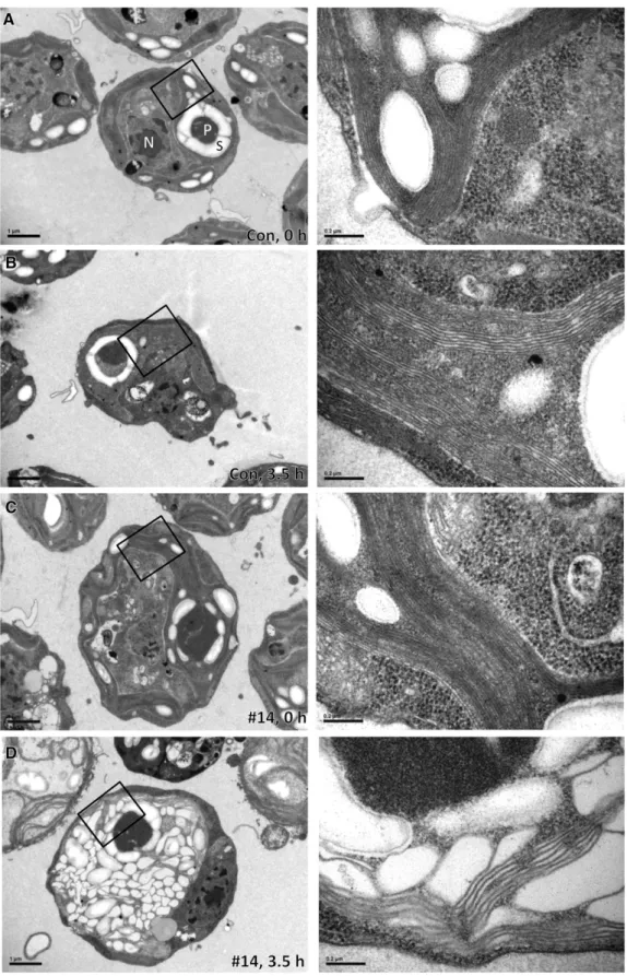
Figure 3. Thylakoids in VIPP1-amiRNA Strains Exposed to High Light Intensities Are Extremely Swollen.
(A) Electron microscopy image of a cell from the control strain grown at low light intensities. Cells were grown at;30mE m 2s 1in TAP-NH
4medium.
grown on nitrate to high light intensities (
;
1000mE m22s21) and observed that bleaching was retarded when compared withVIPP1-RNAi strains grown on ammonium (see Supplemental
Figure 7 online). Also, the loss of PSII maximum quantum efficiency was slower when VIPP1-RNAi cells were grown on nitrate compared with ammonium but still faster and more severe than in control cells grown on either nitrogen source (Figure 5A). Similar effects were observed when cells were exposed to very high light intensities (
;
1800mE m22s21) for 60 min and allowed to recover at low light: The extent of PSII photodamage was highest and recovery slowest in VIPP1-RNAi cells grown on ammonium, followed by VIPP1-RNAi cells grown on nitrate, control cells grown on ammonium, and control cells grown onnitrate (Figure 5B). Interestingly, PSII maximum quantum effi-ciency in control cells was also less severely affected by high light when cells were grown on nitrate as a nitrogen source compared with ammonium.
To test whether the reduced high light sensitivity of VIPP1-RNAi strains grown on nitrate correlated with reduced thylakoid swelling, we took 1015 electron microscopy images from control and VIPP1-RNAi cells, grown on nitrate or ammonium, prior to and after a 3- to 7-h exposure to high light intensities (
;
1000mE m22s21). The observed thylakoid phenotypes were sorted into the categories “ordered,” “disordered,” and “swollen” (examples for these categories are given in Supplemental Figure 8 online). As summarized in Table 1, cells with swollen thylakoids were only observed in VIPP1-RNAi strains grown on ammonium. Here, 8% of the cells displayed swollen thylakoids already at low light intensities (30mE m22s21) and the fraction of cells with swollen thylakoids increased to 68% after the 3 h of high light exposure. Even after 7 h of high light exposure, we observed no thylakoid swelling in VIPP1-RNAi cells grown on nitrate. However, at that time, 36% of the mutant cells were in the process of lysis, whereas this was the case for only 1 to 2% of control cells (see Supplemental Figure 9 online). These results indicate that thyla-koid swelling is not causing lysis of high light–exposed VIPP1-RNAi cells grown on nitrate.A similar picture emerged when we took another 800 electron microscopy images to monitor thylakoid swelling in control and
VIPP1-RNAi cells that were exposed to very high light intensities
(
;
1800mE m22s21) for 60 min and allowed to recover at low light. Here, 38% of ammonium-grown VIPP1-RNAi cells con-tained swollen thylakoids after 60 min at very high light, and this fraction declined to 23% after 60 min of recovery at low light intensities (Table 1). Interestingly, swollen thylakoids were also observed in 11% of ammonium-grown control cells exposed to very high light (thus corroborating the observations from Topf et al., 1992) and this fraction declined to 1% after 60 min of recovery. No thylakoid swelling was observed in nitrate-grown control and VIPP1-RNAi cells exposed to very high light inten-sities (Table 1).Also, at the protein level, we found that exposure to high light (
;
1000mE m22s21) had less severe effects on VIPP1-RNAi cells when they were grown on nitrate as the nitrogen source com-pared with cells grown on ammonium (cf. Figures 1F and 5C). After 7 h of high light exposure, we found a clear decline only in levels of CP43 and perhaps PsaA. While chaperones HSP70B and HSP90C appeared to be normally induced by high light, induction of the LHCSR3 protein still was less pronounced inVIPP1-RNAi cells compared with control cells. Likewise, the
Figure 3. (continued).
starch. Bars in overview images correspond to 1mm and those in zoom-ins to 0.2 mm.
(B) Electron microscopy image of a cell from the control strain exposed to high light. Cells were grown at;30mE m 2s 1in TAP-NH
4medium and
exposed to;1000mE m 2s 1for 3.5 h. Images were taken as in (A).
(C) Electron microscopy image of a cell from a VIPP1-amiRNA strain grown at low light intensities. VIPP1-amiRNA strain #14 was grown at;30mE m 2
s 1in TAP-NH
4medium. Images were taken as in (A).
(D) Electron microscopy image of a cell from a VIPP1-amiRNA strain exposed to high light. VIPP1-amiRNA strain #14 was grown at;30mE m 2s 1in
TAP-NH4medium and exposed to;1000mE m 2s 1for 3.5 h. Images were taken as in (A).
Figure 4. VIPP1-RNAi Cells Are More Severely Photoinhibited at Very High Light and Replace Photodamaged D1 by de Novo–Synthesized D1 Slower Than Control Cells.
Cultures of control and VIPP1-RNAi strain #80 grown in TAP-NH4
medium were split into two parts. One part was supplemented with ethanol-dissolved chloramphenicol (CAP) to a final concentration of 100 mg/mL, while the other received the same volume of pure ethanol. The four cultures were exposed to;1800mE m 2s 1for 30 min
(photo-inhibition [PI]) and shifted back to;30 mE m 2s 1(recovery [rec]).
Maximum quantum efficiency of PSII during photoinhibition and recovery was measured with a PAM fluorometer as variable fluorescence (FV= FM
F0) normalized to FM. Shown is the average of two independent
strong decline of PSII subunits and, to a lesser extent, of PSI subunits observed after exposure of ammonium-grown VIPP1-RNAi cells to very high light intensities (
;
1800mE m22s21) was virtually absent upon exposure of nitrate-grown VIPP1-RNAi cells to very high light (cf. Supplemental Figures 10A and 10B online). However, similar to VIPP1-RNAi cells continuously ex-posed to;
1000mE m22s21, VIPP1-RNAi cells exposed for 60 min to;
1800mE m22s21were still deficient in the full induction of LHCSR3 protein expression, despite growth on nitrate. And in contrast with nitrate-grown VIPP1-RNAi cells exposed to;
1000 mE m22s21, cells exposed to;
1800mE m22s21still appeared to be impaired in the full induction of the HSP70B and HSP90C chaperones and still experienced reduction of initially elevated LHCII levels (see Supplemental Figure 10B online).Taken together, our observations show that thylakoid swelling is indeed only observed in high light–treated cells grown on ammonium as nitrogen source. Moreover, thylakoid swelling enhances, but does not cause, the sensitivity of VIPP1-RNAi strains to high light.
VIPP1-RNAi Strains Are Impaired in the Perception/ Transmission of the High Light State
We wondered whether the reduced accumulation of the LHCSR3 protein in high light–treated VIPP1-RNAi strains was due to a defect in the perception/transmission of the high light conditions or due to a defect in the conversion of the LHCSR3 transcript into a stable protein, for example, by its impaired insertion into the thylakoid membrane. In the former case, we expected a reduced induction of the LHCSR3 transcript in response to high light, and in the latter case, we expected no effect. To distinguish between these possibilities, we exposed cells from control and VIPP1-RNAi strains to high light (
;
1000mE m22 s21) and analyzedLHCSR3 transcripts by RNA gel blot analysis. As shown in Figure
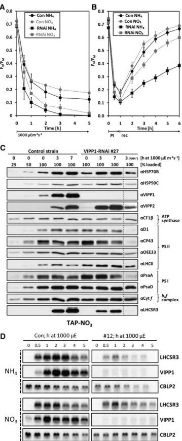
Figure 5. High Light Sensitivity Is Alleviated but Not Abolished in VIPP1-RNAi Strains Grown on Nitrate.
(A) PSII in VIPP1-RNAi strains is less high light sensitive in cells grown on nitrate compared with ammonium. Control (Con) and VIPP1-RNAi strains
#12 and #27 were grown in TAP-NO3or TAP-NH4medium. Cells were
exposed to;1000mE m 2s 1and maximum quantum efficiency of PSII
was measured with a PAM fluorometer as described in Figure 4. Shown is the average of two independent experiments. Error bars representSE. (B) PSII in VIPP1-RNAi strains is less sensitive to photoinhibition in cells grown on nitrate compared with ammonium. Control and VIPP1-RNAi strains #5, #27, and #41 were grown in TAP-NO3or TAP-NH4medium.
Cells were exposed to;1800mE m 2s 1for 1 h (photoinhibition [PI])
and shifted back to;30mE m 2s 1(recovery [rec]). Maximum quantum
efficiency of PSII was measured with a PAM fluorometer as described in Figure 4. Shown is the average of four independent experiments. Error bars representSE.
(C) Subunits of PSII and PSI are less prone to degradation in high light– exposed VIPP1-RNAi strains grown on nitrate. Whole-cell proteins from nitrate-grown control and VIPP1-RNAi strain #27 exposed to;1000mE m 2s 1for 7 h were separated on 14% SDS-polyacrylamide gels and
analyzed by immunoblotting. For comparison, whole-cell proteins from
VIPP1-RNAi strain #27 grown on ammonium and exposed to high light
for 3 h was loaded next to the other samples.
(D) RNA gel blot analysis of high light–exposed control and VIPP1-RNAi strains. Control and VIPP1-RNAi strain #12 were grown in TAP-NO3or
TAP-NH4medium. Cells were exposed to;1000mE m 2s 1for 5 h, and
RNA was extracted from samples taken at the indicated time points and subjected to RNA gel blot analysis. CBLP2 served as loading control.
5D, induction of the LHCSR3 gene was strongly reduced in
VIPP1-RNAi strains grown on ammonium as nitrogen source,
while differences between control and VIPP1-RNAi strains in the high light–induced expression of LHCSR3 were less pronounced in cells grown on nitrate. Similarly, the reduction in LHCSR3 transcript accumulation observed in VIPP1-RNAi strains com-pared with a control strain after exposure to very high light (
;
1800mE m22s21) was less pronounced in cells grown on nitrate instead of ammonium (see Supplemental Figure 10C online). In control cells grown on either nitrogen source, VIPP1 transcripts strongly accumulated in response to high light expo-sure, whereas they were almost completely repressed in VIPP1-RNAi cells.These data suggest that the reduced accumulation of LHCSR3 protein observed in high light–exposed VIPP1-RNAi strains compared with the control strain is mainly based on the reduced expression of the LHCSR3 gene. This in turn suggests that
VIPP1-RNAi strains are defective in the retrograde signaling of
the high light state, which is particularly true for cells grown on ammonium but to a lesser extent also for cells grown on nitrate. Specific Photosynthesis Parameters Are Affected in VIPP1-RNAi Strains
Although we have shown that thylakoid swelling occurs only in high light–exposed cells grown on ammonium and not in cells grown on nitrate as nitrogen source, it is not clear why swelling is so much more severe in VIPP1-RNAi/amiRNA cells compared with control cells. Thylakoid swelling in high light–treated mutant cells may result from their ability to generate a higher proton-motive force (pmf) than control cells, caused, for example, by a reduced ATP synthase activity (Majeran et al., 2001). Alterna-tively, thylakoid swelling may have resulted from the reduced
ability of VIPP1-RNAi/amiRNA strains to counteract the osmotic pressure generated in the thylakoids upon high light exposure in the presence of ammonium.
To test the first hypothesis, we used electrochromic shift measurements to compare control and VIPP1-RNAi strains in respect of the pmf generated at saturating light intensities and with regard to thylakoid membrane conductivity (corresponding to the ATP synthase activity). These measurements were per-formed with cells grown under mixotrophic conditions using ammonium and nitrate as nitrogen sources prior to and after a 3-h exposure to high light intensities of
;
1000mE m22s21. As shown in Figures 6A and 6B, maximum thylakoid membrane energization in saturating light was significantly lower in VIPP1-RNAi cells than in control cells under all conditions tested, while ATP synthase activity was never lower than that of the un-treated control. Hence, thylakoid swelling in high light–exposedVIPP1-RNAi strains is unlikely to be caused by an increased
pmf.
To identify defects in VIPP1-RNAi strains that might cause their increased light sensitivity and the apparently reduced ability of their thylakoids to withstand an increased osmotic pressure, we performed more biophysical measurements. We observed that PSII maximum quantum efficiency was mildly but significantly lower in VIPP1-RNAi compared with control cells (Figure 6C), while cytochrome f reduction and oxidation kinetics were unal-tered, indicating similar electron transport capacities in control and VIPP1-RNAi strains (Figures 6E and 6F). Moreover, the chlorophyll a/b ratio was lower in VIPP1-RNAi strains than in controls (Figure 6D), which may result from the higher levels of chlorophyll b–rich LHCII in VIPP1-RNAi strains (Figures 1F and 5C; see Supplemental Figures 10A and 10B online). A larger antenna cross section in VIPP1-RNAi strains was also supported by the faster fluorescence induction kinetics in mutant versus
Table 1. Categorization of Thylakoid Structure
Condition/Strain
TAP-NH4 TAP-NO3
Ordered Disordered Swollen Ordered Disordered Swollen
High light Con (LL) 100 0 0 99 1 0 Con (1809 HL) 100 0 0 98 2 0 RNAi #27 (LL) 92 0 8 92 8 0 RNAi #27 (1809 HL) 27 5 68 100 0 0 Photoinhibition Con (609 PI) 87 0 11 98 2 0 Con (609 rec) 99 0 1 100 0 0 RNAi #27 (609 PI) 46 16 38 92 8 0 RNAi #27 (609 rec) 74 3 23 100 0 0 Heat shock Con (258C) 96 0 4 n.d. n.d. n.d. Con (408C) 100 0 0 n.d. n.d. n.d. RNAi #32 (258C) 90 1 9 n.d. n.d. n.d. RNAi #32 (408C) 70 9 21 n.d. n.d. n.d.
Transmission electron microscopy images were taken on cells from control (Con) and VIPP1-RNAi strains #27 and #32 grown in TAP-NH4or TAP-NO3
medium under the following conditions: LL, low light intensities of;30mE m 2s 1; 1809 HL, 180 min at high light intensities of;1000mE m 2s 1;
609 PI, 60 min at photoinhibitory light of;1800mE m 2s 1; 609 rec, 60 min at;30mE m 2s 1for recovery from photoinhibition; 258C, 258C and;30
mE m 2s 1; 408C, 408C for 1 h at;5mE m 2s 1; n.d., not determined. On average, 101 images per strain and condition were analyzed and sorted
control cells (Figure 6G). The reduced sigmoidicity of the induc-tion curve in VIPP1-RNAi strains also indicated that cooperativity between PSII centers was lower in VIPP1-RNAi cells compared with control cells. Thermoluminescence measurements revealed a reduced maximum temperature of the Q-band in VIPP1-RNAi cells compared with control cells, a phenotype observed in cells grown on either ammonium or nitrate as nitrogen source (Table 2). Following a saturating single turnover flash at low tempera-ture, a thermoluminescence band, named Q-band, is detected upon heating in the region of 8 to 128C. This luminescence arises from the recombination of the charge pairs S2QA2, with S2being an oxidation state of the Mn cluster and QA2the semireduced
primary quinone acceptor in PSII (Rutherford et al., 1982). The downshift of the maximum temperature of the Q-band in VIPP1-RNAi cells reflects a lowering of the midpoint potential of the redox couple QA/QA2(Krieger-Liszkay and Rutherford, 1998). A lower midpoint potential of QAwas shown to increase the yield of 1O
2generation in the light (Fufezan et al., 2007) and therefore might represent the mechanism underlying high light sensitivity of VIPP1-RNAi cells.
We conclude that thylakoid swelling in ammonium-grown, high light–exposed VIPP1-RNAi cells is not caused by a higher pmf but rather by a disturbance of thylakoid membrane organi-zation. The defect causing this disturbance also might be
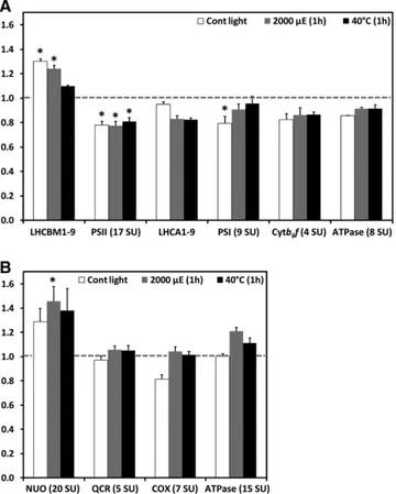
Figure 6. VIPP1-RNAi Strains Are Affected in Some Photosynthesis Parameters.
(A) Thylakoid membrane energization. Control (Con) and VIPP1-RNAi strains #2-7, #2-19, #3-30, #4-19, and #4-49 were grown in TAP-NO3or TAP-NH4
medium and kept in low light (LL) of;30mE m 2s 1or exposed to high light (HL) of;1000mE m 2s 1for 3 h. Maximum thylakoid membrane
energization was then determined by measuring the ECS in saturating light. Shown is the average of eight and six measurements on the control strain, and of 12 and 10 measurements on VIPP1-RNAi strains grown on NO3and NH4, respectively. Error bars representSE. Chl, chlorophyll.
(B) ATP synthase activity. The activity of the ATP synthase was inferred from the decay kinetic of the ECS signal during a short dark interval using the same cells as described in (A).
(C) Maximum quantum efficiency of PSII. FV/FMwas measured after 5 min of far-red illumination, followed by 10 min of dark adaptation of the cell
suspension using control and VIPP1-RNAi strains #4, #9, #40, #53, #72, #111, and #129 grown in low light. Shown is the average of four and two measurements on the control strain, and of 13 and 10 measurements on VIPP1-RNAi strains. Error bars representSE.
(D) Chlorophyll a/b ratios. Chlorophyll was extracted with 80% acetone from the same cells as described in (C), and the chlorophyll concentration was determined spectrophotometrically. Asterisks indicate the significance of the difference to untreated controls grown on nitrate and ammonium as nitrogen source, respectively (t test, P value# 0.05).
(E) Cytochrome f reduction kinetics. Control and VIPP1-RNAi strains #4, #9, and #72 were grown in TAP-NH4medium. Cytochrome f reduction was
initiated by switching off saturating red light at time point zero. The fully reduced state of cytochrome f in the dark was normalized to zero and the fully oxidized state to one. Kinetics were recorded four times for each strain. Averages for the control and all three VIPP1-RNAi strains are shown. (F) Cytochrome f oxidation kinetics. Control and VIPP1-RNAi strains #27, #29, and #30 were grown in TAP-NH4medium. Cytochrome f oxidation was
initiated by switching on a saturating red light pulse at time point zero. Six replicates of the control strain and two each for the VIPP1-RNAi strains were recorded and averages plotted. Again, the fully reduced state of cytochrome f in the dark was normalized to zero and the fully oxidized state to one. (G) Fluorescence induction kinetics. Control and VIPP1-RNAi strains #27, #29, und #30 were grown in TAP-NH4medium. Dark-adapted cells trapped in
state 1 were supplemented with DCMU, and fluorescence was induced by switching on the light. Six replicates of the control strain and two each for the
responsible for reduced PSII maximum quantum efficiency, lowered midpoint potential of the QA/QA2redox couple, reduced PSII cooperativity, and increased antenna cross section in
VIPP1-RNAi strains.
PSII in VIPP1-RNAi Strains Is Sensitive to Heat Shock If VIPP1-RNAi strains indeed have a defect in thylakoid mem-brane organization, we can expect phenotypes to also be in-duced by treatments that do not involve high light intensities but similarly represent a threat to thylakoid membrane integrity. Heat shock at 40 to 428C, combined with low light intensities that are harmless for PSII at 258C, was demonstrated previously to result in loss of PSII activity in C. reinhardtii (Schuster et al., 1988). This finding was explained by cross-linking of the D1 protein with other proteins, presumably induced by the overproduction of radicals by heat-stressed, illuminated PSII.
To test whether PSII was more sensitive to heat stress in
VIPP1-RNAi cells compared with control cells, we shifted mutant
and control cells from 25 to 408C at low light intensities (
;
5mE m22 s21) for 4 h and back to 258C for 2 h for recovery and monitored PSII maximum quantum efficiency. This experiment was done with cells grown mixotrophically on ammonium or nitrate as nitrogen sources (Figure 7A). In control cells grown on ammonium, variable fluorescence/maximum fluorescence (FV/ FM) declined already within the first 20 min at 408C from 0.73 to 0.68 but recovered fast and almost completely after shifting cells back to 258C. The loss of PSII maximum quantum efficiency in nitrate-grown control cells followed kinetics similar to those observed for ammonium-grown control cells within the first hour at 408C, but, surprisingly, the FV/FMvalue in nitrate-grown control cells declined further to 0.58 after 4 h at 408C and did not fully recover at 258C. In ammonium-grown VIPP1-RNAi cells heat-shocked for 4 h, the FV/FMvalue dropped dramatically from 0.68 to 0.44 and at 258C recovered only to 0.47. This effect was less drastic in nitrate-grown VIPP1-RNAi cells, in which the FV/FM value declined from 0.69 to 0.49 after 4 h at 408C and recovered maximally to 0.57 at 258C.To pick up changes induced by heat shock at the ultrastruc-tural level, we took 400 electron microscopy images of ammo-nium-grown control and VIPP1-RNAi strains prior to and after a 60-min heat shock at 408C. Interestingly, the fraction of VIPP1-RNAi cells containing swollen thylakoids increased from 9% under nonstress conditions to 21% after heat shock (Table 1).
Heat stress hardly affected levels of thylakoid membrane com-plex subunits, no matter on which nitrogen source cells were grown (Figure 7B; see Supplemental Figure 11A online). While in control and VIPP1-RNAi strains levels of the VIPP2 protein were strongly induced by high light, they did not further increase during heat stress (cf. Figures 1F and 7B). Also, in contrast with the high light treatments, the HSP70B and HSP90C chaperones appeared to be normally induced by heat stress in VIPP1-RNAi strains grown on either nitrogen source (Figure 7B; see Supple-mental Figure 11A online). In control cells grown on ammonium or nitrate, heat stress led to a slight and transient increase in levels of the LHCSR3 protein. By contrast, we observed no increase in LHCSR3 levels in VIPP1-RNAi strains after 60 min at 408C but a strong decline of LHCSR3 after 180 min of heat stress. Also, the induction of the LHCSR3 gene by heat shock was reduced in VIPP1-RNAi strains (Figure 7C; see Supplemental Figure 11B online).
In summary, the dramatic effect of heat stress combined with very low light intensities on PSII maximum quantum efficiency in
VIPP1-RNAi strains further supports the notion that VIPP1
de-pletion affects the structural organization of thylakoid membrane protein complexes. Again, this defect appears to have a sec-ondary effect on retrograde signaling triggering the induction of the LHCSR3 gene by heat stress.
Quantitative Proteomics Reveals Reduced Levels of Thylakoid Membrane Core Complexes but Increased LHCII Levels in VIPP1-RNAi Cells
The conclusion that thylakoid membranes in VIPP1-RNAi/amiRNA strains must harbor structural defects prompted us to get a more quantitative picture of alterations in the abundance of thylakoid membrane protein complexes. The method of choice for this is shotgun proteomics using15N metabolic labeling, by which a much higher quantification accuracy can be achieved than with quantitative immunoblotting (Mu¨hlhaus et al., 2011). Using this technique, we compared the accumulation of the main protein complexes of the thylakoid membranes in control and VIPP1-RNAi strains that were grown on15NO
3and14NO3as nitrogen sources, respectively (cells were grown on nitrate to avoid the pleiotropic effects caused by thylakoid swelling in cells grown on ammonium). Cells were kept under nonstress conditions or were exposed to 1-h high light (2000 mE m21s21) and heat stress (408C) treatments. For comparison, we also quantified the rela-tive abundances of protein complexes involved in the mitochon-drial respiratory chain. For each of the measured complexes, average values determined for multiple subunits are shown (see Supplemental Data Set 1 online for details). As shown in Figure 8A, thylakoid membranes from unstressed VIPP1-RNAi cells contained 30% more LHCII, but
;
20% less PSII, PSI, and cytochrome b6f complex, and 14% less ATP synthase thancontrol cells, while levels of LHCI were unchanged. High light and heat stress led to a 5 to 15% decline in levels of LHCs and to less reduced levels of PSI (5 to 9%) and ATP synthase (8%) in
VIPP1-RNAi cells compared with stressed control cells, while
levels of PSII and cytochrome b6f complex remained unchanged.
Interestingly, levels of the mitochondrial NADH:ubiquinone oxidoreductase complex were 30% higher and those of the cytochrome oxidase 19% lower in VIPP1-RNAi cells compared
Table 2. Temperature of the Q-Band
Strain
Tmax(8C)
TAP-NO3 TAP-NH4+
Wild type 11.56 0.5 11.16 0.5
VIPP1-RNAi 7.46 1.2 9.06 1.0
Cells were grown in TAP medium containing either ammonium or nitrate as nitrogen source at low light intensities of;30mE m 2s 1. Values
represent averages6SDfrom thermoluminescence measurements on two independent control and four independent VIPP1-RNAi lines (#1-29, #2-29, #4-49, and #3-30).
with control cells, while levels of the ubiquinone-cytochrome c reductase complex and the ATP synthase were unchanged (Figure 8B). High light and heat stress treatments led to increases in levels of all respiratory chain complexes in VIPP1-RNAi cells relative to control cells. Hence, it appears possible that VIPP1-RNAi cells attempt to compensate for the slightly reduced capacity of their thylakoid membrane core complexes by higher levels of the NADH:ubiquinone oxidoreductase complex. VIPP1-RNAi/amiRNA Strains Harbor Aberrant Structures at the Origin of Thylakoid Membranes
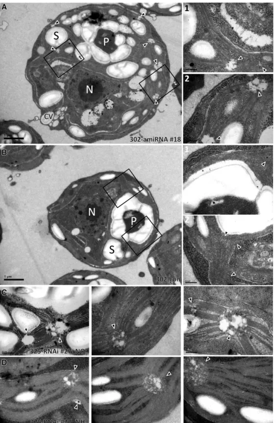
Finally, we set out to gather information on the localization of VIPP1 in C. reinhardtii by immunofluorescence microscopy using an affinity-purified antibody against VIPP1. As shown in Figure 9, VIPP1 was detected as diffuse material throughout the chloro-plast but also in distinct dot-like structures that sometimes also appeared to extend into rods. This localization pattern is in agreement with our previous finding that C. reinhardtii VIPP1 may form rod-like structures, which by the help of molecular chaperones are kept in equilibrium with ring-like structures and smaller assembly states (Liu et al., 2007). We reasoned that rod-like structures potentially formed by VIPP1 might be visible in electron micrographs and therefore revisited the electron micro-graphs taken from control and VIPP1-RNAi/amiRNA strains. Unfortunately, we were unable to distinguish potential VIPP1 rods or rings from thylakoid membranes in control cells. How-ever, we observed aberrant structures in VIPP1-RNAi/amiRNA cells that occurred particularly in regions from which multiple thylakoid membrane layers emerge and which may well corre-spond to the dot-like structures observed by immunofluores-cence microscopy (Figure 10A). The aberrant structures were often found around the pyrenoid but were not restricted to that area. They were very rarely observed in control cells and oc-curred abundantly and consistently in VIPP1-RNAi and amiRNA cells (cw15-325 and cw15-302 backgrounds, respectively) grown under low light or high light on ammonium or nitrate (Table 3, Figure 10).
In summary, we could localize VIPP1 to diffuse material and to distinct spots within the chloroplast. As judged from the distri-bution of these spots, they may correspond to regions from which multiple thylakoid layers emerge and which are aberrant in
VIPP1-RNAi/amiRNA cells.
DISCUSSION
Here, we present a thorough characterization of phenotypes arising in C. reinhardtii as a consequence of VIPP1 depletion mediated by inverted repeat and amiRNA constructs. VIPP1 expression was repressed in two different strain backgrounds: In strain cw15-325, VIPP1 levels were reduced to below 5% of wild-type levels, which, however, resulted in the upregulation of VIPP2
Figure 7. PSII of VIPP1-RNAi Strains Is Highly Susceptible to Heat Stress.
(A) PSII maximum quantum efficiency of control and VIPP1-RNAi strains exposed to heat stress. Control and VIPP1-RNAi strains #5, #20, #27, and #41 were grown in TAP-NO3or TAP-NH4medium, and cells were
exposed to 408C at;5mE m 2s 1. F
V/FMwas measured over time with
a PAM fluorometer as described in Figure 4. Shown is the average of five and seven independent experiments for control and VIPP1-RNAi strains, respectively. Error bars representSE.
(B) PSII subunits are only mildly affected by heat stress. Whole-cell proteins were extracted from control and VIPP1-RNAi strain #27 grown and heat-stressed as described in (A). Whole-cell proteins were separated on 14% SDS-polyacrylamide gels and analyzed by immunoblotting. (C) RNA gel blot analysis of heat-stressed control and VIPP1-RNAi
strains. Control and VIPP1-RNAi strain #20 were grown and heat stressed as described in (A). RNA was extracted from samples taken at the indicated time points and subjected to RNA gel blot analysis.
to
;
20% of total VIPP levels (Figure 2). In strain cw15-302, VIPP2 was not upregulated in response to VIPP1 repression, but VIPP1 only could be reduced to;
25% of wild-type levels. Despite the different contributions of VIPP1 and VIPP2 to the residual VIPP pool in the two strain backgrounds, phenotypes were the same (Figures 1, 3, and 10; see Supplemental Figure 5B online). This allows us to conclude that VIPP1 and VIPP2 largely are func-tionally redundant and that the total VIPP pool in C. reinhardtiicannot be constitutively repressed to below
;
25% of wild-type levels, similar to the situations in Arabidopsis and cyanobacteria (Kroll et al., 2001; Westphal et al., 2001; Fuhrmann et al., 2009a). VIPP1 obviously is the major VIPP species in C. reinhardtii, a conclusion that is supported by the finding that repression of VIPP2 in strain cw15-325 did not result in high light sensitivity or other obvious phenotypes (M. Ru¨tgers and M. Schroda, unpub-lished results). Very similar phenotypes obtained with VIPP1-RNAi and -amiRNA constructs also widely rule out off-target effects in the RNAi lines (Ossowski et al., 2008).Aberrant Structures at the Origin of Multiple Thylakoid Membrane Layers in VIPP1-RNAi/amiRNA Cells Suggest Deficits in Thylakoid Biogenesis
The most striking observation we consistently made in C.
reinhardtii VIPP1-RNAi/amiRNA cells under all growth conditions
tested is the occurrence of aberrant structures in regions from which bundles of thylakoids emerge and often project into different directions of the chloroplast (Figure 10, Table 3). Some of these aberrant structures were reminiscent of prola-mellar body (PLB)–like structures observed in dark-grown C.
reinhardtii y-1 (yellow-in-the-dark) mutants (Hoober and Blobel,
1969; Friedberg et al., 1971). y-1 mutants are defective in nuclear genes that are involved in the posttranscriptional expression or accumulation of the CHLL subunit of the light-independent protochlorophyllide reductase, leading to a block in chlorophyll synthesis in the dark (Cahoon and Timko, 2000). Hence, PLB-like structures are likely made of thylakoid membrane precursors in
Figure 8. Levels of Thylakoid Membrane Core Complexes Are Slightly Lower in VIPP1-RNAi Cells Than in Control Cells.
(A) Ratios of thylakoid membrane core complexes in VIPP1-RNAi cells relative to control cells. A control strain and VIPP1-RNAi strain #111 were metabolically labeled using15NO
3and14NO3, respectively, as nitrogen
source and maintained at;30mE m 2s 1and 258C for 1 h (Cont light),
exposed to photoinhibitory light of;2000mE m 2s 1for 1 h at 258C, or
heat-shocked at 408C for 1 h at;5mE m 2s 1. After mixing control and
VIPP1-RNAi cells from each treatment at a 1:1 ratio, proteins in
mem-brane-enriched fractions were separated by SDS-PAGE and digested tryptically in gel. Peptides were eluted, desalted, and analyzed by liquid chromatography-MS/MS. Peptide identification and quantification was performed as described previously (Mu¨hlhaus et al., 2011). Quantifica-tion values for single core complex subunits (SU) were computed from quantified peptides, and the average ratio of light (VIPP1-RNAi) to heavy (control) subunits was calculated for the different core complexes. Error bars representSE, and asterisks indicate the significance of the differ-ence of the ratio from one (assuming equal variance; t test, P value# 0.05).
(B) Ratios of respiratory chain core complexes in VIPP1-RNAi relative to control cells. Ratios of respiratory chain core complexes were deter-mined as described in (A).
Figure 9. Immunofluorescence Microscopy Detects VIPP1 in Distinct Punctae and as Diffuse Material in the Chloroplast.
Control cells from the cw15-325 background were grown at;30mE m 2
s 1in TAP-NH
4medium and fixed and processed for
immunofluores-cence (IF) microscopy as described in Methods. The signal recognized by the affinity-purified anti-VIPP1 antibody is shown in green. Triangles indicate potential rod-like extensions. Bars = 5mm.
Figure 10. VIPP1-RNAi/amiRNA Strains Harbor Aberrant Structures at the Origin of Thylakoid Membranes. (A) Electron microscopy image of a cell from VIPP1-amiRNA strain #18. Cells were grown at;30mE m 2s 1in TAP-NH
4medium. An overview image is
shown on the left, and zoom-ins of the regions demarcated by black boxes are shown on the right. Triangles indicate regions at the origin of multiple thylakoid membrane ramifications. CV, contractile vacuole; N, nucleus; P, pyrenoid; S, starch. Bars in overview images = 1mm, those in zoom-ins = 0.2 mm. (B) Electron microscopy image of a cell from the control strain. Cells were grown at;30mE m 2s 1in TAP-NH
4medium. Symbols are as in (A).
(C) Electron microscopy image of a cell from VIPP1-RNAi strain #27. Cells were grown at;30mE m 2s 1in TAP-NO
3medium. Abbreviations are as in (A).
(D) Electron microscopy image of a cell from VIPP1-RNAi strain #27. Cells were grown at;30mE m 2s 1in TAP-NH
which the biogenesis of photosynthetic protein complexes is arrested or at least retarded. The aberrant structures in VIPP1-RNAi/amiRNA cells were frequently (but not exclusively) located close to the pyrenoid in regions previously suggested to house translation zones (T-zones; Figure 10) (Uniacke and Zerges, 2007). T-zones were postulated to represent areas where de novo PSII biogenesis and the regulated transport of newly assembled PSII complexes to thylakoid membranes occur. If indeed the aberrant, PLB-like structures in VIPP1-RNAi/amiRNA cells correspond to T-zones, they might be caused by a problem during photosystem biogenesis/assembly.
In cyanobacteria, groups of thylakoids converge at peripheral cytoplasmic points without any apparent connection to the plasma membrane (Kunkel, 1982). Thylakoids at these points are attached to so-called thylakoid centers, which are 30 to 50 nm wide tubular structures of up to 1 mm length that are composed of subunits generating a 14-fold rotational symmetry (Kunkel, 1982; van de Meene et al., 2006). Thylakoid centers have been postulated to be linked to a membrane fraction with a density intermediate to that of thylakoid and plasma membranes (Hinterstoisser et al., 1993). It has also been postulated that these are the sites at which protein/pigment complexes are assembled and incorporated into photosynthetic membranes and that po-tentially are related to T-zones in C. reinhardtii (Nickelsen et al., 2011). A coincidence too striking to be overlooked is that recombinant VIPP1 from Arabidopsis, C. reinhardtii, and
Syn-echocystis forms ring-like structures with an outer diameter of 25
to 37 nm, a 12- to 17-fold rotational symmetry, and the capacity to assemble into rod-like structures of up to 1.4mm (Aseeva et al., 2004; Liu et al., 2007; Fuhrmann et al., 2009b). Hence, it is tempting to speculate that thylakoid centers in fact are VIPP1 rods, which again may correspond to microtubule-like structures reported in plastids of diverse algal and plant species in various types of tissues (Liu et al., 2007, and references therein). Thyla-koid centers may have moved from a peripheral position in cyanobacteria to more central ones in chloroplasts and still serve as sites of thylakoid biogenesis. This speculation is supported by the localization of VIPP1 by immunofluorescence to distinct spots within the chloroplast that sometimes appear to extend into rod-like structures (Figure 9). Support also comes from the
biochemical localization of VIPP1 to low-density membranes (Liu et al., 2005) that are membranes intermediate between envelope and thylakoids at which thylakoid protein biogenesis might occur (Zerges and Rochaix, 1998). Depletion of VIPP1 may impair the formation of proper rods, thereby leading to disordered thylakoid centers and eventually resulting in the aberrant structures ob-served. Alternatively, rods may be formed normally but soluble VIPP1 is depleted and not available for chaperone-mediated cycling between soluble and complexed forms (Liu et al., 2007). If this process was required for the transport of building blocks to T-zones for thylakoid biogenesis (see below), the resulting tail-back may be the cause for the aberrant structures observed, comparable to the generation of PLB-like structures by the absence of chlorophyll in the y-1 mutant.
Is VIPP1 Involved in the Biogenesis of Thylakoid Membranes or of Thylakoid Membrane Core Complexes?
The aberrant, PLB-like structures at the origin of thylakoids point to a deficit in thylakoid biogenesis. However, is the formation of the lipid bilayer itself affected, as proposed previously (Kroll et al., 2001; Westphal et al., 2001; Aseeva et al., 2007), or is the biogenesis/assembly of the photosynthesis complexes affected, as suggested by Gao and Xu (2009)?
Similar to what has been reported for a VIPP1 depleted
Arabi-dopsis mutant (Kroll et al., 2001; Aseeva et al., 2007), we find no
change in lipid composition, but we do observe slightly lower levels of fully assembled photosystems, cytochrome b6f complex, and
ATP synthase (by 14 to 20%) in nonstressed C. reinhardtii VIPP1-RNAi strains (Figure 8A; see Supplemental Figures 2 and 3 online). In contrast with the earlier reports, however, we find neither an obvious reduction of thylakoid membranes nor changes in the number of membranes per granum (Figures 3 and 10). We rather find levels of LHCII increased by 30%, which correlates with a lower chlorophyll
a/b ratio and faster fluorescence induction kinetics (Figures 6D, 6G,
and 8A). Hence, there appears to be no limitation of thylakoid membranes in VIPP1-RNAi/amiRNA strains for housing protein complexes, which points to a role of VIPP1 in the biogenesis/ assembly of core complexes rather than in the formation of the membranes themselves. These data are in support of those from Gao and Xu (2009), who showed in Synechocystis that depletion of VIPP1 first affected PSII activity, then PSI activity, and once pho-tosystem activities were lost, thylakoids degenerated. A role for VIPP1 in core complex biogenesis/assembly is also supported by the previous finding that VIPP1 interacts with Alb3.2 (Go¨hre et al., 2006). Alb3.2 was suggested to play a role in PSI and PSII assembly, as it was found to directly interact with photosystem subunits. Moreover, both photosystems accumulated to reduced levels when Alb3.2 was downregulated by RNAi, while cytochrome b6f and ATP
synthase were barely affected. Therefore, it appears possible that VIPP1 supports Alb3.2 during photosystem assembly.
Phenotypes in Stressed C. reinhardtii VIPP1-RNAi/amiRNA Strains Point to Defects in the Structural Organization of Thylakoid Membrane Complexes
If VIPP1 is involved in the biogenesis/assembly of thylakoid membrane core complexes, which step may be affected in
Table 3. Quantification of Aberrant PLB-Like Structures in
VIPP1-RNAi/amiRNA Cells
Strain Growth Conditions
cw15-325 LL, NH4 3.5 h HL, NH4 LL, NO3 Control 0 n.d. 1 VIPP1-RNAi #27 44 n.d. 64 VIPP1-RNAi #41 63 n.d. 89 cw15-302 Control 2 4 n.d. VIPP1-amiRNA #14 48 69 n.d. VIPP1-amiRNA #18 63 50 n.d. VIPP1-amiRNA #20 51 48 n.d. Cells were grown in TAP medium containing either ammonium or nitrate as nitrogen source at low light (LL) intensities of;30mE m 2s 1or at
high light intensities (HL) of;1000mE m 2s 1. Fifty electron