HAL Id: hal-01000215
https://hal.archives-ouvertes.fr/hal-01000215
Submitted on 29 May 2020
HAL is a multi-disciplinary open access
archive for the deposit and dissemination of
sci-entific research documents, whether they are
pub-lished or not. The documents may come from
teaching and research institutions in France or
abroad, or from public or private research centers.
L’archive ouverte pluridisciplinaire HAL, est
destinée au dépôt et à la diffusion de documents
scientifiques de niveau recherche, publiés ou non,
émanant des établissements d’enseignement et de
recherche français ou étrangers, des laboratoires
publics ou privés.
TOPAZ1, a novel germ cell-specific expressed gene
conserved during evolution across vertebrates
Adrienne Baillet, Ronan Le Bouffant, Jean Nicolas Volff, Alix Luangpraseuth,
Elodie Poumerol, Dominique Thépot, Eric Pailhoux, Gabriel Livera, Corinne
Cotinot, Béatrice Mandon-Pepin
To cite this version:
Adrienne Baillet, Ronan Le Bouffant, Jean Nicolas Volff, Alix Luangpraseuth, Elodie Poumerol, et
al.. TOPAZ1, a novel germ cell-specific expressed gene conserved during evolution across vertebrates.
PLoS ONE, Public Library of Science, 2011, 6 (11), pp.1-15. �10.1371/journal.pone.0026950�.
�hal-01000215�
Conserved during Evolution across Vertebrates
Adrienne Baillet1,2, Ronan Le Bouffant3, Jean Nicolas Volff4, Alix Luangpraseuth1,2, Elodie Poumerol1,2, Dominique The´pot1,2, Eric Pailhoux1,2, Gabriel Livera3, Corinne Cotinot1,2, Be´atrice Mandon-Pe´pin1,2*
1 INRA, UMR 1198 Biologie du De´veloppement et Reproduction, Jouy-en-Josas, France, 2 ENVA, Maisons Alfort, France, 3 CEA, DSV/DRR/SEGG/LDRG, Laboratory of Differentiation and Radiobiology of the Gonads, Unit of Gametogenesis and Genotoxicity, Fontenay aux Roses, France,4 Institut de Ge´nomique Fonctionnelle de Lyon, Universite´ de Lyon, CNRS, INRA, Ecole Normale Supe´rieure de Lyon, Lyon, France
Abstract
Background:We had previously reported that the Suppression Subtractive Hybridization (SSH) approach was relevant for the isolation of new mammalian genes involved in oogenesis and early follicle development. Some of these transcripts might be potential new oocyte and granulosa cell markers. We have now characterized one of them, named TOPAZ1 for the Testis and Ovary-specific PAZ domain gene.
Principal Findings: Sheep and mouse TOPAZ1 mRNA have 4,803 bp and 4,962 bp open reading frames (20 exons), respectively, and encode putative TOPAZ1 proteins containing 1,600 and 1653 amino acids. They possess PAZ and CCCH domains. In sheep, TOPAZ1 mRNA is preferentially expressed in females during fetal life with a peak during prophase I of meiosis, and in males during adulthood. In the mouse, Topaz1 is a germ cell-specific gene. TOPAZ1 protein is highly conserved in vertebrates and specifically expressed in mouse and sheep gonads. It is localized in the cytoplasm of germ cells from the sheep fetal ovary and mouse adult testis.
Conclusions:We have identified a novel PAZ-domain protein that is abundantly expressed in the gonads during germ cell meiosis. The expression pattern of TOPAZ1, and its high degree of conservation, suggests that it may play an important role in germ cell development. Further characterization of TOPAZ1 may elucidate the mechanisms involved in gametogenesis, and particularly in the RNA silencing process in the germ line.
Citation: Baillet A, Le Bouffant R, Volff JN, Luangpraseuth A, Poumerol E, et al. (2011) TOPAZ1, a Novel Germ Cell-Specific Expressed Gene Conserved during Evolution across Vertebrates. PLoS ONE 6(11): e26950. doi:10.1371/journal.pone.0026950
Editor: Renee A. Reijo Pera, Stanford University, United States of America Received May 2, 2011; Accepted October 6, 2011; Published November 1, 2011
Copyright: ß 2011 Baillet et al. This is an open-access article distributed under the terms of the Creative Commons Attribution License, which permits unrestricted use, distribution, and reproduction in any medium, provided the original author and source are credited.
Funding: This work was supported in part by PHASE Division, INRA. AB was supported by a doctoral fellowship from the Ministe`re de´le´gue´ a` l’Enseignement supe´rieur et a` la Recherche, France. The funders had no role in study design, data collection and analysis, decision to publish, or preparation of the manuscript. Competing Interests: The authors have declared that no competing interests exist.
* E-mail: [email protected]
Introduction
Ovarian development in mammals involves coordinated and multiple and complex molecular, cellular and morphogenetic events. Unlike testis differentiation, where germ cells are dispensable, oocytes are required to induce and maintain ovarian somatic cells [1]. Some key events occur during development of the fetal mammalian ovary, including, colonization of the undifferentiated gonad by primordial germ cells (PGCs) and then their proliferation, the entrance of germ cells into meiosis, atresia of germ cells and the formation of primordial follicles [2]. Although animal germline development varies in different species, some general rules have emerged. During the earliest embryonic stages, PGCs proliferate in a niche supported by somatic cells and then differentiate into germline stem cells, which proliferate through self-renewal. Formation of the germ cell nest (or cyst) is an important step in germline differentiation that has been conserved during evolution from insects to vertebrates [3,4]. Oogonia in nests are clonally derived [5] and germline nest formation occurs through incomplete cytokinesis during mitosis, resulting in the connection of daughter cells by intercellular bridges [6,7]. In the
mouse, the maximum number of germ cell clusters is detected on embryonic day 13.5, just prior to the commitment of PGCs to the oocyte developmental program and entry into meiosis [8]. Within germ cell nests, the oogonia enter meiotic prophase [9,10]. Several global approaches using microarray technologies have been used to isolate the genes involved in meiosis by comparing transcrip-tional activity in ovaries with or without germ cells in meiotic prophase I [11–15]. Both germ cell-intrinsic and -extrinsic factors govern meiotic initiation in mouse embryos [16,17]. The RNA-binding protein DAZL (Deleted in AZospermia-like), expressed by both male and female post-migratory germ cells, is one of the first intrinsic factors necessary to enroll germ cells in the meiosis event [17] in response to retinoic acid (RA). Entry into meiosis is then controlled by RA signals that are blocked in the testis by CYP26B1 (cytochrome P450 26B1) [18–20] and fibroblast growth factor 9 (FGF9) [21]. In the fetal ovary, RA signaling acts on germ cells to promote the expression of the cytoplasmic factor Stra8 (Stimulated by Retinoic acid). Stra8 is required for premeiotic DNA synthesis, meiotic initiation and meiotic progression in germ cells [22,23]. Consistent with the central role that RA is believed to play in vertebrate germline development and the sex-specific timing of
entry into meiosis, RA appears to function as one of the principal inducers of Stra8 expression in early germ cells [24–27]. However, another study reported that Stra 8 expression in the mouse fetal ovary does not require RA signaling, and that Stra8 expression is expressed in the absence of physiologically detectable levels of RA [28]. That study demonstrated that another unknown inducer of Stra8 might be involved in Stra8 expression to enable the initiation of mouse meiosis.
Just after birth in the mouse, and during fetal life in ruminants and humans, the nests break down to form individual primordial follicles [8,29,30]. Their formation, and the transition to primary follicles, represents a critical period of follicle formation [31,32]. Ovarian follicles are the functional units within the female gonad that nurture oocyte maturation and enable the production of steroid hormones.
During the past ten years, many of the growth factors and signaling proteins that mediate the early stages of folliculogenesis have been identified using mouse genetic models, in vivo injection studies, and ex vivo organ culture techniques. These studies revealed important roles for the transforming growth factor beta (Tgf-beta) superfamily of proteins in the ovary [33–35]. Moreover, several transcription factors are known to affect the regulation of oocyte-specific genes during early folliculogenesis (Figure S1). Figla (a factor in the germline alpha) regulates the expression of numerous genes in the ovary, including Zp2, Pou5f1, Nlrp14, Nlrp4f, and Nlrp4b [36,37]. Nobox (newborn ovary homeobox gene) is necessary for the expression of several key oocyte-specific genes, including Gdf9 and Pou5f1 [38–40]. Sohlh1 (spermatogenesis- and oogenesis-specific bHLH transcription factor 1) is another germ cell-specific gene that encodes a bHLH protein [41]. Sohlh1 lies upstream of Lhx8, Zp1, and Zp3 and is preferentially expressed in primordial oocytes [41]. More recently, Rajkovic’s team described a novel spermatogenesis- and oogenesis-specific basic helix-loop-helix (bHLH) transcription factor, Sohlh2 [42,43]. Sohlh2-deficient ovaries can form primordial follicles that do not differentiate surrounding granulosa cells into cuboidal and multilayered structures [43].
In addition to these functional relationships, small non-coding RNAs, including microRNAs (miRNAs), small interfering RNAs (siRNAs), and Piwi-interacting RNAs (piRNAs) have been implicated as key regulators, especially in the gonads of eukaryotes [44]. It has been shown that piRNAs become abundant in germ cells at around the pachytene stage of prophase of meiosis I [45], but they may be present at lower levels during earlier stages. It has been shown that piRNAs repress transposable elements (TEs) but their molecular functions are still unknown [46–49]. Unlike
microRNAs, individual piRNAs sequences per se are not conserved.
In order to discover novel genes expressed in the ovary during prophase I meiosis of germ cells and follicle formation, we previously reported on the generation of two sheep fetal ovary cDNA libraries, obtained by applying SSH using mRNAs isolated from 55 and 82 day post-coitum (dpc) female gonads [50]. In this species, meiosis occurs between days 50 and 75 and the first primordial follicles are observed at around days 75–80 of gestation [51]. In the resulting libraries [50], we found several transcripts with sex or stage-specific patterns of expression, such as MOV10L1 (EMBL:CU654655), [52–54], TEX11 (EMBL:CU651916), [55] and Gametocyte-specific factor 1 (GTSF1) (EMBL:CU652509), [56]. Among these transcripts, we isolate an EST encoding a putative protein with a PAZ domain (Piwi/Argonaute/Zwille), namely TOPAZ1 (EMBL: CU654960 and CU655260). During the current study, we characterize the genomic structure of the TOPAZ1 gene and determine its expression pattern during development in sheep and mouse. We show here that TOPAZ1/ Topaz1 is expressed in both the male and female gonads and is highly conserved among vertebrates, including fish, amphibians, birds and mammals. Furthermore, we demonstrate that TOPAZ1/ Topaz1 mRNA and TOPAZ1 protein are localized in both male and female germ cells lineages.
Results
Characterization of TOPAZ1 transcripts
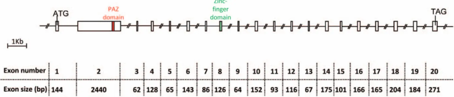
CU654960 and CU655260 ESTs arising from our previously described sheep fetal ovary libraries [50] were aligned with public EST databases and displayed significant similarity with human testis EST (Genbank AI208289): identities = 393/428 (92%); e value = 2e-171. As this sheep EST was expressed in the testis and ovary and contained a PAZ-domain, we decided to call it TOPAZ1. In order to characterize the full-length sequence of the TOPAZ1 transcript in sheep, 59RACE experiments were per-formed using mRNA from female fetal ovaries of 60 days post-coitum (60 dpc). The primers used during the 59RACE experiments are listed inTable S1. Moreover, the rest of the coding sequence of TOPAZ1 cDNA was cloned by successive PCR procedures from sheep fetal ovaries of 60 dpc. The primers used to clone sheep TOPAZ1 cDNA are listed inTable S2. We found that the full length TOPAZ1 cDNA sequence contained 20 exons with an open reading frame sequence of 4803 bp. The number of nucleotides for each exon is shown in Figure 1. The putative TOPAZ1 protein contained 1600 amino acids.
Figure 1. Structure of the sheepTOPAZ1gene. Sheep TOPAZ1 cDNA derived from exons 1–20 and contained 4952 nucleotides. TOPAZ1 was composed of twenty exons in sheep, bovine, human and mouse. The PAZ domain is located in the second exon and the CCCH domain in exon 8. The nucleotide number of each exon of sheep TOPAZ1 is indicated under the Figure.
doi:10.1371/journal.pone.0026950.g001
By searching for sequences with similarities to sheep TOPAZ1 gene within public databases, we found the human TOPAZ1 gene (Official Symbol in GeneBank C3orf77; GeneBank Accession number NM_001145030, Ensembl ENSG00000173769; Uni-Gene Hs.132125; human hypothetical protein LOC375337). Full length human TOPAZ1 cDNA (ORF: 5079 bp) was also made up of 20 exons, as in sheep. Moreover, similarity analysis highlighted the mouse Topaz1 gene (Official Symbol in GeneBank Gm9524; GeneBank Accession number XM_001474362, UniGene Mm.461223; mouse hypothetical protein LOC100032115) but this predicted gene was much shorter (ORF: 1851 bp) when compared to sheep and human TOPAZ1. In order to verify the exact size of the mouse Topaz1 cDNA, we designed oligonucleotide primers (Table S3) and ran successive RT-PCR using mouse adult testis mRNA. We were thus able to deduce the size of the mouse Topaz1 cDNA ORF as being 4962 bp, which was much longer than the predicted sequence available in public databases (GeneBank Accession number XM_001474362: 1919 bp, ORF: 1851 bp).
In these three species of mammals, all exons had almost the same nucleotide size, except for exon 1 (seeFigure S2). Indeed, in its 39 part, the human TOPAZ1 exon 1 contained 141 bp more than the sheep and mouse genes. Moreover, using RT-PCR and sequencing we showed that the location of the predicted start codon (ATG) differed between sheep and mouse. In the latter species, the beginning of the ORF is at the same position as the human protein start codon (UniProtKB: Q8N9V7).
According to the Vista Genome browser (http://genome.lbl. gov/vista/index.shtml), the nucleotide similarity between the ORF of the TOPAZ1 mRNA of sheep and of different mammalian species (cattle: BK008402, human: NM_001145030 and mouse: HM631980) was high, ranging from 79% between sheep and mice and 82% between sheep and human to 97% between sheep and cattle (Figure 2A) and the highest degree of similarity across species existed from 3.25 to 5.0 Kb on the TOPAZ1 cDNA sequences. TOPAZ1 localized to human chromosome HS 3p21.33, its mouse orthologue to chromosome 9, and the cattle gene to chromosome 22.
We have submitted these sheep and mouse TOPAZ/Topaz1 sequences to the GenBank databases and the accession numbers are: HM631979 (Ovis aries) and HM631980 (Mus musculus). We also submitted the predicted cattle TOPAZ1 cDNA (Bos taurus) to Genbank: nucleotide sequence data reported are available in the Third Party Annotation Section of the DDBJ/EMBL/GenBank databases under the accession number TPA: BK008402.
TOPAZ1 protein contains conserved PAZ and zinc finger domains
PAZ domain. TOPAZ1 protein belongs to a functionally uncharacterized protein family (mouse hypothetical protein LOC671232; human hypothetical protein LOC375337) containing an evolutionarily conserved domain named PAZ for Piwi/Argonaut/Zwille (PF02170, Accession IPR003100) located in exon 2 (Figure 1) of all aligned sequences (amino acids 688 to 745 in human; 593 to 650 in ovine; 646 to 703 in bovine; 651 to 708 in mouse). This domain was found in two families of proteins that are involved in post-transcriptional gene silencing, namely the Piwi and Dicer families. The conservation of the PAZ domain in different species was high (between 71% and 96%) and is shown in Figure 2 (Ba).
Other domains. In addition, TOPAZ1 protein contains a conformation typical zinc-finger domain (amino acids 1097 to 1116 in humans; 1005 to 1024 in sheep (Figure 1); 1058 to 1077 in cattle and 1056 to 1075 in mouse) of C-x8-C-x5-C-x3-H type
(PF00642, Accession IPR000571), but which is C-x9-C-x4/5-C-x3-H in the TOPAZ1 proteins of several species (Figure 2 (Bb)).
TOPAZ1 protein is phylogenetically conserved in Vertebrates
To determine whether the TOPAZ1 protein has been conserved during evolution, a phylogenetic analysis was performed on a 620 amino-acid alignment extending from the CCCH zinc finger to the C-terminal end using the neighbor-joining method [57], (1,000 bootstrap replicates). We showed that TOPAZ1 proteins were phylogenetically very close between sheep and cattle, and neighbors with horse and dog orthologs (Figure 3). Moreover, these 620 TOPAZ1 amino acids were aligned within several classes of vertebrates such as fish, amphibians and birds, and displayed a high degree of conservation within the Vertebrate Subphylum. Thus TOPAZ1 proteins are highly conserved across Vertebrate phylogeny.
TOPAZ1 is specifically expressed in gonads
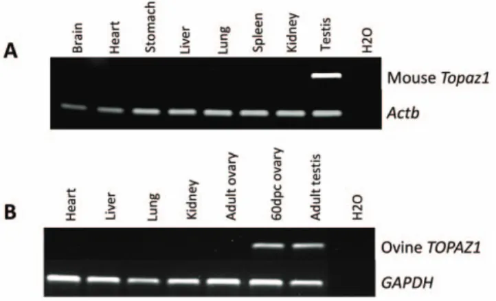
The tissue specificity of both sheep and mouse TOPAZ1/Topaz1 transcripts was tested by semi-quantitative RT-PCR analyses on different adult somatic tissues and gonads (adult and fetal ovaries in sheep). The primers used are listed inTable S4. GAPDH in sheep and beta-actin (Actb) in mouse served as positive controls and were detected in all tissues (Figure 4). Among the different tissues tested, mouse Topaz1 cDNA was exclusively expressed in adult testis (Figure 4A) and sheep TOPAZ1 cDNA was detected in both adult testis and fetal ovary (Figure 4B). None of the somatic tissues tested displayed amplified bands corresponding to TOPAZ1 cDNA in both mouse and sheep species.
TOPAZ1 displays a time-regulated expression profile during gonad development
In order to study the time course of TOPAZ1 mRNA expression, quantitative RT-PCR was performed (primers listed inTable S5) on female and male gonad tissues at different stages of development (fetal, post-natal and adult). The relative expression of sheep female TOPAZ1 mRNA, normalized to HPRT1 mRNA expression (Figure 5A), increased during the meiosis prophase I period (55–65 dpc), decreased until the end of the fetal life and then remained very low in adult ovary. Throughout the fetal period of testicular development, the relative expression of TOPAZ1 mRNA was low. By contrast, TOPAZ1 expression was only high in the adult testis of sheep.
According to the expression profile of ovine TOPAZ1 (Figure 5A) i.e. its increase during the 55–75 dpc period in fetal ovary and in adult testis, we compared its expression with that of DMC1, a gene essential for meiotic homologous recombination during the development of male and female gonads in sheep using quantitative RT-PCR (Figure 5B). In females, DMC1 transcripts were detected as from 55 dpc, peaked between 60–65 dpc and then decreased after 75 dpc. This profile was very similar to that of TOPAZ1. In males, the expression of both DMC1 and TOPAZ1 was strongly observed at the adult stage.
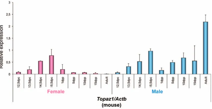
In the mouse (Figure 6), the relative expression profile of Topaz1 mRNA differed from that seen in sheep. Indeed, in developing female and male gonads in fetal mice, the relative expression of Topaz1 mRNA was approximately the same in both sexes, and further increased during development. In females, its relative expression was decreased after birth; it was even quite low at 5 days post-partum (dpp) and almost null in mouse adult ovaries. In males, Topaz1 mRNA increased as from 5 dpp to become strongly expressed in the adult mouse testis.
Figure 2. (A) Nucleotide sequence conservation of mammalianTOPAZ1transcripts and (B) Conservation of PAZ and CCCH domains of TOPAZ1 protein in mammalian species. (A) The percentage nucleotide identity between sheep TOPAZ1 cDNA and that of other mammalian species is indicated on the right-hand side. These alignments were obtained using the Vista Genome browser (http://genome.lbl.gov/vista/index. shtml). (Ba) The PAZ domain surround (in red) consisted of 58 amino acids. The percentages of identity were 96%, 78%, and 71% between sheep-cattle, sheep-human and sheep-mouse, respectively. (Bb) The CCCH domain is indicated in the green box, and consisted of 20 amino acids. Amino acid identity was 90%, 85%, and 80% between sheep-cattle, sheep-human and sheep-mouse, respectively. Identical residues are noted with an asterisk.
doi:10.1371/journal.pone.0026950.g002
Thus TOPAZ1/Topaz1 was a gonad-specific expressed gene in the sheep and mouse. However, its expression profile differed slightly between these two species. Contrary to the mouse, the sheep TOPAZ1 transcript was preferentially expressed in females during fetal life when prophase I meiosis occurred (Figures 5A and 5B). Indeed, we had previously shown that the leptotene and zygotene stages were clearly identifiable in sheep ovogonia as from 55 dpc [51]. At this stage, the transcription of DMC1, SPO11, and MSH4 was initiated. A strong signal was observed until 75 dpc, after which expression decreased more or less rapidly and was found to be present at a low level at 94 dpc. At birth, all ovocytes were at the dictyate stage of meiosis [51]. The sheep TOPAZ1 transcript presented a profile similar to that of meiotic genes.
Topaz1 is a germ cell-specific gene
In order to determine whether Topaz1 expression in gonads is restricted to germ cells, we analyzed germ cell fractions from mouse testis and ovary. Enrichment in germ cells was ensured by the immunomagnetic isolation of SSEA1+ cells (SSEA1-enriched germ cells) from fetal gonads at 13 dpc. Stage-Specific Embryonic Antigen 1 (SSEA1) is regulated developmentally during early embryogenesis and is widely used as a marker to monitor the
differentiation of pluripotent EGCs, ESCs and embryonic carcinoma cells (ECCs) [58]. To quantify Topaz1 gene expression, we performed RT-qPCR on whole gonads versus SSEA1-enriched germ cells from male and female 13 dpc gonads (Figure 7). Topaz1 transcripts were abundant in both male and female SSEA1-enriched cells but were rare in whole gonads, showing that the Topaz1 gene was germ cell-specific in the mouse.
To consolidate our findings, we used homozygous fetuses with null mutation for the c-Kit gene [59]. The absence of Kit prevents the migration and survival of PGC, so that gonads from these fetuses contain no germ cells. To produce these WlacZ/WlacZ embryos, we intercrossed WlacZ/+ mice (obtained from Trans-genesis Institute of Orle´ans-Villejuif Resource Center, (http:// transgenose.cnrs-orleans.fr/eng/intragene/presentation.php). An analysis of Topaz1 mRNA expression within a pool of gonads from WlacZ/WlacZ embryos (from 17.5 to 19.5 dpc) was compared with the expression of Vasa (or Mhv, germ cell marker) and Wt1 (Wilms tumor 1, somatic cell marker) (Figure 8). Topaz1 transcripts (such as Vasa) were clearly absent from male and female germ cell-deprived gonads. On the other hand, Wt1, a gene with somatic expression, was highly expressed in these gonads. These findings reinforced our previous data indicating that Topaz1 is a germ cell-specific gene in the mouse.
Topaz1 expression is not RA-dependant in mouse fetal testis
In order to determine whether TOPAZ1 could be regulated by the retinoic acid (RA) pathway, in vitro mouse fetal testis culture experiments were performed [18]. Indeed, whereas in vivo male meiosis only starts after birth, RA has been shown to induce meiosis in vitro in mouse fetal germ cells from both 11 and 12.5 dpc XY gonads [18]. In these in vitro cultures, about 20% of the germ cells displayed histological characteristics typical of the leptotene and zygotene stages. After such in vitro culture experiments on 11 dpc mouse testis, the expression of Rec8 (a meiosis-specific phosphoprotein involved in recombination events), and of Topaz1, was quantified using real-time quantitative RT-PCR (Figure 9). We showed that a high level of RA (1026M) induced the onset of meiosis (as attested by the increase in Rec8 expression) after three days of culture, when compared with the control culture (without
Figure 3. Molecular phylogeny of TOPAZ1. Phylogenetic analysis was performed on a 620 amino-acid alignment extending from the CCCH zinc finger domain to the C-terminal end using the neighbor-joining method [57]; (1,000 bootstrap replicates). The tree was not rooted. Accession numbers: Homo sapiens (human) NP_001138502; Pan troglo-dytes (chimpanzee) XP_526186; Macaca mulatta (rhesus monkey) XP_001114967; Mus musculus (house mouse) EDL09106; Rattus norvegicus (rat) EDL76795; Bos taurus (cattle) NW_001494083.2 (genomic); Ovis aries (sheep) HM631979; Equus caballus (horse) NW_001867381.1 (genomic); Canis familiaris (dog) NW_876276.1 (genomic); Monodelphis domestica (gray short-tailed opossum) XP_001381211; Ornithorhynchus anatinus (duck-billed platypus) NW_001794448.1 (genomic); Gallus gallus (chicken) NW_001471633.1 (genomic); Anolis carolinensis (green anole, lizard) ENSEMBL scaffold_98 (genomic); Xenopus tropicalis (frog) ENSEMBL scaffold_320 (genomic); Ictalurus punctatus (catfish) ABD91555; Danio rerio (zebrafish) NW_001877999.1 (genomic); Tetraodon nigroviridis (pufferfish) ENSEMBL Chr. 21.
doi:10.1371/journal.pone.0026950.g003
Figure 4. Expression ofTOPAZ1mRNA in different sheep (A) and mouse (B) tissues. (A) The expression of sheep TOPAZ1 mRNA was tested using RT-PCR in four adult somatic tissues (heart, liver, lung and kidney) and in gonads (60 dpc ovary and adult testis). (B) The expression of the mouse Topaz1 gene was tested in seven adult somatic tissues (brain, heart, stomach, liver, lung spleen and kidney) and in adult testis. GAPDH (glyceraldehyde-3P-dehydrogenase) and Actb amplifica-tion served as loading controls.
RA). By contrast, the expression of Topaz1 was not modified by RA treatment and Topaz1 expression in the mouse fetal testis did not require RA signaling.
Production of a specific TOPAZ1 antibody
To determine the cell type and sub-cellular localization of TOPAZ1 protein, we produced a polyclonal antibody from two 15-mer peptides. The specificity of this antibody was tested by Western blot analysis on protein extracts from different sheep tissues (cytosolic fractions of adult testis, fetal ovary at 60 dpc and liver). This analysis showed that TOPAZ1 antibody bound specifically to adult testis and fetal ovary extracts, but not to liver extracts (Figure 10). The molecular weight of TOPAZ1 protein was approximately 199 kDa, slightly higher than the predicted
molecular weight of 180 kDa. This difference may have resulted from post-translational modifications. No similar signals were detected in nuclear extracts from sheep gonads (data no shown). The antibody against BETA-ACTIN (ACTB) detected a specific 42 kDa band in all the extracts tested, thus serving as a positive control. TOPAZ1 protein was exclusively expressed in the cytosolic compartment of gonad cells, and the protein level was higher in sheep adult testis than in the fetal ovary. These results were consistent with our RT-qPCR analysis (Figure 5A).
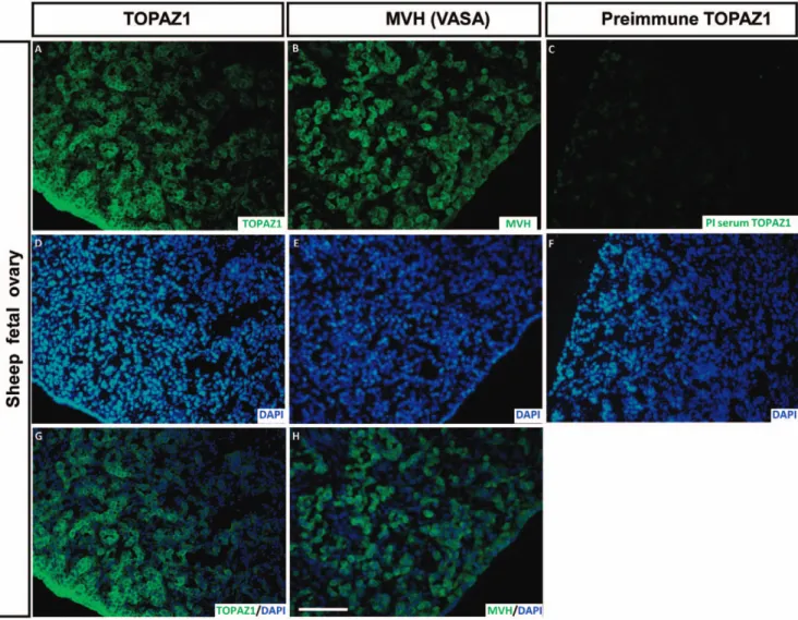
TOPAZ1 protein is present in germ cells of sheep fetal ovary
The TOPAZ1 antibody was used on sheep fetal ovary sections at 60 dpc (Figure 11A, G), the stage displaying the peak expression of
Figure 5. Expression ofTOPAZ1andDMC1mRNA in sheep gonads at different developmental stages. Quantification of TOPAZ1 (A) and DMC1 (B) genes expression using quantitative real-time RT-PCR analysis at several fetal stages (44, 55, 60, 65, 75, 82, 90, 114 dpc) and in adult ovary (red histograms) and testis (blue histograms). The HPRT1 (hypoxanthine phosphoribosyltransferase) gene was used as a reporter gene. Values indicated on the graph are means 6 SEM of two independent RT experiments at each stage.
doi:10.1371/journal.pone.0026950.g005
TOPAZ1 mRNA (Figure 5A). We also performed immunodetection of MVH (VASA) (a germ cell marker expressed in the cytoplasm of germ cells) on adjacent sections of 60 dpc ovary (Figure 11B, H). Immunofluorescence was mainly detected in the germ cells. When merged with DAPI, TOPAZ1 staining was clearly localized in the cytoplasm of the cells (Figure 11G). When comparing TOPAZ1 and MVH immunostaining, we concluded that TOPAZ1 was expressed in the cytoplasm of 60 dpc female germ cells in sheep. Pre-immun TOPAZ1 was also tested on sheep 60 dpc ovary sections and did not reveal any staining (Figure 11C).
TOPAZ1 protein is present in germ cells of mouse adult testis
The localization of TOPAZ1 in mouse adult testis was determined using immunofluorescence (Figure 12A, E). The
peptides used for immunization were designed according to the sheep sequence and were not fully homologous with the mouse Topaz1 sequence. In order to enhance antibody specificity, purified anti-TOPAZ1 antibody was used on mouse sections. TOPAZ1 displayed clear staining in germ cells of from mouse adult testis (Figure 12A). The same degree of staining was achieved using MVH antibody (Figure 12B). Moreover, when merged with DAPI, the TOPAZ1 protein appeared to be clearly localized in the cytoplasm of these germ cells (Figure 12G). Our results thus confirmed the cytoplasm localization of TOPAZ1 in the germ cells of mouse adult testis, a funding in agreement with those obtained by the expression of Topaz1 transcripts in the germ cell enriched fraction (Figure 7) and in germ cell-deprived gonads (Figure 8). TOPAZ1/Topaz1 mRNA and TOPAZ1 protein were localized in the germinal cell lineage of sheep and mice of both sexes.
Discussion
During this study, we isolated and characterized a new gene encoding for a putative protein containing a PAZ domain. Until now, the evolutionarily conserved PAZ (Piwi/Argonaute/Zwille) domain had been found in two families of proteins that are involved in post-transcriptional gene silencing [60], the PIWI and DICER families. The three-dimensional structure of this domain has been solved [61–63] and studies have revealed a unique mode of single-stranded nucleic acid binding in which the two 3-terminal nucleotides are buried in a hydrophobic cleft. It has been proposed that the PAZ domain contributes to the specific recognition of siRNAs [64].
In addition, TOPAZ1 contained a zinc-binding domain of the Cys-X9-Cys-X4-Cys-X3-His type (termed the CCCH domain).
Berg et al. defined that the CCCH family is a group of zinc-finger proteins consisting of a canonical C-X6–14-C-X4–5-C-X3-H motif
[65]http://www.ncbi.nlm.nih.gov/pubmed/8599083. After
ana-Figure 6. Expression ofTopaz1mRNA in mouse gonads at different developmental stages. Quantification of Topaz1 gene expression using quantitative real-time RT-PCR analysis at several fetal stages (12.5, 13.5, 14.5, 16.5 dpc), after birth (1, 5, 10, 15 dpp) and in adult ovary (pink histograms) and testis (light blue histograms). The Actb gene was used as a reporter gene. Values indicated on the graph are means 6 SEM of three independent RT experiments at each stage.
doi:10.1371/journal.pone.0026950.g006
Figure 7. Expression of theTopaz1transcript in mouse enriched germ cell fractions. Germ cells were isolated from 13 dpc gonads by means of SSEA1+ immunomagnetic isolation. Quantification of Topaz1 gene expression using quantitative real-time RT-PCR analysis in whole ovaries (pink histogram) and testes (light blue histogram) and in germ cell fractions (hatched histograms). Values indicated on the graph are means 6 SEM of three independent samples.
Figure 8. Expression ofTopaz1, MvhandWt1transcripts in mouse WlacZ/+ or WlacZ/WlacZ gonads. (A) Expression of Mvh, a gene only expressed in germ cells, was used to verify the absence of germ cells from WlacZ/WlacZ gonads (F2/2 and M2/2, hatched histograms) in contrast to WlacZ/+ gonads (F+/2 and M+/2, plain histograms). (B) Expression of the Wt1 gene in somatic cells was used to verify the integrity of somatic cells. (C) The expression of Topaz1 was determined by qRT-PCR analysis in WlacZ/+ or WlacZ/WlacZ ovary and testis. The Hprt1 gene was used as a reporter gene. Values indicated on the graph are means 6 SEM of two independent samples.
doi:10.1371/journal.pone.0026950.g008
lyzing the entire CCCH family in Arabidopsis and rice, Wang et al. re-determined that CCCH proteins are characterized by one to six C-X4–15-C-X4–6-C-X3-H motifs which constitute glycine-rich and
phenylalanine-rich sequences [66]. CCCH-type zinc-finger pro-teins are RNA binding propro-teins with regulatory functions at all stages of mRNA metabolism, including pre-mRNA splicing, mRNA transportation, sub-cellular localization and stability/ degradation, transcription, ubiquitination, and poly ADP-ribosyla-tion [67,68]. It has been shown that different CCCH-type ZnF proteins interact with the 39-untranslated region of various mRNA [69], and that a single CCCH domain is capable of binding single-stranded RNA with considerable affinity and selectivity [70]. The presence of these two domains in the TOPAZ1 protein thus strongly suggested a role for this factor in RNA recognition and binding.
Phylogenetic analysis has revealed a fish-specific TOPAZ1 clade within vertebrates. Among mammals, eutherians are separated into two groups, rodents and others. This divergence could explain the difference in expression patterns that we observed between mice and sheep during gonad development. The strong conser-vation of the ZnF_CCCH domain in fish and amphibians suggests that the TOPAZ1 molecules of lower vertebrates possess the structural features required for RNA binding. Whilst the C-terminal from the ZnF_CCCH domain is well conserved among
all Topaz1 molecules, the N-terminal before the ZnF_CCCH domain is more divergent between fish, amphibians, monotremes and eutherian mammals (data not shown).
During the current study, we showed that the TOPAZ1/Topaz1 gene was specifically expressed in the gonads of sheep and mice, in a sex-specific manner. Its expression was stronger in females than in males during fetal gonad development. However, this pattern was reversed after birth, with strong expression in adult males only and the disappearance of Topaz1 expression from the adult ovary. This time course was suggestive of a relationship between Topaz1 expression and the occurrence of germ cell meiosis. To reinforce this hypothesis, we analyzed the expression profile of DMC1 in the same samples and compared its expression pattern in sheep. DMC1 is a meiosis-specific RecA/Rad51 homolog required for the recombinational repair of meiotic Double Strand Breaks (DSBs). The DMC1 protein was detected in mouse leptotene-to-zygotene spermatocytes at the initiation of homolog pairing [71]. In Saccharomyces cerevesiae and mice of both sexes, DMC1-deficient mutants arrest in the late zygoneme/early pachyneme of meiotic prophase I, with an accumulation of DSBs and defective formation of the synaptonemal complex [72,73]. The comparison of sheep TOPAZ1 and DMC1 expression patterns revealed a very similar profile. Furthermore, we demonstrated that Topaz1 was germ line-specific using two different approaches; the enrichment of mouse germ cells by SSEA1 antibody and the use of gonads from WlacZ/ WlacZ mutant mice devoid of germ cells. Moreover, we showed that TOPAZ1/Topaz1 mRNA encoded a protein present in germ cells from both male and female gonads. Taken together, these results reinforce the idea that TOPAZ1/Topaz1 could be involved in germ cell development.
Our study also showed that Topaz1 expression was not stimulated by retinoic acid in cultured male fetal gonads from mice. This could have been due either to the incomplete meiosis prophase I obtained during our organ culture experiments [18] or to the need for non-retinoid pathways in order to regulate Topaz1 expression [28].
In addition to the recombination events and chromatin exchange that occur during meiosis prophase I, it is of paramount importance for germ cell genomes to be protected from the uncontrolled propagation of mobile genetic elements. These mobile elements must be distinguished from endogenous genes and then selectively silenced. In Drosophila, germ cells express a class of small RNAs (piRNAs) that are specialized in the repression of mobile elements [74–76]. Germ cells in mice express two types
Figure 9. Effect of retinoic acid (RA) onTopaz1expression in mouse fetal testis. Mouse testes at 11 dpc were cultured for 3 days with (RA+, hatched histograms) or without (RA2, plain histograms) retinoic acid (1026M). At the end of the culture period, total RNA was extracted and Rec8 (A)
and Topaz1 (B) mRNA expressions were measured by real-time quantitative RT-PCR. Values indicated on the graph are means 6 SEM of three independent samples.
doi:10.1371/journal.pone.0026950.g009
Figure 10. Specificity of anti-TOPAZ1 antibody by Western blot. Western-blotting experiments were performed with cytosolic extracts from sheep adult testis, fetal ovary and liver. Anti-TOPAZ1 antibody recognized a 199 kDa protein in sheep adult testis and 60 dpc ovary, but no band was detected in sheep liver. As a positive control, ACTB expression was detected in each sample.
of piRNAs: (i) a small fraction of piRNAs derived from the repetitive genomic regions expressed during mouse embryogene-sis, and (ii) a larger fraction of piRNAs derived from non-repetitive regions that start to be expressed during the pachytene stage of meiosis (pachytene piRNAs). [46,47,77]. Pachytene piRNAs interact with MIWI (mouse PIWI) and MILI (MIWI-like), two of the three mouse PIWI proteins [47]. In mice, the PIWI clade contains three members: Miwi2, Mili, and Miwi, all of which display distinctive developmental expression patterns [78–81]. In the male germline, a deficiency in one of two Piwi family members, Mili or Miwi2, results in the loss of DNA methylation marks on transposons, and mutant animals display a phenotype similar to that of Dnmt3L-deficient mice [81–84]. These data have led to the hypothesis that Piwi/piRNA complexes might serve as sequence-specific guides that direct the de novo DNA methylation machinery to transposable elements [81,82,85]. The fact that Topaz1 possesses two RNA interaction domains and an expression profile related to meiosis prophase I supports the hypothesis that Topaz1 might be involved in the piRNA pathway. It is noteworthy
that MOV10L1, a transcript previously isolated in our sheep fetal ovary libraries [50] has recently been confirmed as an essential component in the Piwi-interacting RNA pathway in the mouse [53,54]. On the other hand and by comparison with MVH immunostaining, we showed that TOPAZ1 displayed a similar cellular localization. Consequently, TOPAZ1 could be expressed in primordial germ cells and in germ cells undergoing gametogenic processes until the post-meiotic stages in both males and females. In adult testis, MVH and TOPAZ1 proteins were located in the cytoplasm of spermatogenic cells. It has recently been shown that MVH plays a crucial role in piRNA processing and in the gene silencing of retrotransposons [86]. The similarity of the expression profiles and cell localization of both genes, in addition to the presence of PAZ and RNA-binding domains in Topaz protein, reinforce the possible role of TOPAZ1 in the regulation of retrotransposons in the germ line.
Further studies are necessary to investigate the functional role of TOPAZ1 in mammalian gametogenesis, and for this purpose we are currently generating a Topaz1 knockout mouse.
Figure 11. Immunodetection of TOPAZ1 protein in sheep fetal ovary. Immunofluorescence was performed on transversal sections of sheep 60 dpc ovaries to detect TOPAZ1 (A) and MVH (B) proteins. Pre-immune serum (PI) was also tested (C). For each section, DAPI was performed to detect nuclei (D-E-F). TOPAZ1 showed similar cellular localization as MVH (A, B). The immunolabeling of TOPAZ1 and MVH was merged with DAPI (G, H). TOPAZ1 can be detected in the cytoplasm of germ cells in ovine fetal ovary. PI serum of TOPAZ1 did not reveal any staining (C). Scale bars = 200 mm in H (applies to A–G).
doi:10.1371/journal.pone.0026950.g011
In conclusion, TOPAZ1 is a new evolutionarily conserved gene, specifically expressed in gonads and probably involved in gametogenesis. The presence of a PAZ domain in Topaz1 suggests that it could be involved in the RNA silencing pathway. It constitutes a potential new actor in piRNA-directed retrotranspo-son silencing in germ cells that contributes to a safeguard mechanism for genetic information.
Materials and Methods Animal and tissue samples
Pregnant Pre´-Alpes female sheep were obtained as previously described [50,51]. Sheep fetuses were collected at 44, 55, 60, 65, 75, 82, 90 and 114 days post coitum (dpc). The gonads of adult animals were collected at slaughter, cut into small pieces, flash frozen in liquid nitrogen and then stored at 280uC. Other organs from sheep fetuses, such as the liver, kidney, lung and heart, were
treated similarly. C57/Bl6, 129/Sv and NMRI mouse fetuses and adult organs were also collected, frozen immediately and then stored at 280uC. Experiments were performed in accordance with the International Guiding Principles for Biomedical Research involving animals, as promulgated by the Society for the Study of Reproduction and with the European Convention on Animal Experimentation. All researchers working directly with the animals possessed an animal experimentation license delivered by the French veterinary services, and the permit number for our studies is C91332101. The study also involved the use of 129/Sv mice carrying the KitW/LacZ allele. The KIT gene was inactivated and the first exon of Kit was replaced by an nlsLacZ-neo cassette. Full details on this construction can be found in the paper by Bernex and colleagues [59]. Ovaries and testes from heterozygous (WlacZ/+) and homozygous (WlacZ/WlacZ) mice were collected at between 17.5 dpc and 20.5 dpc.
Figure 12. Immunodetection of TOPAZ1 protein in mouse adult testis. Immunofluorescence was performed on sections of mouse adult testis to detect TOPAZ1 (A) and MVH (B) proteins. Anti-TOPAZ1 antibody raised against sheep protein was used to detect mouse Topaz1. For each section, DAPI was performed to detect nuclei (C–D). Mouse TOPAZ1 displayed a localization similar to that of MVH (A, B). The immunostaining of TOPAZ1 and MVH was merged with DAPI (E, F). TOPAZ1 can be detected in the cytoplasm of germ cell in mouse testis. Scale bars = 200 mm in H (applies to A–F).
RNA extraction, reverse transcription (RT), semi-quantitative RT-PCR and real-time PCR
Total sheep RNAs were extracted from each sample using TrizolH reagent (Invitrogen Life Technologies, Cergy-Pontoise, France] plus the RNeasy Mini kit (QIAGEN SA, Courtaboeuf, France), following the manufacturer’s instructions. In sheep, we used six ovaries and four testes at 45 dpc, four gonads at 55 dpc, two gonads at 60-65-75-82 dpc, one gonad at 90–114 dpc and a sample of adult gonads. RT was performed on each sample using 5mg Dnase-treated RNA incubated with random hexanucleotide primers with Superscript II (Invitrogen, Cergy-Pontoise, France), according to the manufacturer’s instructions.
Briefly, semi-quantitative RT-PCR was performed on 1mL (corresponding to 25 ng reverse-transcribed total RNA) of each RT, which was amplified using 0.5 U Taq polymerase (TaKaRa, Lonza, Verviers, Belgium). The primer sequences and PCR conditions used are shown inTable S4. In sheep, the GAPDH gene was used as a control. The PCR conditions were presented inTable S4.
Real-time PCR analysis of TOPAZ1 was performed using the ABI Prism 7700 HT apparatus (Applied Biosystems). Briefly, PCR was performed with the ABsolute blue QPCR SYBR Green ROX mix (Abgene, Les Ulis, France), using 50 ng of cDNA from the RT. The primers used for real-time PCR are presented inTable S5. We have performed all control experiments to ensure that our primers could not amplify any genomic products. All expression data were normalized using the HPRT1 expression level for sheep samples. Real-time PCR on sheep involved two different RT experiments for each stage and the SEM was calculated from these two independent samples. Real-time PCR in the mouse was performed on three different RT samples for each stage.
In the mouse, total RNA was extracted using the RNeasy Mini kit (Qiagen) in C57/Bl6 and NMRI mice or the RNeasy Micro kit (Qiagen) in 129/Sv WlacZ/+ and WlacZ/WlacZ mice, depending on the size of the gonads. Ovaries from WlacZ/+ or WlacZ/ WlacZ mice at between 17.5 dpc and 20.5 dpc were pooled in two separate tubes (14 and 18 ovaries, respectively). The same procedure was used for WlacZ/+ and WlacZ/WlacZ testes (4 and 6 testes for each condition). Reverse transcription was carried out with the Omniscript kit (Invitrogen), according to the manufacturer’s instructions. The expression of Topaz1 or Rec8 genes was assessed by real-time PCR using the SYBR Green Universal PCR Master Mix 26 (Applied Biosystems, Foster City, CA). Reactions were carried out and fluorescence was detected on an ABI Prism 7000 apparatus. In the mouse, the amount of each cDNA detected was normalized using Actb, Hprt1 or Mvh. All primers are presented inTable S5.
59RACE-PCR in sheep
The full-length cDNA of sheep TOPAZ1 was determined using the 59 RACE System (Invitrogen, Cergy-Pontoise, France). Total RNA was extracted from 60 dpc female fetal gonads. One microgram of total RNA was reverse-transcribed into first-strand cDNA using a specific oligo for TOPAZ1 (TOPAZ1_RT,Table S1) and Superscript II (Invitrogen, Cergy-Pontoise, France). First-strand cDNA was treated with RNase H (Invitrogen, Cergy-Pontoise, France) and purified on a S.N.A.P column (Invitrogen, Cergy-Pontoise, France). TdT tailing was performed on cDNA using dCTP and terminal transferase (Invitrogen, Cergy-Pontoise, France). Thirty two PCR cycles were performed using the AAP anchor primer (a non-specific primer) and TOPAZ1 specific primer 1, with or without 5% formamide. A second PCR were performed on 5mL of the PCRI samples diluted at 1/100, with two different TOPAZ1-specific primers (Topaz1–2 or Topaz1–3 nested probe, Table S1) and AUAP (a non-specific primer). Both PCR
procedures were performed according to the following protocol: 94uC for 30 s, 55uC for 30 s, and 72uC for 1.5 min for 32 cycles, followed by an extension for 10 min at 72uC. All RACE-derived PCR products were subcloned into the pGEMTeasy vector and the clones were then sequenced.
Primers for TOPAZ1 cDNA in sheep and mouse were designed to verify the size of the full-length cDNA sequences on adult testis. They are listed inTables S2 and S3. The PCR conditions were 94uC for 30 s, 60uC for 30 s, and 72uC for 1.5 min for 35 cycles, followed by an extension for 10 min at 72uC.
NMRI Mouse Testis cultures with retinoic acid (RA)
Pregnant female mice were killed by cervical dislocation at 11 dpc and their fetuses removed from the uterine horns. Gonads and their mesonephros were isolated from the fetuses under a binocular microscope and kept in Ham F12/DMEM (1:1) (Life Technologies, Inc., Grand Island, NY, USA) until explantation. The sex of the fetus was determined by PCR amplification of Sry, as previously described [87]. The gonads were cultured on Millicell-CM Biopore filters (Millipore, Billerica, MA, USA) in 0.3 ml of Ham F12/DMEM (1:1) for 3 days under a 5% CO2/
95% air atmosphere. The medium was changed every 48 h. One gonad from each fetus was cultured in a medium containing all-trans-RA (1026M, Sigma, St Louis, MO), and the other was cultured in a control medium.
Purification of NMRI mouse germ cells
About 50 gonads from 13.5 dpc fetuses were digested first in 0.25% trypsin-0.02% EDTA (Trypsin/EDTA solution, Sigma-Aldrich, St. Louis, MO) at 37uC. The samples were then centrifuged and digested with 2 mg/ml collagenase and 0.02 mg/ml DNase I in HBSS. For the purification of SSEA1-positive cells, dispersed cells were incubated with anti-SSEA1 (1/5, anti-SSEA1 monoclonal antibody, DSHB, Iowa) in PBS, 0.5% BSA (PB) for 20 min at 4uC. The cells were then centrifuged and washed once with 1 ml PB, before being incubated with 20mL microbead-linked donkey anti-mouse IgM antibody (Miltenyi Biotec, Germany) in 300mL PB with 2 mM EDTA (PBE) for 15 min at RT. The column was rinsed three times with 500mL PBE to wash out unbound cells, which represented the SSEA1-cell fraction. After removal from the magnet, the column was flushed with 1 ml PB, which allowed collection of the SSEA1+ cell fraction. The cells were pelleted, RLT buffer was added and total RNA was extracted as previously described.
Phylogenetic analysis
TOPAZ1 sequences were retrieved by BLAST analysis from the databases accessible via the NCBI (http://blast.ncbi.nlm.nih.gov/ Blast.cgi) and ENSEMBL (http://www.ensembl.org/Multi/blast view) servers. Sequence analysis was performed using the GCG Wisconsin package (Version 11.1, Accelrys Inc., San Diego, CA). Gene structure and protein sequences were predicted from genomic sequences using FGENESH [88]; (http://linux1.softberry.com/ berry.phtml?topic = fgenesh&group = programs&subgroup = gfind). Multiple sequence alignments were generated using ClustalX [89]. Phylogenetic analysis was performed on a 620 amino-acid alignment extending from the CCCH zinc finger to the C-terminal end using the neighbor-joining method [57]; (1,000 bootstrap replicates; Kimura two-parameter correction) as implemented under Seaview (Version 4.0) [90].
Proteins Extraction
Frozen tissues (sheep 60 dpc liver and ovaries and adult testis) were pounded into liquid nitrogen. The tissue powder was then
mixed with 1 ml lysis buffer in a Dounce, and 0.2% NP40 was added. Samples were centrifuged at 13,000 rpm for 1 hr at 4uC, and the supernatant corresponding to the cytosolic extract was removed. The nucleus pellet was resuspended in 100 ml nuclear protein extraction buffer (20 mM Hepes pH 7.7, 1.5 mM MgCl2, 0.2 mM ethylenediaminetetraacetic acid (EDTA), 25% glycerol, 1 mM NaF, 1 mM Na3VO4, 10 mM ammonium molybdate, 0.5 mM dithiothreitol (DTT), 2 mM benzamidine, 0.5 mM phenylmethyl sulfonyl fluoride (PMSF)) to which 10 ml of 4 M NaCl was added. After 1 hr of gentle mixing at 4uC, the samples were centrifuged for 30 min at 13,000 rpm, and the supernatant corresponding to nuclear extracts was collected. The Bradford method was used for protein quantification and samples were conserved at 280uC.
Generation of an anti-TOPAZ1 antibody
Anti-TOPAZ1 antibody was generated by Eurogentec Compa-ny. Two peptides of 15 amino acids were used to immunize a rabbit: TEKRKEINAKSSERE (sheep amino acids 601–615) and SLSGAEVRQSRTKEN (sheep amino acids 625–639), chosen for their potential high antigenicity. Their homologies with mouse Topaz1 counterparts were 60% and 66%, respectively. The antibody was affinity purified with both peptides by Eurogentec Company. The non-purified antibody was used in sheep and the purified antibody was used in mouse.
Western Blotting
For Western-blot analyses, (11 mg) proteins were separated in NuPAGE 4–12% Bis-Tris polyacrylamide gels according to the manufacturer’s instructions (Invitrogen) and transferred onto a PVDF membrane (Millipore). The blots were incubated with primary antibodies (rabbit polyclonal anti-TOPAZ1 antibody 1:300, or rabbit monoclonal anti-ACTB, 1:500, Sigma) overnight at 4uC and were then incubated for 1 h with the corresponding peroxidase-conjugated antibody (anti-rabbit immunoglobulin, 1:5000, Biosystem). Immu-noreactive bands were detected by ImmobilonTM western Chemi-luminescent HRPSubstrate (Millipore).
Immunofluorescence
Freshly dissected gonads were fixed in 4% paraformaldehyde in phosphate saline buffer (PBS) at 4uC overnight. After washing in PBS with increasing concentrations of sucrose (0, 12%, 15%, and 18%), the tissue specimens were embedded in Jung Tissue Freezing Medium (Leica Instruments) and frozen at 80uC. Cryosections (5mm) were obtained and stored at 280uC. The
sections were air-dried, treated with chloroform for 30 sec, and then rehydrated in PBS for 10 min. The TOPAZ1 antibody (1:1000) and VASA antibody (or MVH, 1:500, ab13840, Abcam) were diluted in 1% BSA/PBS and applied to tissue sections overnight at 4uC. After several washes, the sections were incubated with a secondary anti-rabbit IgG-FITC antibody against rabbit
(1:200, Vector) for 1 h at room temperature. The slides were then rinsed in PBS, mounted in Vectashield mounting medium with DAPI (Vector) and observed with a Leica DMRB epifluorescence microscope coupled to a DP50CCD camera (Olympus).
Supporting Information
Figure S1 Main genes involved in follicle formation and preservation of germ cell resting pool. Several factors are involved in primordial (FIGLA, NOTCH2, SOHLH1 and 2, FOXL2), primary (NOBOX, LHX8, cKIT/KL) or secondary (GDF9, BMP15) follicle formation. Others support the resting pool of primordial follicles and avoid their differentiation (AMH, PTEN, FOXO3A).
(PDF)
Figure S2 TOPAZ1 exon 1 in human, sheep and mouse. (PDF)
Table S1 59 RACE primer sequences used in sheep. (PDF)
Table S2 Sequences of semi-quantitative RT-PCR prim-ers used to sequence full-lengthTOPAZ1 cDNA in sheep. (PDF)
Table S3 Sequences of semi-quantitative RT-PCR prim-ers used to sequence and/or verify the size of mouse Topaz1 cDNA.
(PDF)
Table S4 Primer sequences and conditions used for RT-PCR analyses.
(PDF)
Table S5 Sequences of qPCR primers. Oligonucleotides for qPCR were designed using the PrimerExpress Designer software (Perkin Elmer).
(PDF)
Acknowledgments
The authors would like to thank Dr J.J. Panthier for providing the Wlacz mutant mice. We thank the staff at the UCEA animal facility for ensuring care of the sheep and IERP staff members responsible for our mouse facility. We also are indebted to Dr Mae¨lle Pannetier from our laboratory for providing training in immunohistological techniques for AB and AL.
Author Contributions
Conceived and designed the experiments: CC BM-P. Performed the experiments: AB RLB AL EPailhoux GL BM-P. Analyzed the data: AB EPailhoux CC BM-P. Contributed reagents/materials/analysis tools: EPoumerol. Wrote the paper: AB CC BM-P. Phylogenetical analyses and the in silico characterization of non-mammalian Topaz genes: JNV. In silico analyses: AB DT.
References
1. McLaren A (2000) Germ and somatic cell lineages in the developing gonad. Mol Cell Endocrinol 163: 3–9.
2. Capel B (2009) Gonad Morphogenesis in Vertebrates: Somatic-Germ Cell Interactions. Annu Rev Cell Dev Biol.
3. Pepling ME, de Cuevas M, Spradling AC (1999) Germline cysts: a conserved phase of germ cell development? Trends Cell Biol 9: 257–262.
4. Pepling ME (2006) From primordial germ cell to primordial follicle: mammalian female germ cell development. Genesis 44: 622–632.
5. Wartenberg H, Hilscher B, Hilscher W (1998) Germ cell kinetics during early ovarian differentiation: an analysis of the oogonial cell cycle and the subsequent changes in oocyte development during the onset of meiosis in the rat. Microsc Res Tech 40: 377–397.
6. Gondos B, Zamboni L (1969) Ovarian development: the functional importance of germ cell interconnections. Fertil Steril 20: 176–189.
7. Gondos B, Bhiraleus P, Hobel CJ (1971) Ultrastructural observations on germ cells in human fetal ovaries. Am J Obstet Gynecol 110: 644–652.
8. Pepling ME, Spradling AC (2001) Mouse ovarian germ cell cysts undergo programmed breakdown to form primordial follicles. Dev Biol 234: 339–351. 9. Menke DB, Koubova J, Page DC (2003) Sexual differentiation of germ cells in
XX mouse gonads occurs in an anterior-to-posterior wave. Dev Biol 262: 303–312.
10. Bullejos M, Koopman P (2004) Germ cells enter meiosis in a rostro-caudal wave during development of the mouse ovary. Mol Reprod Dev 68: 422–428. 11. Herrera L, Ottolenghi C, Garcia-Ortiz JE, Pellegrini M, Manini F, et al. (2005)
Mouse ovary developmental RNA and protein markers from gene expression profiling. Dev Biol 279: 271–290.
12. Grimmond S, Van Hateren N, Siggers P, Arkell R, Larder R, et al. (2000) Sexually dimorphic expression of protease nexin-1 and vanin-1 in the developing
mouse gonad prior to overt differentiation suggests a role in mammalian sexual development. Hum Mol Genet 9: 1553–1560.
13. Small CL, Shima JE, Uzumcu M, Skinner MK, Griswold MD (2005) Profiling gene expression during the differentiation and development of the murine embryonic gonad. Biol Reprod 72: 492–501.
14. Nef S, Schaad O, Stallings NR, Cederroth CR, Pitetti JL, et al. (2005) Gene expression during sex determination reveals a robust female genetic program at the onset of ovarian development. Dev Biol 287: 361–377.
15. Olesen C, Nyeng P, Kalisz M, Jensen TH, Moller M, et al. (2007) Global gene expression analysis in fetal mouse ovaries with and without meiosis and comparison of selected genes with meiosis in the testis. Cell Tissue Res 328: 207–221.
16. Swain A (2006) Sex determination: time for meiosis? The gonad decides. Curr Biol 16: R507–509.
17. Lin Y, Gill ME, Koubova J, Page DC (2008) Germ cell-intrinsic and -extrinsic factors govern meiotic initiation in mouse embryos. Science 322: 1685–1687. 18. Trautmann E, Guerquin MJ, Duquenne C, Lahaye JB, Habert R, et al. (2008)
Retinoic acid prevents germ cell mitotic arrest in mouse fetal testes. Cell Cycle 7: 656–664.
19. Suzuki A, Saga Y (2008) Nanos2 suppresses meiosis and promotes male germ cell differentiation. Genes Dev 22: 430–435.
20. Li H, MacLean G, Cameron D, Clagett-Dame M, Petkovich M (2009) Cyp26b1 expression in murine Sertoli cells is required to maintain male germ cells in an undifferentiated state during embryogenesis. PLoS One 4: e7501.
21. Bowles J, Feng CW, Spiller C, Davidson TL, Jackson A, et al. (2010) FGF9 suppresses meiosis and promotes male germ cell fate in mice. Dev Cell 19: 440–449.
22. Anderson EL, Baltus AE, Roepers-Gajadien HL, Hassold TJ, de Rooij DG, et al. (2008) Stra8 and its inducer, retinoic acid, regulate meiotic initiation in both spermatogenesis and oogenesis in mice. Proc Natl Acad Sci U S A 105: 14976–14980.
23. Mark M, Jacobs H, Oulad-Abdelghani M, Dennefeld C, Feret B, et al. (2008) STRA8-deficient spermatocytes initiate, but fail to complete, meiosis and undergo premature chromosome condensation. J Cell Sci 121: 3233–3242. 24. Bowles J, Knight D, Smith C, Wilhelm D, Richman J, et al. (2006) Retinoid
signaling determines germ cell fate in mice. Science 312: 596–600.
25. Koubova J, Menke DB, Zhou Q, Capel B, Griswold MD, et al. (2006) Retinoic acid regulates sex-specific timing of meiotic initiation in mice. Proc Natl Acad Sci U S A 103: 2474–2479.
26. Bowles J, Koopman P (2007) Retinoic acid, meiosis and germ cell fate in mammals. Development 134: 3401–3411.
27. Ohta K, Lin Y, Hogg N, Yamamoto M, Yamazaki Y (2010) Direct Effects of Retinoic Acid on Entry of Fetal Male Germ Cells into Meiosis in Mice. Biol Reprod 83: 1056–1063.
28. Kumar S, Chatzi C, Brade T, Cunningham TJ, Zhao X, et al. (2011) Sex-specific timing of meiotic initiation is regulated by Cyp26b1 independent of retinoic acid signalling. Nat Commun 2: 151.
29. Konishi I, Fujii S, Okamura H, Parmley T, Mori T (1986) Development of interstitial cells and ovigerous cords in the human fetal ovary: an ultrastructural study. J Anat 148: 121–135.
30. Sawyer HR, Smith P, Heath DA, Juengel JL, Wakefield SJ, et al. (2002) Formation of ovarian follicles during fetal development in sheep. Biol Reprod 66: 1134–1150.
31. Tingen C, Kim A, Woodruff TK (2009) The primordial pool of follicles and nest breakdown in mammalian ovaries. Mol Hum Reprod 15: 795–803. 32. Nicholas CR, Haston KM, Pera RA (2010) Intact fetal ovarian cord formation
promotes mouse oocyte survival and development. BMC Dev Biol 10: 2. 33. Edson MA, Nagaraja AK, Matzuk MM (2009) The mammalian ovary from
genesis to revelation. Endocr Rev 30: 624–712.
34. Trombly DJ, Woodruff TK, Mayo KE (2009) Roles for transforming growth factor beta superfamily proteins in early folliculogenesis. Semin Reprod Med 27: 14–23.
35. Roy A, Matzuk MM (2006) Deconstructing mammalian reproduction: using knockouts to define fertility pathways. Reproduction 131: 207–219. 36. Soyal SM, Amleh A, Dean J (2000) FIGalpha, a germ cell-specific transcription
factor required for ovarian follicle formation. Development 127: 4645–4654. 37. Joshi S, Davies H, Sims LP, Levy SE, Dean J (2007) Ovarian gene expression in
the absence of FIGLA, an oocyte-specific transcription factor. BMC Dev Biol 7: 67.
38. Rajkovic A, Pangas SA, Ballow D, Suzumori N, Matzuk MM (2004) NOBOX deficiency disrupts early folliculogenesis and oocyte-specific gene expression. Science 305: 1157–1159.
39. Choi Y, Qin Y, Berger MF, Ballow DJ, Bulyk ML, et al. (2007) Microarray analyses of newborn mouse ovaries lacking Nobox. Biol Reprod 77: 312–319. 40. Choi Y, Rajkovic A (2006) Characterization of NOBOX DNA binding
specificity and its regulation of Gdf9 and Pou5f1 promoters. J Biol Chem 281: 35747–35756.
41. Pangas SA, Choi Y, Ballow DJ, Zhao Y, Westphal H, et al. (2006) Oogenesis requires germ cell-specific transcriptional regulators Sohlh1 and Lhx8. Proc Natl Acad Sci U S A 103: 8090–8095.
42. Ballow DJ, Xin Y, Choi Y, Pangas SA, Rajkovic A (2006) Sohlh2 is a germ cell-specific bHLH transcription factor. Gene Expr Patterns 6: 1014–1018.
43. Choi Y, Yuan D, Rajkovic A (2008) Germ cell-specific transcriptional regulator sohlh2 is essential for early mouse folliculogenesis and oocyte-specific gene expression. Biol Reprod 79: 1176–1182.
44. Lau NC (2010) Small RNAs in the animal gonad: guarding genomes and guiding development. Int J Biochem Cell Biol 42: 1334–1347.
45. Ma L, Buchold GM, Greenbaum MP, Roy A, Burns KH, et al. (2009) GASZ is essential for male meiosis and suppression of retrotransposon expression in the male germline. PLoS Genet 5: e1000635.
46. Aravin A, Gaidatzis D, Pfeffer S, Lagos-Quintana M, Landgraf P, et al. (2006) A novel class of small RNAs bind to MILI protein in mouse testes. Nature 442: 203–207.
47. Girard A, Sachidanandam R, Hannon GJ, Carmell MA (2006) A germline-specific class of small RNAs binds mammalian Piwi proteins. Nature 442: 199–202.
48. Grivna ST, Beyret E, Wang Z, Lin H (2006) A novel class of small RNAs in mouse spermatogenic cells. Genes Dev 20: 1709–1714.
49. Lau NC, Seto AG, Kim J, Kuramochi-Miyagawa S, Nakano T, et al. (2006) Characterization of the piRNA complex from rat testes. Science 313: 363–367. 50. Baillet A, Mandon-Pepin B, Cabau C, Poumerol E, Pailhoux E, et al. (2008) Identification of transcripts involved in meiosis and follicle formation during ovine ovary development. BMC Genomics 9: 436.
51. Mandon-Pepin B, Oustry-Vaiman A, Vigier B, Piumi F, Cribiu E, et al. (2003) Expression profiles and chromosomal localization of genes controlling meiosis and follicular development in the sheep ovary. Biol Reprod 68: 985–995. 52. Wang PJ, McCarrey JR, Yang F, Page DC (2001) An abundance of X-linked
genes expressed in spermatogonia. Nat Genet 27: 422–426.
53. Frost RJ, Hamra FK, Richardson JA, Qi X, Bassel-Duby R, et al. (2010) MOV10L1 is necessary for protection of spermatocytes against retrotransposons by Piwi-interacting RNAs. Proc Natl Acad Sci U S A 107: 11847–11852. 54. Zheng K, Xiol J, Reuter M, Eckardt S, Leu NA, et al. (2010) Mouse MOV10L1
associates with Piwi proteins and is an essential component of the Piwi-interacting RNA (piRNA) pathway. Proc Natl Acad Sci U S A 107: 11841–11846.
55. Yang F, Gell K, van der Heijden GW, Eckardt S, Leu NA, et al. (2008) Meiotic failure in male mice lacking an X-linked factor. Genes Dev 22: 682–691. 56. Krotz SP, Ballow DJ, Choi Y, Rajkovic A (2009) Expression and localization of
the novel and highly conserved gametocyte-specific factor 1 during oogenesis and spermatogenesis. Fertil Steril 91: 2020–2024.
57. Saitou N, Nei M (1987) The neighbor-joining method: a new method for reconstructing phylogenetic trees. Mol Biol Evol 4: 406–425.
58. Henderson JK, Draper JS, Baillie HS, Fishel S, Thomson JA, et al. (2002) Preimplantation human embryos and embryonic stem cells show comparable expression of stage-specific embryonic antigens. Stem Cells 20: 329–337. 59. Bernex F, De Sepulveda P, Kress C, Elbaz C, Delouis C, et al. (1996) Spatial and
temporal patterns of c-kit-expressing cells in WlacZ/+ and WlacZ/WlacZ mouse embryos. Development 122: 3023–3033.
60. Cerutti L, Mian N, Bateman A (2000) Domains in gene silencing and cell differentiation proteins: the novel PAZ domain and redefinition of the Piwi domain. Trends Biochem Sci 25: 481–482.
61. Song JJ, Liu J, Tolia NH, Schneiderman J, Smith SK, et al. (2003) The crystal structure of the Argonaute2 PAZ domain reveals an RNA binding motif in RNAi effector complexes. Nat Struct Biol 10: 1026–1032.
62. Yan KS, Yan S, Farooq A, Han A, Zeng L, et al. (2003) Structure and conserved RNA binding of the PAZ domain. Nature 426: 468–474.
63. Lingel A, Simon B, Izaurralde E, Sattler M (2003) Structure and nucleic-acid binding of the Drosophila Argonaute 2 PAZ domain. Nature 426: 465–469. 64. Lingel A, Sattler M (2005) Novel modes of protein-RNA recognition in the
RNAi pathway. Curr Opin Struct Biol 15: 107–115.
65. Berg JM, Shi Y (1996) The galvanization of biology: a growing appreciation for the roles of zinc. Science 271: 1081–1085.
66. Wang D, Guo Y, Wu C, Yang G, Li Y, et al. (2008) Genome-wide analysis of CCCH zinc finger family in Arabidopsis and rice. BMC Genomics 9: 44. 67. Brown RS (2005) Zinc finger proteins: getting a grip on RNA. Curr Opin Struct
Biol 15: 94–98.
68. Hall TM (2005) Multiple modes of RNA recognition by zinc finger proteins. Curr Opin Struct Biol 15: 367–373.
69. Lai WS, Carballo E, Strum JR, Kennington EA, Phillips RS, et al. (1999) Evidence that tristetraprolin binds to AU-rich elements and promotes the deadenylation and destabilization of tumor necrosis factor alpha mRNA. Mol Cell Biol 19: 4311–4323.
70. Michel SL, Guerrerio AL, Berg JM (2003) Selective RNA binding by a single CCCH zinc-binding domain from Nup475 (Tristetraprolin). Biochemistry 42: 4626–4630.
71. Yoshida K, Kondoh G, Matsuda Y, Habu T, Nishimune Y, et al. (1998) The mouse RecA-like gene Dmc1 is required for homologous chromosome synapsis during meiosis. Mol Cell 1: 707–718.
72. Bishop DK, Park D, Xu L, Kleckner N (1992) DMC1: a meiosis-specific yeast homolog of E. coli recA required for recombination, synaptonemal complex formation, and cell cycle progression. Cell 69: 439–456.
73. Pittman DL, Cobb J, Schimenti KJ, Wilson LA, Cooper DM, et al. (1998) Meiotic prophase arrest with failure of chromosome synapsis in mice deficient for Dmc1, a germline-specific RecA homolog. Mol Cell 1: 697–705.
74. Brennecke J, Aravin AA, Stark A, Dus M, Kellis M, et al. (2007) Discrete small RNA-generating loci as master regulators of transposon activity in Drosophila. Cell 128: 1089–1103.
75. Saito K, Nishida KM, Mori T, Kawamura Y, Miyoshi K, et al. (2006) Specific association of Piwi with rasiRNAs derived from retrotransposon and heterochromatic regions in the Drosophila genome. Genes Dev 20: 2214–2222. 76. Vagin VV, Sigova A, Li C, Seitz H, Gvozdev V, et al. (2006) A distinct small RNA pathway silences selfish genetic elements in the germline. Science 313: 320–324.
77. Aravin AA, Hannon GJ, Brennecke J (2007) The Piwi-piRNA pathway provides an adaptive defense in the transposon arms race. Science 318: 761–764. 78. Deng W, Lin H (2002) miwi, a murine homolog of piwi, encodes a cytoplasmic
protein essential for spermatogenesis. Dev Cell 2: 819–830.
79. Aravin AA, Sachidanandam R, Bourc’his D, Schaefer C, Pezic D, et al. (2008) A piRNA pathway primed by individual transposons is linked to de novo DNA methylation in mice. Mol Cell 31: 785–799.
80. Unhavaithaya Y, Hao Y, Beyret E, Yin H, Kuramochi-Miyagawa S, et al. (2009) MILI, a PIWI-interacting RNA-binding protein, is required for germ line stem cell self-renewal and appears to positively regulate translation. J Biol Chem 284: 6507–6519.
81. Kuramochi-Miyagawa S, Watanabe T, Gotoh K, Totoki Y, Toyoda A, et al. (2008) DNA methylation of retrotransposon genes is regulated by Piwi family members MILI and MIWI2 in murine fetal testes. Genes Dev 22: 908–917.
82. Aravin AA, Sachidanandam R, Girard A, Fejes-Toth K, Hannon GJ (2007) Developmentally regulated piRNA clusters implicate MILI in transposon control. Science 316: 744–747.
83. Carmell MA, Girard A, van de Kant HJ, Bourc’his D, Bestor TH, et al. (2007) MIWI2 is essential for spermatogenesis and repression of transposons in the mouse male germline. Dev Cell 12: 503–514.
84. Kuramochi-Miyagawa S, Kimura T, Ijiri TW, Isobe T, Asada N, et al. (2004) Mili, a mammalian member of piwi family gene, is essential for spermatogenesis. Development 131: 839–849.
85. Zamudio N, Bourc’his D (2010) Transposable elements in the mammalian germline: a comfortable niche or a deadly trap? Heredity 105: 92–104. 86. Kuramochi-Miyagawa S, Watanabe T, Gotoh K, Takamatsu K, Chuma S,
et al. (2010) MVH in piRNA processing and gene silencing of retrotransposons. Genes Dev 24: 887–892.
87. Petre-Lazar B, Livera G, Moreno SG, Trautmann E, Duquenne C, et al. (2007) The role of p63 in germ cell apoptosis in the developing testis. J Cell Physiol 210: 87–98.
88. Salamov AA, Solovyev VV (2000) Ab initio gene finding in Drosophila genomic DNA. Genome Res 10: 516–522.
89. Thompson JD, Gibson TJ, Plewniak F, Jeanmougin F, Higgins DG (1997) The CLUSTAL_X windows interface: flexible strategies for multiple sequence alignment aided by quality analysis tools. Nucleic Acids Res 25: 4876–4882. 90. Galtier N, Gouy M, Gautier C (1996) SEAVIEW and PHYLO_WIN: two
graphic tools for sequence alignment and molecular phylogeny. Comput Appl Biosci 12: 543–548.