HAL Id: hal-01215434
https://hal.sorbonne-universite.fr/hal-01215434
Submitted on 14 Oct 2015
HAL is a multi-disciplinary open access
archive for the deposit and dissemination of
sci-entific research documents, whether they are
pub-lished or not. The documents may come from
teaching and research institutions in France or
abroad, or from public or private research centers.
L’archive ouverte pluridisciplinaire HAL, est
destinée au dépôt et à la diffusion de documents
scientifiques de niveau recherche, publiés ou non,
émanant des établissements d’enseignement et de
recherche français ou étrangers, des laboratoires
publics ou privés.
perturbed in cortical malformations
Sara Bizzotto, Fiona Francis
To cite this version:
Sara Bizzotto, Fiona Francis. Morphological and functional aspects of progenitors perturbed in
cortical malformations. Frontiers in Cellular Neuroscience, Frontiers, 2015, 9, pp.30.
�10.3389/fn-cel.2015.00030�. �hal-01215434�
Morphological and functional aspects of progenitors
perturbed in cortical malformations
Sara Bizzotto1,2,3andFiona Francis1,2,3* 1INSERM UMRS 839, Paris, France
2Sorbonne Universités, Université Pierre et Marie Curie, Paris, France 3Institut du Fer à Moulin, Paris, France
Edited by:
Yoko Arai, Université Paris Diderot, France
Reviewed by:
Silvia Cappello, Ludwig Maximilian University of Munich, Germany Veronique Marthiens, Institut National de la Santé et de la Recherche Médicale, France
*Correspondence:
Fiona Francis, Inserm UMRS 839, Institut du Fer à Moulin, 17 rue du Fer à Moulin, 75005 Paris, France e-mail: fiona.francis@inserm.fr
In this review, we discuss molecular and cellular mechanisms important for the function of neuronal progenitors during development, revealed by their perturbation in different cortical malformations. We focus on a class of neuronal progenitors, radial glial cells (RGCs), which are renowned for their unique morphological and behavioral characteristics, constituting a key element during the development of the mammalian cerebral cortex. We describe how the particular morphology of these cells is related to their roles in the orchestration of cortical development and their influence on other progenitor types and post-mitotic neurons. Important for disease mechanisms, we overview what is currently known about RGC cellular components, cytoskeletal mechanisms, signaling pathways and cell cycle characteristics, focusing on how defects lead to abnormal development and cortical malformation phenotypes. The multiple recent entry points from human genetics and animal models are contributing to our understanding of this important cell type. Combining data from phenotypes in the mouse reveals molecules which potentially act in common pathways. Going beyond this, we discuss future directions that may provide new data in this expanding area.
Keywords: neurodevelopment, mouse mutant, radial glial cells, proliferation, epilepsy, intellectual disability, lamination
INTRODUCTION
Cortical malformations (Figure 1) are usually detected during pregnancy (fetal ultrasound), and are obvious after birth due to developmental delay, epilepsy and intellectual deficits. In human, magnetic resonance imaging (MRI) is used to classify the defects and if a genetic origin is suspected, this classification directs potential genetic screens. New variants of these disorders, unexplained by known genes, are currently the subject of exome sequencing projects. Studies in the mouse, as well as in other organisms, try to model these disorders. Knockdown or knockout of genes of interest reveals the cellular mechanisms. Alternatively, mouse mutants arise spontaneously and their characterization subsequently helps reveal both new genes and mechanisms. In general there are many different forms of cortical malformation, and many variants in each category. This review aims not to be exhaustive, but to resume general notions related to the abnormal functioning of progenitor cells. We start here by briefly describing the malformations of interest at the morphological level. We then group different gene mutations, classifying by similar phenotypes observed in mouse mutants, and in so-doing, dissect different aspects of progenitor cell function. Finally, we discuss and integrate all this information in order to have a more global current view of the cellular mechanisms related to malformations.
CORTICAL MALFORMATIONS
For so-called disorders of neuronal migration, neurons derived from zones of proliferation close to the ventricles, do not reach their correct destination in the cortical plate (CP), either because they are arrested in the white matter (subcortical band heterotopia, SBH or “double-cortex”, online mendelian inheritance in man OMIM 300067), and/or form a disordered, often thickened, CP (Barkovich et al., 2012). A thicker cortex is often associated with abnormal cortical gyri, leading either to a simplified or abnormal gyral pattern or, in their absence, to a smooth appearance of the cortical surface (lissencephaly). The type 1 lissencephaly spectrum (e.g., OMIM 607432, 300067) hence includes a smooth, thickened and disorganized cortex (agyria), or simplified, thickened and abnormal gyri (pachygyria), or SBH. X-linked inheritance, different gene mutations and different genes (Table 1;Des Portes et al., 1998; Jaglin and Chelly, 2009) explain the spectrum of phenotypes. In general these latter disorders may involve intrinsic functions in migrating neurons which are not mentioned here, although some genes play multiple roles including in neural progenitors, which we discuss in Section Mouse and Human Mutations and Mechanisms Important for RGC Function.
Overmigration of neurons at the pial surface (so-called cobblestone or type II lissencephaly, e.g., OMIM 236670, 253800, Cormand et al., 2001; van Reeuwijk et al., 2005),
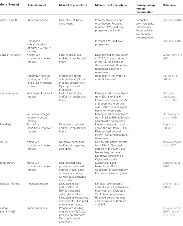
FIGURE 1 | MRI schemas of malformations. (A) Control brain,
(B) Cobblestone lissencephaly, where neuronal overmigration (represented by
gray patches at the surface of the brain) can arise due to breaks of the basement membrane. (C) Periventricular nodular heterotopia, some neurons (represented by gray nodules) remain stuck at the ventricular surface, most probably due to breaks and disorganization of the ventricular lining.
(D) Microcephaly, several mechanisms may give rise to this malformation
leading to a greatly reduced size of the brain. In pure forms, brain architecture is relatively well-preserved, in other forms (microcephaly with simplified gyral pattern, MSGP, not shown), brain organization and cortical folds are also affected. (E) Globular or ribbon-like heterotopia, represented by gray globular masses. In this case the heterotopia starts at the level of the ventricles and fills up the white matter in some brain areas. The heterotopia can appear to have gyri. Modified fromFrancis et al. (2006).
represents a different set of disorders involving perturbed progenitors (Section Basal Processes of RGCs, Shedding Light on Heterotopias, Polymicrogyria and Type II Lissencephaly). Walker Warburg syndrome (WWS) is a severe autosomal recessive disorder of this nature characterized by muscular dystrophy, eye and neuronal migration defects. Overmigration gives rise to disorganized cerebral and cerebellar cortices and multiple coarse gyri with agyric regions (cobblestone lissencephaly). As well as this, the structural brain anomalies include agenesis of the corpus callosum, cerebellar hypoplasia and hydrocephalus. WWS is grouped within a series of disorders which include Fukuyama congenital muscular dystrophy (FCMD;
Kobayashi et al., 1998), and Muscle-Eye-Brain disease (MEB). Mutations in a number of related genes have been associated with the various types of cobblestone lissencephaly (Section Basal Processes of RGCs, Shedding Light on Heterotopias, Polymicrogyria and Type II Lissencephaly, Table 1;Godfrey et al., 2007).
Polymicrogyria (e.g., OMIM 615752, 610031, 606854,
Leventer et al., 2010) is considered as a separate entity, although there can be overlapping features with cobblestone lissencephaly (Bahi-Buisson et al., 2010). This disorder is
characterized by multiple small folds at the surface of the brain, either diffuse or restricted to one brain region. The mechanisms causing this disorder initially remained elusive and they have for some time been described as affecting the end stages of migration. With the identification of various mutant genes (Table 1; Squier and Jansen, 2014), and studies in the rodent, it has become clear that a number of genes play a role in progenitors (Section Basal Processes of RGCs, Shedding Light on Heterotopias, Polymicrogyria and Type II Lissencephaly), especially the end, basal attachment of radial glial cells (RGCs) forming the pial surface of the cortex.
Periventricular heterotopia (PH, e.g., OMIM 300049,
Parrini et al., 2006) is associated with large clusters of neurons present at the ventricular surface. These are often observed on MRI as visible gray matter nodules extending into the ventricles. Since neurons are known to be generated in these regions during development, it is assumed that some neurons, after being produced, do not migrate at all. Mouse models reveal abnormalities at the ventricular lining, which is made up of apical RGC end-feet with intricate cell-cell junctions (Section Apical Adhesive Interactions and
Table 1 | Genes and human malformations. Malformation
Microcephaly1 ASPM2 CDK5RAP2 MCPH1 CENPJ WDR622 STIL CEP152 CEP63
MSGP1 WDR622 NDE1 TUBB32 ASPM2 KIF5C2
Syndrome including microcephaly TBR2 CENPE DYNC1H1 TUBG1 KIF2A KIF5C2 PLK4
(e.g., complex MCD3or associated with PMG4)
Periventricular heterotopia FLNA ARFGEF2 C6orf70 FAT4 DCHS1
Lissencephaly (type I) LIS1 DCX TUBA1A TUBB32 RELN
PMG4 TUBB2B GPR56 TUBA8 TUBB32 TUBA12A WDR622 NHEJ1 KBP
Lissencephaly (type II) FKTN POMT1 POMT2 POMGNT1 FKRP LARGE LAMB1
Atypical heterotopia5 EML1
1All genes code for proteins with centrosomal-related activities, MSGP, microcephaly with simplified gyral pattern;2Some genes appear in multiple categories
depending on the patient and gene mutation;3Complex malformation of cortical development (MCD) associated with microcephaly;4PMG, Polymicrogyria; 5Associated with macrocephaly and sometimes hydrocephaly.
Mechano-Transduction, Shedding Light on PH and Ciliopathies). Each of the PH genes (Table 1; Sheen, 2014), shows potential roles linking the plasma membrane to the cytoskeleton and some of these genes may also be important during neuronal migration.
Hydrocephaly (e.g., OMIM 307000) is associated with an abnormal quantity of cerebrospinal fluid in the ventricles, causing them to be larger than normal. Genetic causes related to specific human hydrocephaly phenotypes are still relatively unknown, with the notable exception of L1-CAM mutations (Adle-Biassette et al., 2013), involved in a syndrome including hydrocephalus due to aqueductal stenosis. Although causative mechanisms are indeed heterogeneous, hydrocephaly can arise because fluid movement is impaired. One defect is related to motile cilia on neuroependymal cells, as ciliary beating drives fluid flow (Tissir et al., 2010; Tong et al., 2014). Mouse mutations that affect motile ciliogenesis can thus lead to hydrocephalus, disruptions in neurogenesis and brain tumor formation (Han et al., 2009; Tissir et al., 2010; Hildebrandt et al., 2011). Primary non-motile cilia are known to act as mechano-transducers, transmitting signals to the developing tissue (Paridaen et al., 2013). Abnormalities in these processes may or may not be associated with hydrocephaly. Abnormal cilia, and the cycle where cilia components are disassembled to be re-used during mitosis, can also be associated with other cortical malformations. Various mouse mutants with progenitor defects show hydrocephaly (Sections Apical Adhesive Interactions and Mechano-Transduction, Shedding Light on PH and Ciliopathies,
Table 2), although often the exact causes of this remain
unidentified.
Microcephaly (e.g., OMIM 251200, 605481, Gilmore and Walsh, 2013) in human refers to a disorder in which the brain at birth is found to be significantly (−2.5–3 standard deviations below the mean) smaller than control brains. This condition leads to intellectual disability. In microcephalia vera, or primary microcephaly, although the brain is proportionally smaller, brain architecture seems not to be dramatically changed and the brain exhibits cortical folds. Its small size is indicative of a highly reduced number of neurons, premature neurogenesis or excessive cell death is likely, and most of the genes identified suggest roles in centrosomal-associated activities during division (see Table 1 and Section RGCs and Cell Division, Mechanisms Leading to Microcephaly, Gilmore and Walsh, 2013). There are a number of related disorders with microcephaly and additional cortical malformations, such as microcephaly with a simplified gyral pattern (MSGP, Adachi et al., 2011), or complex cortical malformations and polymicrogyria (e.g., OMIM 603802, 604317, e.g., Bilgüvar et al., 2010; Yu et al., 2010). These less “pure” forms show, as well as a reduced brain size, more noticeably affected gyrations (simplified or multiple small gyri), implying parallel changes in neuron production, organization and brain architecture. There is now known to be overlap between “pure” forms and those more obviously affecting gyri, as shown by unbiased genetic studies revealing mutations in genes previously identified mutated in other variants of the pathology (Poulton et al., 2014). Whole exome or genome sequencing is extremely useful in this respect, revealing
unexpected genes associated with wider phenotypes than initially thought.
Macrocephaly (e.g., OMIM 600302) potentially has multiple origins related either to increased neuron number (inverse situation compared to microcephaly) but also to increased neuropil (e.g., dendritic arborizations), the latter linked to conditions such as autism spectrum disorder (OMIM 605309). Macrocephaly due to increased neuron number is not yet as clearly elucidated as conditions such as microcephaly, probably related to the multiple potential causes of this disorder, and different types of progenitors found in primate, which are not easily studied in the rodent (Hansen et al., 2010; Wang et al., 2011). Future studies with human genetics as a starting point (and see e.g., Keeney et al., 2014a,b) will almost certainly shed further light on this condition.
MOUSE AND HUMAN MUTATIONS AND MECHANISMS IMPORTANT FOR RGC FUNCTION
GENERAL CHARACTERISTICS OF RGCs
One characteristic aspect of RGCs is their intrinsic highly polarized structure with the cell body confined to the ventricular zone (VZ), and two processes that depart from it: a long basal process reaching the pial surface, and a short apical process descending to the ventricular lining. RGCs need both processes to exert their function: the basal process constitutes the scaffold for migration of newly born neurons through the intermediate zone (IZ), while the apical process is responsible for attachment to the ventricular lining and contains key elements of signaling pathways. These are important to control the balance between proliferation and differentiation, and for cellular specification. RGCs, which are Pax6-positive, as well as self-renewing, can give rise to basal progenitors in the subventricular zone (SVZ) which are Tbr2-positive, these then give rise to post-mitotic neurons. These and further progenitor types are greatly expanded in the primate cortex (reviewed by LaMonica et al., 2012).
As RGCs present a very specialized morphology and dynamics, which are strictly linked with the function they exert during cortex development, every minor perturbation involving their structure or behavior is susceptible to lead to major developmental problems. Indeed, numerous genes coding for proteins influencing RGC morphology and function have been found mutated in mouse models and in cortical malformation patients. We try to bring together here mouse mutant data related to these genes (also resumed in Table 2), classifying these data by different RGC compartments and cellular mechanisms (resumed in Figures 2, 3).
BASAL PROCESSES OF RGCs, SHEDDING LIGHT ON HETEROTOPIAS, POLYMICROGYRIA AND TYPE II LISSENCEPHALY
Perturbations that affect basal process structure can cause subsequent problems of neuron migration and lead to cortical malformations such as heterotopia or cobblestone lissencephaly. Breaks of the cortical basement membrane (BM; Figure 2) have been associated with RGC basal process end-feet that are not well attached to the extracellular matrix (ECM). The
Table 2 | Specific genes mouse mutants.
Gene (Protein) Animal model Main RGC phenotype Main cortical phenotype Corresponding human malformation
Reference
Gpr56 (Gpr56) Knockout mouse Disruption of basal
attachment
Irregular thickness and organization; Reduced number of VZ and SVZ progenitors at E14.5 Restricted polymicrogyria; Cobblestone lissencephaly-like neuronal over-migration Bae et al. (2014) Transgenic overexpression of human GPR56 in mouse Increased VZ and SVZ progenitors Bae et al. (2014)
Itgb1 (β1-integrin) Nestin-Cre
conditional knockout mouse
Lack of basal glial endfeet; irregular glial fibers
Disorganized cortical layers at E15.5; Ectopic neurons in the MZ and deep in the cortical wall; Defective meningeal basement membrane Graus-Porta et al. (2001) Antibody-mediated blocking at E12.5 and E15.5 in mouse cortex
Progenitors divide outside the VZ; Apical process detachment; Dystrophic basal processes
Reduction in the width of cortical layers I-V
Loulier et al. (2009)
Itga (α-integrin) α6-integrin knockout
mouse
Lack of basal glial endfeet; irregular glial fibers
Disorganized cortical layers from E13.5 to E18.5; Ectopic neurons in the MZ and deep in the cortical wall; Defective meningeal basement membrane Georges-Labouesse et al. (1998) α3- and α6-integrin double knockout mouse
Disorganized cortical layers from E13.5 to E16.5; Ectopic neuroblastic outgrowths
De Arcangelis et al. (1999)
Fak (Fak) Emx1-Cre
conditional knockout mouse
Defective basal glial endfeet; irregular glial fibers
Neuronal ectopia in and above the MZ from E14.5; Disorganized cortical layers; Disrupted basement membrane
Beggs et al. (2003)
Ilk (Ilk) Emx1-Cre
conditional knockout mouse
Defective basal glial endfeet; disorganized glial fibers
Cortical lamination defects from E14.5; Neuronal ectopia in the MZ; Basal lamina fragmentation; Defective positioning of Cajal-Retzius cells
Niewmierzycka et al. (2005)
Rhoa (RhoA) Emx1-Cre
conditional knockout mouse Disorganized basal processes; neuronal ectopia in MZ; Loss of apical anchoring; Mitotic cells scattered ectopically
Subcortical band heterotopia (SBH); Cobblestone lissencephaly-like neuronal over-migration;
Cappello et al. (2012)
Marcks (Marcks) Knockout mouse Disrupted radial
glial scaffold at E15.5; Abnormal radial glial endfeet; Disrupted apical polarity components; Disrupted mitotic orientation;
No clear delineation of cortical layers; Cobblestone lissencephaly; Disrupted VZ; Ectopic progenitors; Reduced cellular density and thickness of both VZ and SVZ
Weimer et al. (2009)
Lama2
(Laminin-α2)
Knockout mouse Progenitors dividing outside the VZ; Apical process detachment; Dystrophic basal processes Miyagoe et al. (1997); Loulier et al. (2009) (Continued)
Table 2 | Continued
Gene (Protein) Animal model Main RGC phenotype Main cortical phenotype Corresponding human malformation Reference Mltt4 or Cdh2 (Afadin and Cdh2) Emx1-Cre conditional knockout mouse Disruption in adherens junctions, Progenitors divide outside the VZ; Shorter cell cycle and reduced cell cycle exit
Double cortex-like phenotype
Gil-Sanz et al. (2014)
FlnA (FlnA) Knockout mouse Loss of adherens
junctions
Focal disruptions of the VZ/SVZ and cell expansion into the ventricular space; Disruptions of the VZ surface
Periventricular heterotopia (PH)
Feng et al. (2006)
Mekk4 (Mekk4) Knockout mouse Periventricular heterotopia
(PH); Focal disruptions of the VZ/SVZ and cell expansion into the ventricular space; Thinner IZ; Subpial ectopia and polymicrogyria; Decrease in CP thickness; Sarkisian et al. (2006) E14.5 mouse RNAi-mediated knockdown by in utero electroporation Laminin disruption-mediated disorganization of radial glial fibers
Sarkisian et al. (2006)
Fat4 (Fat4) E13.5 and
E14.5 mouse RNAi-mediated knockdown by in utero electroporation Increased progenitor proliferation in VZ and SVZ; Block in differentiation between the Pax6+ and Tbr2+ states
Progenitor displacement, accumulation of neural precursors, and periventricular heterotopia (PH) Van Maldergem syndrome with a partially penetrant PH phenotype Cappello et al. (2013) Dchs1 (Dchs1) E13.5 and E14.5 mouse RNAi-mediated knockdown by in utero electroporation Increased progenitor proliferation in VZ and SVZ; Block in differentiation between the Pax6+ and Tbr2+ states
Progenitor displacement, accumulation of neural precursors, and periventricular heterotopia (PH) Van Maldergem syndrome with a partially penetrant PH phenotype Cappello et al. (2013) Efnb1 (Ephrin-B1) E13.5 mouse overexpression by in utero electroporation Blocking in progenitor differentiation Qiu et al. (2008) E13.5 mouse RNAi-mediated knockdown by in utero electroporation Increased progenitor differentiation Qiu et al. (2008)
Knockout mouse Loss of RGC radial organization; Reduced progenitor number
Irregular appearance with micro-invaginations at the apical surface of the neuroepithelium; Misplacement of mitotic nuclei within the cortical wall
Davy et al. (2004); Qiu et al. (2008); Arvanitis et al. (2013)
Ar13b (Arl13b) Knockout mouse
(null allele)
RGC inverted polarity; RGC somas ectopically located near the pial surface; ciliary-based signaling perturbed
Neurons ectopically located near the ventricular surface; Reversed Reelin localization; Discontinuous pial membrane; Disrupted apical adherens junctions; Marked disruption of neuronal layer organization
Joubert syndrome with disrupted neurogenesis and malformed cerebral cortex Cantagrel et al. (2008); Higginbotham et al. (2013) FoxG1-Cre conditional knockout mouse Abnormal organization of the RGC scaffold; Loss of RGC apical-basal polarity Perturbed neuronal positioning and layer formation
Higginbotham et al. (2013)
Table 2 | Continued
Gene (Protein) Animal model Main RGC phenotype Main cortical phenotype Corresponding human malformation Reference α-E-catenin (α-E-catenin) Emx1-Cre conditional knockout mouse Disruption of radial glial morphology and ventricular lining architecture; Formation of rosette-like structures
Formation of a large SBH and thinner layered cortex
Schmid et al. (2014) Nestin-Cre conditional knockout mouse Disruption of RGC apical-junctional complexes; Loss of RGC polarity; At E13.5 abnormal activation of the Shh pathway causing: cell cycle shortening, increased number of mitotic cells, decreased apoptosis
Massive dysplasia; VZ cell dispersion; Increase in cortical thickness and size
Lien et al. (2006) Ctnnb1 (β-catenin) FoxG1-Cre conditional knockout mouse Disruption of progenitor apical adherens junctions
Telencephalon size severely reduced at E10.5; Loss of neuroepithelial integrity; Progenitor delamination and apoptosis Junghans et al. (2005) Transgenic overexpression in mouse progenitors Tangential increase in progenitors number and formation of cortical folds
Chenn and Walsh (2003)
Pard3 (Par3) E12 mouse
RNAi-mediated knockdown by
in utero lentiviral
injection
Perturbation in the apical Par polarity complex
Decreased progenitor proliferation; Premature cell cycle exit and neuronal differentiation
Costa et al. (2008)
Pard6 (Par6) E13 mouse
overexpression by in utero retroviral injection Increased progenitor number Prolonged maintenance of VZ progenitors Costa et al. (2008) Cdc42 (Cdc42) Emx1-Cre conditional knockout mouse Gradual disappearance of adherens junctions; Detachment of RGCs from the ventricular surface and conversion into basal progenitors
Severely disorganized cortex; Increased cortical thickness due to increased neurogenesis
Cappello et al. (2006)
Numb (Numb) and Numbl (Numbl)
Nestin-Cre Numb
and Numbl double conditional knockout mouse
Loss of progenitor radial organization
RGC clustering in neurogenic foci along the cortex; Increased progenitor number; Increased apoptosis; Reduced neuronal differentiation; Disruption of neuroepithelium integrity Petersen et al. (2002, 2004),Li et al. (2003)
Aspp2 (Aspp2) Knockout mouse Disruption of both tight
and adherens junctions; Progenitor expansion and mislocalization Hydrocephalus; Drastic ventricular dilatation Sottocornola et al. (2010)
Lgl1 (Lgl1) Knockout mouse Disruption of apical tight
and adherens junctions; Progenitor expansion; Loss of RGC polarity; Disruption of RGC apical domain
Hydrocephalus; Disruption of the VZ surface; Reduced differentiated neuronal cell population
Klezovitch et al. (2004)
Table 2 | Continued
Gene (Protein) Animal model Main RGC phenotype Main corticalphenotype Correspondinghuman malformation
Reference
aPKCλ (aPKCλ) Nestin-Cre
conditional knockout mouse
Loosely packed RGCs; Disrupted adherens junctions and apical processes
Rough ventricular surface at E15.5; VZ, SV and IZ severely disorganized and difficult to distinguish at E16.5
Imai et al. (2006)
Mcph1 (Mcph1) Knockout mouse Spindle misorientation
favoring asymmetric divisions; Delayed and imbalanced centrosomal maturation; Abnormal spindles and chromosome misalignment; Lengthening of cell cycle progression
Small brain due to 20% decrease in thickness and lateral dimensions; Decrease in progenitor proliferation and premature neurogenesis Autosomal recessive primary microcephaly type 1 Gruber et al. (2011) Cdk5rap2 (Cdk5Rap2) Homozygous mouse mutant
Mitotic delay; Aneupolar spindle poles; Spindle orientation defects
Decreased cortical thickness mainly in superficial layers; Decrease in the size of apical and basal progenitor populations; Progenitor and neuronal cell death Autosomal recessive primary microcephaly Lizarraga et al. (2010)
Cenpj (Cenpj) Nestin-Cre
conditional knockout mouse
Mitotic delay and cell death of delocalized progenitors
Decreased brain size and cortical thickness
Autosomal recessive primary microcephaly Insolera et al. (2014) Plk4 (Plk4) Nestin-Cre conditional overexpressing mouse Supranumerary centrosomes; Multipolar spindles; Prolonged mitosis; Frequent aneuploidy and apoptosis
Drastically reduced brain size; Decreased radial thickness; Reduced numbers of apical progenitors, basal progenitors and post-mitotic neurons Microcephaly, growth failure and retinopathy Marthiens et al. (2013),Martin et al. (2014)
Htt (Huntingtin) E14.5 mouse
RNAi-mediated knockdown by in utero electroporation Spindle orientation defects Increased neuronal differentiation at the expense of progenitors Godin et al. (2010) Nestin-Cre conditional knockout mouse Spindle orientation defects Increased neuronal differentiation at the expense of progenitors Godin et al. (2010) Homozygous mutant huntingtin (glutamine expansion) carrying mouse Spindle orientation defects
Thinner VZ and thicker CP; Thinner total cortical thickness
Molina-Calavita et al. (2014)
Tcof1 (Treacle) Heterozygous
mouse
Spindle orientation defects; M-phase extension and mitotic delay
Brain hypoplasia; Reduced number of neurons; Reduced apical progenitor population Treacher Collins Syndrome (TCS) showing microcephaly Sakai et al. (2012)
Nde1 (Nde1) Knockout mouse Mitotic spindle defects
resulting in mitotic delay/arrest; Increased horizontal cleavage orientation
Small-brain phenotype; Thinning of the cortex more pronounced in superficial cortical layers
Micro-lissencephaly syndrome due to defects in neuron production and cortical lamination
Feng and Walsh (2004)
Table 2 | Continued
Gene (Protein) Animal model Main RGC phenotype Main cortical phenotype Correspondinghuman malformation
Reference
Nde1, Lis1 (Nde1,
Lis1)
Nde1 null and Lis1
heterozygous double mutant mouse
Decreased self renewal and accelerated cell cycle exit; Increase in horizontal mitosis; Defects in metaphase plate formation; Failed mitotic spindle function; Disruption of apical
Severely disorganized and thinner cerebral cortex; Lack of distinct cellular layers; Reduced radial unit number
Micro-lissencephaly
Pawlisz et al. (2008)
integrity and lateral contacts during mitosis
Lis1 (Lis1) hGFAP-Cre
conditional knockout mouse Spindle orientation defects; Premature reduction of RGC population
Thinner cortex; Less organized cellular structure
Lissencephaly Yingling et al. (2008) PP4c (PP4C) Emx1-Cre conditional knockout mouse Spindle orientation defects; Premature reduction of RGC population
Thinner and disorganized cortical layers
Xie et al. (2013)
Vangl2 (Vangl2) Knockout mouse Spindle orientation
defects
Reduced size of the cortex; Premature progenitor differentiation; Decreased neuronal production
Lake and Sokol (2009)
Insc (Insc) Nestin-Cre
conditional knockout mouse
Spindle orientation defects
Thinner cerebral cortex; Decreased neurogenesis Postiglione et al. (2011) Ubiquitous overexpression in mouse Spindle orientation defects
Thicker cerebral cortex; Increase in basal progenitor number; Increased neurogenesis
Postiglione et al. (2011)
Wnt3a (Wnt3a) E13.5
overexpression by in
utero electroporation
in mouse
Progenitor expansion Increased VZ thickness; Increased basal progenitor number and differentiation into neurons; Neuronal heterotopia; Thinner cerebral cortex
Munji et al. (2011)
Pax-6 (Pax-6) Homozygous loss of
function mouse Altered spindle orientation; Unequal inheritance of apical membrane domains; Decrease in apical complex proteins transcription Decreased tangential expansion of the cerebral cortex; Ectopic progenitors
Asami et al. (2011)
LGN (LGN) Knockout mouse Randomized mitotic
orientation
Decreased thickness of the VZ; Ectopic Pax6+ and Tbr2+ progenitors
Konno et al. (2008)
Eml1 (Eml1) Homozygous loss of
function mouse
Defects in mitotic spindle orientation
Slightly decrease in VZ thickness; Both Pax6+ detached and Tbr2+ ectopic progenitors Periventricular and globular ribbon-like subcortical heterotopia; Macrocephaly; Hydrocephaly Kielar et al. (2014)
Kif20b (Kif20b) Homozygous loss of
function mouse
Abnormally shaped and misaligned midbodies
Reduced cortical thickness; Greatly reduced output progeny of apical progenitors
Janisch et al. (2013)
meningeal BM is located immediately below the pia matter and serves as an anchor point for the end-feet of RGCs and as a physical barrier to migrating neurons. Through human genetics studies, it has been shown that cobblestone lissencephalies are
associated with reduced glycosylation of alpha-dystroglycan, a basal process dystrophin-associated glycoprotein that is crucial to act as anchor between the dystrophin complex and the ECM (e.g., laminin, van Reeuwijk et al., 2005; Roscioli et al.,
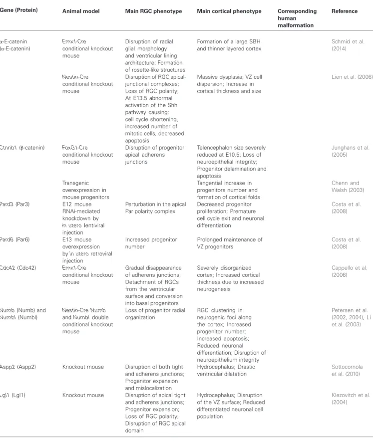
FIGURE 2 | RGC mechanisms leading to mouse malformations. (A) Control situation, apical progenitors (containing blue nuclei) divide by
interkinetic nuclear migration (INM) in the VZ, neurons (burgundy nuclei) migrate radially on RGC basal processes across the IZ, to settle in the CP. Cajal Retzius cells (ovals) present in the MZ secrete signals to the migrating neurons. End-feet of RGC basal processes receive signals from ECM molecules in the BM close to the pial surface. (B) Cobblestone lissencephaly phenotype, in this case some RGC basal processes are not well attached to the pial surface, possible breaks in the BM potentially cause neurons (burgundy nuclei at the surface of the brain) in some regions to overmigrate into the meningeal space. (C) Periventricular disorganization, some neurons (burgundy nuclei) remain stuck at the ventricular surface, most probably due to breaks in the ventricular lining where apical end-feet of RGCs normally
attach. (D) Microcephaly phenotype, two potential mechanisms may give rise to this malformation leading to a greatly reduced size of the brain. Some mouse models suggest that premature differentiation of progenitors into post-mitotic neurons (burgundy nuclei within radially migrating neuron close to VZ) depletes the progenitor pool (represented by red cross over blue nuclei in VZ). Other studies show instead increased cell death of abnormal progenitors (red cross over blue nuclei present in IZ). (E) Globular heterotopia (e.g., HeCo mice), in this case a proportion of apical progenitors detach from the ventricular surface (represented by blue nuclei without apical attachment to the ventricular lining) and retain proliferation capacity, providing a local source of neurons in the IZ (burgundy nuclei). A subcortical heterotopia subsequently arises. VZ, ventricular zone; IZ, intermediate zone; CP, cortical plate; MZ, marginal zone; BM, basement membrane.
2012; Buysse et al., 2013). Six major genes have been identified encoding putative or demonstrated glycosyltransferases, POMT1, POMT2, FKTN, FKRP, LARGE and POMGnT1. Post-mortem studies have helped characterize this disorder, however, mouse models for these genes are often not viable, which has led to the difficulty of studying the exact mechanisms involved (Brockington et al., 2001; Yoshida et al., 2001; Beltrán-Valero de Bernabé et al., 2002; Longman et al., 2003; van Reeuwijk et al., 2005; Manzini et al., 2012; Roscioli et al., 2012; Willer et al., 2012). Fragmentation of the BM is however, also frequently seen after deletion of other ECM components or receptors. Laminins are major secreted glycoproteins found in the BM, where they influence cell proliferation, differentiation, migration, and adhesion. LAMB1, encoding the laminin subunit beta-1 is involved in basal process attachment to the pial surface and also
found mutated in a cobblestone brain malformation (Radmanesh et al., 2013). Thus, mutations in laminin subunit genes, as well as glycosyltransferases, may both lead to detachment of RGC processes from the pial surface leading to breaches of the BM, disintegration of the scaffold mediating neuronal migration, subcortical heterotopias and neuronal overmigration phenotypes. These studies hence reveal the progenitor origin of certain “neuronal migration” defects (but see also Moers et al., 2008, for intrinsic problems in migrating neurons, affecting potentially their ability to stop migrating).
Similarly, the GPR56 gene encodes a heterotrimeric guanine nucleotide-binding adhesion protein (G-protein)-coupled receptor that is highly expressed in progenitors, is localized to the basal process, and binds ECM proteins at the pial surface (Li et al., 2008; Luo et al., 2011). Disruption of
FIGURE 3 | Radial glial cell and different genes. Schematic representation
of interphase and dividing RGCs (in green) and neurons (in gray) migrating along basal RGC processes. Higher magnifications show RGC structural details such as basal attachment to the pial surface (represented in red), apically located adherens junctions (black), apical attachments and midbody (purple), centrosomes (pink), primary cilia (blue), and a mitotic cell with the MTs organized in the mitotic spindle (light blue) and the DNA aligned at the metaphase plate (black). The ventricular surface is represented as a gray line. The different genes are represented close to the structure in which they have
clearly been shown to be involved based on the classification proposed in this review, with a color corresponding to the color of the structure. Genes involved in RGC basal process attachment to the pial surface are represented in red; in black are genes linked with adherens junctions; in purple are genes participating in apical polarity, attachment to the ventricular surface, and in the apically-located midbody; in blue are genes essential for the primary cilium; in pink are represented centrosome-related genes, and in light blue genes participating in the regulation of mitotic spindle function. Genes that are involved in transcriptional regulation are represented in green.
GPR56 was found to selectively and bilaterally perturb
the human cortex surrounding the Sylvian fissure with a strikingly restricted polymicrogyria (Bae et al., 2014). Loss
of GPR56 disrupts RGCs pial anchorage and causes breaks in the BM, through which some neurons over-migrate (Li et al., 2008; Bahi-Buisson et al., 2010). Studies involving this
gene hence link these phenotypes to mechanisms leading to polymicrogyria. Moreover, even when the pia is intact, as observed in Gpr56 knockout mice, cortical thickness and organization are irregular with periodic thinner regions. Such defects suggest proliferation problems and indeed in these mice there are less mitotic progenitors in both the VZ and SVZ at embryonic day (E) 14.5. Conversely, in mice carrying a transgene that overexpresses human
GPR56, the opposite effect was observed (Bae et al., 2014). These data show that disruption of basal processes and overmigration, can be intimately linked with a perturbation of proliferation.
RGC basal processes have been postulated to regulate progenitor proliferation via integrin signaling (Radakovits et al., 2009; Fietz et al., 2010, see Section Apical Adhesive Interactions and Mechano-Transduction, Shedding Light on PH and Ciliopathies). GPR56 influences attachment to ECM proteins, such as collagen type III, and tetraspanins, which are known to also bind integrins expressed by basal end-feet (Xu and Hynes, 2007; Li et al., 2008; Luo et al., 2011). Studies in conditionalβ1-integrin knockout mice showed a wavy appearance of cortical layers at E15.5, indicative of defects in the organized laminar cytoarchitecture and abnormal positioning of cortical neurons. Neurons either invaded the marginal zone (MZ) or accumulated deep in the cortical wall, resembling cobblestone lissencephaly. RGC fibers in the mutants terminated at varying positions close to the BM and were highly irregular ( Graus-Porta et al., 2001). All these observations, together with the finding, by conditional ablation specifically in neurons, that β1-integrins are not essential for neuron-glia interactions and neuronal migration per se (Belvindrah et al., 2007), indicated that they are likely to primarily be required for anchorage to the BM. Defects similar to those found in β1-integrin knockout mice were also found in mice with mutations in the genes encoding the integrin α6-subunit or both α3 and α6 (Georges-Labouesse et al., 1998; De Arcangelis et al., 1999; Hynes, 2002); other components of the BM (Miner et al., 1998; Halfter et al., 2002; Pöschl et al., 2004); and the integrin downstream effectors focal adhesion kinase (FAK;Beggs et al., 2003) and integrin-linked kinase (ILK; Niewmierzycka et al., 2005).
Studies involving β1-integrin, the small GTPase RhoA and the protein Marcks (Myristoylated alanine-rich substrate protein) also highlight the fact that a number of proteins are likely to have a role in both apical and basal processes. The conditional deletion of RhoA and Marcks in the developing mouse cortex leads to a prominent tissue mass (heterotopia) found underneath an apparently layered but thinner cortex. Moreover, in the case of RhoA deletion, there was a phenotype reminiscent of cobblestone lissencephaly. Analysis of progenitor morphology also revealed already at E12.5, that mitotic cells were scattered in the cortex instead of being neatly aligned at the ventricular lining. At E16 mitotic cells were assembled into a broad band located abnormally in the middle of the cortex between the pial and the ventricular surfaces. Moreover, RGCs had mis-oriented processes and had lost their apical anchoring. While RhoA-depleted neurons still migrated fairly normally in a wild-type
environment, they followed a largely non-radial path when the RGC scaffold was disturbed by RhoA depletion (Cappello et al., 2012). RhoA plays a role in polymerizing actin into fibers (F-actin) (Etienne-Manneville and Hall, 2002). Thus, loss of RhoA destabilized the actin and microtubule (MT) cytoskeletons in both neurons and RGCs but the most severe consequences were in RGC positioning and in the proper formation of the basal scaffold (Cappello et al., 2012). Knockout mice for Marcks, an actin-cross-linking protein and subcellular substrate of protein kinase C (PKC), also presented a disorganized RGC scaffold, impaired cell polarity, disorganized VZ, and ectopic progenitors. Marcks is a potent upstream regulator of the localization and function of cell polarity complexes, and its mutation leads to disrupted VZ organization and mitotic orientation (Blackshear et al., 1997; Weimer et al., 2009). At E15.5, the RGC scaffold was severely disrupted and basal end-feet failed to branch appropriately and instead had a club-like, balled-up appearance as they reached the pial surface. There was a reduction in the thickness of both the VZ and SVZ, and perturbed migration. Phenotypes combining apical and basal perturbation thus show misplaced progenitors, perturbed proliferation and either inability of neurons to reach the cortex or overmigration. In the different models whether the basal detachment causes the detachment of the apical process, or vice versa, is not always clear. These studies also emphasize that the cytoskeleton is essential for the maintenance of RGC structure.
APICAL ADHESIVE INTERACTIONS AND MECHANO-TRANSDUCTION, SHEDDING LIGHT ON PH AND CILIOPATHIES
Perturbations of the apical domain of RGCs, a complex and relatively well-studied cell compartment, are becoming recognized as leading to neuroepithelial disorganization, different types of heterotopia (this section), or micro- and macro-cephaly (Section RGCs and Cell Division, Mechanisms Leading to Microcephaly), depending on the affected cell mechanisms. The consequence of the perturbation can appear as obvious breaks in the ventricular lining and changes in VZ architecture, or be more subtle. To help explore these phenotypes, we further focus in this section on molecules which have been described to play a role in intercellular and ECM contacts in the VZ.
We re-mention here β1-integrins which have also been shown to be involved in end-feet anchoring to the ventricular surface, binding with laminins, located also in the apical ECM. These anchors are reinforced by cadherin–catenin-based adherens junctions, which help attach apical end-feet of adjacent RGCs to each other (Kadowaki et al., 2007). Blocking β1-integrin’s function by injection of specific antibodies into the lateral ventricles of embryos at E12.5 and E15.5 showed a significant increase in dividing cells due to a larger abventricular (localized outside the VZ) dividing progenitor population (Loulier et al., 2009). This phenotype was linked to the detachment of apical processes from the ventricular surface and alterations in mitotic spindle orientation showing thatβ1-integrin plays a critical role in the adhesion that maintains the progenitor cells within their niche and preserves the architecture of the VZ. The same effect was observed in laminin-α2 deficient mice (Miyagoe et al., 1997; Loulier et al., 2009). Another link between apical VZ integrity and
heterotopia formation is represented by the conditional deletion in the cortex of the apical junction moleculeα-E-catenin. Due to the disruption of RGC morphology, caused by impaired actin cytoskeletal organization, progenitors were found disorganized in rosette-like structures, associated with a large heterotopia and a thin layered cortex (Schmid et al., 2014). Another component of adherens junctions is Afadin, which interacts with cadherins and stabilizes them (Sato et al., 2006). Its conditional inactivation in the developing cortex leads to disruption of adherens junctions, dispersion of dividing progenitors with a shorter cell cycle and reduced cell cycle exit, and formation of a double cortex-like phenotype (Gil-Sanz et al., 2014). In the same study, conditional knockout mice for a cadherin subunit, Cdh2, showed a very similar phenotype, close also to that described previously for RhoA-knockout mice (Cappello et al., 2012). Thus intercellular contacts and downstream pathways seem to be essential to maintain the integrity of the VZ and to regulate proliferation.
Similarly, the Eph/ephrin signaling pathway (Nievergall et al., 2012) activates signal transduction cascades, and exhibits extensive cross-talk with other receptors, including cadherins and integrins (Arvanitis and Davy, 2008). Ephrin B1 is expressed in apical progenitors from the neuroepithelial stage in a ventricular-high to pial-low gradient (Stuckmann et al., 2001). Prolonged Ephrin B1 activity was shown to prevent progenitor differentiation, while loss of function had an opposite effect promoting differentiation and leading to loss of progenitor cells (Qiu et al., 2008; Murai et al., 2010). Indeed, Ephrin B1 reverse signaling controls the switch between progenitor maintenance and neuronal differentiation (Arvanitis et al., 2010). In knockout mice, and more severely in heterozygous mice, the neuroepithelium had an irregular appearance with formation of micro-invaginations due to abnormal folding of the VZ without changes in apico-basal polarity of progenitors. Progenitor detachment was also observed. In absence of Ephrin B1, local alterations of the apical surface might weaken the rigidity and cohesion of the neuroepithelium. The reason why in heterozygous embryos the phenotype is more severe might be that sorting between Ephrin B1-positive and Ephrin B1-negative cells leads to discontinuous rigidity, which is more detrimental to the morphogenesis of this tissue than a homogeneous decrease in rigidity. Thus, the normal function of Ephrin B1, via an interaction with EphB2 on neighboring cells, is to maintain morphology and localization of progenitors in the VZ by promoting apical integrin-based adhesion (Arvanitis et al., 2013).
Mutations in the Filamin A (FLNA) gene were found in 100% of families with X-linked bilateral PH and in 26% of sporadic patients with PH (Fox et al., 1998; Parrini et al., 2006). β1-integrin mediated adhesion to the ECM was also found to be dependent on the binding of FLNA to vimentin and PKC epsilon (PKC1, Kim et al., 2010) allowing vimentin phosphorylation by PKC1. This step is crucial for the activation and trafficking of β1-integrin to the plasma membrane. FLNA encodes a large phosphoprotein that crosslinks actin filaments into orthogonal networks, reorganizing them by interacting with several proteins at the membrane (Stossel et al., 2001; Nakamura et al., 2007).
It may play a role both in progenitors and migrating neurons. The ventricular surface has been shown to be disrupted in FlnA knockout mice (Feng et al., 2006). PH formation and alterations in the neuroepithelial lining were also shown in FlnA-knockdown brains where disruption of both the polarized RGC scaffold and the neuroepithelial lining were the likely cause of the PH (Carabalona et al., 2012). Also, loss of mitogen-activated protein kinase kinase kinase 4 (MEKK4) in mice, a regulator of FlnA phosphorylation, leads to a similar phenotype (Sarkisian et al., 2006). A second human PH gene, ARFGEF2, coding for brefeldin-A-inhibited guanine exchange factor-2 (BIG2) is likely to play a role in endocytosis, regulating levels of Arf1 at the plasma membrane, which is known to regulate cell-cell contacts (Zhang et al., 2013). PH was also induced by knockdown of C6orf70 in the developing rat cortex, a gene of unknown function, mutated in a PH patient, and coding for a protein with a vesicle-like subcellular localization (Conti et al., 2013). These combined data suggest a coordinated role for actin and vesicle trafficking in controlling cell adhesion in apical regions in the VZ (Sheen, 2014).
Related to this, protocadherins Dchs1 and Fat4 (Cappello et al., 2013) are important for an apically located adhesive complex (Ishiuchi et al., 2009). Van Maldergem syndrome, an autosomal-recessive multiple malformation syndrome, shows a partially penetrant PH phenotype caused by mutations in FAT4 or DCHS1. Dchs1 is the ligand of the Fat4 receptor and the complex they constitute is situated apically, closer to the ventricle relative to adherens junctions. Fat4 and Dchs1 knockdown studies in mice also showed an increased cell proliferation in the VZ and SVZ, a block in differentiation between the Pax6+ and Tbr2+ states, and an accumulation of neuronal precursors, showing that this adhesive complex normally suppresses continued proliferation (Cappello et al., 2013). Adhesion and proliferation hence seem to be interlinked themes related to these phenotypes.
Another very important characteristic of the apical domain of RGCs is the presence of the primary cilium, an MT-based, slender projection from the cell that is thought to be important for sensing signaling factors present in the cerebrospinal fluid, and with a guiding role in the establishment of apical-basal polarity of the RGC scaffold. The importance of cilia function for cortical development is evident in developmental brain disorders such as Joubert, Meckel-Gruber, orofaciodigital and Bardet-Biedl syndromes (commonly referred to as ciliopathies), where disrupted cilia and the resulting changes in cortical formation may underlie cognitive deficits and intellectual disability (Cantagrel et al., 2008). Mutations in a gene encoding the centrosome-associated protein CEP290, important for ciliogenesis (Kim et al., 2008), have been found in both Meckel-Gruber and Joubert syndromes (Valente et al., 2006; Frank et al., 2008). Also, Arl13b, a small GTPase of the Arf/Arl family that is mutated in Joubert syndrome, is specifically localized to cilia and controls the MT-based, ciliary axoneme structure. Deletion of Arl13b impairs the cilium’s ability to convey critical extracellular signals such as Shh (Caspary et al., 2007). In constitutive mutant mice, and in E9 conditional knockout mice, early neuroepithelial progenitors showed markedly perturbed polarity with the soma located near the pial surface and the basal
end-feet located near the VZ. These cells divided ectopically at or near the pial surface, instead of adjacent to the ventricular surface (Higginbotham et al., 2013). These studies show that primary cilia play an important role in both signal transduction and polarity.
Indeed, RGC polarity is a crucial issue for cortical development. We resume this only briefly here (see details in Table 2 and Figure 3). Conditional mutagenesis in the mouse or focal knockdown experiments, have often been necessary to reveal the role of a particular polarity protein, in this case it remains difficult to directly link these data with malformations. What clearly comes out of the different studies is the relationship between apical polarity complexes (Par-Complex and its regulators), maintenance of the structure of the ventricular lining and neuroepithelium, and regulation of cell proliferation/differentiation and fate. Thus, defects in the polarity complexes have been studied both prior to neurogenesis and during the neurogenic period. Changes in the balance between expansion of RGCs, production of basal progenitors, and differentiation of post-mitotic neurons have been identified but are still little-understood. This imbalance can be the cause of the incapacity of the brain to form an ordered layer structure and/or a brain of the correct size (see also Section RGCs and Cell Division, Mechanisms Leading to Microcephaly). Diverse mechanisms can be affected by the perturbation of different polarity molecules, related to the complex interactions that link the different players. The variable consequences are also likely to be due to the different importance these molecules have during neuroepithelial progenitor expansion and/or the neurogenic phase when RGCs have a major role. Also, cell adhesion complexes, strictly related to polarity components, change during the transition from early neuroepithelial cells to RGCs (Götz and Huttner, 2005), adding to the complexity. Mechanisms leading to hydrocephaly identified in some mutant mice remain complex, however, disruption of the early neuroepithelium and polarity changes are clearly associated.
RGCs AND CELL DIVISION, MECHANISMS LEADING TO MICROCEPHALY
We previously discussed how cell junctions and the integrity of apical polarity domains are important for regulating the structure of the ventricular lining and the balance between proliferation and differentiation. However, there are other cellular mechanisms that are more solely linked with proliferation/differentiation and cell fate. RGC centrosome behavior, mitosis, the regulation of spindle orientation (which has effects on symmetric and asymmetric division and cell localization), cytokinesis and interkinetic nuclear migration (INM) are finely regulated processes, and several cortical malformation genes or mouse mutants associated with these mechanisms have been studied. We have classified these phenotypes within separate sub-categories, but these can still often be considered as overlapping.
Microcephaly genes and centrosome function
The centrosome is important for correct spindle assembly and function during mitosis. Centrosomes influence the morphology of the MT cytoskeleton, function as the base for the primary cilium and integrate important signaling pathways.
At the core of a typical centrosome are two cylindrical MT-based structures termed centrioles, which recruit a matrix of associated pericentriolar material (Nigg and Stearns, 2011). RGC centrosomes are located at the extremity of the apical process and are aligned at the ventricular surface. This position influences cell polarity and anchoring in the VZ. Once a cell enters mitosis, centrosome duplication takes place and these move more basally to help form the spindle poles and the bipolar mitotic spindle. A set of proteins related to centrosome behavior has been identified, whose mutation was found to cause microcephaly. Mutations in ASPM (abnormal spindle-like microcephaly associated) are the most common cause of primary microcephaly in humans (Kumar et al., 2004; Pichon et al., 2004; Shen et al., 2005; Gul et al., 2006). Aspm has been shown to exert a critical role at the spindle poles of neuroepithelial cells, maintaining spindle position during mitosis and, consequently regulating the precise cleavage plane orientation required for symmetric, proliferative divisions (Fish et al., 2006). Microcephalin (MCPH1) mutations also cause primary microcephaly type 1 (Woods et al., 2005).
Mcph1 is expressed at high levels in the VZ and SVZ at E13.5
and E15.5 (Gruber et al., 2011) and Mcph1-deficient mice have a small brain. Characterization of the mutant cortex revealed premature production of neurons and exhaustion of progenitors. Mcph1 deficiency specifically caused a delayed and imbalanced centrosomal maturation, leading to a lengthening of the cell cycle due to abnormal spindles and chromosome misalignment (Gruber et al., 2011). Another example of a gene mutated in primary microcephaly is SCL-interrupting locus protein (STIL), encoding a centriole-duplication factor that localizes to the procentriolar cartwheel region, a key structure in procentriole assembly. STIL depletion was shown to completely block centriole formation, whereas its overexpression resulted in extensive centriole amplification (Arquint and Nigg, 2014).
Human primary microcephaly is also caused by mutations in
CDK5RAP2 (cyclin-dependent kinase 5 related activator protein
2, Bond et al., 2005). In somatic cells, CDK5RAP2 promotes centrosomal cohesion (Graser et al., 2007) and recruits the γ-tubulin ring complex (γ-TuRC)—the MT nucleator—to the centrosome (Fong et al., 2008). In a homozygous mouse model of Hertwig’s anemia (an), the disease is caused by a mutation in Cdk5rap2 (Lizarraga et al., 2010). Brain size was reduced and an increased ventricular size and decreased cortical thickness were already detected at E13.5. Mutant animals had fewer total neurons and the last-born superficial neurons were particularly reduced. The premature decrease in progenitors was due to problems encountered during mitosis causing cell death affecting both progenitors and neurons and, possibly, changes in cell fate. Indeed, an increase in pro-metaphase and metaphase precursor cells with mono-, tri-, and other aneupolar spindle poles, together with defective spindle orientations, were detected (Lizarraga et al., 2010). Mutations in centromere protein J (CENPJ) also cause microcephaly (Bond et al., 2005). This gene is involved in the maintenance of centrosome and spindle integrity. A recent study described conditionally inactivated Cenpj also known as SAS-4 (Insolera et al., 2014). This led to mitotic delay, p53 activation and cell death of delocalized progenitors. Keeping cells alive
by p53 inactivation showed many RGCs in the IZ, which were multipolar but could still divide, self-renewing and producing also basal progenitors and neurons. Under these conditions small heterotopias formed in the IZ. This study showed that cell death was not due to aneuploidy or other chromosomal abnormalities, unlike hypomorphic Cenpj mutants (McIntyre et al., 2012), instead delocalized RGCs were often remarkably deficient in centrioles and cell death was most probably due to mitotic delay.
Centrosome amplification may also cause microcephaly by affecting the correct formation of the spindle and continuation through mitosis. Polo-like kinase 4 (Plk4) is a centriole duplication protein whose overexpression leads to cells with supernumerary centrosomes. Conditional overexpression of Plk4 specifically in progenitors, led to reduced brain size, accompanied by a reduction of both apical and basal progenitors, and the neuronal population. In Plk4 overexpressing embryos, cells with extra centrosomes showed bipolar, as well as multipolar, spindle configurations, and spent more time in mitosis. This was at the origin of p53-dependent cell death and could be one of the main causes of brain reduction in this model. Deletion of p53 showed accumulation of aneuploid daughter progenitors, and these underwent premature neuronal differentiation, with subsequent depletion of the progenitor population (Marthiens et al., 2013). Related to this work, another two MCPH proteins, CEP63 and CEP152, form a complex that is an essential part of the molecular machinery controlling centrosome numbers, and defects in either component result in a diminished pool of precursors that cannot provide an adequate supply of neurons (Sir et al., 2011). Thus centrosome formation, numbers, maturation and function are all important for maintaining a correct progenitor number.
Spindle genes and mitosis
The cell cycle of RGCs is characterized by an oscillatory behavior called INM. Mitosis occurs apically close to the ventricular surface, nuclei then migrate basally during G1 to reach the most basal side of the VZ where they undergo S-phase, and migrate apically during G2 to reach the ventricular surface before undergoing mitosis again. This behavior of the nuclei of RGCs gives the VZ the appearance of a pseudo-stratified epithelium. A variety of molecules (Kif1a, Dynein (Tsai et al., 2010); Lis1 (Cappello et al., 2011); Tag-1 (Okamoto et al., 2013); Rnd3 (Pacary et al., 2013); Dock7, Tacc3 (Yang et al., 2012); SUN-KASH protein complex (Zhang et al., 2009); Tpx2 (Kosodo et al., 2011)) have been reported to play a role in this process, although since no cortical malformation in human has been shown to our knowledge to be caused directly by abnormal INM (but see discussion below for dynein and LIS1, and Asp in Drosophila (Rujano et al., 2013)), we do not mention them further here. We focus instead on mitosis itself and division occurring at the ventricular lining.
Even if the mechanisms of mitosis are still not clear, the orientation of the mitotic spindle was previously linked with symmetric or asymmetric modes of cell division and, consequently, also with the progenitor state or cell cycle exit. This remains a little-understood area. Mitotic division planes
are coordinated with the polarized expression of cell fate determinants such as Numb,β-catenin, Par3 and Notch (Zhong et al., 1996; Chenn and Walsh, 2002; Bultje et al., 2009). In order to be RGCs, daughters of dividing progenitors need to inherit both the apical and the basal attachments, this is favored when the spindle is oriented parallel to the ventricular lining with a cleavage plane that bypasses both the apical and basal domains (Taverna et al., 2014). The molecular mechanisms that govern the mode of cell division in RGCs are still not clear (Knoblich, 2001; Lancaster and Knoblich, 2012). Orientations of the spindle other than parallel may favor asymmetric divisions and the generation of neurons or basal progenitors, which do not inherit the apical attachment, and migrate to the SVZ to undergo a symmetric final division and generate two neurons (Postiglione et al., 2011). However, other studies concern models in which ectopic RGC progenitors result from perturbations of spindle orientation and the unequal inheritance of apical attachment sites upon division, with the retention however, of the molecular signature of apical progenitors (Konno et al., 2008; review by Lancaster and Knoblich, 2012; Kielar et al., 2014). This suggests that the primary role of planar spindle orientation in apical divisions is to maintain daughter cells attached to the ventricular surface, but not directly to influence the choice between symmetric and asymmetric outcomes (Peyre and Morin, 2012). The size of the apical domain corresponds to only 1– 2% of the total membrane surface. This is why minor changes in spindle orientation may decide whether the cleavage plane would dissect or bypass the small apical domain and result in its equal or unequal repartition and the distribution of cell fate determinants between the daughter cells (Kosodo et al., 2004; Marthiens and ffrench-Constant, 2009; Peyre and Morin, 2012). Moreover, defects in mitotic spindle assembly, dynamics and function have often been linked with mitotic delay, changes in cell cycle length and, consequently, of daughter cell fate. The cell cycle length of wild-type progenitor cells increases from 8.1 h at E11 to 18.4 h at E17 in mouse embryos. In contrast, the period of the G2/M-phase, is very rigidly controlled and remains constant at 2 h throughout brain development (Takahashi et al., 1995; Sakai et al., 2012). Therefore, altering M-phase progression is likely to influence cell survival and fate determination. Thus, even if the role of spindle orientation in cell fate and mode of division are not clear, its timely and mechanistic regulation are finely controlled processes, and mutations have been found in several genes which severely perturb the formation of the cortex, often causing different versions of microcephaly.
Interestingly, Huntingtin (Htt), the protein whose mutation leads to Huntington’s disease (HD), is one such gene. During mitosis, Htt was found specifically located at the spindle poles and at the spindle midzone (Godin et al., 2010). Htt was shown to control spindle orientation by ensuring the proper localization of several key components of the spindle and, as a consequence, its position. The MT-dependent transport of the dynein/dynactin complex to the spindle was reduced in Htt-depleted cells, and the localization of Protein Numa1 (NuMA) was modified. In mammalian cells, NuMA by assembling with dynein/dynactin is essential for the organization of MTs at the spindle pole (Merdes et al., 1996; Fant et al., 2004) and the regulation of astral
MT interactions with the cell cortex (Du and Macara, 2004). Depletion of Htt by RNAi in progenitors in vivo led to spindle mis-orientation and promotion of premature neurogenesis (Godin et al., 2010; Molina-Calavita et al., 2014).
Spindle orientation is regulated by the interaction of astral MTs with the cellular membrane, and the polymerization of MTs directed toward the chromosomes assures their proper segregation. Related to this, mutations in the Treacher Collins Syndrome Treacle Protein (TCOF1) gene cause Treacher Collins Syndrome (TCS), which, amongst other defects, is associated with microcephaly. TCOF1 codes for a nucleolar phosphoprotein known as Treacle (The Treacher Collins Syndrome Collaborative Group, 1996). Tcof1 heterozygous mice exhibited considerable brain hypoplasia, with a reduced RGC population and cells already committed to neuronal fate. Vertical cleavage planes in dividing RGCs were found dramatically reduced showing that Treacle is important for correct spindle orientation. This was accompanied by an extension of M-phase and mitotic delay. Treacle was found to localize to centrosomes of RGCs during interphase and in mitotic cells, it co-localized with CENP-E at the kinetochore, and was also found at the midzone in anaphase cells and the midbody in telophase. In Tcof1 knockdown cells, mitotic spindles were found disorganized, and chromosome assembly at the metaphase plate incomplete, suggesting roles for the Treacle protein in chromosome movement and spindle formation. Loss of Plk1 function, which phosphorylates Treacle, also resulted in perturbation of mitotic spindle orientation and mitotic delay (Sakai et al., 2012).
Thus, multiple human microcephaly proteins can take part in the assembly of the mitotic MT structure and its dynamics (Bond and Woods, 2006; Fish et al., 2006; Sun and Hevner, 2014; Valente et al., 2014). However, further similar function proteins seem also important for cortical layering. WD repeat-containing protein 62 (WDR62) encodes a centrosome- and spindle pole-associated protein in which mutations cause microcephaly with simplified gyri and abnormal cortical architecture (Bilgüvar et al., 2010; Yu et al., 2010). WDR62 accumulated strongly at the spindle poles during mitosis and the murine version, Wdr62 was found expressed in the neuroepithelium exclusively in apical precursors undergoing mitosis at the ventricular surface (Nicholas et al., 2010). Also, centromere-associated protein E (CENPE), the gene coding for centromere-associated protein E was found mutated in patients with microcephalic primordial dwarfism (MPD), featuring microcephaly and a simplified gyral pattern (OMIM 616051). Mutations in CENPE were shown to alter spindle dynamics and chromosome segregation leading to delayed mitotic progression (Mirzaa et al., 2014). CENPE is a core kinetochore component functioning initially to mediate the bringing together of misaligned chromosomes, and subsequently to capture spindle MTs during mitosis (Abrieu et al., 2000; Yao et al., 2000). Indeed, the stable propagation of genetic material during cell division depends on the congression of chromosomes to the spindle equator before the cell initiates anaphase (Kapoor et al., 2006). A replicated chromosome possesses two discrete, complex, dynamic, macromolecular assemblies, known as kinetochores that are positioned on opposite sides of the
primary constriction of the chromosome. Proper chromosome congression depends on MT bundles (K fibers) that connect sister kinetochores of each chromosome to opposite spindle poles (Rieder and Salmon, 1998). CENPE is clearly involved in these processes, although the reason why layering is also affected with CENPE (or WDR62) mutations still remains unclear.
Similarly, NDE1 is one of the known spindle-associated genes and mutations also cause a severe microlissencephaly syndrome that reflects both morphological and quantitative defects in RGCs. In apical cells, Nde1 was found enriched at the centrosome in interphase and early mitosis and then reduced during metaphase and telophase during which it was present at the mitotic spindle and at the level of kinetochores. NDE1 was shown to be important for normal mitotic spindle function (Alkuraya et al., 2011; Bakircioglu et al., 2011). Nde1 knockout mice showed a small-brain phenotype from birth (Feng and Walsh, 2004). The thinning of the cortex in these mice was much more pronounced in superficial cortical layers, which are formed near the end of neurogenesis. Mitotic spindle defects were described to result in mitotic delay/arrest and shifted orientation towards horizontal cleavage. Nde1 self-associates and has a scaffolding function in mitotic spindle assembly. Blocking its self-association induced defective centrosomal duplication, and this defect was at least partially responsible for observed spindle mis-assembly (Feng and Walsh, 2004). NDE1 is also a critical binding partner of LIS1 (Feng et al., 2000), a gene causative of neuronal migration defects and type I lissencephaly (Dobyns et al., 1993; Sicca et al., 2003). Nde1 null and Lis1 heterozygous double mutant mice showed not only a thinner but also a severely disorganized cortex where all the distinct cellular layers were lacking and reduced numbers of radial neuronal units were caused by loss of progenitors in early ages due to failed mitotic spindle function (Pawlisz et al., 2008). Lis1 is also a cytoplasmic scaffold protein that functions as an adaptor that controls the organization of the MT cytoskeleton and MT-associated motors, confirming their importance for spindle orientation (Faulkner et al., 2000; Yingling et al., 2008). Indeed, mutant RGCs were able to establish apical junctions and overall polarity, but failed to maintain apical cell shape and intimate association with the ventricular surface in particular during mitosis (Pawlisz et al., 2008). Thus, Nde1-Lis1 is essential for mitotic orientation determination, but also critically required for maintaining apical cell integrity and lateral contacts of RGCs during mitosis, showing that the polarity and morphology of metaphase progenitors must be co-regulated with mitotic spindle orientation for correct neuron number and organization (Pawlisz et al., 2008).
It is still unclear how LIS1 works in the human cortex, and if heterozygote gene dosage defects found in human lissencephaly patients affect more neuronal migration or progenitor proliferation. However, a role of Lis1 in spindle orientation was confirmed by studies in conditional knockout mice (Yingling et al., 2008). Complete Lis1 loss was found to have a deleterious effect early in development during symmetric divisions of neuroepithelial stem cells, however its loss specifically in RGCs was also shown to give rise to a