HAL Id: tel-00441925
https://tel.archives-ouvertes.fr/tel-00441925
Submitted on 17 Dec 2009HAL is a multi-disciplinary open access
archive for the deposit and dissemination of sci-entific research documents, whether they are pub-lished or not. The documents may come from teaching and research institutions in France or abroad, or from public or private research centers.
L’archive ouverte pluridisciplinaire HAL, est destinée au dépôt et à la diffusion de documents scientifiques de niveau recherche, publiés ou non, émanant des établissements d’enseignement et de recherche français ou étrangers, des laboratoires publics ou privés.
Role of membrane microparticles in angiogenesis
Raffaella Soleti
To cite this version:
Raffaella Soleti. Role of membrane microparticles in angiogenesis. Pharmaceutical sciences. Université d’Angers, 2008. English. �tel-00441925�
UNIVERSITÉ D’ANGERS UNIVERSITA’ DEGLI STUDI DI BARI
Année 2008 N° d’ordre 900
ROLE OF MEMBRANE MICROPARTICLES IN
ANGIOGENESIS
RÔLE DES MICROPARTICULES MEMBRANAIRES DANS L’ANGIOGENÈSE
THÈSE DE DOCTORAT EN COTUTELLE Spécialité : Pharmacologie expérimentale et clinique
ÉCOLE DOCTORALE BIOLOGIE SANTÉ
Présentée et soutenue publiquement
le 18 décembre 2008 à Bari
par Raffaella SOLETI
Devant le jury ci-dessous :
Professeur Bernard MULLER Rapporteur externe Professeur Dario Domenico LOFRUMENTO Rapporteur externe Professeur Georges LEFTHERIOTIS Examinateur Professeur Antonella MUSCELLA Examinateur
Professeur Maria Antonietta PANARO Co-directeur de thèse Docteur Carmen MARTÍNEZ Directeur de thèse
Laboratoire de Biologie Neuro-Vasculaire Intégrée UMR CNRS 6214-INSERM U771
ACKNOWLEDGEMENTS ... 1
ABBREVIATIONS ... 5
PUBLICATIONS ... 10
PART I ... 13
MICROPARTICLES... 14
I. General aspects... 14
I.1. Generation of Microparticles ... 15
I.2. Composition of Microparticles ... 18
II. Microparticles in health and disease... 21
II.1. Microparticles in coagulation ... 22
II.2. Microparticles in inflammation ... 23
II.3. Effects of Microparticles on cardiovascular system... 24
II.4. Microparticles and preeclampsia... 28
II.5. Microparticles and sepsis ... 29
II.6. Microparticles and diabetes ... 30
II.8. Microparticles and cancer... 31
II.9. Microparticles and AIDS ... 33
ANGIOGENESIS... 35
I. General aspects... 35
I.1. Angiogenic mediators ... 38
I.1.1. Vascular endothelial growth factor... 38
I.1.2. Fibroblast growth factor... 39
I.1.3. Hepatocyte growth factor... 39
I.1.4. Nitric oxide... 40
I.1.5. Angiopoietins ... 41
I.1.6. Other angiogenic mediators ... 41
I.2. Angiogenesis process... 43
I.2.1. Vasodilatation, increased endothelial permeability and extracellular matrix (ECM) degradation ... 43
I.2.2. Endothelial cells proliferation and migration... 46
I.2.3. Endothelial cells assemblage, lumen formation and stabilization... 47
II. Angiogenesis and Microparticles ... 48
MANUSCRIPT I ... 55
Microparticles harbouring Sonic Hedgehog promote angiogenesis
through the up-regulation of adhesion proteins and pro-angiogenic
factors ... 56
MANUSCRIPT II ... 86
In vivo pro-angiogenic effects exhibited by sonic hedgehog carried by
microparticles... 87
REVIEW ... 114
DISCUSSION and CONCLUSION ... 115
ANNEXE ... 125
Genes analyzed by qRT-PCR ... 125
Questa tesi è un lavoro collettivo. E’ perciò naturale esprimere la mia riconoscenza a tutti i coloro che hanno guidato i miei passi, mi hanno offerto la loro esperienza e il loro sapere, insegnato il rigore, la perseveranza e la pazienza, donato consigli giudiziosi, evitato le insidie, motivato nei momenti di debolezza, protetto dai miei errori, sostenuto nelle avversità, offerto la loro amicizia, le loro orecchie e le loro spalle per i miei sfoghi ed anche per avermi aperto il loro cuore!!
Ringrazio il Prof. Vincenzo Mitolo che mi ha offerto possibilità di scoprire nuovi orizzonti, permettendo la mia partenza ad Angers.
Ringrazio il Dott. Carmen Martinez che ha diretto questo lavoro, per avermi seguito con consigli e confronti che mi hanno aiutato ad intraprendere, ogni volta, le scelte più appropriate, per la continua disponibilità e prontezza nei chiarimenti e suggerimenti, per la lettura critica della tesi e per avermi guidato con i suoi suggerimenti fino alla conclusione di questo percorso formativo.
Ringrazio il Dott. Ramaroson Andriantsitohaina che, con il suo ottimismo, i suoi
incoraggiamenti e consigli, l’entusiasmo contagioso e la passione per la ricerca, ha saputo orientarmi verso direzioni piene di risultati e sostenuto la realizzazione di questo lavoro e la sua revisione.
E’ grazie a Carmen e Naina che ho scoperto le microparticelle!! Ed è loro che ringrazio per l’affetto dimostratomi, il tempo trascorso fuori dal laboratorio ed il loro appoggio certo.
Ringrazio la Prof. sa Maria Antonietta Panaro che ha sostenuto la mia permanenza ad Angers, per la continua disponibilità e gentilezza che mi ha mostrato in ogni momento.
Ringrazio il Dott. Daniel Henrion per avermi accolto nel sua unità di ricerca.
Leftheriotis e la Prof. sa Antonella Muscella per aver accettato di giudicare questo lavoro. Ringrazio Chiara, collega, ma soprattutto amica, che ha costruito le solide basi di questo lavoro e che con estrema pazienza ha sopportato i miei sbalzi di umore e le mie paranoie. Se ho raggiunto questo traguardo lo devo anche alla sua continua presenza.
Ringrazio Tarek, il mio compagno di viaggio, con cui condivido questo studio e traguardo!!
Ringrazio “le italiane” Mariele, Angela,Mirella e Daniela con cui ho trascorso la maggior parte del mio tempo, allegre serate, ed a cui ho esternato i miei nervosismi più estremi, ma soprattutto per avermi mostrato affetto e amicizia.
Ringrazio Simon, Matthieu, Ahmed, Abdel, Vannina e Silvia per gli scambi, gli aiuti ed i bei momenti trascorsi insieme.
Ringrazio Emmannuelle per la sua gentilezza e cortesia in ogni momento.
Desidero ringraziare Tita, Rosetta, Tonia, Pasqua, Angela, Annamaria, Margherita, Sabrina, con le quali ho iniziato questo cammino, scambiato pensieri e risate, trovato sostegno e amicizia.
Per ultimi, ma di certo non per importanza, ringrazio la mia famiglia e gli amici che mi sono stati molto vicini in tutto questi periodo.
Il mio primo pensiero, ovviamente, va ai miei genitori, a cui dedico questo lavoro di tesi: senza il loro aiuto non avrei mai raggiunto questa meta. Sono davvero grata per tutto il loro sostengo! Spero tutti i sacrifici spesi siano in questo modo, almeno parzialmente, ripagati.
Ringrazio i miei fratelli che,seppure lontani, mi sono stati vicini ed hanno sempre tifato per me!!
ed ancor più i miei rientri.
ActD: actinomycin D; Ang: angiopoietin;
bFGF: basic-fibroblast growth factor;
cGMP: Guanosine 3’,5’-cyclic monophosphate; Cav-1: caveolin-1;
COX-2: cyclooxygenase-2; ECM: extracellular matrix; EC(s): endothelial cell(s);
EGFR: epidermal growth factor receptor; EMP(s): endothelial microparticle(s); eNOS: endothelial nitric oxide-syntase; EPC(s): endothelial progenitor cell(s); ERK: extracellular-regulated kinase; FAK: focal adhesion kinase;
Flt: VEGFR;
FGF: fibroblast growth factor;
FGFR: fibroblast growth factor receptor; GP: glycoprotein;
HGF: hepatocyte growth factor; HIF: hypoxia induced factor;
HIV: human immunodeficienty virus; HSP: heat shock protein;
ICAM-1: intracellular adhesion molecule-1; IGF-1: insulin-like growth factor 1;
IL: interleukin;
iNOS: inducible nitric oxide-syntase; LMP(s): lymphocytic microparticle(s); JNK: jun N-terminal kinase;
MAPK: mitogen-activated protein kinase; MCP-1: monocyte chemotactic protein 1; MEK: MAPK-ERK-kinase;
MMP(s): matrix metalloproteinase(s); MP(s): microparticle(s);
MPsShh+: microparticles bearing sonic hedgehog; NADPH: nicotinamide adenine dinucleotide phosphate; NF-κκκκB: nuclear factor κB;
NO: nitric oxide; NOX: NADPH oxidase; PC: phosphatidylcholine;
PDGF: platelet-derived growth factor; PE: phosphatidylethanolamine;
PECAM 1: platelet-endothelium adhesion molecule 1; PGE2: prostaglandin E2;
PHA: phytohemagglutinin;
PI3K: phosphatidyl-inositol 3 kinase; PKC: protein kinase C;
PMA: phorbol-myristate-acetate; PMP(s): platelet microparticle(s); PS: phosphatidylserine;
ROCK: Rho-associated kinase; ROS: reactive oxygen species; S1P: sphingosine 1-phosphates; Shh: sonic hedgehog;
SMC(s): smooth muscle cell(s); SOD: superoxide dismutase; TF: tissue factor;
TGFββββ: transforming growth factor β;
TIMP(s): tissue-localized inhibitors of metalloproteinase(s); TNF-ααα: tumour necrosis factor-α; α
TSP1: thrombospondin 1; TXA2: thromboxane A2;
tPA: tissue plasminogen activator; uPA: urokinase plasminogen activator;
uPAR: urokinase plasminogen activator receptor; VEGF: vascular endothelial growth factor;
Publications
S. Lisi, M. Sisto, R. Soleti, C. Saponaro, P. Scagliusi, M. D’Amore, M. Saccia, AB. Maffione, V. Mitolo. Fcγ receptors mediate internalization of anti-Ro and anti-La autoantibodies from Sjögren’s syndrome and apoptosis in human salivary gland cell line A-253. J Oral Pathol Med 2007 36 (9), 511–523.
M. Pricci, JM. Bourget, H. Robitaille, C. Porro, R. Soleti, A. Mostefai, FA. Auger, MC. Martinez, R. Andriantsitohaina, L. Germain. Applications of human tissue engineered blood vessel models to study the effects of shed membrane microparticles from T-lymphocytes on vascular function. Tissue Eng.doi:10.1089/ten.tea.2007.0360.
Review:
C. Porro, R. Soleti, T. Benameur, AB. Maffione , R. Andriantsitohaina, MC. Martinez MC. 2009. Sonic hedgehog pathway as a target for therapy in angiogenesis-related diseases. Current Signal Transduction Therapy. In press.
Publication in revision
:R. Soleti*, T. Benameur*, C. Porro, MA. Panaro, R. Andriantsitohaina, MC. Martínez.
Microparticles harbouring Sonic Hedgehog promote angiogenesis through the up-regulation of adhesion proteins and pro-angiogenic factors. *These authors participated equally in this work.
Publication submitted:
ML. Mastronardi, HA. Mostefai, R. Soleti, A. Agouni, MC. Martinez, R. Andriantsitohaina. Microparticles from apoptotic monocytes enhance NO and reactive oxygen species in human endothelial cells concomitantly with angiogenesis.
Publication in preparation:
T. Benameur*, R. Soleti*, C. Porro, MA. Panaro, R. Andriantsitohaina, MC. Martínez. In vivo pro-angiogenic effects exhibit by Sonic Hedgehog carried by microparticles. *These authors participated equally in this work.
MICROPARTICLES
I. General aspects
Transfer of information between cells is pivotal for proper functioning of multi-cellular organisms, which have evolved elaborate communication strategies to coordinate cell activities. Much of this crosstalk occurring at cell-cell contacts, is regulated by complex structural interfaces and also involves secreted extracellular bioactive molecules. In addition to this classical and well characterized network, in the last years, microparticles (MPs) have been considered as efficient vectors of biological information from one cell type to another and hence, it has been proposed that MPs may account for long-range intercellular communication (Mostefai et al. 2008a).
MPs are plasma membrane vesicles released from various cell types during activation by agonists or physical or chemical stress, including apoptosis (Martinez et al. 2005). They expose at their surface phosphatidylserine (PS) and express antigenic profile characteristic of the phenotype of the cell they stem from. The most abundant particle species in peripheral blood are platelet-derived MPs (PMPs), but circulating MPs can also arise from lymphocytes, monocytes, endothelial cells (ECs), and other cell types. Besides, they are normal constituents of blood plasma, but their concentration increases during pathological states.
The variable composition and amount may explain, at least in part, the beneficial or deleterious effects elicited by MPs in physiological or pathological conditions (Freyssinet 2004). Moreover, the greater ability to circulate throughout the vasculature confers them another characteristic that aids in transmission of biological messages. Additionally, the ways by which they may mediate intercellular communication are
different. They can bear combinations of ligands that would engage different cell-surface receptors simultaneously. Thus, MPs may provide interaction between cells without the need for direct cell contact. They can bind to target cell membrane, which would then bear new surface antigen and acquire new biologic properties and activities. Alternatively, MPs can also fuse with target cells and transfer bioactive molecules.
Altogether, these evidences support the hypothesis that MPs constitute vectors able to transport transcellular messages and thus, they can be considered as real actors in the regulation of physiological and pathological process.
I.1. Generation of Microparticles
Microparticles are a heterogeneous population of small membrane-coated vesicles released virtually from any cell types during activation or apoptosis. Their generation seems to be a well-regulated process, although these vesicles are highly variable in size (0.05-1 µm), composition and function. In eukaryotic cells, the transbilayer distribution of lipids across biological membranes is asymmetric (Bretscher 1972).
The choline-containing lipids, phosphatidylcholine (PC) and sphingomyelin (SM), are enriched primarily on the external leaflet of the plasma membrane. In contrast, the amine-containing glycerophospholipids, phosphatidylethanolamine (PE) and phosphatidylserine (PS), are located preferentially on the cytoplasmic leaflet. Loss of transmembrane phospholipid asymmetry, with consequent exposure of PS in the external monolayer, occurs in both normal and pathologic conditions. PS externalization is induced early in the process of apoptosis (Fadok 1992) and during cell activation
(Bevers et al. 1982). This perturbation results in a change in cell surface properties, including conversion to a procoagulant state (Lubin et al. 1981), increased adhesion (Schlegel et al. 1985), increased aggregation (Wali et al. 1987), and recognition by phagocytic cells (Fadok et al. 1998; 2001). While these processes are essential for normal cell development and homeostasis, unregulated loss of PS asymmetry may contribute significantly to heart disease and stroke and has been associated with diseases that have high cardiovascular risk (Wali et al. 1988; Wilson et al. 1993). Because passive lipid transbilayer diffusion is slow, a number of proteins have evolved to either dissipate or maintain this lipid gradient. These proteins fall into three classes: cytofacially-directed, ATP-dependent transporters, translocases; exofacially-directed, ATP-dependent transporters, floppases; and bidirectional, ATP-independent and Ca2+ -dependent transporters, scramblases. The translocase is highly selective for PS and functions to keep this lipid sequestered from the cell surface. Floppase activity has been associated with the ATP-binding cassette class of transmembrane transporters. Scramblases are inherently non-specific and function to randomize the distribution of newly synthesized lipids in the endoplasmic reticulum or plasma membrane lipids in activated cells (Bevers et al. 1999). The combined action of these proteins and the
physical properties of the membrane bilayer that generate and maintain transbilayer lipid asymmetry (Fig. 1).
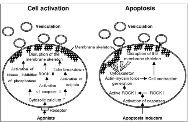
During cell activation by different agents (thrombin, collagen, ADP, calcium ionophores), the asymmetrical distribution of phospholipids is lost. The intracellular mechanisms underlying the release of MPs seem to be associated to sustained increase of Ca2+, which favours scramblases and floppases activities and concomitantly inhibits the translocase. As consequence, became the exposure of PS at the external leaflet of the
plasma membrane and, probably, this is the most prominent feature of the collapse of transbilayer asymmetry in mammalian cells. These modifications are followed by kinase activation, phosphatase inhibition, cytoskeleton degradation by Ca2+-dependent proteolysis and, an increase in bleb formation takes place (Fig. 2).
Fig. 1: Regulation and physiology of membrane phospholipids asymmetry. This model describes how
membrane phospholipid asymmetry is generated and maintained. Membrane lipid asymmetry is regulated by the cooperative activities of three transporters. Translocase, which rapidly transports PS and PE from the cell outer-to-inner leaflet; floppase, which slowly transports lipids from the cell inner-to-outer leaflet; and scramblase, which allows lipids to move randomly between both leaflets. The model predicts that the translocases are targets for Ca2+ that directly regulates the transporter activities. Elevated intracellular
Ca2+ induces PS randomization across the cell membrane by providing a stimulus that positively and
negatively regulates scramblase and translocase activities, respectively. At physiologic Ca2+
concentrations, PS asymmetry is promoted because of an active translocase and floppase but inactive scramblase. Increased cytosolic Ca2+ result in cell and/or calpain activation, which facilitate membrane
blebbing and the release of PS-expressing MPs. The appearance of PS on outer leaflet promotes coagulation and thrombosis and marks the cell as a pathologic target for elimination by phagocytes. Recognition of the PS-expressing targets can occur by both antibody-dependent and direct receptor-mediated pathways (Fom Zwaal and Schroit 1997).
MP formation during apoptosis results from Rho-associated kinase (ROCK I) activity, due to caspase 3 activation. ROCK I promotes increased actin-myosin force
generation, couples actin-myosin filaments to the plasma membrane and, as consequence, leads to disruption of membrane skeleton structure and formation of membrane blebs (Coleman et al. 2001) (Fig.2).
Conversely, other authors have shown that MP shedding induced by thrombin from ECs involves ROCK II activation via caspase 2 pathway, despite an absence of cell death (Sapet et al.2006), illustrating the complexity of pathways that lead to the formation of MPs.
Fig. 2: Schematic representation of general mechanisms involved in MP formation during cell activation and apoptosis (Modified from VanWijk et al. 2003).
I.2. Composition of Microparticles
Microparticle bilayer consists mainly of phospholipids and proteins and results negatively charged because of presence of PS and PE. Their composition differs
between cell types and process triggering their formation.
As examples, the phospholipids composition of MPs from healthy humans consists mainly of PC (60%) (Weerheim et al. 2002); whereas MPs from synovial fluid of inflamed joints of arthritis patients contain PC, PE, SM, lysophospholipids (all 20-25%) and small amounts of PS (Fourcade et al. 1995). Also, MPs from ECs exposed to an oxidative stress present oxidized phospholipids, whereas there are absent in the same cells stimulated with calcium ionophore (Huber et al. 2002).
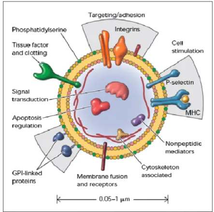
Fig. 3: Cellular MPs: a disseminated storage pool of bioactive effectors.. MPs are shed from the plasma membrane of stimulated cells. They harbor membrane and carry cytoplasmic proteins as well as bioactive lipids implicated in a variety of fundamental processes. This representation does not intend to be exhaustive with respect to the different hijacked components. MHC, major histocompatibility complex; GPI, glycosylphosphatidylinositol (From Hugel et al. 2005).
In addition, on the surface, MPs bear antigens characteristic of the cell from which they are released and carry other membrane and cytoplasmic constituents (Fig. 3). Besides, these antigens can be used for determination of cell origin using antibodies directed against specific epitopes. PMPs expose glycoproteins (GP)Ib (CD42b), platelet-endothelium adhesion molecule-1 (PECAM 1; CD31) and the fibrinogen receptor, the integrin αIIbβ3 (GPIIb-IIIa). In addition, they can expose markers of activated cells such as P-selectin (CD62P) (Diamant et al. 2004).
Global composition of MP proteins can be related to stimulus at their origin. MPs generated from activated (by phytohemagglutinin, PHA and phorbol-myristate-acetate, PMA) and apoptotic (actinomycin D, ActD) CEM T lymphocytes (cell line) or lymphocytes from diabetic patients expose on their surface the morphogen Sonic
Hedgehog (Shh) (protein implicate in embryonic and adult development). Whereas
treatment of same cells with PHA alone, PMA alone and ActD alone generates MPs lacking in Shh, as well as diabetic patient MPs elicited under apoptotic conditions
(Martinez et al. 2006). Moreover, thecomparison of protein composition obtained from
the CEM T-cell line MPs either in mitogenic (PHA)and apoptotic (ActD) conditions shows several differences. In total, 390 proteins were identified in MPs, among which 34% were described to be associated or localized in the plasma membrane.Only very few nuclear, mitochondrial, Golgi, or endoplasmic reticulum proteins were detected (less than 10% in total). Histone proteins (H2A, H2B, H3) were only identified in apoptotic conditions, as expected following cell death induction. Half of the detected proteins are intracellular proteins.
They can be grouped into cytoskeleton or cytoskeleton-associated proteins (actins, actinins, tubulins, myosins, ezrin, filamins, ARP2/3 proteins, destrin), heat shock proteins (HSP90 and 71), translation-associated proteins (ribosome proteins and elongation factors), and metabolism enzymes (e.g., lactate dehydrogenases, peroxiredoxins, glyceraldehyde 3-phosphate dehydrogenase). The differentially expressed proteins were essentially cytoplasmic proteins: ribosomal proteins, which were dramatically increased under apoptotic stimulation, elongation factors, and nuclear histones. Only three plasma membrane CD antigens were differentially detected, (CD81, CD99, CD107) among which CD99 can be attributed to apoptosis triggering
(Miguet et al. 2006).
II. Microparticles in health and disease
Despite being previously considered inert dust without specific function, MPs actively orchestrate important physiological and pathophysiological processes in vascular diseases (table 1). MPs have been implicated in hemostasis and thrombosis, diabetes, inflammation, atherosclerosis, angiogenesis, tumour progression, apoptosis, vascular cell proliferation and outgrowth of transplanted haematopoietic stem cells (Baj-Krzyworzeka et al. 2002; Janowska-Wieczorek 2001; Azevedo et al. 2007).
In vitro, the release of MPs has been shown from ECs, smooth muscle cells (SMC), platelet, leukocytes, lymphocytes and erythrocytes. Some of these MP populations occur in the blood of healthy individuals and patients. There are obvious alteration in number, cellular origin and composition of MP population in various disease states. However, the real impact of these changes on their in vivo effect is still
not fully understood.
MP ORIGIN EFFECTS PATHOLOGY
PLATELET Endothelium activation Vascular hyperreactivity Hematopoietic cell proliferation Angiogenesis enhancement
Hypertension Myocardial infarction Diabetes
Cancer
ENDOTHELIAL Angiogenesis enhancement (low concentration)
Angiogenesis inhibition (high concentration)
Endothelial dysfunction
Acute coronary syndrome Type I diabetes mellitus Lupus anticoagulant
LEUKOCYTE Vascular hyporeactivity Endothelial stimulation Endothelial dysfunction
AIDS Preeclampsia
Type II diabetes mellitus Severe trauma, sepsis
Table 1: Depending on their origin, examples of MP-evoked effects and associated pathologies.
II.1. Microparticles in coagulation
Negatively charged phospholipids, mainly PS, exposed by MPs promote the aggregation of coagulation cascade system. PMPs exhibit these anionic phospholipids, which yield high-affinity binding sites for coagulation factors factor VIII, factor IXa and factor Va (Gilbert et al. 1991; Hoffman et al. 1992) and giving PMP procoagulant properties. PMPs harbour major membrane glycoproteins, including functional adhesive receptors and consequently disseminate procoagulant potential (Fox 1994). The procoagulant ability is also extended to monocytes, lymphocytes and endothelial MPs (EMPs), which present PS at their surface.
Moreover, monocytic, fibroblast and EMPs expose tissue factor (TF), which is the trigger of coagulation cascade that culminate in generation of fibrin clot (Morrissey
2001). Besides, on EMP surface other effectors are detected, for instance von
Willebrand factor and E-selectin. In addition to their support of the fluid phase of coagulation, MPs also have a role in the recruitment of cells to developing thrombi. Furthermore, under certain conditions, MPs can also exhibit anticoagulant properties dependent on their origin and the stimulus to release. Accordingly, MPs may contribute to the complex regulation of balance between an anti- or prothrombotic vasculature (Lynch and Ludlam 2007).
II.2. Microparticles in inflammation
MPs play a role as actors in inflammatory process because they promote cells to produce potent proinflammatory mediators and they carry them. MPs can harbour
interleukin-1β (IL-1β) (MacKenzie et al. 2001), a proinflammatory cytokine, or
substrates of phospholipase A2 for the generation of lysophosphatic acid, a potent proinflammatory mediator and platelet agonist (Fourcade et al. 1995). PMPs which deliver arachidonic acid to ECs may initiate inflammation, encouraging the up-regulation of intracellular adhesion molecule-1 (ICAM-1) and cyclooxygenase-2 (COX-2) which modulates the vascular and platelet functional interaction and activate a membrane-liked signalling (Barry et al. 1997; 1999).
Blood-derived MPs can stimulate release of cytokines from ECs and up-regulation of TF expression at their surface. PMPs also enhance expression of cell adhesion molecules in monocytic and ECs and induce production of 8, 1β and
IL-6 by ECs as well as IL-8, IL-1β and tumour necrosis factor-α (TNF-α) by monocytes
(Nomura 2001).
II.3. Effects of Microparticles on cardiovascular system
Effects elicited by MPs are not restricted to inflammation and coagulation, but also involve changes on alteration in endothelial function and vascular contraction. In fact, they can affect EC (Martin et al. 2004) and SMC (Tesse et al. 2005) responses and vasoreactivity, as well as, angiogenesis (Tarabolletti et al. 2002; Kim et al. 2004). In addition, MPs may attenuate or exacerbate cardiovascular disorders. Moreover, endothelial responses trigger by MPs can be acute, by releasing several factors, or delayed, implying changes in expression of genes involved in structural and functional regulation of vascular wall (Martinez et al. 2005).
Under several pathological conditions the level of circulating EMPs increases and manifests EC damage and dysfunction, which may be also aggravated by MPs themselves. EMPs impair endothelium-dependent relaxation in rat aorta by altering nitric oxide (NO) production. This effect results in enhancing production of superoxide anion that reduces the bioavailability of NO (Brodsky et al. 2004).
Moreover EMPs, which bear protease activities, can elicit angiogenesis inducing the degradation of extracellular matrix (ECM), an essential step of neovascular structure formation (Tarabolletti et al. 2002).
PMPs stimulate platelets and ECs through modification of arachidonic acid metabolism and generation of thromboxane A2 (TxA2). They induce expression of the proinflammatory inducible isoform of COX-2 and the generation and release of
prostacyclin (Barry et al. 2007). Furthermore they enhance arachidonic-induced contraction in the aorta and methacholine-induced contraction in rabbit pulmonary arteries (Pfister 2004). PMPs from healthy individuals promote proliferation, migration and tube formation in cultured ECs and these effects are mediated by their lipid component, notably sphingosine 1-phosphates (S1P) (Kim et al. 2004). Another study shows that proangiogenic effect exerted by PMPs is mediated by vascular endothelial growth factor (VEGF), basic fibroblast growth factor (bFGF) and platelet-derived growth factor (PDGF) and also via activation of phosphatidylinositol 3-kinase (PI3K), Src kinase and extracellular-regulated kinase (ERK) pathways (Brill et al. 2005).
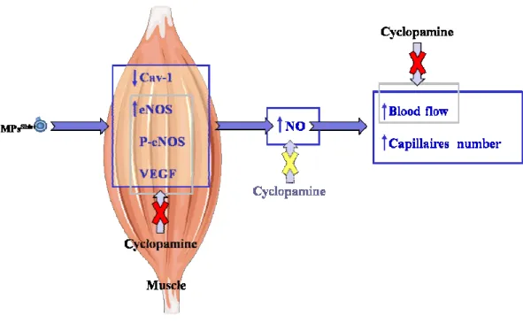
Lymphocytic MPs isolated from diabetic patients lead to endothelial dysfunction, especially by decreasing the endothelial NO-syntase (eNOS) expression and increasing caveolin-1 (Cav-1) expression (Martin et al. 2004). Little information is available regarding the effect of MPs on the regulation of vascular tone via direct action on SMCs.
Nevertheless, in vitro generated MPs from apoptotic lymphocytes are also able
to act directly on SMCs through the activation of the transcription nuclear factor κB
(NF-κB), leading to enhanced expression of inducible NOS (iNOS) and COX-2 with
subsequent increased NO and prostacyclin production respectively, ending in vascular hyporeactivity (Tesse et al. 2005). These MPs are able to decrease NO production and increase oxidative stress in ECs. They active multiple pathway related to NO and reactive oxygen species (ROS) production, mainly though PI3K. Beside, PI3K controls
the activation of ERK cascade, which counteracts the increase of xanthine
oxidase-derived ROS production by the former. Furthermore, the NF-κB pathway regulates both
endothelial dysfunction (Mostefai et al. 2008a). Additionally, MPs from apoptotic lymphocytes potently suppress neovascularization in vivo and in vitro by augmenting ROS generation via NADPH oxidase (NOX) and interfering with the VEGF signalling pathway (Yang et al. 2007). MPs generated in vitro from human activated/apoptotic T-lymphocytes, express Shh morphogen. This morphogen is involved in many biological processes during embryonic development, but it may be recruited postnatal mainly in response to tissue injury (Porro et al. 2009). MPsShh+ are able to induce NO production from cultured ECs and organs taken from MPsShh+-treated mice; and this effect was mediated directly by Shh pathway. Shh cascade is complex, as provide by its cross-talk with other pathways. In fact, it can activate non canonical signals, like PI3K and Akt and, at the same time, it is subjected to regulation by other signal cascades, like protein
kinase C (PKC)δ/MEK-1 pathway (Kessaris et al. 2004).
In ECs, MPsShh+ exert the concomitant effect of enhancement of NO and decrease of ROS production, which might result in an increasing of the bioavailability of generated NO by reducing oxidative stress and the subsequent scavenging of NO. The increase in NO release is associated with an enhancement of eNOS expression and activity, as reflected by the increase in eNOS phosphorylation, and with changes in the expression and phosphorylation of Cav-1. All of these effects of MPsShh+ on pathways involved in NO production, except on Cav-1 expression, are dependent on the PI3K pathway. By contrast, the reduction in ROS induced by MPs is dependent on PI3K and ERK pathways (Agouni et al. 2007). Moreover, in a model of mice coronary arteries subjected to ischemia/reperfusion MPs bearing Shh restore endothelial dysfunction, probably through their dual ability to increase NO and reduce ROS (Agouni et al. 2007). Accordingly, modulation exerted by MPsShh+ on different pathways leads to
beneficial potential effect on the cardiovascular system. In addition, MPsShh+ are involved in differentiation process which concern either developmental and regenerative phenomena of vascular cells, in fact they addressed human K562 pluripotent erythroleukemic cells toward megakaryocytic lineage and facilitate the progression from S to G2/M phase of cell cycle (Martinez et al. 2006).
Few studies have shown the role of MPs shed from SMCs. Probably these MPs fuse with injured arterial wall and atherosclerotic plaques and release TF harboured at their surface (Schecter et al. 2000). Moreover the ability of apoptotic smooth muscle MPs to enhance thrombus formation correlates to the functional TF carried (Brisset et al. 2003).
The majority of in vivo circulating MPs derive from platelet compared with MPs from other circulating or vascular cells (Martinez et al. 2005). Under several pathological conditions, the number of total MPs, as well as the proportion of their different origins, can change (VanWijk et al. 2003). Thus, in diseases such as atherosclerosis, congestive heart failure, diabetes, preeclampsia and cancer, the level of circulating MPs is considerably enhanced compared with healthy patients.
Moreover, the phenotype of circulating MPs is altered in different pathological states. Indeed, PMPs are enhanced in myocardial infarction (Mallat et al. 2000), hypertension (Preston et al. 2003), diabetes (Nomura et al. 1995), metabolic syndrome (Agouni et al. 2008), sepsis (Mostefai et al. 2008c) and cancer (Kim et al. 2003), whereas EMPs are most abundant in acute coronary syndromes (Mallat et al. 2000), metabolic syndrome (Agouni et al. 2008), sepsis (Mostefai et al. 2008c) and type I diabetes mellitus (Sabatier et al. 2002). In diabetic patients, the number of MPs of leukocyte origin is threefold higher than in healthy donors (Sabatier et al. 2002). In
addition, human immunodeficient virus (HIV)-infected patients show elevated levels of MPs bearing CD4 antigen (Aupeix et al. 1997). Elevated levels of MPs from granulocytes and lymphocytes have been reported in preeclampsia (VanWijk et al. 2002). Also, in severe trauma, circulating levels of MPs generated from activated leukocytes and harbouring adhesion marker are enhanced (Fujimi et al. 2003).
Because of the variety of MPs, it is plausible that they may exert pleiotropic effects on the vascular wall. Moreover, depending on the MP composition, one can speculate that different subpopulations of MPs may serve as vectors of exchange of specific message in regulating vascular function and dysfunction (Martinez et al. 2005).
Below are briefly described such pathologies in which MPs may exert deleterious or beneficial effects in regarding endothelial and/or vasomotor function.
II.4. Microparticles and preeclampsia
Preeclampsia is a pregnancy disorder due to association of hypertension and proteinuria. Total number of circulating MPs from healthy and preeclamptic pregnant women is not significantly altered despite an increase in number of T cells and granulocytes in blood. Moreover, a close relationship between endothelial dysfunction and circulating level of EMPs is reported in preeclamptic patients (Redman et al. 2007). Women affected by preeclampsia display elevated levels of MPs from lympho-monocytes and platelets in their blood stream compared with normal pregnancy. Furthermore, MPs from preeclamptic women are able to induce reduced vascular responsiveness to a vasoconstrictor agent, because of up-regulation of proinflammatory protein expression, iNOS and COX-2. Among MPs from preeclamptic women, the
platelet subset is able to stimulate only the release of NO, whereas the non-platelet ones induced both the release of NO and COX-2 vasoconstrictor products.
Thus, MPs in preeclampsia could act as vectors to stimulate intracellular cascades in vascular cells, leading to an enhanced NO production to counteract increased COX-2 vasoconstrictor metabolites by taking into account pregnancy (Meziani et al. 2006; Tesse et al. 2007).
II.5. Microparticles and sepsis
Sepsis is an acute and systemic immune response mainly to bacterial infection. During sepsis, it has been reported the generation of procoagulant MPs from endothelial, platelet, erythroid and leukocyte origin (Itakura Sumi et al. 2003; Nieuwland et al. 2000). MPs active inflammatory process, cellular apoptosis and promote multiorgan failure (Kobayashi et al. 2001). Also, MPs contribute to increase of TF expression and activity, which support the spread of coagulopathy, microcirculatory thrombosis, and tissue hypoxia and then generation of lactates. Furthermore MPs generated during sepsis participate in modulation of oxidative status in several tissues, because of greater production of ROS originated by NOX subunits present on PMPs (Ogura et al. 2001; 2004). However, recent data show that MPs from septic patients exert protective effects. Firstly, it has been described that, in patients with severe sepsis, high levels of platelet, leukocyte and endothelial MPs are associated with higher survival rate. Moreover, a negative correlation between MPs and organ dysfunction exists, suggesting decreased cell activation in patients with higher morbidity and mortality (Soriano et al. 2005).
Secondly, Mostefai et al. (2008c) have shown that MPs from septic patients are able to counteract sepsis-associated hyporeactivity through a mechanism sensitive to the TxA2 receptor antagonist, SQ-29548. Then, although septic MPs may be link between inflammation and thrombosis observed in sepsis, they may rather protective against vascular hyporeactivity in order to maintain a tonic pressor response in septic shock patients. These data bring mechanistic basis of the correlation between increased circulating MPs and better survival rate in early phase of septic shock patients.
II.6. Microparticles and diabetes
The development of vasculopathies in diabetes involves multifactorial process including pathological activation of vascular cells, which may explain the elevated levels of circulating MPs found in blood of patients affected by both type I and type II diabetes.
The cellular origin and the procoagulant activity of circulating MPs differ according to the type of diabetes (Sabatier et al. 2002). PMPs, as well as monocyte MPs, are elevated in patients with type I diabetes mellitus. Especially, monocyte MPs were highly elevated in patients with diabetic nephropathy and could be an indicator of vascular complications in diabetes (Nomura et al. 1995; Omoto et al. 2002). By contrast, type II diabetic mellitus patients present significantly higher amount of PMPs and EMPs. Indeed, in type II diabetic patients only the total PS-positive blood cell MPs is increased, whereas in type I diabetic patients the total MPs, EMPs and PMPs and the procoagulant activity are also elevated.
EMPs could be markers of the endothelial damage associated with microvascular complications and nephropathy in these patients (Sabatier et al. 2002).
II.7. Microparticles and hypertension
High blood pressure is often associated with high concentrations of circulating MPs, in particular monocytic MPs and PMPs. In such patients, treatment with an inhibitor of calcium voltage-dependent channels, significantly reduces the levels of circulating MPs. This reduction is important in the prevention of cardiovascular complications caused by adhesion molecules, activated platelets and monocytes, and high levels of circulating PMPs and monocytic MPs (Nomura et al. 2002). By contrast, Preston et al., (2003) have shown that in severe uncontrolled hypertension an increased pressure-dependent release of EMPs and PMPs takes place.
II.8. Microparticles and cancer
Tumour cells are able to generated MPs both in vitro and in vivo. Through proteins, such as urokinase, CD147 or SM, harbored by MPs from tumour cells, MPs can modify the adhesive and invasive properties of tumour target cells (Angelucci et al. 2000), or the angiogenic activity of ECs (Millimaggi et al. 2007). Moreover, it has been shown that PMPs enhance the in vitro invasive potential of breast cancer cell lines, and
induce metastasis and angiogenesis in lung cancer.
Indeed, injection of PMPs resulted in metastatic foci in lung mice (Janowska-Wieczorek et al. 2005). These data suggest that MPs transfer a transcellular signal that
could allow tumour progression. Accordingly, it has been show that epidermal growth factor receptor (EGFR) carried by MPs from glioma cells can merge with the plasma membrane of cancer cells lacking this receptor. As consequence, transfer of oncogenic activity occurs, including activation of EGFR downstream signalling pathways, such as mitogen-activated kinase (MAPK) and Akt cascades, increased production of VEGF, and expression of the anti-apoptotic protein Bcl-xL, decreased level of p27/kip1 cyclin-dependent kinase inhibitor, morphological transformation and increase in anchorage-independent growth capacity. These events lead to propagation of an enhanced proliferative, survival, motogenic and angiogenic capacity (Al-Nedawi et al. 2008).
Circulating MPs have also been studied in patients with cancer so their diagnostic and prognostic utility has been suggested. In patients with stage IV gastric cancer, PMPs were significantly elevated when compared with patients with stage I or II/III. PMPs count had also more than 90% sensitivity and specificity in the prediction of distant metastasis in these patients (Kim et al. 2003). Another study showed that PMPs and monocyte MPs are elevated in patients with lung cancer as compared with control subjects. Also, these MPs are significantly higher in patients with non-small cell lung cancer as compared with patients with small cell lung cancer (Kanazawa et al. 2003).
In addition, levels of P-selectin associated to PMPs and TF generated from cancer cells are increased indicating that proteins involved in hemostasis are elevated in patients with cancer (Yu and Rak 2004) and may represent a tool for exacerbated thrombosis.
II.9. Microparticles and AIDS
MPs seem to play several roles in AIDS, including implication in HIV infection, as well as in the propagation of the virus and its escape from classical vaccine strategies.
Few years ago, a mechanism allowing HIV to infect cells lacking the chemokine receptor CCR5 was suggested. CCR5 is released through MPs from the surface of CCR5-positive cells and is transferred to deficient peripheral blood mononuclear cells, rendering them CCR5 positive and susceptible to HIV-1 infection (Mack et al. 2000). A recent study confirms the existence of such a process by showing that platelet- and megakaryocyte-derived MPs can also transfer CXCR4 receptor to CXCR4-null cells (Rozmyslowicz et al. 2003).
In addition, one cannot exclude a role for CD4+-derived MPs observed in augmented proportions in some HIV-positive patients, especially in a situation in which individuals with high levels of circulating MPs and low circulating CD4+ cell counts seem to be protected from the classical complications of AIDS (Aupeix et al. 1997).
ANGIOGENESIS
I. General aspects
In multicellular organisms, all cells require a dependable, finely controlled supply of oxygen and nutrients. During the early stages of embryonic development, in absence of vascularization, this need takes place by diffusion, but rapidly has evolved an elaborate network of capillary plexuses and blood vessels. The initial events in vascular growth in which endothelial progenitor cells (EPCs), also called angioblasts, migrate to discrete locations, differentiate in situ and assemble in solid endothelial cords, forming later a plexus with endocardial tube, is referred to as vasculogenesis (Noden 1989). The subsequent growth, expansion and remodelling of primitive vessels into mature vessels, in embryonic life, as well as the formation of new blood vessels by sprouting of capillaries from existing vasculature, in pre- and postnatal life is referred to as angiogenesis. This process is based on endothelial sprouting or intussusceptive (non-sprouting) microvascular growth (Ausprunk and Folkman 1977; Risau 1997). The latter represents an additional and/or alternative mechanism and is not dependent on local EC proliferation or sprouting: a large sinusoidal capillary divides into smaller capillaries, which then grow separately (Djonov et al. 2000). The functional modifications of largest arteries, such addiction of a thick muscular coat concomitant with acquisition of viscoelastic and vasomotor properties, are referred to as arteriogenesis.
New vessels in the adult arise mainly through angiogenesis, although vasculogenesis also may occur. Even if, as a general rule, establishment of the vasculature of most organs occurs by angiogenesis, development of the vascular network of certain endodermal organs, including the liver, lungs, pancreas, stomach/
intestine and spleen, occurs by vasculogenesis (Pardanaud and Dieterlen-Lievre 1999). The existence of a postnatal vasculogenesis is supported by the evidence that both ECs and EPCs co-exist in the circulation. Moreover, EPCs are also recruited to sites of neovascularization in mature mammals from a circulating, marrow-derived population of progenitor cells (Asahara et al. 1997). The distinction between vasculogenesis and angiogenesis is not absolute and they overlap. Both require EC proliferation, migration, three-dimensional reorganization of newly formed aggregates and use similar extracellular matrix adhesive mechanisms (Drake et al. 1995). Moreover, vasculogenesis and angiogenesis are not mutually exclusive, inasmuch as angioblasts can be incorporated into expanding pre-existing blood vessels (Auerbach and Auerbach 1997).
Sprouting angiogenesis develops through a highly orchestrated multi-step process. The initial vasodilatation of existing vessels is accompanied with increasing of endothelial permeability and basement membrane degradation by the action of proteolytic enzymes, such as matrix metalloproteinases (MMPs) and plasminogen activators secreted by ECs, resulting in the formation of tiny sprouts penetrating the perivascular stroma. This allows migration of the ECs at the sprout tip toward the angiogenic stimulus and proliferation of the ECs below the sprout. The subsequent canalization, branching, and formation of vascular loops, lead to the development of a functioning circulatory network. Finally, perivascular apposition of pericytes and SMCs to support the abluminal side and de novo synthesis by ECs and pericytes of the basement membrane constituents lead to vessels stabilization.
Angiogenesis is subject to a complex control system with proangiogenic and antiangiogenic factors. In adults, angiogenesis is tightly controlled by this "angiogenic
balance", a physiological balance between the stimulatory and inhibitory signals for blood vessel growth; this switch depends on a local change in the balance.
In normal circumstances, the formation of new blood vessels occurs during wound healing, organ regeneration and in the female reproductive system during ovulation, menstruation, and the formation of the placenta (Hoeben et al. 2004). Also, a large number of different and non-related diseases are associated with impairment or excess of new vasculature formation. Among the pathologies in which angiogenesis is impaired, tissue damage after reperfusion of ischemic tissue or cardiac failure or diabetes needs formation of new collateral vessels to improve disease conditions (Carmeliet et al. 1999; Ferrara and Alitalo 1999). In several diseases, excessive angiogenesis is part of the pathology. These diseases include cancer (both solid and hematologic tumours), cardiovascular diseases (atherosclerosis), chronic inflammation (rheumatoid arthritis, Crohn's disease), diabetic retinopathy, psoriasis and endometriosis (Griffioen and Molema 2000).
Angiogenic growth factors, under both physiological and pathological conditions, induce, promote and/or interfere with all steps of angiogenesis (Losordo and Dimmeler 2004; Ng and D’amore 2001; Post et al. 2001). A variety of growth factors
plays significant role in cell proliferation, maturation and differentiation leading to the formation of mature blood vessels. These factors act as signalling molecules between cells, and bind to specific receptors on the surface of their target cells. Also, hypoxia is the most important environmental factor that leads to neovascularization.
I.1. Angiogenic mediators
Angiogenesis is driven by numerous mediators produced by different cells under a large variety of conditions. These mediators are either soluble, ECM or membrane bound growth factors, or components of ECM themselves. The best-known factors with proven angiogenic potency are the family of VEGF and FGF (Tunyogi-Csapo et al. 2007; Jacobs 2007), hepatocyte growth factor (HGF) (Tong et al. 2006), NO (Lau and Ma 1996), and angiopoietins (Ang1 and 2) (Asahara et al. 1998).
I.1.1. Vascular endothelial growth factor
The VEGF family comprises seven members. The most abundant and potently mitogenic is the VEGF-A. The two VEGF-specific tyrosine kinase receptors, VEGFR-1 (Flt-1) and VEGFR-2 (KDR-Flk-1), are expressed on vascular endothelium. Activation of VEGFR-2 by VEGF interaction is a critical requirement to induce the full spectrum of VEGF responses. VEGF transcription is stimulated greatly by hypoxia, as a result of
hypoxia inducible factor (HIF-α) binding to a hypoxia response element within the
VEGF promoter (Semenza 2001). VEGF production is also increased by inflammatory
mediators, such as IL-1α and IL-1β, transforming growth factor β (TGFβ),
prostaglandin E2 (PGE2), or COX-2 activation (McColl et al. 2004), as well as mechanical forces of shear stress and cell stretch (Milkiewicz et al. 2001; Li et al 1997). VEGF promotes EC survival through activation of PI3K/Akt pathway and through
association with αvβ3 integrin and activation of focal adhesion kinase (FAK) (Zachary
2003). VEGF induces EC proliferation and migration through numerous pathways, including activation of the MAPK, ERK, p38 and c-jun N-terminal kinase (JNK), and RhoGTPase family members (Zachary 2003).
I.1.2. Fibroblast growth factor
Members of the FGF family are also potent inducers of angiogenesis. Cellular responses mediated by FGFs include cell migration, proliferation and differentiation
(Kanda et al. 1997). The FGF family consists of nine structurally related polypeptides, of which FGF-1 (acid-FGF) and FGF-2 (basic-FGF) are most extensively studied. The cellular effects of FGFs are mediated via specific binding to high-affinity tyrosine kinase receptors (FGFRs) (Klein 1996). Binding of FGF-2 to these receptors initiates PKC dependent signalling and/or GRB2/SOS mediated activation of MAPK pathway. FGFR1 signalling stimulates migration and differentiation whereas FGFR2 only migration (Kanda et al. 2004). A major contribution of FGF signalling may be through the recruitment of other growth factor pathways.
I.1.3. Hepatocyte growth factor
HGF is secreted by mesenchyme-derived cells as an inactive precursor that is activated by proteolytic cleavage by urokinase or tissue plasminogen activator (uPA or tPA) (Zhang et al. 2003). HGF binds to a tyrosine kinase receptor, c-met, found predominantly on epithelial and ECs. Through activation of c-met, HGF is a potent mediator of angiogenesis, in part because it induces production of VEGF by the endothelium (Reisinger et al. 2003; Zhang et al. 2003). In addition, HGF can induce
angiogenesis independently of the VEGF pathway (Sengupta et al. 2003). HGF-α
dependent activation of the transcription factor Ets-1 causes further production and release of HGF, creating a positive feedback loop that perpetuates the state of EC activation (Hashiya et al. 2004). HGF also activates sphingosine kinase, increasing production of S1P, a molecule known to increase cell motility (Duan et al. 2004).
Another effect of HGF signalling is the negative regulation of thrombospondin 1 (TSP1) in tumour cells (Zhang et al. 2003). TSP1 is an extracellular matrix protein associated with anti-angiogenesis signalling, including activation of EC apoptosis.
I.1.4. Nitric oxide
NO is an important mediator of angiogenesis, in addition to its well-recognized vasodilatory properties. NO triggers capillary EC growth and differentiation via cyclic GMP-dependent gene transcription. The regulation of capillary growth by NO is complex because both angiogenic and angiostatic effects of NO have been demonstrated (Lau and Ma 1996; Murohara et al. 1998; RayChaudhury et al. 1996; Ziche et al. 1994). Low concentrations of NO stimulate capillary-like tube formation and cell migration through activation of PKC and ERK and c-Jun phosphorylation, whereas high NO concentrations inhibit these angiogenic responses (Jones et al. 2004). NO is a component of the pathways underlying VEGF-activated EC proliferation. However, whether NO acts as an upstream or a downstream mediator of VEGF is controversial. VEGF induces the expression of eNOS and promotes the release of NO via activation of the MAPK cascade (Papapetropoulos et al. 1997; Parenti et al. 1998). NO has been
shown to both increase and decrease VEGF production via modulation of HIF-1α
expression. Some authors report that NO mediates suppression of hypoxia-induced
production of VEGF by decreasing HIF-1α DNA binding activity (Huang et al. 1999;
Sogawa et al. 1998), whereas others describe enhanced HIF-1α binding activity in
response to NO (Dulak et al. 2000; Kimura et al. 2000). Interestingly, while VEGF-induced angiogenesis is mediated by NO, the capillary growth stimulated by FGF-2 can be both NO-independent and inhibited by NO (RayChaudhury et al. 1996; Ziche et al. 1997).
I.1.5. Angiopoietins
The two isoforms of angiopoietins (Ang), Ang1 and 2, play a role in vascular stabilization. The former is associated with developing vessels and its absence leads to
defects in vascular remodeling (Thurston 2003); the latter antagonizes Ang1 action, causing destabilization of preexisting vessels. Ang2 is found in tissues such as ovary, uterus, and placenta that undergo transient or periodic growth and vascularization, followed by regression (Maisonpierre et al. 1997).
Ang and receptor tyrosine kinase Tie1 and Tie2 play critical role in the later stages of angiogenesis as well. Ang1 and Ang2 are Tie2-specific ligands that activate or antagonize Tie2 signalling in endothelium, respectively (Asahara et al. 1998). They are required for communication of ECs with the surrounding mesenchyme to establish stable cellular and biochemical interaction (Maisonpiere et al. 1997). Tie1 function is related to EC differentiation and the establishment of blood vessel integrity. Tie2, on the other hand, is particularly important for vascular network formation (Dumont et al. 1994; Puri et al. 1995; Sato et al. 1995).
I.1.6. Other angiogenic mediators
Other angiogenic factors involve in the switch are TGF-β1 and PDGF. When
mesenchymal cells are treated with TGF-β1, they express SMC markers, indicating
differentiation toward a SMC lineage, and the differentiation can be blocked by
antibodies against TGF-β1 (Hirschi et al. 1998). TGF-β1 has been also reported to
direct neural crest cells toward a vascular SMC lineage (Shah et al. 1996). PDGF-B is secreted by ECs, presumably in response to VEGF and facilitates recruitment of mural cells. PDGF-B gene mutation may cause failure of pericyte recruitment (Lindhal et al.
1997).
It is important to note that the activity of an angiogenesis-regulating cytokine depends on the presence and concentration of other factors or cytokines in the environment of the responding endothelium (Pepper et al. 1998). As example, exogenous factors such as hormones can affect condition leading to angiogenesis (Schiffenbauer et al. 1997). Moreover it is well appreciated that immune system cells such as monocytes/macrophage, lymphocytes and mast cells can affect pro- and
antiangiogenic balance (Sunderkotter et al. 1996; Blair et al. 1997).
In recent years, evidence has accumulated that, in addition to the classic factors, many other endogenous peptides (erythropoietin, angiotensin II, endothelins, proadrenomedullin-derived peptides, urotensin II, adipokines, neuropeptide-Y, vasoactive intestinal peptide, pituitary adenylate cyclase-activating polypeptide and substance P) play an important regulatory role in angiogenesis, especially under pathological conditions (Ribatti et al. 2007).
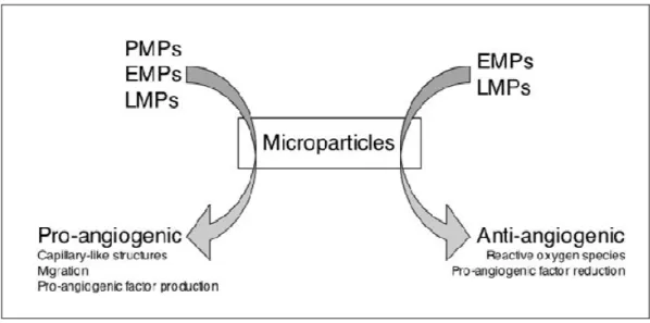
Moreover, recent data show that MPs are implicated in modulation of neovascularization. PMPs promote proliferation and survival of ECs as well as tube formation (Kim et al. 2004); they induce angiogenesis and improve revascularization after chronic ischemia in vivo (Brill et al. 2005). Furthermore, MPs derived from tumour cells also promote tumour angiogenesis (Kim et al. 2002), and MPs from endothelial origin may contribute to angiogenesis because of the MMPs activities they carry (Taraboletti et al. 2002). Major details on MPs involvement in angiogenic process are discussed below.
I.2. Angiogenesis process
I.2.1. Vasodilatation, increased endothelial permeability and extracellular matrix (ECM) degradation
Vasodilatation of existing vessels is one of the earliest steps in angiogenesis. VEGF is a major player in neovessels initiation; based on its ability to induce vasodilatation via endothelial NO production and its EC permeability increasing effect
(Ziche et al. 1997). The observation that VEGF production is under control of HIF-α
strengthens the suggestion of an early involvement of VEGF in the angiogenic response. Moreover, VEGFR expression is up-regulated under hypoxic or ischemic conditions as well (Forsythe et al. 1996). This allows redistribution of intracellular adhesion molecules, including PECAM-1 and vascular endothelial (VE)-cadherin, and alteration in cell membrane structure via induction of a series of kinases (Eliceiri et al. 1999a; Gale and Yancopoulos 1999). Then, the extravasion of plasma proteins follows, which creates a provisional network support (Dvorak 1986) that leads subsequently to the migration of activated ECs. Consequently permeability changes must be tightly regulated. For instance, Ang1 is a natural anti-permeability factor, which provides protection and balance against excessive plasma leakage (Thurston et al. 2000).
Endothelial sprouting is further enhanced by Ang2, which, appearing at angiogenic and vascular remodelling sites, is involved in detaching SMC and loosening underlying matrix, thereby allowing ECs to migrate as inter-EC contacts are relieved (Gale and Yancopoulos 1999; Maissonpierre et al. 1997).
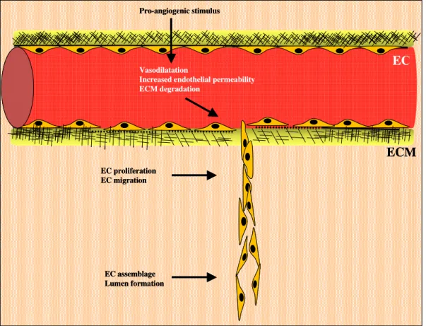
Degradation of ECM involves an array of proteinases which not only provides way for the migrating cells, but also results in the liberation of growth factors, including bFGF, VEGF and insulin-like growth factor-1 (IGF-1), which otherwise remain sequestered within the matrix. Over twenty MMPs have been described and implicated in angiogenesis, tumourogenesis and cell proliferation (Nelson et al. 2000). Inhibitors of MMPs include circulating protease inhibitors, such as tissue-localized inhibitors of metalloproteinases (TIMPs) (Brew et al. 2000). It is, at least partly, through the secretion of MMP-2, MMP-3 and MMP-9, and suppression of TIMP-2 that Ang1 induces sprouting (Kim et al. 2000). Similarly, MMP-3, MMP-7 and MMP-9 have been shown to induce angiogenesis in neonatal bones and tumours (Vu et al. 1999). However, MMPs do not uniformly enhance angiogenesis; temporal and spatial factors likely dictate their function. An interaction with other proteins also alters their roles in angiogenesis. TSP1 is believed to be antiangiogenic by preventing activation of MMP-2 and MMP-9 (Bein and Simons 2000). Other proteinases, such as plasmin, have also implicated in matrix degradation enabling endothelial migration (Pepper 2001) (Fig. 4).
EC
ECM
Pro-angiogenic stimulusVasodilatation
Increased endothelial permeability ECM degradation EC proliferation EC migration EC assemblage Lumen formation
EC
ECM
Pro-angiogenic stimulus VasodilatationIncreased endothelial permeability ECM degradation EC proliferation EC migration EC assemblage Lumen formation
EC
ECM
Pro-angiogenic stimulus VasodilatationIncreased endothelial permeability ECM degradation
EC proliferation EC migration
EC assemblage Lumen formation
Fig. 4: Early events implicated in new vessel formation. Angiogenic stimuli (hypoxia, growth factor,
inflammation or mechanical factors) cause vasodilatation, and increased endothelial cell (EC) permeability. Degradation of extracellular matrix (ECM), controlled mainly by MMPs, promote EC invasion into surrounding interstitial matrix concomitantly with remodelling of cytoskeleton, which provides directional migration of EC sprouting. EC proliferation occurs early in angiogenesis and continues as new capillary sprout elongates. Once migrate, ECs assemble to form a multi-cellular structure, which forms a lumen. New vessel will be formed when the sprout anastamoses with a pre-existing capillary.
I.2.2. Endothelial cells proliferation and migration
When the physical barriers are dissolved, proliferating ECs are free to migrate to distant sites. This stage involves interplay between the various forms of VEGF, Ang, bFGF and their receptors, all of which are responsible in mediating angiogenesis, although additional factors have also been implicated. Besides its effect on angiogenesis initiation, VEGF also affects EC proliferation. This effect can be partly attributed to the ability of VEGF to release NO and activate MAPK family through cGMP (Yu and Sato 1999). Ang1, via phosphorylation of Tie2, is chemotactic for ECs, induces sprouting and stimulates the interaction between endothelial and peri-endothelial cells (Gale et al. 2002; Suri et al. 1996). Ang2, in concert with VEGF is also angiogenic, although in absence of VEGF may induce vessel regression (Maisonpiere et al. 1997). FGF stimulate EC growth and recruit mesenchymal and/or inflammatory cells, producing many angiogenic factors (Carmeliet 2000). PDGF is angiogenic for microvascular sprouting ECs and recruits pericytes and SMCs around nascent vessel sprout (Lindhal et al. 1998; Hellström et al. 1999). Several chemokines, including monocyte chemotactic protein 1 (MCP-1), have been demonstrated to induce endothelial growth (Belperio et al. 2000). When the ECs proliferate and migrate, in part by signalling through integrins αvβ3 and α5β1 (Eliceiri and Cheresh 1999b), PECAM-1 (Ilan et al. 1999) and Eph/ephrin receptor-ligand pairs (Huynh-Do et al. 1999; Shima and Mailhos 2000; Wilkinson 2000), they contact with other ECs (Fig. 4).
EC junctions are established with gap proteins such as VE-cadherin and members of the connexin family (Corada et al. 1999; Knudsen et al. 1998).
I.2.3. Endothelial cells assemblage, lumen formation and stabilization
Once ECs migrate into the ECM, they assemble in solid cords. Sprouting of one or two cells may form a lumen by intracellular canalization, which occurs through fusion of cytoplasmic vesicles, or by the alternative process in which a lumen is created by the membrane apposition of two different cells (Eggiton Gerritsen 2003). Lumen diameter is tightly regulated by interactions between various VEGF isoforms, Ang, and their receptors as well as different integrins (αvβ3 and α5β1) (Suri et al. 1998; Bayless 2000). Finally, there are also several endogenous inhibitors of lumen formation, including TSP1 (Gendron et al. 2000).
EC interaction with ECM and mesenchymal cells is a prerequisite to form a stable vasculature. Therefore, after EC proliferation and maturation, and the formation of endothelial tube structures, a surrounding vessel layer composed of mural cells (pericytes in small vessel and SMCs in large vessels) is required. ECs may accomplish this via the synthesis and secretion of PDGF, a mitogen chemoattractant for a variety of mesenchymal cells. Subsequent differentiation of mural precursor cells into pericytes and SMC is believed to be a cell-cell contact dependent process (Griffioen Molena
2000). On endothelial cell-mural cell contact, a latent form of TGFβ, producing by both