HAL Id: tel-01093377
https://hal.archives-ouvertes.fr/tel-01093377v2
Submitted on 6 Jan 2015
HAL is a multi-disciplinary open access archive for the deposit and dissemination of sci-entific research documents, whether they are pub-lished or not. The documents may come from teaching and research institutions in France or abroad, or from public or private research centers.
L’archive ouverte pluridisciplinaire HAL, est destinée au dépôt et à la diffusion de documents scientifiques de niveau recherche, publiés ou non, émanant des établissements d’enseignement et de recherche français ou étrangers, des laboratoires publics ou privés.
Solid-state NMR studies of protein dynamics: New
approaches and applications to crystalline proteins and
large molecular assemblies
Paul Schanda
To cite this version:
Paul Schanda. Solid-state NMR studies of protein dynamics: New approaches and applications to crystalline proteins and large molecular assemblies. Life Sciences [q-bio]. Université Joseph Fourier Grenoble, 2014. �tel-01093377v2�
Habilitation à Diriger des Recherches (HDR) Université Joseph Fourier, Grenoble
UFR de Chimie
Solid-state NMR studies of protein dynamics
New approaches and applications to crystalline proteins and
large molecular assemblies
Dr. Paul Schanda
MEMBERS OF THE HABILITATION JURY:
Dr. Fabien Ferrage
(rapporteur)
Prof. Frans Mulder
(rapporteur)
Dr. Guido Pintacuda
(rapporteur)
Prof. Mikael Akke
(examinateur)
Dr. Dominique Bourgeois
(examinateur)
Prof. Tatyana Polenova
(examinatrice)
Prof. Bernd Reif
(examinateur)
Date of habilitation (soutenance): 3.12.2014Table of Contents
1 CHARACTERIZING PROTEIN MOTION AT ATOMIC RESOLUTION: INTRODUCING THE
POSSIBILITIES OF SOLID-STATE NMR ... 6
1.1 INTRODUCTION –PROTEIN FUNCTION RELIES ON FLEXIBILITY... 6
1.2 NEW POSSIBILITIES TO STUDY PROTEIN MOTION BY SOLID-STATE NMR... 8
1.3 DIPOLAR COUPLINGS IN SOLID-STATE NMR: DIRECT INSIGHT INTO AMPLITUDES OF MOTION... 10
1.4 NUCLEAR SPIN RELAXATION AND PROTEIN DYNAMICS... 13
1.5 MOTION ON MICRO-TO-MILLISECOND TIME SCALES:PROBING DYNAMICS ON BIOLOGICAL FUNCTIONAL TIME SCALES... 16
1.6 QUANTIFYING DYNAMICS IN THE SOLID STATE: EXPERIMENTAL CHALLENGES... 16
2 DEUTERATION AND FAST MAGIC-ANGLE SPINNING: OVERCOMING CHALLENGES OF DYNAMICS STUDIES IN SSNMR... 18
2.1 LONG COHERENCE LIFE TIMES IN DEUTERATED PROTEINS... 18
2.2 NOVEL TYPES OF CORRELATION EXPERIMENTS: FROM ASSIGNMENT TO HYDROGEN-BOND SCALAR COUPLINGS... 20
2.3 STRUCTURE DETERMINATION OF DEUTERATED PROTEINS FROM UNAMBIGUOUS 1H-1H DISTANCE RESTRAINTS... 21
3 SOLID-STATE NMR MEASUREMENTS OF ASYMMETRIC DIPOLAR COUPLINGS PROVIDE INSIGHT INTO PROTEIN SIDE-CHAIN MOTION ... 24
3.1 INTRODUCTION... 24
3.2 EXPERIMENTAL OBSERVATION OF ASYMMETRIC DIPOLAR COUPLINGS IN UBIQUITIN SIDE CHAINS.. 25
3.3 MODELS OF SIDE CHAIN MOTION FROM DIPOLAR COUPLINGS... 29
3.3.1 Valines ... 29
3.3.2 Leucines... 30
3.3.3 Isoleucines ... 31
3.3.4 Possibilities of studying other side chains, and characterizing side chain motion in even more detail 32 4 TIME SCALES AND AMPLITUDES OF PROTEIN BACKBONE DYNAMICS IN THE SOLID-STATE ... 33
4.1 PROTON-DETECTED SPECTROSCOPY ON UBIQUITIN: ASSIGNMENTS AND QUALITATIVE CONCLUSIONS ABOUT DYNAMICS... 34
4.2 INSIGHT INTO SUB-MICROSECOND BACKBONE MOTION FROM EXPERIMENTAL DATA: THEORETICAL CONSIDERATIONS... 35
4.3 AMPLITUDES OF BACKBONE MOTION FROM NH DIPOLAR COUPLINGS... 38
4.4 TIME SCALES OF BACKBONE MOTION: INSIGHT FROM DIFFERENT SPIN RELAXATION PARAMETERS. 41 • 15NR 1 AT FIELD STRENGTHS CORRESPONDING TO 500,600 AND 850MHZ 1HLARMOR FREQUENCY.. 41
4.5 COMPARISON TO SOLUTION-STATE NMR: DOES THE CRYSTAL LATTICE IMPACT PROTEIN DYNAMICS? 45 4.6 THE IMPACT OF THE CRYSTALLINE ENVIRONMENT FROM COMPARISONS OF DIFFERENT CRYSTAL POLYMORPHS... 47
4.7 CONCLUSIONS... 47
5 SITE-RESOLVED MEASUREMENT OF MICROSECOND TO MILLISECOND CONFORMATIONAL-EXCHANGE PROCESSES IN PROTEINS BY SOLID-STATE NMR SPECTROSCOPY... 50
5.1 INTRODUCTION... 50
5.2 RESULTS AND DISCUSSION... 51
5.2.1 Differential zero- and double-quantum 1H-15N line broadening reveals conformational exchange. ... 51
5.2.2 15N CPMG relaxation dispersion experiments. ... 55
5.2.3 Comparison to conformational exchange in solution. ... 57
6 STRUCTURAL INSIGHT INTO TRANSIENT CONFORMATIONAL STATES OF PROTEINS
BY SOLID-STATE R1Ρ RELAXATION-DISPERSION NMR SPECTROSCOPY ... 61
6.1 INTRODUCTION... 61
6.2 THE PROPERTIES OF R1Ρ EXPERIMENTS IN MAS SOLID-STATE NMR: INSIGHT FROM NUMERICAL SIMULATIONS... 61
6.3 EXPERIMENTAL IMPLEMENTATION OF R1Ρ RELAXATION DISPERSION... 63
6.4 KINETICS AND THERMODYNAMICS OF CONFORMATIONAL EXCHANGE: FITTING THE BLOCH -MCCONNELL-REGIME OF R1Ρ DISPERSION CURVES... 64
6.5 BEYOND CHEMICAL SHIFT INFORMATION: BOND ORIENTATION CHANGES UPON THE STRUCTURAL TRANSITION TO THE EXCITED STATE... 67
7 ATOMIC MODEL OF A CELL-WALL CROSS-LINKING ENZYME IN COMPLEX WITH AN INTACT BACTERIAL PEPTIDOGLYCAN... 69
7.1 INTRODUCTION... 69
7.2 EXPERIMENTAL SECTION... 70
7.2.1 Sample preparation ... 70
7.2.2 NMR spectroscopy and structure calculation ... 70
7.3 RESULTS... 71
7.3.1 LdtBs tightly binds to peptidoglycan ... 71
7.3.2 Peptidoglycan dynamics in the protein-bound state ... 73
7.3.3 Atomic model of the complex from 1H-detected ssNMR experiments ... 74
7.3.4 Mutants and isolated domains have different binding affinities than full-length wild-type LdtBs 76 7.4 DISCUSSION... 79
7.5 CONCLUSIONS... 80
Preface
This habilitation manuscript outlines some of the work I performed since the beginning of my research on solid-state NMR in Beat H. Meier’s laboratory in 2008, and at IBS Grenoble since 2011. The particular emphasis of this work is on the use of solid-state NMR spectroscopy for the study of protein dynamics. This field has seen great improvements and developments over the last 10-15 years, to which I hope was able to contribute somewhat.
The field of protein dynamics has fascinated me since my undergraduate studies. This fascination arose from interactions I had with several persons that have shared their passion for protein dynamics and structural biology NMR with me. I therefore would like to express here my gratitude to these persons, and many others that have been influential for my work, but which would be too numerous to be exhaustively listed here. My early fascination for NMR has been triggered by Robert Konrat and Bernhard Brutscher, and although none of the work described here was done with them, I am grateful for their guidance. I am particularly grateful to the people that have taught me solid-state NMR spectroscopy at ETH Zurich, most notably Matthias Ernst, Beat H. Meier and René Verel.
Matthias’ scientific rigor and ability to explain NMR theory, down to the last detail, and his ability to “walk through Floquet space” better than some of us wander around in 3D space, leaves me impressed. I am grateful for his support during my time at ETH. I would also like to acknowledge Beat’s support, who let me develop all the proton-detection approaches on my own in his lab. I am particularly grateful also for the excellent NMR courses that Beat and Matthias were giving at ETH.
Some of the proton-detected experiments were done together with Matthias Huber, and I thank him for numerous discussions.
I would like to thank particularly my colleagues at IBS Grenoble. Special thanks goes to Bernhard Bruscher, Jerome Boisbouvier and Jean-Pierre Simorre for their support when setting up my research activity at IBS. I have and had the great chance to work with excellent colleagues, post-docs, PhD students and exchange students. Jens Haller, although he has been in my group for just a few months, made impressive progress and was the main actor in some of the ubiquitin dynamics work presented in this work. Astrid Sivertsen, Peixiang Ma and Vilius Kurauskas were brave enough to embark on a difficult membrane project, and I am grateful for their endurance and efforts. Peixiang has been a very valuable member of the team also for projects which are rather far from his initial education: he managed to make sense out of our work on relaxation dispersion, and I want to express my gratitude for his wide range of activities. I would also like to thank Audrey Hessel, who has set up the activity that I myself know least – membrane protein biochemistry – and has been a major driving force, together with Vilius and Peixiang, to bring forward the membrane protein project. Special thanks goes to Martin Tollinger, with whom I had the chance to collaborate at several instances, which has always been a very stimulating experience.
My work on proton-detected solid-state NMR spectroscopy started out when I entered the solid-state NMR field in 2008. At that time Beat gave me the freedom to choose my research topic by myself, and among the most promising approaches I found in the literature for measuring dynamics were reports on deuteration and proton detection. The ideas that were around at that time, particularly put forward by Bernd Reif, but also others, shaped the way I approached solid-state NMR. Thanks to the impressive methodological developments, NMR studies of protein dynamics have made great progress over the last decade, and I am happy to witness these exciting steps of the field.
I am glad that some of the researchers that actively shape NMR these days, and that have been inspiring for my work have accepted to take the time to read and evaluate the present HDR manuscript and HDR defense.
Dr. Paul Schanda September 2014
1 Characterizing protein motion at atomic resolution:
introducing the possibilities of solid-state NMR
1.1 Introduction – Protein function relies on flexibility
One of the key goals of contemporary science is the understanding of cellular processes on a molecular level. The determination of the three-dimensional structure of biomolecules has been extremely important to understand the molecular basis of processes in living cells. The hallmark feature of all processes in living systems, however, is molecular motion. Thus, the static pictures provided by traditional structural biology approaches fail to grasp an important aspect of biomolecules, namely how these structures interconvert between alternate conformations, thereby possibly sampling states that are the actual functionally relevant ones.
Characterizing protein motion at atomic resolution is challenging. Dynamics occur on a wide range of time scales, calling for versatile methods sensitive to different time scales and types of motions. Furthermore, functionally relevant structural excursions may be infrequent, and involve short-lived conformational states that are populated to only minute extents in equilibrium with a major conformation. Detecting minor forms in the presence of an overwhelming conformer represents a severe challenge in terms of sensitivity. In addition, one wants to study protein motion in the environment where the molecule actually performs its function, i.e. possibly in the cellular context, or at least in near-native solution or in the native membrane environment. Given all these different requirements, advanced experimental methods, or combinations of methods are often required to characterize protein dynamics. A number of experimental techniques have been developed to study protein dynamics. While exhaustively reviewing these techniques is not within the scope of this work, one may cite in particular methods based on detailed analysis of X-ray diffraction data, possibly collected at physiologically relevant temperature1-3, that visualize alternate conformations (although without providing insight into thermodynamics/kinetics or the processes). Kinetic crystallography represents a very attractive route to studying off-equilibrium dynamics in systems in which a reaction can be triggered,4 which in many cases is difficult to achieve. Lower-resolution methods, such as IR spectroscopy,5 also provide powerful tools to monitor global structural changes, although without atomic details.
Arguably, NMR spectroscopy in solution state stands out among many experimental techniques for studying protein dynamics. NMR spectroscopy delivers information about motions on a wide range of time scales at atomic resolution. Not only does it provide information at the atomic scale about the presence of motion, but it also gives detailed information about time scales and relative populations of states, and thus gives access to a range of physico-chemical parameters.
Solution-state NMR methods nowadays routinely probe the bond librational motions of the protein backbone and side chains, that can be used as a direct measure of conformational entropy, thus providing insight into the thermodynamics of proteins.6
Excitingly also, NMR spectroscopy can probe motions that are on time scales at which proteins actually perform their function, which are mostly millisecond time scales. A growing list of beautiful examples highlights this power of NMR spectroscopy to directly see proteins in action. NMR techniques have, for example, allowed to get insight into enzyme function, where it could be directly shown that the rate at which an enzyme performs its action corresponds to the rate at which it undergoes conformational exchange, and that this exchange process is present even without substrate.7
Motion also plays an important role in membrane proteins, where substrate transport can often be accomplished only if the membrane protein undergoes significant structural rearrangement. In this
context, a recent work has beautifully explained how the small multidrug resistance transporter EmrE dynamically exchanges between two states that are open to one side of the membrane, thus allowing to transport its substrates.8
Dynamic exchange processes also play a key role in allosteric regulation. Figure 1 shows an example of a dynamic process in an allosteric protein, the small transcription co-activator domain KIX, which can bind two transcription factors. In vitro, binding of the MLL activation domain to KIX cooperatively enhances the interaction with c-Myb: KIX in complex with MLL displays a ∼2-fold higher affinity for the c-Myb activation domain than the KIX domain alone. While this allostery has been detected by thermodynamic methods, the molecular mechanism that mediates this cooperativity remained unclear. NMR methods could shed light onto this phenomenon: in the presence of only one of the two binding partners, the binary complex (KIX+MLL) is in continuous exchange between a major conformational state (of which a structure had been determined), and a minor state, that is too transient and low-populated to be seen in a crystal structure or conventional NMR structure. Our solution-state NMR data could reveal that it is this minor state that is pre-formed to bind the second transcription factor.9 In other words, the actual function state of this
protein does not correspond to the main conformer, that is generally in the focus of structural biology. Rather, a dynamically sampled alternate conformation is the functionally relevant one. These few examples (and many many more could be cited here) emphasize the importance of characterizing “hidden” states, which generally are overlooked by the static views of structural biology. Understanding the jiggling and wiggling of biomolecules in detail will provide important clues about their functions.
Figure 1. A dynamic process underlies allosteric signal transmission in the KIX domain. (A) Structure of the transcription co-activator KIX (blue) in complex with two transcription factors, MLL and c-Myb (red, green). (B) In the presence of only one of the two binding partners (MLL, red), KIX is in a dynamic exchange between a major state and a transient minor state on a millisecond time scale. The minor state is preformed to accommodate the second binding partner, explaining the allostery of binding.
1.2 New possibilities to study protein motion by solid-state NMR
Biological processes occur in complex cellular environments. Many processes involve very large biomolecular complexes and assemblies, such as ribosomes, membrane-embedded proteins and enzymatic complexes of hundreds of kilodalton in size. Solution-state NMR spectroscopy is significantly challenged in these cases: many of these objects may inherently not be soluble (e.g. membrane proteins), or their large size and associated slow molecular tumbling in solution leads to rapid signal loss and line broadening.
Magic-angle spinning solid-state NMR (MAS ssNMR) does not face these inherent limitations. The perspectives of gaining atomic-resolution insight into objects that were so far out of reach (for solution-state NMR) have been a major driving force for the development of the technique. While the technically more demanding requirements of MAS ssNMR have retarded the widespread use of ssNMR in structural biology for some time, relative to its solution-state counterpart, the development of ssNMR over the last decade is impressive. Protein structures of increasing size and complexity are nowadays being determined by ssNMR,10 and, importantly also, interactions can be
probed in complex environments, including entire cells and cell walls.11
Figure 2. Time scales of protein dynamics (a) and corresponding solid-state NMR approaches to probe these motions (b). The approaches shown in (b) will be discussed in the present manuscript. The averaging of dipolar couplings (red) is introduced in section 1.3 and in more detail in chapter 3. Relaxation approaches (shown in orange) are discussed in section 1.4 and their application to studying protein backbone is discussed in chapter 4. Slower motional processes can be studied using methods shown here in green and blue, discussed in chapters 5 and 6, respectively.
The development of MAS ssNMR has also opened the way to probe protein dynamics in these samples. In addition to probing motion in systems that are out of reach for solution-state NMR, solid-state NMR also allows addressing interesting biophysical questions. For example, solid-state NMR can monitor motion in (micro-)crystalline proteins, thus allowing to answer to questions that arise in crystallography, such as the link to crystallographic temperature factors.
Figure 2 shows time scales of protein dynamics, and MAS ssNMR approaches to study these motions, whereby, for simplicity, only the methods that were used and developed in this present
fs ps ns s ms
Vibration
Libration
Loop motion
Nuclear spin relaxation (R1)
Nuclear spin relaxation (R1) CCR: dipole-CSA
CCR: CSA-CSA
CCR: isotropic CS - isotropic CS
R1 relaxation dispersion CPMG relaxation dispersion Averaging of dipolar couplings
s (a) Sidechain rotation Folding, misfolding Allosteric regulation Enzymatic catalysis (b)
habilitation are shown. (The reader is referred to reviews on protein dynamics for a more comprehensive treatment of the subject.12-15)
In the remainder of this chapter, different techniques for measuring dynamics in solids are introduced. The main aim of this introduction is to allow the reader getting an intuitive understanding of the approaches. The discussion deliberately avoids deriving the theory behind the methods in a formal way. A rigorous formal introduction of the theory (e.g. Redfield theory or dipolar coupling Hamiltonians) would by far exceed the scope of the present discussion, and I apologize for having to refer the reader to more thorough treatments of these theories. I shall also apologize for the fact that I cannot (by far!) discuss all of the approaches that have been proposed for studying dynamics, but rather focus on those that seem most relevant for my work.
1.3 Dipolar couplings in solid-state NMR: direct insight into amplitudes of motion
The dipolar coupling is the dominant spin-spin interaction in the solid state. It is used as the primary source of structural information, and it is also a very useful tool for studies of dynamics.
In the absence of an external magnetic field, the dipolar interaction between two spins is isotropic: it depends exclusively on the distance between the two spins as well as the types of spins involved, but not on their relative orientation in space (Figure 3A). This situation is never encountered in NMR spectroscopy, where a strong external magnetic field dictates a preferential orientation. This external magnetic field truncates the dipolar-coupling interaction, which becomes anisotropic (i.e. orientation-dependent, Figure 3B). In the case of the heteronuclear dipolar coupling between two spins, k and n, the Hamiltonian describing the interaction can be written as:
where θ denotes the orientation of the inter-nuclear vector relative to the laboratory frame (given by the orientation of B0), rkn is the inter-nuclear distance, and γi is the gyromagnetic ratio of spin i. Due
to the functional form of the dependence of the dipolar coupling on the orientation, (3cos2θ-1)/2,
the splitting due to a heteronuclear dipolar coupling takes values ranging from +1!δD to -0.5!δD,
where δD is the anisotropy of the dipolar coupling tensor given as δD= −2µ0
4π γkγn
rnk3 .
The dipolar coupling tensor depicted in Figure 3B reflects this situation. Here, the orientation of the tensor reflects the orientation of the inter-nuclear vector, i.e. in the example in Figure 3B the internuclear vector is parallel to the B0 axis. The dipolar splitting can be found from the component
of the tensor along the B0 axis.
Of particular relevance for this manuscript is the impact of dynamics on the dipolar coupling. Dynamics, i.e. the interconversion of numerous structures, reorients a given inter-nuclear vector in a molecule, and thus modulates the orientation of the dipolar coupling between the involved nuclei. As a consequence, the dipolar coupling tensor is given by the time-averaged tensor over all involved orientations. For a given inter-nuclear vector in a single molecule, oriented at a certain angle with respect to the external magnetic field strength, this averaging of orientations can, in principle, increase or decrease the effective dipolar coupling (i.e. the anisotropy of the dipolar-coupling tensor). Dynamics will result in an increased dipolar dipolar-coupling strength for an internuclear vector aligned at the magic angle (where the dipolar coupling strength vanishes), but dynamics will decrease the coupling strength for most orientations. When considering an ensemble of randomly oriented molecules within the sample (a “powder”), the dynamic process leads to an overall reduction of the dipolar coupling strength. This averaging in schematically shown in Figure 3D.
Figure 3. The dipolar coupling in solid-state NMR. (A) In the absence of an external magnetic field, the dipolar coupling between two spins is isotropic, i.e. independent of the relative orientation of the two interacting spins. (B) The presence of an external magnetic field B0 renders the dipolar coupling anisotropic, and its orientation-dependence is described by a rank 2 tensor, that is depicted. The interaction strength can be seen as the component of this tensor along the B0 field axis. (C) In a sample of randomly oriented molecules, a given internuclear vector in different molecules samples all possible orientations, leading to a distribution of coupling strengths that results in a powder-pattern (left). (D) In the presence of motion, the coupling strength is reduced. Note that (C) and (D) depict the situation of static samples (without MAS).
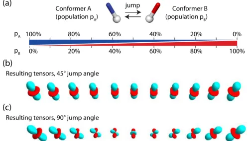
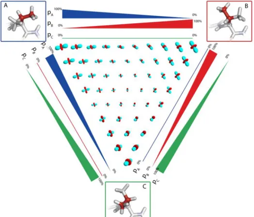
Figure 4. The effect of motion on the dipolar coupling tensor, shown for a two-site exchange process. In panels (b) and (c) the resulting dipolar coupling tensors are shown for the exchange depicted in panel (a). The different tensors in (b) and (c) correspond to the averaged tensors resulting from the two-site exchange with relative populations of the two states ranging from 100% state A to 100% state B. Positive and negative lobes of the dipolar coupling tensors are shown in red and cyan.
The measurement of dipolar couplings in solid therefore gives a very direct access to the amplitude of the motion sampled by a given inter-nuclear vector: a comparison of the experimentally measured dipolar-coupling tensor with the rigid-limit dipolar-coupling tensor reveals the space sampled by the vector under consideration. Generally this amplitude of motion is described by a single value, the “order parameter”, S, that compares the anisotropies δD of the dipolar coupling
tensors, S = δ /δ . The rigid-limit tensor δ depends only on the types of nuclei and
pA 100% 80% 60% 40% 20% 0%
pB 0% 20% 40% 60% 80% 100% jump
Conformer A
(population pA) (population pConformer BB)
Resulting tensors, 45° jump angle
Resulting tensors, 90° jump angle
(a)
(b)
the inter-nuclear distance. For directly bonded nuclei, where the distance is given by the bond length, the rigid-limit is thus readily computed.
This order-parameter description of a dynamic process is of course an oversimplification, because motion is in the general case asymmetric, i.e. its amplitude is orientation-dependent. Figure 4 illustrates the effect of a simple two-site exchange model on the dipolar-coupling tensor. As can be seen by comparing the dipolar-coupling tensors depicted in Figure 4b and c with the rigid-limit coupling tensor (Figure 3B), the motional process not only changes the strength of the dipolar coupling (i.e. the anisotropy of the tensor), but it also makes this tensor asymmetric. This often-overlooked fact can be put to good use: the characterization of the anisotropy AND asymmetry of the dipolar-coupling tensor allows extracting information of the motional process at a fair level of detail. Chapter 3 shows the measurement of the asymmetry of dipolar couplings, and illustrates how such measurements provide access to side chain motional processes, such as rotamer jumps.
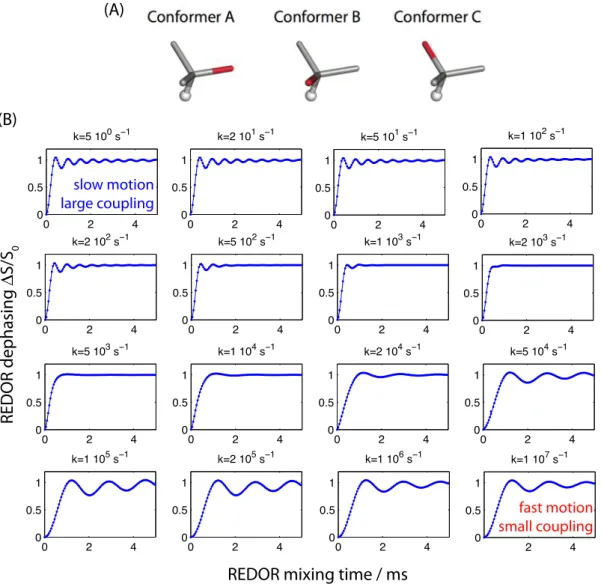
The above discussion illustrated how motion averages dipolar couplings. So far we have assumed that all the orientations that a given inter-nuclear vector samples contribute to this averaging. However, it is of course important to consider the time scale over which this averaging is effective. In Figure 5 we consider the averaging process through explicit numerical spin simulations, i.e. simulations of a REDOR-experiment under MAS, assuming a three-site exchange process. REDOR is a recoupling experiment which re-introduces the dipolar coupling under MAS, and the coupling strength is reflected by the oscillation frequency of the curve, where fast oscillation reflects a large dipolar-coupling strength δD.
As Figure 5 reveals, a fast motional process leads to an averaging, and thus a reduced dipolar coupling strength, see lower right panel. This is the averaging process discussed above. The other extreme, very slow motion effectively means that the involved conformational states never inter-convert. Thus, each of the involved states has its full rigid-limit dipolar coupling (top left panel). The transition between these two extremes occurs when the rate matches the (rigid-limit) dipolar-coupling strength, as Figure 5 shows.
This situation resembles the coalescence observed in chemical-shift averaging between two (or more) states with different chemical shifts: exchange with a rate slower than the chemical-shift difference between the states results in two separate peaks, while fast exchange (kex>>Δω) results in
a single peak. In between these two extremes, broadening of the peak occurs, similar to the situation in the coalescence regime in Figure 5.
Figure 5. Motional averaging of heteronuclear dipolar coupling, as seen by explicit numerical simulations of a system undergoing three-site exchange. In this model, depicted in (A), a given internuclear vector, as shown in red, jumps between three distinct conformations, which are here assumed to have tetrahedral symmetry. It is assumed that the relative populations of the three states are equally distributed (an assumption that is lifted in chapter 3). In (B), the resulting REDOR recoupling curves are shown for different rates of exchange between the states, from very slow motion (top left) to very fast motion (bottom right), as indicated above the panels. The coupling strength assumed here is 20 kHz (approximately a 1.02Å N-H distance). The REDOR recoupling is performed as in reference16.
Taken together, dipolar couplings provides a rich source of information about motional amplitudes. They possibly can reveal details of the motion, such as asymmetric motion, as long as the motional process takes place on time scales shorter than the inverse of the coupling strength. For typical heteronuclear systems (H-C, H-N, where δD is approximately 10 to 25 kHz) this means that
amplitudes of motions faster than about 10-20 µs can be accessed. The development of methods to accurately measure dipolar couplings, and applications thereof, are shown in chapters 3, 4 and 7. 1.4 Nuclear spin relaxation and protein dynamics
Nuclear spin relaxation rates are sensitive to molecular motion. In solution state, where anisotropic interactions (dipolar couplings and chemical-shift anisotropies) are averaged by molecular tumbling, nuclear spin relaxation rates are the primary source of information about molecular motion. Nuclear spin relaxation is sensitive to the amplitude and time scales at which anisotropic
0 2 4 0 0.5 1 k=5 100 s 0 2 4 0 0.5 1 k=2 101 s 0 2 4 0 0.5 1 k=5 101 s 0 2 4 0 0.5 1 k=1 102 s 0 2 4 0 0.5 1 k=2 102 s 0 2 4 0 0.5 1 k=5 102 s 0 2 4 0 0.5 1 k=1 103 s 0 2 4 0 0.5 1 k=2 103 s 0 2 4 0 0.5 1 k=5 103 s 0 2 4 0 0.5 1 k=1 104 s 0 2 4 0 0.5 1 k=2 104 s 0 2 4 0 0.5 1 k=5 104 s 0 2 4 0 0.5 1 k=1 105 s 0 2 4 0 0.5 1 k=2 105 s 0 2 4 0 0.5 1 k=1 106 s 2 4 0 0.5 1 k=1 107 s
REDOR mixing time / ms
REDOR dephasing S/S 0 (A) (B) slow motion large coupling fast motion small coupling
interactions (dipolar couplings, CSAs) are modulated. Through the well-established Redfield theory17
A detailed treatment of the theory of spin relaxation is not within the scope of this manuscript, and the reader is referred to excellent text books that cover in particular the situation in solution state (see, for example reference 18)
Likewise, relaxation rates also provide valuable information about motion in solids.
In this discuss we will not treat relaxation theory, but rather briefly introduce the main differences of solid-state relaxation approaches as compared to the (more widespread) solution-state relaxation analyses.
The main difference to solution-state NMR is the fact that in solid proteins do not have overall tumbling. This fact results in a number of important consequences:
1. Solution-state NMR measurements of internal molecular motion are restricted to time scales shorter than the overall molecular tumbling correlation time scale. This comes from the fact that overall tumbling averages all correlation functions of motion, and therefore any internal motion that is slower than the overall tumbling (typically in the low nanosecond range) becomes masked by the overall motion. In solids this is not the case, due to the absence of tumbling. Thus, solid-state NMR can probe motions over much wider ranges of time scales. Figure 6 shows this situation for 15N relaxation rates. The left panels show the situation in solution state: any internal motion that is slower than the overall motion does not lead to any alteration of the relaxation rates.
Figure 6. Relaxation rates in solution and solid state, arising from internal motion, as a function of the parameters of the internal motion. Shown are 15N R1 and R2 relaxation rates in solution-state, assuming an overall tumbling correlation time of 5 ns, and in the solid state (right), in the absence of overall motion. The relaxation rates are calculated as a function of the squared order parameter (x-axis), describing the motional amplitude, and the correlation time of internal motion.
2. The overall motion in solution state is generally the dominant process leading to relaxation. As a consequence, in solution state the relaxation rates e.g. of 15
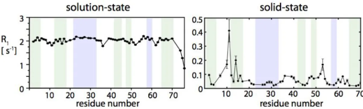
N spins are dictated by this overall motion, which is the same for the entire molecule. Figure 7 depicts this situation with the example of 15N R1 relaxation rates in ubiquitin. As these data show, the relaxation rates are very uniform, and site-to-site variations are only of the order of 10 %. This is completely different in solids, where relaxation is uniquely due to internal motion. The right panel in Figure 7 shows 15
N R1 rates in the same protein, ubiquitin, in crystals. Site-to-site variations of relaxation rates are much larger, about one order of magnitude. Thus, solid-state NMR has the potential to reveal the internal motion with much higher precision that solution-state NMR.
Figure 7. 15N R1 relaxation rates in ubiquitin in solution and in crystals. Note that the site-to-site variation of relaxation rates in solution is less than 10%, whereas relaxation rates vary by more than one order of magnitude in solids.
3. The absence of tumbling also leads to serious experimental and theoretical complications. On the theoretical side, the validity of the generally used theory of spin relaxation, Redfield theory17
, is not generally granted. In the derivation of Redfield theory, it is assumed that the relaxation rates are much smaller than the rate of the dynamic processes that leads to the relaxation. In solution state, there is always molecular tumbling on ps-ns time scales, and relaxation rates are always in the ms-s time scale. Thus, in solution state the validity of Redfield’s derivation is always given. In solids this is not necessarily the case. Figure 6 (right panel) shows the R2 relaxation rates in solids, derived with Redfield theory. In the top of this panel i.e. at long correlation times of microseconds, the R2 rates become very large, and comparable to the correlation time. In this regime Redfield theory starts losing its validity. Numerical simulation of spin relaxation may be one way of deriving theoretical relaxation rates in this regime.
4. On the experimental side, the absence of tumbling has also severe consequences, that arise because anisotropic interactions – most notably dipolar couplings – are not averaged out any more, unlike in solution. The dipolar couplings can thus lead to evolution of the density operators. This coherent evolution gives rise to spin-diffusion of longitudinal spin density operators, and can thus interfere with the accurate measurement of “real”, i.e. incoherent longitudinal relaxation processes that are due to stochastic motion. This situation is thus a challenge for measuring R1 rate constants. The situation is even more severe for transverse relaxation. In solids the fate of nuclear spin coherences is generally largely dominated by dipolar dephasing. For this reason, transverse relaxation parameters in solids are very difficult to access. Solutions to this problem have been proposed for 15N spins,{Chevelkov:2007dc}{Lewandowski:2011gw} but so far accessing transverse relaxation rates of carbons or protons remains a challenge.
Taken together, solid-state relaxation measurements provide potentially a very rich source of information about sub-microsecond motion in (bio-)molecules. There are challenges, however, with respect to how one can access relaxation rates properly, and how to interpret them.
The main challenge on the experimental side is due to the strong dipolar couplings to protons. The advent of deuteration for analyzing dynamics in solids, pioneered by Reif and co-workers, and also described in this manuscript, as well as fast magic-angle spinning, explored by a number of groups, has thus lead to an improvement, and has allowed to measure relaxation rates in a quantitative manner.
1.5 Motion on micro-to-millisecond time scales: Probing dynamics on biological functional time scales
Many biomolecular reactions take place on microsecond to millisecond time scales. Over the last 15 years solution-state NMR has witnessed a very strong interest in measuring dynamics on these time scales, and impressive examples have related such dynamics to biomolecular function and protein folding.9,21-23
Figure 8. The impact of microsecond to millisecond exchange on NMR spectra in solution, and the well-established solution-state CPMG methodology to access such motions. Panel (A) shows spectra of a system that exchanges between two states with different chemical chemical shifts (separated by Δω=50 Hz), as a function of the time scale of the exchange process. In this case the two states are equally populated. Panel (B) schematically depicts the principle of CPMG experiments. Transverse relaxation is measured in the presence of a train of refocusing pulses. The spacing of these pulses is changed (resulting in different pulsing frequencies νCPMG), and in the presence of an exchange process the effective transverse relaxation rate is altered. The right panel shows the effective relaxation rates as a function of the νCPMG.
Figure 8 shows the effect that an exchange process between two states with different chemical shift has on an NMR spectrum. Exchange between different conformations leads to line broadening, and in many cases it becomes impossible to detect minor conformational states in exchange with a major state, because the effect of the exchange broadens the resonances beyond detection. Nonetheless, the presence of minor conformational states is detectable through the line broadening of the major-state peak that remains observable. Carr-Purcell-Meiboom-Gill (CPMG) relaxation dispersion experiments (Figure 8B) or R1ρ relaxation-dispersion experiments essentially quantify this line broadening, and thereby provide access to conformational exchange processes. Quantitative analysis of relaxation-dispersion data (right panel in Figure 8B) allow the quantification of the kinetics of the exchange process, the relative populations and the chemical-shift difference. These approaches are well established in solution-state NMR.
While a theoretical treatment is not within the scope of this manuscript, it is worth highlighting the particularities of solid-state NMR approaches that aim at quantifying exchange processes.
The primary challenge for quantifying conformational exchange in solids comes from the fact that line widths in the solid state are generally dominated by factors that are unrelated to dynamics. In particular, as mentioned in the previous section, dipolar dephasing represents the primary factor of line broadening. As a consequence, line widths are generally not suited for the analysis of conformational exchange.
1.6 Quantifying dynamics in the solid state: experimental challenges
The previous sections have introduced approaches to measure dynamics in the solid state. Although solid-state NMR has a great potential for studying dynamics, a couple of experimental challenges have hampered in the past its widespread use. As discussed in the previous sections, these challenges are:
• Transverse relaxation parameters are difficult to access. The reason for this is the dipolar dephasing, i.e. a decay of magnetization through coherent processes rather than incoherent
stochastic (i.e. dynamic) processes. On the one hand, this situation hampers the measurement of motion on the time scale of hundreds of nanoseconds to microsecond (through the zero-frequency spectral density). On the other hand, the fact that transverse decay parameters are most often not reflecting dynamics also hampers that analysis of μs-ms dynamics, which are routinely measured in solution through relaxation-dispersion experiments.
• Longitudinal relaxation rates may also be corrupted by the presence of coherent evolution of polarization, i.e. by spin diffusion.
• The measurement of dipolar couplings is in principle possible through methods that have been in use for decades. However, measuring dipolar couplings quantitatively is also challenging, because experimental imperfections (e.g. pulse imperfections) as well as couplings to remote spins may interfere with the accurate measurement.
On top of these difficulties to access dynamics quantitatively, it should also be mentioned that spectral resolution and sensitivity are always a serious challenge in solid-state NMR, and also interfere with the extraction of dynamic parameters on a residue-by-residue basis.
As this manuscript will show, the use of deuteration and fast magic-angle spinning resolves several of the above-mentioned problems. The following chapter briefly introduces recent developments of the use of deuteration and fast-MAS in biomolecular solid-state NMR.
2 Deuteration and fast magic-angle spinning: overcoming
challenges of dynamics studies in ssNMR
The main challenges for quantifying dynamics in solids, and also for line widths (and thus resolution), can be ascribed to dipolar dephasing, i.e. the evolution of the density matrix due to large dipolar couplings. Magic-angle spinning does not allow to eliminate multiple non-commuting interactions, but these interactions are reduced by a scaling factor of 1/νMAS (where νMAS is the MAS
frequency). Increasing the spinning speed is thus one viable approach for reducing the effects of dipolar dephasing. An alternative and fully complementary approach to reduce the effect that the multiple couplings to protons have is to chemically remove protons and replacing them by deuterons.
The use of deuteration has become very popular over the last 10 years, following pioneering work by Zilm,24 Rienstra25 and Reif26 and their respective co-workers. When I started solid-state NMR in
2008 these works have been influential for my work, and we further developed methods and applications of deuterated proteins, some of which are outlined in this manuscript. More recently, Pintacuda and co-workers have shown very elegantly the power of deuteration, fast magic-angle spinning and highest magnetic field strengths for assignment and structure determination of proteins, and applications to complex systems such as membrane proteins are rapidly emerging.27-29
The reader is referred to recent reviews on the topic for further information.30,31
This chapter briefly introduces the opportunities that deuteration and fast magic-angle spinning have opened for biomolecular solid-state NMR.
2.1 Long coherence life times in deuterated proteins
In most instances, protein deuteration is combined with re-protonation of sparse sites in the protein, most often the amide sites. As a consequence, the distances between the few remaining protons are large, and accordingly the dipolar couplings between them very weak. Due to these drastically reduced dipolar-coupling network, dipolar dephasing is greatly reduced, and coherence life times are thus longer. This leads to narrow line widths of heternuclei (15N, 13C), but also of protons, and
therefore allows direct proton detection. The high gyromagnetic ratio of protons then leads to an additional sensitivity gain. As the effect of dipolar interactions are scaled down by fast magic-angle spinning, this approach is ideally performed at fast MAS. However, even at rather moderate MAS frequencies of ~20-25 kHz very high resolution spectra may be obtained already, given high levels of deuteration.32
Figure 9 shows examples of proton-detected solid-state NMR spectra of microcrystalline ubiquitin, a small 8 kDa model protein. This protein is used in many of the developments and applications discussed in this manuscript. These spectra show very high resolution in heteronuclear dimensions (13C, 15N, line widths down to ~15 Hz), but also in proton dimensions. In addition, the sensitivity is generally high, even though these spectra were recorded with a rather small amount of sample (2.5 – 3 mg).
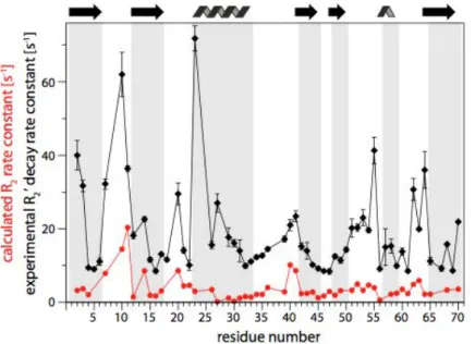
Figure 10 investigates 15N transverse relaxation rates in a highly deuterated sample of ubiquitin. Coherence life times up to 100 ms are found for a majority of residues, which reflects the high resolution obtained in spectra in Figure 9. Remarkably, however, these coherence life times are not yet the “true” relaxation-limited coherence life times. As shown in Figure 10 (red and blue data sets), the actual expected R2 are of the order of about 5 s-1. In other words, even in highly
deuterated proteins at an MAS frequency nowadays dubbed “ultra-fast”, dipolar dephasing still dominates the coherence decay. Nonetheless, these T2’ , which are obtained with only moderate 1H
decoupling, are significantly longer than in protonated proteins, where 15N T2’ are mostly below
30-50 ms,33 and only if high-power (~100 kHz 1H decoupling is used).
The high sensitivity and resolution offered by proton-detected solid-state NMR of deuterated proteins offers a number of exciting possibilities for studying biomolecular structure, dynamics and interactions.
Figure 9. High-resolution proton-detected solid-state NMR spectroscopy by deuteration and fast magic-angle spinning. These data have been recorded at a static magnetic field strength of 19.9 T (850 MHz) and MAS frequencies of 50-55 kHz. Typical experimental times are of the order of 20-30 mintues.
Figure 10. Transverse relaxation rate constants in a highly deuterated sample of microcrystalline ubiquitin. The experimental R2’ (black) is of the order of 10-20 s-1, i.e. coherence life times of up to 100 ms. Details about the measurement scheme are presented elsewhere.34 The red data set shows expected R2’, based on the backbone dynamics derived from dipolar-coupling and relaxation experiments (chapter 4).
Figure 11. Sequential assignment experiments based on proton detection in the solid state. The right panels show the nuclei that are correlated in the 3D experiments, of which one plane is shown on the left. This set of three 3D experiments was obtained within 7 days of experimental time (data unpublished).
2.2 Novel types of correlation experiments: from assignment to hydrogen-bond scalar couplings
Assignment experiments in solid-state NMR traditionally relied exclusively on correlation spectra of heteronuclei, i.e. on CANCO, NCACX, NCOCX correlations and variants thereof. The possibility of detecting protons allows having an additional nucleus for increasing resolution and spreading signals. Figure 11 shows representative examples of 3D assignment experiments, recorded in 2011 on a 39 kDa protein (unpublished data). As pointed out very recently, 1H-detected assignment experiments are rather rapid, and a 3D data set (hCANH, hCONH) can be obtained within less than 12 hours with less than 3 mg of proteins. This is significantly faster than 13 C-detected experiments, that typically require >15 mg of protein and several days of experimental time for a 3D data set.
The long coherence life times of protonated proteins at fast MAS allow also scalar-coupling based coherence transfer experiments, rather than cross-polarization experiments. Linser et al have shown that in highly deuterated proteins it is possible to construct experiments that are based exclusively on scalar-coupling mediated transfer steps.35 This type of experiments make it possible to use the
scalar couplings along the backbone for assignment purposes. These couplings are of the order of 7-13 Hz, and the transfer delays are thus of the order of 25 ms. While in protonated proteins the coherence life times are typically too short for efficient transfer throughout the ~25 ms long transfer delays, the long coherence life times in deuterated samples make these experiments straightforward.
Figure 12. Direct detection of hydrogen-bond scalar couplings (3hJNC’) in proteins in the solid state. (A) pulse sequence employed. The transfer delay 2T is 66.6 ms. (B, C) Experimental results obtained on microcrystalline ubiquitin at a MAS frequency of 57 kHz. Hydrogen-bond scalar couplings are evident from the cross-peaks encircled in panel (C), and are highlighted on the structure.
The most challenging experiments among biomolecular scalar-based transfer experiments are those experiments that aim at measuring very weak interaction: hydrogen-bond scalar couplings between backbone 15N and hydrogen-bonded carbonyl 13C are of the order of 1 Hz or less, i.e. about one order of magnitude lower than the experiments correlating nuclei along the backbone. Accordingly, the delays required for coherence transfer are long, typically of the order of hundred milliseconds. Thus, trans-hydrogen-bond scalar coupling transfer is a particular challenge. Figure 12 shows that when combining high levels of deuteration with fastest possible MAS, it is indeed possible to measure such couplings, i.e. identify the nuclei involved in the hydrogen bond, and even quantify the coupling strength, which reflects the H-bond geometry.34
2.3 Structure determination of deuterated proteins from unambiguous 1H-1H distance restraints
Protein structure determination does not have a very long and/or impressive history so far, regarding the number of structures solved or the size of the proteins for which structures were obtained (although important biological assemblies have been resolved). Structure determination has so far mostly relied on the extraction of 13C-13C or 15N-13C internuclear distances in 13
C-detected experiments (although 1H-1H distances are indirectly used in CHHC-type experiments36
on protonated samples). Challenges of such experiments are typically related to the large number of structurally irrelevant intra-residue correlation peaks, and low sensitivity. The availability of highly resolved proton-detected spectra opens new opportunities for structure determination through 1H-1H
distance measurements.
A number of advantages might arise from the use of deuterated samples. (i) When sparsely protonated samples are used, then 1H-1H distances necessarily correspond to long-range contacts (at least to a different residues). This is generally not the case when using 13C-13C contacts in uniformly
13C-labeled molecules, where the majority of observed distances arise from (trivial) connectivities
relatively large couplings even for distant nuclei, i.e. one can expect to see longer distances than with 13C-13C experiments.
Structure determination using proton detection in deuterated proteins has been pioneered by Rienstra and co-workers,25 and, simultaneously Reif and co-workers37, and our group#.38
Figure 13 shows the approach we have chosen to obtain the structure of microcrystalline ubiquitin, which consists of measuring 1H-1H distances between amides, as well as between methyls in specifically 13CHD
2 labeled and otherwise deuterated samples. The high sensitivity of proton
detection, a consequence of the long coherence life times and the high gyromagnetic ratio of 1H, enabled us to obtain four-dimensional spectra, akin to solution-state HSQC-NOESY-HSQC data, which have essentially no assignment ambiguity. Details of this approach are not within the scope of this manuscript.
Figure 13. Protein structure determination from 1H-1H distance measurements for deuterated proteins at fast magic-angle spinning. (A) Pulse sequence used for obtaining methyl-methyl distance restraints from 4D data. (B) and (C) Examples of methyl-methyl and amide-amide distance restraints, respectively. (D) Structure obtained from these distance restraints, along with secondary structure information from TALOS.
In summary, the advent of proton-detected experiments, enabled by deuteration and fast MAS has opened a range of new possibilities, and has had a great impact on biomolecular solid-state NMR. In this chapter we have shown how coherence life times are prolonged in deuterated samples, and what benefit can be obtained for assignment and structure determination.
In the remainder of this manuscript we will primarily focus on opportunities to study protein dynamics with deuterated proteins.
Chapter 3 shows an approach that allows measuring dipolar couplings at unprecedented level of detail, thus enabling to characterize protein side chain mobility.
Chapter 4 focuses on time scales and amplitudes of protein backbone motion on sub-microsecond time scales. Different experimental observables are critically analyzed, and finally the impact of the protein crystal on protein motion is investigated.
# work presented at the International Workshop on high-field solid- and solution-state NMR, Les Houches, France. June
Chapters 5 and 6 investigate how one can obtain information about slower processes, in which a predominant state undergoes transient excursions to low-populated “higher-energy” conformers. Chapter 7 presents the application of (mostly) proton-detected NMR to the study of a complex of a protein with an intact peptidoglycan cell wall in terms of its structure and dynamics.
3 Solid-state NMR measurements of asymmetric dipolar
couplings provide insight into protein side-chain motion
#3.1 Introduction
Understanding conformational flexibility is of critical importance for understanding protein function, folding, and interactions with other proteins and ligands. NMR spectroscopy is an important tool for such investigations in solution[1] and increasingly also in the solid state[2] since it
allows site-resolved studies of dynamic processes. An experimental characterization of all motional modes of a protein is a great challenge and simplified models are necessary. In NMR studies of dynamics, motional amplitudes are generally expressed in terms of a single order parameter,[3]
discarding the details of the motion, such as the motional asymmetry. In the following, we show a significant extension of this description, by detecting asymmetric motion of side chains in a protein in the solid state.
Dipolar couplings are particularly powerful probes of local molecular dynamics in the solid state. In the absence of motion, the tensor describing the dipolar interaction between two nuclei is a traceless axially-symmetric second-rank tensor. It can be characterized by a single parameter, namely its anisotropy δD,rigid which depends only on the internuclear distance and isotope type of the nuclei
involved.
In the presence of “fast” motional processes, i.e., processes with a correlation time shorter than approximately 1/δD,rigid, (typically 10-100 µs), the dipolar-coupling tensor becomes partially
averaged. In the case of a motional process with three-fold (C3) or higher symmetry, for example,
an isotropic motion within a cone, the averaged tensor remains axially symmetric and is fully characterized by the effective anisotropy δD which has a reduced value compared to δD,rigid. In this
case, the motional amplitude can be expressed by a single order parameter[4] S = δD/δD,rigid, such that
the dipolar couplings asymmetry is given as: δD = S−2µ0
4π
γkγn!
rkn3
where S and rkn are the order parameter and the internuclear distance, respectively.
However, in the case of a general fast motion, the characterization solely by S is incomplete because the averaged dipolar tensor is no longer axially symmetric[5] and one additional tensor parameter, the asymmetry ηD is needed for a complete description. The asymmetry ηD varies
between zero (symmetric tensor) and one.[5a]
In this general case, the dipolar coupling tensor thus takes the following general form:
! D=δD1 2 −1−η 0 0 0 −1+η 0 0 0 2 ⎡ ⎣ ⎢ ⎢ ⎢ ⎤ ⎦ ⎥ ⎥ ⎥
Here, The value of the asymmetry is 0≤η ≤ 1, implying the following ordering of principal components of
!D:
# This work was published in: P. Schanda, M. Huber, J. Boisbouvier, B. H. Meier and M. Ernst. Solid-State NMR
Measurements of Asymmetric Dipolar Couplings Provide Insight into Protein Side-Chain Motion. Angew. Chem. Int. Ed. Engl. 2011, 50, 11005.
Dyy ≤ Dxx ≤ Dzz . Thus, ηD= Dyy− Dxx
Dzz .
Note that in the case of axially symmetric motion (η=0) the dipolar coupling tensor reduces to the often assumed form, where the diagonal elements are -1, -1 and 2, as is the case for a rigid-limit coupling.
In solution-state NMR, dipolar couplings can be measured as residual couplings (RDCs) in anisotropic media. The evaluation of motional amplitudes from RDCs is challenging because RDCs also depend on the (a priori unknown) degree of molecular alignment and the orientation of a given vector relative to the alignment frame and usually data from different alignment media must be combined[6]. The situation is much simplified in solid-state magic-angle spinning (MAS) NMR, where overall molecular tumbling is absent, allowing the direct measurement of dipolar couplings that only depend on the interatomic distance and dynamics. For the case of one-bond dipolar couplings (C-H, N-H, or C-N) the rigid-limit dipolar-coupling tensor is known from the bond lengths. Thus, measurements of the dipolar-coupling tensor provide direct access to the amplitude and axial symmetry of the motion sampled by the bond vector. However, due to the limited precision and accuracy of the currently available experimental data, dynamically-averaged dipolar-coupling tensors have, so far, always been analyzed in terms of a single order parameter S. In this communication, we demonstrate the first direct measurement of asymmetric dipolar-coupling tensors in MAS NMR, providing a more detailed picture of motional amplitudes. We exemplify the measurement of asymmetric dipolar couplings by studying side-chain motions in the protein ubiquitin, using a combination of appropriate sample labeling with sensitive and precise NMR measurement techniques. We find that the asymmetry ηD of the dipolar-coupling tensor of several
methyl C-H moieties deviates indeed significantly from zero and provides useful information about the details of the motional processes.
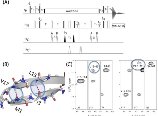
In order to obtain the necessary accuracy and precision in the measurement of 1H-13C dipolar-coupling tensors, we extend a recently developed experimental approach with greatly improved accuracy.[7] In brief, it consists of (i) the selective introduction of isolated 1H-13C spin pairs in an otherwise perdeuterated protein, using specifically protonated precursors, and (ii) a REDOR recoupling technique in combination with sensitive proton detection. REDOR has a built-in normalization[8], such that the recoupling data are expressed in a manner that is independent of the peak intensity or the coherence loss during the recoupling period and can be fitted using only δD (or
S) and ηD as free parameters.[7]
3.2 Experimental observation of asymmetric dipolar couplings in ubiquitin side chains
We have prepared two samples of perdeuterated ubiquitin carrying 1H-13C spin pairs on a single methyl group of either Ile (δ1) or Val (γ1 or γ2) and Leu (δ1 or δ2) residues and we study side-chain dynamics as probed by the methyl C-H dipolar-coupling tensor. The labeling follows established protocols[9] (see Supporting Information for details). The low 1H density in such samples largely eliminates 1H-1H couplings and couplings from the 13C spins to remote 1H spins, thus excluding one source of potential systematic errors in dipolar-coupling measurements. Furthermore, the low proton density allows the acquisition of high-resolution proton-detected correlation spectra with high sensitivity.[10] In combination with fast magic-angle spinning, coherences in such samples are long lived[11] leading to a further increase in sensitivity and thus precision of dipolar-coupling measurements. The improved measurement precision, the suppression of systematic errors and the normalized nature of REDOR recoupling curves are crucial to detect dipolar tensor asymmetry. Figure 14 shows how the presence of discrete jumps that result in asymmetric dipolar coupling tensors impacts REDOR recoupling curves, and Figure 15 depicts the
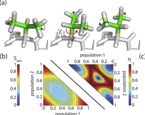
tensors resulting from conformational exchange in a tetrahedral geometry, such as a side chain performing rotations around the χ1 angle.
Figure 14. The effect of discrete bond jumps on REDOR curves, exemplified here for two-site (a,b) and three-site (c) cases.
Figure 15. Dipolar coupling tensors in a system undergoing exchange between three sites (tetrahedral geometry). This is typically the case for e.g. a valine side chain. Along the three axes the relative populations of the three rotamer states are changed, and the symbols in the center depict the resulting dipolar coupling tensors. Symmetric tensors are obtained if either only one rotamer is populated (the three extremities of this plot), or if all are populated equally (center).
0°/180° 30°/150° 60°/120° 90° 0 0.5 1 1.5 2 0 0.2 0.4 0.6 0.8 1 1.2 0 0.5 1 1.5 2 0 0.2 0.4 0.6 0.8 1 1.2 jump angle state B (40%) state A (60%) 60° state Bp B state A pA populations pA/pB S/S 0 S/S 0 time / ms time / ms 0.5/0.5 0.6/0.4 0.7/0.3 0.9/0.1 1/0 0 0.5 1 1.5 2 0 0.2 0.4 0.6 0.8 1 1.2 time / ms S/S 0
2-site jump models
3-site jump models
side view top view
top view side view 1/3 1/3 1/3 1/3 1/3 1/3 side view top view
0.6 0.3 0.1
only one orientation no jumps (a)
(c)
Figure 17a shows experimental REDOR curves for a number of representative methyl groups in ubiquitin, measured using the pulse sequence of Figure 16. The full set of experimental recoupling curves is shown in Fig. S2, and representative two-dimensional spectra are shown in Fig. S3 of the SI. The results of a two-parameter fit (anisotropy δD and asymmetry ηD, red curves in Figure 2a) are
shown in Figure 3 and listed in Table S1. Reduced-chi-square (χ2
red) surfaces are shown in Fig. 2b.
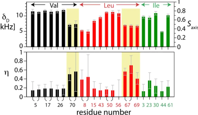
Comparing the different methyl groups in ubiquitin, a large variation in the fitted anisotropies δD is
observed, ranging from 4.9 to 11.8 kHz, indicating that site-to-site variations of side chain motional amplitudes are large. We also detect significant variation of the asymmetries ηD between different
side chains (between 0 and 0.58). Based on the fit of the anisotropy and the asymmetry to the REDOR data, the methyl groups can be classified into three groups: (i) methyl groups that have a low χ2
red value and an asymmetry that is not significantly different from 0, (ii) methyl groups that
have a low χ2
value but a value of the asymmetry parameter that is significantly different from 0, and (iii) methyl groups that cannot be properly fitted (with correspondingly large χ2
value) by a single asymmetric dipolar tensor. The majority of the methyl groups in ubiquitin (22 out of 29),
such as Val5, Val17, Val26, Leu50 (Fig. 1) fall into category (i) with an almost symmetric dipolar-coupling tensor, i.e. an asymmetry below about 0.2. These methyl groups can be described by the conventional symmetric-tensor assumption. A significant asymmetry with values of η ≥ 0.4 (category ii) is observed for the methyl groups of Val70, Leu67, and Leu69 (5 out of 29). For two methyl groups the asymmetric dipolar-coupling model does not result in satisfactory fits (category (iii), δ1 of Ile13 and Ile36), pointing to slow motional processes (vide infra). Thus, for several sites the traditional symmetric tensor model clearly does not apply, and the reported asymmetric tensors provide further insight into the motion of these side chains.
The observed dipolar tensor parameters δD and ηD for a methyl group are the result of several
averaging processes. Invariably, at room temperature, the methyl groups are in fast rotation around the local three-fold axis with correlation times typically in the picosecond range. This rotation leads to an averaged axially-symmetric tensor with an asymmetry δD,rigidaxis = δD,rigid/3 ≈ 14.53 kHz (based
on the canonical tetrahedral angle θHCC = 109.47° and a C-H bond length of 1.115 Å). This tensor is further affected by motional processes involving the direction of the three-fold methyl axis, which result from librational motions, and, more importantly in terms of amplitude, jumps between discrete rotamer states. Under such fast (<10-100μs) rotamer jumps, the observed tensor is the average of the involved orientations, and will, thus, be generally asymmetric, characterized by its asymmetry η and the anisotropy δD or the axis order parameter Saxis= δD/ δD,rigidaxis. Rotamer jumps
should thus have a measurable impact on tensor anisotropies and asymmetries, and information about rotamer equilibria should be contained in the dipolar tensors. Furthermore, rotamer jumps equally affect the methyl groups γ1 and γ2 attached to a given Val, and the δ1/δ2 methyls attached to a given Leu side chain, and the tensors should thus be identical, provided that librational motions are negligible or similar to both sites. Indeed, we find that the tensor parameters for methyls attached to the same side chain always agree within error bars.
Figure 16. Pulse sequence used in this study for the measurement of 1H-13C dipolar couplings in highly deuterated, sparsely 1H,13C labeled samples. Filled and open rectangles denote 90° and 180° pulses,
respectively. Pulse phases are set to ϕ1=2(x),2(-x), ϕ2=x,-x, ϕ3=y,-y, ϕ4=4(x),4(-x), ϕrec=x,-x,-x,x,-x,x,x,-x. 1H pulse phases during REDOR recoupling are incremented (decremented) according to the XY-16 scheme in the first (second) half of the REDOR block. The delays Δ, τ and ζ were set to 1.5ms, 4µs and 10ms, respectively. All pulses were applied at a field strength of γB1/2π=100kHz, except for 1H 180° pulses during the REDOR train, which were applied at 125kHz (4µs). The 1H saturation pulse (“sat.”) was applied for 80ms at a field strength of about 10kHz. 2.5kHz/3kHz WALTZ-16 decoupling was applied on 2H/13C, respectively.
Figure 17. a. REDOR recoupling curves for methyl 1H-13C sites in crystalline ubiquitin. Each data point was obtained from 2D recoupling and reference spectra, recorded in 95 minutes per spectrum. Experimental points are shown along with error bars, based on twice the standard deviation of the spectral noise. Black curves show fits assuming an axially-symmetric dipolar-coupling tensor, red curves use asymmetric tensors, and green curves (only Ile13/36) assume a superposition of two general tensors. b. Plots of the reduced chi-square χ2red for the two-parameter fits of the (red) REDOR curves of Fig. 1a. Shown are three contours at the values of χ2red corresponding to the minimum of χ2red +1, +2 and +3. Thus, the innermost contour denotes the confidence interval. The full set of χ2red plots is shown in Figure S4. Saxis is defined as δD/δD,rigidaxis=δD/14.53kHz.
Figure 18. Dipolar tensor parameters (top: anisotropy, bottom: asymmetry) for methyl groups in ubiquitin. Two data points per residue denote methyls at positions γ1/γ2 (Val) or δ1/δ2 (Leu). Side chains with large tensor asymmetries are highlighted (yellow background).
0 5 10 0 0.2 0.4 0.6 0.81
Val Leu Ile
5 17 26 70 8 15 43 50 56 67 693 23 30 44 61 0 0.2 0.4 0.6 0.8 1 D [kHz] Saxis residue number Val 5 2 Val 17 1 Val 26 2 Val 70 1 Leu 67 1 Leu 69 1 Ile 30 1 Ile 44 1 0 0.5 1 5 10 5 10 5 10 0 0.5 1 0 0.5 1 recoupling time / ms nor maliz ed REDOR dephasing S/S 0 0.2 0 0.4 0.6 0.81 0.2 0 0.4 0.6 0.81 0.2 0 0.4 0.6 0.81 0.2 0 0.4 0.6 0.81
(a)
asymmetr y 0 0.2 0.4 0.6 0.81 0 0.2 0.4 0.6 0.81 0 0.2 0.4 0.6 0.81 0 0.2 0.4 0.6 0.81 anisotropy D / kHz 0.2 0.4 0.6 0.8 1 0.2 0.4 0.6 0.8 1 0.2 0.4 0.6 0.8 1 axis order parameter Saxis(b)
Ile 13 1 Ile 36 1 Val 70 1 Leu 50 2 Val 26 2 Val 17 1Val 5 2 Leu 50 2 Ile 13 1
Ile 30 1 Leu 67 1 Leu 67 2 Leu 69 1 Ile 44 1 Leu 67 2 2 red 5 10 15 20 25 0 Ile 36 1