HAL Id: tel-00714984
https://tel.archives-ouvertes.fr/tel-00714984
Submitted on 6 Jul 2012
HAL is a multi-disciplinary open access
archive for the deposit and dissemination of sci-entific research documents, whether they are pub-lished or not. The documents may come from teaching and research institutions in France or abroad, or from public or private research centers.
L’archive ouverte pluridisciplinaire HAL, est destinée au dépôt et à la diffusion de documents scientifiques de niveau recherche, publiés ou non, émanant des établissements d’enseignement et de recherche français ou étrangers, des laboratoires publics ou privés.
Creating and Use of an New Experimental Preclinical
HLA Transgenic Mice Model to Mapping
HLA-restricted T Cells Epitopes for Polyepitopes
Vaccine Design
Zhitao Ru
To cite this version:
Zhitao Ru. Creating and Use of an New Experimental Preclinical HLA Transgenic Mice Model to Mapping HLA-restricted T Cells Epitopes for Polyepitopes Vaccine Design. Immunology. Université Paris Sud - Paris XI, 2012. English. �NNT : 2012PA11T007�. �tel-00714984�
Université Paris-11 Paris SUD
Ecole doctorale de signalisation et réseaux intégratifs en biologie
Doctorat Immunologie
Présenté et soutenu par
Ru Zhitao le 21 Février 2012
Exploitation d’un modèle expérimentale préclinique de souris HLA transgénique pour l’identification des épitopes T HLA-restreint afin de
concevoir des vaccins poly-epitopiques
Thèse dirigée par Dr Yu-Chun LONE
JURY
M. Antoine Durrbach Président du jury M. Eric Tartour Rapporteur Mme. Monica Sala Rapporteur M. Sebastien.Lacroix-Desmazes Examinateur M. Guy Duc Huynh thien Examinateur M. Yu-chun Lone Examinateur
Je tiens tout d’abord a remercier les membres du jury : Les rapporteurs, Monsieur le Professeur Eric Tartour et Madame le Docteur Monica SALA pour m’avoir consacré un temps précieux à l’examen de cette thèse et m’avoir apporté leur commentaires. Je remercie également aux examinateurs Monsieur le Docteur Sebastien Lacroix-Desmazes et Monsieur le Docteur Guy Duc Huynh thien pour leur expertises et leurs conseils. Enfin, j’exprime mes remerciements au Professeur Antoine Durrbach qui a accepté de présider le jury de cette thèse.
Je remercie à Monsieur Aime Vazquez pour m’avoir accueilli au sein de son laboratoire et à Monsieur Yu Chun Lone, mon directeur de thèse de sa bonne humeur et de son soutien permanent. Et je lui remercie tout particulièrement de m’avoir encadré ce travail avec patience, et surtout m’avoir transmis sa passion pour la recherche.
Je tiens à remercier tous les membres de l’unité de la Régulation de la Survie Cellulaire et des Allogreffes et toutes les personnes qui ont collaboré avec moi.
Un grand merci a mes parents et ma femme pour leur soutien de toujours et leur confiance.
Merci a tout les ›acteursfilistés ici par ordre alphabétique qui me sont chers et qui ont participé de près ou de loin à ce travail : Anthony Pajot, Bernard Maillere, Christelle Martin ,Chaobin Zhu, David M.Ojcius, Guangyu Zhao, Kongli Zhu,Nathalie Mérillon, Wenjun Xiao ,Xiaoming Zhang , Yusen Zhou, Zhihua Kou
Je remercie enfin vivement Cancéropole d'Ile de France, INCA et Service pour la science et la technologie de l’ambassade de France en Chine qui m’ont financé durant cette thèse.
Index
Abbreviation... 3
Table of illustrations ... 5
Introduction... 6
I. Major Histocompatibility Complex (MHC) ... 7
A. Structure of MHC antigen ... 8
B. Immunological function of MHC molecule ... 9
II. Murine MHC (H-2)... 15
III. Human MHC (Human leukocyte antigen, HLA)... 17
A. HLA structure ... 18
B. HLA Nomenclature... 20
C. Polymorphism ... 21
IV. MHC mediated immunology ... 21
A. Cytotoxicity T Lymphocytes response (CTL)... 22
B. Helper T lymphocytes response (Th) ... 25
C. CD4+ T cells epitope ... 27
V. Humanized mouse Model (HLA transgenic mice)... 30
A. HLA- class I transgenic mouse models ... 32
B. Optimization of the HLA-I transgenic mouse model ... 32
C. HLA class II transgenic mouse models ... 37
E. HLA-I/II double transgenic mouse models ... 41
VI. Highly pathogenic Influenza A virus (H5N1) ... 43
A. Structure of HPAIV H5N1 virus ... 44
B. Pathogenesis ... 47
C. Prophylaxis ... 48
VII. Hepatitis B Virus (HBV) ... 50
A. Structure of hepatitis B virus... 51
B. Pathogenesis ... 53
C. Prophylaxis ... 54
Issues ... 57
Discussion ... 70
DP4 is the most frequent allele worldwide ... 72
Significance of DP4 restricted epitope identification ... 74
DP4 restricted epitope mapping in vitro and in vivo... 77
Immunological characteristics of HLA-A2/DP4 transgenic mice 80 HLA-A2/DP4 mice application in DP4 epitope identification ... 84
The application of HLA-A2/DP4 mouse model... 86
Reference ... 93
Annexes... 106
Article 1... 107
Article 2... 133
Abbreviation
Abbreviation Full Name Aa Amino acid
APC Antigen presenting cell bp Paire de bases
BSA Bovine serum albumin CTL Cytotoxic T Lymphocyte CPM Counts per minute DNA Deoxyribonucleic Acid DC Dendritic cell
D-MEM dulbecco's minimum essential medium ELISA enzyme-linked immunosorbent assay ELISPOT enzyme-linked immunospot
EBV Epstein-Barr Virus FBS fetal bovine serum
g gram h hour HA Hemagglutinin HBc HBV Capsule HBs HBV envelope Protein hCD4 Human CD4 molecule HLA Human leucocyte antigen HRP horseradish peroxidase
IC50 half maximal inhibitory concentration
IPTG isopropyl β-D-thiogalacoside IgG immunoglobulin G IFN-γ Interferon γ IL Interleukine Kb Kilobase kDa Kilodalton LB Luria-Benrtani medium min Minute mg milligram ml milliliter
mM millimole per liter M1 Matrix protein 1 NP Nucleoprotein NA Neuraminidase
OD450 Absorption value at 450nm
OVA Albumin from chicken egg white PAGE Polyacrylamide gel electrophoresis PBS phophate buffered saline
PBMCs Peripheral blood mononuclear cells PCR polymerase chain reaction
PEG polyethylene glycol pfu plaque forming unit PMA phorbol myristate acetate rpm rotate per minute
SDS sodium dodecyl sulfate SI Stimulation index T CD4+ CD4+ T lymphocyte T CD8+ CD8+ T lymphocyte TBS tris buffered saline
TAP Transporter associated with antigen processing TCR T cell receptor
WB western-blot µg microgram µl microliter µm micrometer
Table of illustrations
Table 1. Three MHC gene subgroups and their functions………8
Figure 1. Mechanism of MHC class I-mediated CTL response………..10
Figure 2. Mechanism of MHC class II-mediated CTL response……….11
Figure 3.Schematic picture of an MHC class II molecule and a peptide.12 Table 2. HLA and disease susceptibility………..13
Figure 4 .Gene structure of mouse MHC (H-2)………15
Table 3. Mouse Leukocyte Alloantigens Chart……….17
Figure 5. Gene map of the human leukocyte antigen (HLA) region……19
Figure 6. Perforin/Granzyme mechanism of cytotoxic T lymphocytes to kill target cells……….………23
Figure 7. The mechanism of CTL killing target cells……….24
Table 4. Cumulative number of confirmed human H5N1 infected cases ………..44
Figure 8. Molecular structure of H5N1 virus………..45
Figure 9. Molecular structure of hepatitis B virus………...52
I. Major Histocompatibility Complex (MHC)
Major histocompatibility complex (MHC) is a complicated antigen system which is widely present in mammalian cells and it was encoded by a large gene family in all vertebrate. Currently, the function of MHC antigens has been basically stated: it posses an important role in the differentiation and maturation of peripheral T-cell, initiation and regulation of T immune response and it closely related to the autoimmunity. Therefore, it is one of the most important molecules in the immune system and it’s valuable to further exploit its potential function.
MHC gene families varies from different species, for example, chickens MHC (B antigen) having among the smallest known MHC regions (19 genes) [1]; the murine MHC gene region spans approximately 4 Mb of chromosome 17 (23.0 cM, cytoband B–C) and contains 3 major classes of highly polymorphic gene sets: class I (H-2-K, H-2-D, Q, H-2-T18 genes), class II (H-2-I genes), and class III (H-2-S genes)[2]; in humans the MHC region located on the chromosome 6, between the flanking genetic markers MOG and COL11A2, and contains 140 genes spanning 3.6 mega base pairs(3.6 Mb ) [3].
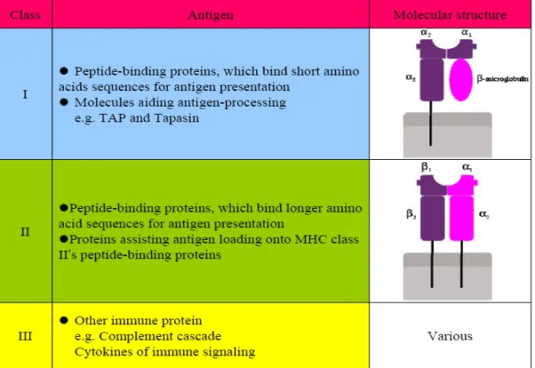
The MHC gene family consists of three subgroups: class I, class II and class III, which are shown in Table 1, take charge of encoding corresponding antigen to mediate immunity.
Table 1. Three MHC gene subgroups and their functions.
A. Structure of MHC antigen
In three kinds of MHC antigens, MHC class I and class II were proved to be more significant in specific cellular immunity and both of antigens are the most studied antigen in MHC antigen.
MHC class I antigen presents on the surface of all nucleated cells –in essence all cells but red blood cells. Actually, there are two kinds of MHC class I molecules, including classical MHC molecules and nonclassical molecules; however, we mainly focused on the classical MHC molecules. MHC class I occurs as an α chain composed of three domains-α1, α2, α3. The α1 subunit stands with a unit of the non-MHC molecule β2m which encoded on human chromosome 15. The α2 subunit form a
peptide-binding groove with α1 (α1/α2 heterodimer) to present epitope to CTL. The α3 subunit is transmembrane, anchoring the MHC class I molecule to the cell membrane. What’s more, α3 subunit allow CD8 molecule of CTLs dock on its surface and initiate cellular immunity.
MHC class II antigen can be conditionally expressed by all cell types, but normally occurs only on professional antigen-presenting cells (APC), such as macrophages, B cells and especially dendritic cells (DCs). MHC class II molecule is constructed by two chains, α and β, and each chain contain two domains-α1 and α2 and -β1 and β2. Among them, α1 and β1 form a heterodimer and the peptide being presented is held by the floor of the peptide-binding groove, in the central region of the α1/β1 heterodimer; α2 and β2 is transmembrane domain that anchoring the MHC class II molecule to the cell membrane.
B. Immunological function of MHC molecule
a. MHC restriction
During the development of T cells in the thymus, immature T lymphocytes have to go through a positive selection, which make sure T lymphocytes can get self MHC restriction and subsequently develop into CD4+ or CD8+ T lymphocytes, and a negative selection, which avoid T lymphocytes recognize the self-antigen and MHC complex. In this way, T cell receptor (TCR) of all mature T lymphocytes possesses dual
specificity: recognizes self MHC as well as nonself antigens. b. Antigen processing and presentation
For MHC class I pathway, proteins in the cytosol are cleaved into fragments by proteasome, liberating peptides internalized by TAP (transporter associated antigen presenting) channel in the Endoplasmic reticulum(ER), then assembles with freshly synthesized MHC-I. These peptide-MHC-I complexes are following glycosylated, secreted out of cell membrane and finally recognized by TCR of CD8+ T lymphocytes. Once CD8 molecule in CTLs dock to the MHC-I accompany with the recognition of TCR to the matching epitope, the CTLs transduce signals prompting the target cell’s apoptosis. In this way, MHC-I antigen participates to mediate cellular immunity (Figure 1).
Figure 1. Mechanism of MHC class I-mediated CTL response. Several experiments demonstrated that peptides that can be bound by MHC-I molecule with a bias: they are mostly a linear structure; from 9 to
10 amino acids for the length, because it was reported that HLA binding affinity of 9-amino acids peptide is 100-1000 times greater than that of more or less 9 amino acids; the anchor residues of these peptides are mainly located on the first N-terminal 2 and 9 to bind HLA molecule through hydrogen bonds; their hydrophobic carboxyl-terminal is constituted by aliphatic amino acids.
Figure 2. Mechanism of MHC class II-mediated CTL response. For MHC class II pathway, antigen processing is mainly performed by phagocytes, such as macrophage, immature dendritic cells, mononuclear phagocytes, B lymphocytes, endothelial cells and epithelium of thymus. These phagocytes uptake entities into phagosomes and the uptaken proteins are cleaved into many different short peptides. Then the peptides assemble with MHC-II in endoplasmic reticulum (ER) and further present on the surface of phagocytes to be recognized by T cell receptors (TCR)
of CD4+ T lymphocytes. In this way, CD4+ T lymphocytes are stimulated and differentiate into effector T cells to mediate specific cellular response (shown in Figure 2).
As distinct from MHC class I epitopes, peptides that can be bound by MHC-II molecules display peptides of variable length(usually 12-26 mers), often encompassing a common binding core sequence, which is variably extended in both the N- and C-terminal directions [4].Crystal structure of complexs between human MHC class II molecules and peptides have indicated that the peptide binding grooves of the different MHC class II isotype are superimposable and that the backbone of peptides bound to the binding grooves is highly conserved. The binding grooves are mainly characterized by properties of the so-called P1, P4, P6 and P9 pockets (as shown in Figure 3), which confer the specificity to the anchor residues of the peptides bound to the groove[5]. This 9 peptide binding core, or nonamer, is sufficient to bind MHC class II molecule [6].
Figure 3.Schematic picture of an MHC class II molecule and a peptide. c. MHC and diseases susceptibility
found at a much higher frequency in those suffering from certain diseases than in the general population. Most of these diseases associated with particular MHC alleles, for instance, as autoimmune disorders, certain viral diseases, disorders of the complement system, some neurologic disorders, and several different allergies, more detail was shown in Table
2.
*Relative risk is calculated by dividing the frequency of the HLA allele in the patient population by the frequency in the general populations:
Table 2. HLA and disease susceptibility.
From the discovery of human HLA antigen, it is discovered that more than 50 human diseases are related to one or more human HLA antigens.
For example, the HLA-B27 antigen arises from about 90% ankylosing spondylitis disease [7]; crowd carrying HLA-A29 monomer tends to suffer
from birdshot chorioretinopathy[8]; IDDM is related to HLA-B8,
HLA-Bw15 and HLA-B18 and so on [9]; the patients with psoriasis carries HLA-B13 or HLA-B17 alleles and so on [10], therefore, specific HLA clinically becomes the genetic markers for some diseases. Now, the correlation of HLA alleles and a given disease can be predicted by determining the frequency of the HLA alleles in patients afflicted with the disease, then comparing these data with the frequency of the same alleles in the general population. In this way, the susceptibility can be predicted and at present, HLA typing in autoimmunity is being increasing used as a tool in diagnosis.
The built HLA transgenic mouse model can be used for the researches on the relevance between specific HLA and diseases. For example, Breban et al built the HLA-B27 transgenic mouse model to study the relevance between this haploid and spondylarthritis, and determined the protection and susceptibility of specific HLA for diseases through understanding of the disorder mechanism of the immune system, so as to provide references for clinical prevention and therapeutic schedule and so on.
d. MHC in transplant rejection
transplant procedure and it can stimulate immune response in the recipient. Normally, even though high MHC polymorphism at the population level, an individual bears at most 18 MHC I or II alleles. For each person, around six HLA class I alleles and six to eight HLA class II alleles simultaneously occur in human haplotype. Thus, serology-based HLA typing is the most important determinant factor in transplantation.
In summary, MHC antigen is one of the most crucial molecules in the immunity. It is necessary to further exploit its biological function as well as mechanism in initiation and regulation of immunity.
II. Murine MHC (H-2)
Mouse MHC antigen is also called H-2 antigen. Its gene locates on mouse chromosome 17. Gene structure of mouse MHC (H-2) is shown in Figure 4. Most studied H-2 antigen is two major classes: class I and class II.
Figure 4 .Gene structure of mouse MHC (H-2)
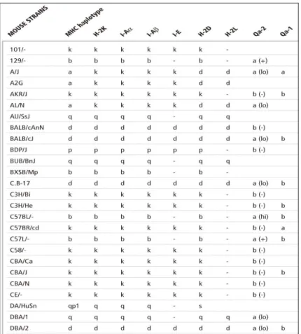
MHC class I” (also called MHC-Ia) that comprises H-2D, H-2K and H-2L subclasses; the “non-classical MHC class I” (MHC-Ib) that comprises H-2Q, H-2M and H-2T subclasses; the “classical MHC class II”(MHC-II a) that includes H-2A(I-A) and H-2E(I-E) subclasses, and the “non-classical MHC class II” (MHC-II b) comprises H-2M and H-2O. H-2 molecules are highly polymorphic. Each laboratory mouse strain is homozygous and has a unique MHC haplotype. The MHC haplotype in these strains is designated by a small letter (a, b, d, k, q, s, etc.). Take the two most useful animal models BALB/ and C57BL/ for example, the mouse leukocyte alloantigen of BALB strains is d, it has H-2Kd, H-2Dd, H-2Ld, IAd, I-Ed, while for C57BL strains is b, it only has H-2Kb, H-2Db for class I and I-Ab for class II. Mouse Leukocyte Alloantigens Chart is shown in Table 3. Thus; the MHC background of C57BL strains is less complicated than BALB.
H-2 class I molecules consist of a 45 kD highly glycosylated heavy chain non-covalently associated with a 12kD β2-microglobulin, a polypeptide that is also found free in serum. H-2 class I antigens are expressed on almost all nucleated murine cells and are reported in many animal trials that they play an important role in presentation of altered self cell antigens (virally infected or tumor cells) to CD8+ cytotoxicity T cells.
Table 3. Mouse Leukocyte Alloantigens Chart
Murine MHC class II molecules are composed of a 33 kD α chain and a 28 kD β chain. H-2 class II antigens are expressed on antigen presenting cells (B cells, monocytes/ macrophages, dendritic cells, and Langerhans cells, etc.) and they involve in presentation of processed peptide antigens to CD4+ cells.
III. Human MHC (Human leukocyte antigen, HLA)
The discovery of MHC in 20th century, especially the discovery of the first human HLA antigen in 1958, was the important milestone in the immunological development course. At present, function of the HLAantigen is practically illustrated as follows: it not only participates in the maturation of peripheral T cell and starts and regulates the function of the immune state together with TCR, but also is related to many autoimmune diseases, and is one of the most important links in the immune system.
A. HLA structure
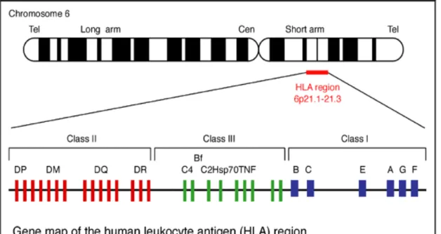
The MHC antigen in humans was firstly found on the surface of human leukocyte, so it was called Human Leukocyte Antigen (HLA). The encoding super locus contains a large number of genes related to immune system function in humans and it locates in the short arm of human chromosome 6, encoding cell-surface antigen-presenting MHC proteins and other proteins.
MHC class I allele exists of three loci: HLA-A, -B and -C and presents peptides on all nucleated cells in the body. MHC class II allele also exists of three loci: HLA-DP, -DQ and -DR and presents peptides on special antigen presenting cells (APC) like macrophages, dentric cells and B-lymphocytes. Other proteins, such as TAP (Transporter associated with Antigen Presentation) and proteasome are also encoded in the MHC class II region. MHC class III is not involved in antigen-presenting but it closely related to immune functions: complements of the complement system (such as C2, C4), cytokines (such as TNF-α, LTA, LTB), and heat shock proteins (hsp). Therefore, HLA studies mainly focus on the HLA
class I and class II. Gene map of HLA is shown in Figure 3.
In the figure 5, it is illustrated that HLA class I contains HLA-A, HLA-B, HLA-C, HLA-E, HLA-F, and HLA-G; HLA class II contains HLA-DR, HLA-DQ, and HLA-DP. According to the structure of HLA class II, each of HLA class II molecules is constituted by two Chains, such as HLA-DRA1 and HLA-DRB1, HLA-DQA1 and HLA-DQB1, HLA-DPA1 and HLA-DPB1. As other MHC class I molecules, HLA-A, HLA-B, and HLA-C present peptides from inside the cell, while HLA-DR, HLA-DM, HLA-DQ, and HLA-DP present peptides from outside of the cell to T lymphocytes, leading to stimulate the differentiation of T-helper cells, which in turn stimulate B-cells to produce antibodies, to that specific antigen.
B. HLA Nomenclature
At present, there are two parallel systems of nomenclature that are widely applied to HLA. The first nomenclature is based on serology recognition. In this system, antigens were eventually assigned letters and numbers (e.g., HLA-A02 or, shortened, A2). The other nomenclature will provide more information. In this system, most designations begin with HLA- in conjunction with a letter “*”, following four-or-more number of digits specifying the allele, such as HLA-A*02:01, HLA-DP*04:01 and HLA-DP*04:02. However, antigen serotype is a crude measure of identify of cells. For instance, HLA-A9 serotype recognizes cells of HLA-A23- and A24- bearing individuals [11].
Actually, many other kinds of nomenclature are also developing. Cellular assay based on the mixed lymphocyte culture (MLC) was used to determine the HLA class II types [12]. This kind of typing used “Dw” to designate and it is more sensitive than serotype due to minor differences unrecognized by alloantisera can also stimulate T cells. Together with difficulty of cellular assay in generating and maintaining cellular typing reagents, cellular assay is being replaced by DNA-based typing method.
Another method is called sequence specific primer (SSP-PCR) [13]. Sequence specific primers are designed to a variant region of HLA region and these overlapping primers will amplify the specific sequence from the different core sequence. Finally, the production of PCR will be separated
and observed by electrophoresis.
C. Polymorphism
The specificity of HLA haplotype varies from race, such as in the Middle Easterners, Africans and other countries. In Caucasians, three polymorphism types, HLA-DR01, HLA-DR03 and HLA-DR04, account approximately 80% of the population; for Middle Easterners, HLA-DR03, HLA-DR04 and HLA-DR07 are dominated, while for Chinese, the predominant genotypes are HLA-DR09, HLA-DR15 and HLA-DR12, accounting for about 70-80% of the total population. In Asia, the Japanese population shares a similar HLA haplotype distribution with Chinese population, while Asian Muslim population and Indian population differ significantly from the Chinese and Japanese population in MHC polymorphism. Therefore, there is a great HLA polymorphism in different races in the world.
IV. MHC mediated immunology
Cellular immunity mainly includes specific cytotoxicity T lymphocytes responses, helper T lymphocytes response as well as unspecific NK killing response. For specific immunity, it was widely accepted that MHC molecule not only participate in antigen presentation to directly mediate
cellular immunology, but also indirectly regulate humoral response by secreting cytokines. Several studies reported that lack of CD8+ CTL delays viral clearance and increases mortality after infection with a virulent strain of influenza virus [14], some CD4+ T cells can also kill target cells as CD8+ T cells [15,16]. However, mice lacking both CD4+ and CD8+ CTL do not clear virus or survive [17]. Therefore, MHC mediated immunity play an important role in antigen specific immunity.
A. Cytotoxicity T Lymphocytes response (CTL)
Cytotoxicity T Lymphocytes is not a single population of lymphocytes but a group of lymphocytes with the specific killing activity. It contains CD8+ T lymphocytes and some CD4+ T lymphocytes. All of these lymphocytes can specifically and rapidly kill target cells.
The procedure of CTLs cytolysis is divided into 4 phases. 1. Recognition and initiation: TCR of precursor CTL recognize the peptide- MHC-I complex together with other ligands combination is the first step of cytotoxicity. 2. Proliferation: after the recognition between precursor CTLs and p-MHC, much IL-2R will be expressed on the surface of precursor CTLs. Meanwhile, Th cells are activated and many kinds of cytokines, such as IL-2, IL-4, IL-7, IL-10, IL-12 and IL-15 are secreted by Th cells will help precursor CTLs develop into effective CTLs to enhance the cytotoxicity. 3. Initiation and dissociation: After TCR
recognize p-MHC, CD11a/CD18 (LFA-1) combine the ICAM-1 or ICAM-2 that expressed on the surface of T lymphocytes, some cytoskeleton composition coupling with TCR, such as tubule and actin, will make organelles in effective T cells rearrange and move to the conjunction between CTLs and target cells. Further, cytoplasmic granules that contain the proteins perforin and granzymes from effective CTLs will attack membranes of target cells to form pores in the target cell membrane and then CTLs dissociate from target cells to search other target cells. 4. Apoptosis: target cells will be killed or initiate apoptosis. The whole procedure is illustrated in Figure 6.
Figure 6. Perforin/Granzyme Killing mechanism of cytotoxic T lymphocytes to kill target cells
There are two kinds mechanism for killing the target cells. Apart form Perforin/Granzyme Killing, the other way is FasL/Fas killing pathway. It
is known that most potential CTL target cells express a receptor for FasL designated Fas. FAS/APO-1(CD95) is a transmembrane protein that widely expressed on the activated T, B cells, NK cells, monocytes and fibroblast; while CTLs express a transmembrane protein that called FasL on their surface, which is widely expressed on the lymphocytes, macrophage, dendritic cells and monocytes. When cytotoxic T cells recognize (bind to) their target, they produce more FasL at their surface to binds with the Fas on the surface of target cell leading to its death by apoptosis, as shown in Figure 7.
Figur e 7. The mechanism of CTL killing target cells.
B. Helper T lymphocytes response (Th)
Helper T Lymphocytes are a cell subset which regulates the body’s immune functions. This cell subset generally has no cytotoxic or phargocytic activity, and therefore could not kill infected cells or pathogens directly, but these cells play key regulatory roles through other types of lymphocytes. For example, they stimulate B lymphocytes to perform humoral immunity and stimulate CD8 lymphocytes to perform cellular immunity, and enhance the killing activity of macrophages. With the depth of helper T cells studies, it has become an important direction of study in immune prevention in better utilizing Helper T cells’ immune regulatory role in vaccine development.
Helper T cells play important roles in many aspects such as infection immunity, tumor immunity, autoimmunity and immune regulation. In pathogen infections, in particular viral infections, Helper T cells play a vital role in recognizing the Th epitopes of pathogen proteins and regulating CTL and humoral immune response.
Through complicated regulatory mechanisms, an organism’s immune system performs two basic biological functions: protecting itself from invasion by various exogenous pathogens and inhibiting immune responses toward its own antigens. When the immune system is certain of an immune response, it has to produce appropriate functional response to each and every different pathogen. One key cell type performing this
function of mediation and regulation is CD4+ T cells, and it has been demonstrated that cytokines secreted by CD4 T cells play important roles in the process of fine tuning its functions [18]. Based on the cytokines they produced, CD4+T cells are categorized into two types: Helper cell type 1 (Th1) and type 2 (Th2). When the T cell receptor (TCR) molecules on the surface of CD4+ T cells bind with antigen-MHC-II complex presented by antigen presenting cells (APC), a first signal is released which results in abundant expression on CD4+ T cell surfaces multiple cytokine receptors such as IL-1R and IL-2R; meanwhile, CD4+ T cells recognize the co-stimulatory molecule on APC surface and form the second signal which is the secretion of multiple cytokines, mainly IL-2 but also including some IL-4, IL-10, IL-12, interferon-γ and others. Subsequently, the IL-2R on CD4+ T cell surface binds to IL-2 secreted in the autocrine or paracrine fashion, which activates the cell to produce many progeny clone Th0 cells. Th0 cells differentiate into Th1 or Th2 cells under the actions of different cytokines. IFN-γ promotes Th0 cells to differentiate into Th1 cells which, through IL-2 and IFN-γ they secreted, enhance the cellular immune response of CD8 T cells. IL-4 promotes Th0 cells to differentiate into Th2 cells which, through cytokines such as IL-4 and IL-5 they secreted, enhance the humoral immune response of B cells [19]. Activated Th1 cells secrete IFN-γ, which activates macrophages, TNF-β, which kills chronically infected macrophages; IL-2, which induces T cell
proliferation; IL-3 and GM-CSF, which act synergistically to induce bone marrow monocytes to differentiate; LT and MCT, which synergistically activate endothelial cells and induce infiltration of macrophages; MCF and MIF, which synergistically attract macrophages to move toward infected foci. Under these physiological responses, Th1 cells upregulate immune functions to resist infection of intracellular pathogens. In contrast, activated Th2 cells have a main function which is to promote B cells to produce antibodies to play the role of humoral immunity [20]. Therefore, systematic and in-depth study of Th epitopes of pathogen proteins is of theoretically significance for understanding the immunity to infection, vaccine reaction characteristics and immune protection mechanisms.
C. CD4+ T cells epitope
Helper T cell epitope is the molecular basis for Th cells to perform immune regulatory functions, and plays important roles in preventing pathogen infection. Systematically studying the Th epitopes of major viruses such as HIV and HBV is of scientific significance for elucidating immune reactions to viral infections and immune protection mechanism. There are two types of T cell epitopes: MHC-I restricted and MHC-II restricted. Studies on MHC-I restricted epitopes mediated CD8+ T cell response have been quite systematic while it is less so for MHC-II epitopes, both in relevance of MHC restricted epitopes to CTL or humoral
response mechanism, and in epitope screening and application. However, as more studies gradually reveal more about Th cell functions and biological significances, the study of Th has attracted wide attention, and in particular, the field has been moving fast in identifying new MHC-II molecules and MHC-II restricted epitopes.
MHC-II restricted epitopes are helper T cell epitopes formed when antigens are cleaved inside the host organism and are 8-25 amino acids in length. The epitopes and MHC-II molecules on APC surface form antigen epitope peptide-MHC-II complex, which binds to TCR-CD3 on CD4+ T cell surface; at the action of the second signal (CD8 or CD86 molecules on APC surface binds to CD28+ molecules on CD4+ cell surface), they promote cells to differentiate into effecter cells, and stimulate cells to highly express a series of cytokines such as IL-2, IL-12, IFN-γ, IL-4, IL-10 and other immune regulatory factors. The microenvironment formed by these cytokines is the messenger which mediates the activation of Th cells and ultimate realization of the immune regulatory function of Th cells.
Many studies have demonstrated Th epitopes derive from the HBV are essential for the immune response against HBV infection. In chronic HBV infected individuals, inefficient binding between hepatocyte surface MHC-II molecules with HBsAg antigen epitopes might account for the dysfunction or weak specific CTL of HBV infected patients [21], which
further cause persistent HBV infection. Similar studies on the NS3 region of HCV revealed that specific Th epitopes on NS3 can contribute to the clearance of HCV infection [22], Th epitopes of HIV viral proteins could enhance CTL cell toxicity toward infected cells and are associated with long-term non-progression of HIV infected individuals [23].Therefore, Helper T cell epitopes could effectively enhance protective cellular and humoral immune response, and have important applications in immune preventives and therapies such as polypeptide vaccines, multiple-epitope recombinant vaccines and therapeutic vaccines.
Developing safer, more effective and more economic vaccines is the trend in vaccine R&D. Novel vaccines have advantages in safety, cost and stability and are the main directions of vaccines in the twenty-first century. Traditional vaccines mainly refer to those biological products which are produced from attenuation or killing of pathogenic microorganisms and can stimulate the body to produce microorganism -specific antibody or cellular immunity. Novel vaccines refer to subunit vaccines, polypeptide vaccines, nucleic acid vaccines, live vector vaccines and therapeutic vaccines. It is a trend for the future to modify and develop novel vaccines based on traditional ones. As studies go into depth, the role of Th epitopes in vaccines, in particular in novel vaccines, has received increasingly more attention. Appropriate Th epitopes will not only enhance cellular immune response such as CTL, but also could raise the level of humoral
immune response.
V. Humanized mouse Model (HLA transgenic mice)
At present, animal models used for preclinical studies mainly include thestrongly repetitive low-cost small animal models, such as inbreeding mice or birds, and large animal model with closer genetic background to the human, such as monkey, orangutan and other nonhuman primates. However, many small animal models are unable to accurately forecast the human response because their genetic background is greatly different from that of the human; the nonhuman primate and other large animal model suffers from high cost, limited sample number and ethical dispute. Moreover, both models are unable to eliminate the influence of species specificity on preclinical studies, so that many preclinical animal experiment results are greatly different from the human clinical response, and therefore neither of the above two kinds of animal models are the ideal animal models for predicting the human immune response.Several studies indicated that the hereditary difference of interspecies MHC molecules is one of the important factors resulting in the difference. Therefore the accuracy for the animal model to forecast the human immune response can be effectively enhanced by replacing the animal MHC molecules with the human HLA molecules in the experimental animal body.
Mouse shares as high as 95% genes and 80% genetic products with human. The genetic background of mice were well decrypted and selecting the inbreeding mouse as the human HLA carrier can effectively reduce the influence of other genetic backgrounds when we are observing the regulation of the cellular immune response with HLA molecules. In the 1980s, the rapid development of molecular biology and maturation of transgenic technology [24] provides technical support for inserting the human specific HLA genes cloned in vitro into the mouse genome [25]. What’s more important, each individual inherited several different HLA haplotype due to the polymorphism and linkage of MHC; it results in the difficulty to observe the single HLA restricted response in human body. Thus, transferring of certain HLA molecule into mouse body can separately inspect the immunological characteristics of this HLA molecule in vivo to avoid the competition from other HLA molecules in antigen presenting.
HLA transgenic mouse models are created by introducing typical human MHC gene into the mouse and knocking out murine H-2 gene. Therefore, the immune response in these transgenic mouse models is only restricted by HLA molecules and the cellular immune responses should be consistent with those of humans. Doubtlessly, HLA transgenic mouse model is an effective tool to enhance the accuracy of preclinical studies. At present, the reported humanized MHC transgenic animal model
includes HLA-I and II transgenic mouse models, which respectively focuses on inspecting the function of cytotoxic T lymphocyte (CTLs) and helper T cell (Th).
A. HLA- class I transgenic mouse models
Kievits F et al reported the first humanized MHC class I transgenic mouse model HLA-B27 [26] in 1987 and proved that the cytotoxic T lymphocyte (CTLs) of this model can specifically recognize virus proteins using the humanized HLA-B27 restricted molecules. Later, despite researches demonstrated that the epitope library presented by the mouse H-2-I molecules is not exactly the same as that presented by the human HLA-I molecules [27,28], Sesma L et al proved through in vitro experiments that among the 1551 epitopes presented by mouse-based cells and 1372 epitopes presented by humanized cells, 1161 epitopes are the same [29].Therefore, the micro environment in mouse body can satisfy the normal replication, modification and transportation of the human HLA class I molecules, and perform the antigen presentation process similar to the human body.
B. Optimization of the HLA-I transgenic mouse model
and are composed of a glycosylated α heavy chain and a β light chain through non-covalent bonding. With deep understanding of the structure and immunological function of HLA-I molecule, HLA class I transgenic mice were unceasingly optimized to solve the problems that encounter in application of HLA class I transgenic mice. This procedure can be divided into three stages.
Stage 1:
The independent α heavy chain or α heavy chain and light chain β2m of human HLA molecules are directly transferred to the wild mouse genome through transgene, so as to realize the expression of human HLA molecules under the mouse-based background, and enable the mouse T cell to recognize the extraneous HLA molecules and the HLA restricted epitopes presented thereof. Where, the representative mouse model includes HLA-B27, HLA-B7, HLA-A2, HLA-Cw3 and HLA-B35 and so on [30-35]. In these models, TCR on the rearranged mature mouse-based T cell surface can recognize the extraneous peptide presented by the human HLA-I molecules, and give rise to the HLA-I restricted T cell response [36-38]
. But the HLA heavy chain genes (α1, α2 and α3) are completely originated from the human, the interaction between functional area of humanized α3 and mCD8 is not as strong as that of the mouse-based α3, and the majority of HLA restricted T cell response is competitively inhibited by the mouse H-2-I restricted T cell response [39-40], therefore,
the mouse CTL still mainly produces the mouse-based H-2-I restricted CTL response.
Stage 2:
The secondary lymphatic organ of the first generation of HLA-I transgenic mouse model has limited quantity of HLA restricted T cells, and the interspecies hereditary difference affects the interaction between mouse-based co-receptor CD8 and the human I-type molecules, therefore enhancing the utilization of HLA-I molecules in producing the CTL response is the key to improve the humanized HLA-I transgenic mouse model. The improved model includes HLA-A2, HLA-B27, HLA-B7, HLA-A24 and HLA-A11 and so on [41-46]. One strategy is to transfer human hCD8+ gene into the mouse genome, so that its T lymphocyte simultaneously expresses mouse-based and humanized CD8+ molecules. Under the circumstances, extraneous HLA-I molecules have more opportunities to combine with CD8+ on mouse CTL surface, and efficiently start the second signal. Another strategy is to optimize the molecular structure of extraneous HLA, replace the humanized intracellular α3 functional area in the transmembrane area with mouse-based one, and build chimeric HLA molecules, namely the chimerism between humanized α1 and α2 functional areas and the mouse-based α3 functional area, HHM for short. This structure can enhance the bonding of mouse CD8 molecules and human MHC class I
molecules [41-43]. The mouse model optimized with the above method can produce non-specific and HLA restricted specific response, but the majority of CTL response is still the mouse-based H2 restricted response [42, 43]
.
Stage 3:
In view of the fact that the first two generations of mouse models mainly generate the mouse-based H-2-I restricted CTL response, it is necessary to eliminate the competitive inhibition of endogenous H-2-I molecules on HLA-I molecules in the HLA-I transgenic mouse model. In the 1990s, with the maturation and application of the gene knockout technology, the mouse H-2-I system was knocked out. Barra C et al reported [47] that after the mouse H-2-I gene is knocked out, the HLA-I restricted CTL response is still very strong. In the event that no H-2-I molecule exists, the HLA-I transgenic mouse model can form a huge TCR cell library using the normal differentiation of TCRVβ [48]. Therefore, the Lemonnier team proposed to build the HLA-I transgenic mouse model with the H-2-I gene knockout, so that the CTL maturation and activation only relies on the human HLA-I molecules. At present, the mouse H-2-I genes can be knocked out with two methods: the first method is to knock out the heavy chain gene encoding the mouse H-2-I molecules, namely first respectively knock out the mouse-based H-2Kb and H-2Db, then mate with HHM mouse, and finally inbred to
homozygous mouse model [49, 50]. But this method suffers from a time-consuming complex process, and will still produce the antigen presentation participated by non-classical H-2-I molecules [51] and CD1 restricted NKT cell response (CD1 expression depends on β2m) as a result of the existence of mouse-based β2m molecules. Therefore, at present, the third generation of HLA-I transgenic mouse model created by using another simpler method, namely first knock out the mouse-based β2m, so that the H-2-I molecules are unable to form the functional unit due to lack of light chain, thereby achieving the purpose of silencing H-2-I molecule functions; meanwhile construct an HHD structure with heavy α chain of HHM combine with human light chain β2m through a linker, so as to prevent the binding of human β2m and endogenous H-2-I heavy chain. Even though the mouse model built with this method could still conduct background expression of H-2-I antigen [52, 53], it does not participate in the classical and non-classical antigen presentation of endogenous H-2-I molecules, therefore the HHD mouse model mainly produces the HLA-I restricted CTL response. It was demonstrated that both the third generation of mouse models possess not only CD8+T cell library with diversified TCR Vβ and Vα on their T cell surface, but also stronger antigen specific HLA-I restricted response than the second generation of mouse model [54,55]. Therefore, such transgenic mouse models are widely applied in related fields of preclinical studies.
C. HLA class II transgenic mouse models
Experimental therapy in humans is limited by technological and ethical considerations; however, no effective wide animal models can instead human cases to meet this requirement. In this case, HLA-II transgenic mouse models were originally developed in order to study the susceptibility between HLA and some diseases, and further used to observe the CD4+ T cells response against virus, parasites, tumour as well as autoimmunity. So far, a range of transgenic mouse models humanized for MHC II molecules, such as HLA-DR01, HLA-DR03,
HLA-DP4, HLA-DR15, DQ6 and DQ8[56], were created to study
rheumatoid arthritis, multiple sclerosis, insulin-dependent diabetes mellitus or celiac disease.
Similar as the HLA-class I transgenic mice, the development of molecular biology and transgenic technology lay a foundation for the feasibility of creation of HLA-class II transgenic mice. First HLA-class II transgenic mice were created by introducing HLA -DR gene into murine cells in 1983[57]. After 2 years, HLA DP transgenic mice were reported and their HLA-DP restricted immunological function of CD4+ T cells was observed as expected [58]. However, these humanized mouse models expressed both human class II and murine MHC class II antigens. It brought great difficulty to distinguish that the immune response were
initiated by the murine MHC class II molecules or that were mediated by the human MHC class II molecules. In this situation, many strategies were used to optimize these mouse models in following aspects.
D. Optimization of the HLA-II transgenic mouse model
As other MHC class II molecules, both HLA class II and H-2 class II molecules are heterodimer which are composed of a α chain and a β chain, and each chain contain two domains-α1, -α2 and -β1, -β2. In antigen presentation, α1 and β1 form a groove to bind epitopes and take charge of antigen presenting; α2 and β2 form transmembrane domain that anchoring the MHC class II molecule to the cell membrane. Therefore, the optimization of HLA-II transgenic mouse model was designed on basis of MHC-II molecule structure.
Stage 1:
In this stage, HLA-class II transgenic mouse models only transgene class II single α chain or β chain of human MHC, and coupled with murine complementary chain in vivo. For example, initial HLA-DR transgenic mouse models were only DRα chain transgenic and paired with murine Eβ ,with absence of DR β chain[59],or only DR β chain transgenic and paired with murine Eα, with absence of DR α chain[60]. In this case, murine T cells can be partially educated under human HLA class II molecule.
Subsequent improvement was fulfilled by simultaneously introducing both human α chain and β chain genes into murine genome so as to allow the integrated HLA antigen can take action in murine background, such as HLA-DR1 and HLA-DR2 transgenic mice. Except the structure of HLA genes, the promoter of HLA was also considered. Some mouse models, such as HLA-DQw6, DR3, DRw17 and DR1, used the promoter from human MHC region [61-63], while some other mouse models used the murine MHC region promoter for expressing DR4 [64, 65]. All of these mouse models were still reported to be low efficient in HLA restricted response.
Stage 2:
In addition to the optimization of human MHC class II molecule expression, the interaction between CD4 molecule of T lymphocytes and MHC class II molecules of target cells was considered. Some groups reported that the interaction between mCD4 and exogenous HLA molecule was relatively weak [66-68], compared with that between hCD4 and exogenous HLA molecule [69]. Therefore, there are two alternatives, one is to modify MHC class II molecule as a chimeric structure, Woods team took this strategy and replaced human α2 and β2 domain by murine α2 and β2 domain in HLA-DR transgenic mice, they reported that the chimeric structure (DR-α1, β1 and I-Eα2, β2) help to stimulate a stronger CD4+ T cells response in mice [64, 65].The other method is to replace
mCD4 by hCD4. Even though Altmann et al reported that the mobilization of CD4+ T cells repertoire of HLA-DR1/hCD4 transgenic mice was the same as that of HLA-DR1 transgenic mice [63], more groups reported that the interaction between HLA and hCD4 was better than that between HLA and mCD4. What’s more, the promoters for hCD4 were from murine genome, such as murine CD3δ promoter [70] in HLA-DR4
mice and murine CD2 promoter [71] in HLA-DQ6 mice.
Stage 3:
As mentioned above, much effort was done to improve the similarity of HLA class II transgenic mice with human. However, many studies demonstrated that the co-expression of H-2 with HLA molecule would lead to the murine immunity preference. Thus, in 1991, Cosgrove et al [72] developed a mouse model that was deficient in H-2 class II, and in 1999, Madsen et al [73] generated another knockout mouse that lacked all four of the classic murine MHC class II gene via a large (80-Kb) deletion of the entire class II region that was engineered by homologous recombination and Cre recombinase-mediated excision. These mouse models subsequently were widely used for developing mouse models that expressed only human MHC class II molecules with absence of all murine H-2 class II molecules to highlight the HLA class II restricted immunity. The human MHC class II molecules in these new mouse models have been shown to be functionally actived and participated in
shaping the CD4+ T cell repertoire in thymus and mediate CD4+ T cell responses in the periphery. It was reported that, mature CD4+ T cells repertoire in HLA transgenic and H-2 class II knock out mouse model is different from that in C57BL/B6 mice [74-76]. Importantly, peptides presented to CD4+ T cells in these mouse models were shown to be similar to peptides presented to CD4+ cells in patients who carry the same MHC class II haplotype [77].
In summary, the currently available HLA class II transgenic mice have high capacity to mimic human cellular response. However, there are still some limitations to be solved. HLA class II molecule in these mice is only one of the HLA class II antigens; whereas CD4+ T cells response in human is the mutual result of different HLA class II molecules. Therefore, more different types of mouse models should be developed for experimental therapy.
E. HLA-I/II double transgenic mouse models
Compared with HLA-I or II single transgenic mice, HLA-I and II double-transgenic mice would allow the observation of synergy effects of MHC-I and -II molecules in immune response, which is uniquely advantageous either studying viral pathogenesis or screening for anti-viral vaccines. In 2004, Anthony et al successfully established a human MHC HLA-A2/DR1 transgenic mouse model by introducing human HLA-A2
and HLA-DR1 gene and knocking out the mouse H-2 class I and class II molecules [78]. For these mice, their cellular immune responses are restricted completely by human HLA molecules and extremely consistent with human cellular immune responses. Especially, HLA-DR1 molecule is capable of regulating mouse B cells mediated humoral immune response and further influence the CTL response. Even though the percentage of CD8+ T cells population is lower than wild-type mice, expression of functional transgenic HLA-A2.1 molecules led to an increase in the size of the peripheral CD8+ T cell population, which reached 2-3% of the total splenocytes in HLA-A2/DR1 mice, compared to 0.6-1% in β2m-KO MHC-I deficient mice. Furthermore, the size of functional TCR repertoire from peripheral CD8+ T cell is highly diverse as comparing to wild type B6 mice, this suggest that CD8 T cell activity is highly competent. Similarly, HLA-DR1 molecule leads to an equivalent expression in HLA-A2/DR1 transgenic mice, which CD4+ T cells represented 13–14% of the splenocytes population. In contrast, only 2–3% of the cells were CD4+ T cells population in H-2 class II-KO mice; in agreement with the initial report on mice lacking MHC class II molecules [72]. Therefore, when this mouse model are used to study T cell epitopes and to evaluate cellular immunity based protective vaccines, it could reflect the human natural immune response, and represent a rather ideal technology and model for vaccine research and evaluation.
Moreover, this double transgenic and double knockout mouse model is a moderate small animal model for transplanting and reconstructing human hematolymphoid cells and tissues.
VI. Highly pathogenic Influenza A virus (H5N1)
HPAIV is a highly pathogenic subtype of the avian influenza A virus. Its mortality rates in infected flocks often approach 100%. Even though many reports indicated that H5N1 presented a highly poultry-specific infectivity and seldom infect humans, there are an increasing number of human cases infected by H5N1. According to the statistics provided by World Health Organization (WHO) (shown in Table 4), H5N1 has totally caused 573 human infected cases worldwide, including 336 deaths since 2003 [79]. Moreover, the influenza H1N1 pandemia in 2009, which partial segment derive from H5N1 virus and arose worldwide panic and ever be speculated to generate the third human pandemic disease like 1918 Spanish pandemia, alerted people to draw more attention on the potential risk of H5N1. Once H5N1 virus spread through species barrier to infect humans and carries its 59% mortality, there will be a disaster. Therefore, for the high lethality and virulence of HPAIV (H5N1), huge potential host repertoire, and its significant ongoing mutations, the H5N1 is doubtlessly the world’s largest current pandemic threat.Table 4.Cumulative number of confirmed human cases for avian
influenza A (H5N1) reported to WHO, 2003-2011
A. Structure of HPAIV H5N1 virus
H5N1 is a subtype of the species Influenza A virus and belonging to Influenza A genus of the orthomyxoviridae family. Like other subtypes of influenza A virus, H5N1 is an single stranded, negative-sense RNA virus that composed of 8 negative segments which encoding 10 proteins, as shown in Figure 8, including hemagglutinin (HA), neuraminidase (NA), matrix protein 1(M1) and matrix protein 2(M2), nucleoprotein (NP), polymerase basic 1(PB1) and PB2, nonstructural protein NS1 and NS2 and polymerase acidic (PA) [80]. There are comprehensive studies on every viral antigen up to date. In our studies, we mainly focus on the HA, NA, M1, M2 and NP related immunity and thus, other proteins we will not make a further introduction.
Figure 8. Molecular structure of H5N1 virus.
Hemagglutinin (HA) and the neuraminidase (NA) proteins are the two surface antigens of H5N1; they play key roles in interactions between the virus and host cells. Structure analysis has shown that HAs are trimeric glycoproteins while NAs present on viral cell surface as tetramers [81, 82]. Both HAs and NAs can bind to sialic acids in cell surface receptors. After protease cleaves HAs into two chains, HA1 and HA2, HA2 can mediate the entry of viruses into host cells [83]. After the HA is cleaved by a protease, the cell imports the virus by endocytosis. NAs enhance the cleavage of sialic acids from virus and host cell glyco-junctions and facilitate the release of mature virions [84]. It is demonstrated that HA cleavage is the premise of H5N1 infection [85].
Matrix protein is encoded by RNA segment 7 and has two forms, including M1 and M2. M1 protein is the most abundant viral protein and it takes up nearly 40% articles in the whole viral protein .Thus, its
antigenic specificity in different influenza strains could be used for diagnosis and virus typing [86]. ThereforeJM1 protein poses a vital role in maintaining virus structure and integrity. Moreover, M1 protein binds to RNP cores and therefore takes significant functions in the virus life cycle, such as inhibits RNA synthesis by the RNP-associated transcriptase of influenza virus and participates in assembly and budding of progeny virions [87].The other form of matrix protein, M2-protein, is a tetrameric, type III transmembrane protein scarcely present on virus particles, but abundant on virus-infected cells [88, 89]. It can form a proton channel during infection which is essential for infection. The extracellular domain of M2 protein contains a 24 amino acid residues that called M2e-sequence. Significantly, this short sequence has remained nearly unchanged since the first human influenza strain was isolated in 1933, despite numerous epidemics and two major pandemics since then [90].
Nucleoprotein is encoded by RNA segment 7 and make up of 498 amino acids. It is one of the most important structural proteins, according to the NP antigenicity; influenza virus can be divided into three subtypes A, B and C. Hence, it is the basic protein that used as classification and diagnosis.In the infected cell newly synthesized NPs are imported into the nucleus where they take part in a RNA elongation/RNP assembly process. According to a favoured assembly model one or a few NPs bind to newly synthesized viral RNA, which will then stimulate further NP
associations by cooperative NP-NP and NP-RNA interactions[91].In this way, NP participate in the virus replication.
B. Pathogenesis
The transmission of Human influenza A virus infection can be easily fulfilled result from the enrichment of hemagglutintin proteins locating in the upper part of the respiratory tract to infect epithelial cells, typically in the mouth, nose as well as throat and it can be easily transmitted by people coughing and sneezing. However, the symptom and characteristic of HPAIV infection is different from that of human influenza A virus. Because the avian influenza hemagglutintin binds to avian cell alpha 2-3 sialic acid receptors(SA α-2,3), while human influenza hemagglutintin bind to human cell alpha 2-6 sialic acid receptors(SA α-2,6)[92]. Once hemagglutinin fuses the viral envelope with the vacuole’s membrane, then the M2 ion channel allows protons to move through the viral envelope and acidify the core of the virus, which lead the core to dissemble and release the viral RNA and core proteins. Owing to more alpha 2-6 sialic acid receptors (SA α-2, 6) exist in the deeper respiratory tract of human body, the severe tissue damage even death occurs more often in patients after H5N1 virus infection due to inflammatory cascade that triggered by H5N1 virus [93].This so called a “cytokines storm” might be aroused by what seems to be a positive feedback process of damage to
the body resulting from immune system stimulation. It also account for the reason that H5N1 virus can replicate in the lower respiratory tract and further cause viral pneumonia [94, 95] in the lungs.
Much concerns about the H5N1 pandemia due to the high mutation of HA. It was widely demonstrated that the ability of various influenza strains to show species-selectivity is largely due to variation in the hemagglutintin genes. Even a single amino acid substitution might dramatically change the ability of viral HA to bind to receptors on the surface of host cells, which further make virus strains being inefficient at infecting human cells change into a more contagious virus strains as the seasonal human influenza viruses. Another apprehension about the H5N1 pandemia is attributing to the ability of H5N1 genetic recombination by segments reassortment in the host who had been infected by more than two kinds of subtype, which alter H5N1 virus from not pathogenic in humans to become pathogenic in humans. Take A/2009/H1N1 for example, the special structure of influenza virus allows the segments from swine flu, bird flu and human flu reconstitute a new H1N1 influenza virus which emerged in Mexico, the United States, and several other nations and caused a public panic [96].
C. Prophylaxis
the treatment and prophylaxis of influenza A infections: two M2 inhibitors (amantadine and rimantadine) and two neuraminidase inhibitors (zanamivir and oseltamivir) [97, 98]. However, there are many factors should be considered, such as drug resistance and costs. Mass vaccination is the best form protection to prevent and reduce the chance of severe illness or death in people during a H5N1 pandemia.
The development and potentially imminent availability of approved human vaccines against H5N1 marks are of great interest to address this threat. Although, the history of human H5N1 vaccines is a relatively young in many aspects, such as immunogenicity, safety, dosage, degree and duration of their protective effects, at least 16 different manufacturers have an H5N1 vaccine in relatively advanced development based on a range of approaches (including egg and cell culture grown viruses, live virus and inactivated vaccines, whole and split antigen and vaccines with and without different adjuvants). Additional novel vaccines are also under consideration and development [99].
Current two licensed H5N1 vaccines by Food and Drug Administration (FDA) of USA are the conventional inactivated virus vaccine (CIV) and the live-attenuated vaccine (LAV). Both of them have been demonstrated to be effective for protection. However, whether these traditional vaccines against H5N1 can provide protection against new emerging H5N1 virus is hard to be speculated because of the high mutation of H5N1 surface
antigens Hemagglutinin (HA) and Neuraminidase (NA). Therefore, many novel vaccines were developed to deal with the disadvantages of current licensed vaccines, including split vaccine, subunit vaccine [100], VLPs vaccine [101] and DNA vaccine [102], however, most of these vaccines still focus on stimulating the neutralizing antibodies against external glycolproteins HA and NA and related reports largely published. Several researches reported that the vaccines based on the H5N1 Hemagglutinin has lower efficiency than seasonal human influenza vaccines and unadjuvanted inactivated whole virus H5N1 vaccines appear to be more immunogenic than unadjuvanted split or subunit vaccines. In more recent years, “universal flu vaccines” [103], which based on the conserved viral antigens or epitopes, are developing to potentially provide longer-lasting protection against different subtypes [104,105]. For this reason, less mutated proteins nucleoprotein, matrix protein, polymerase base as well as many conserved CTL epitopes, B-cell epitopes and Th epitopes are essential targets for next generation vaccine design.
VII. Hepatitis B Virus (HBV)
Hepatitis B virus is a member of the hepadnavirus family. It was reported that 2 billion people had been infected by hepatitis B virus (HBV), more than 350 millions of people worldwide are chronic carrier of the virus and 600,000 people die each year due to the acute or chronic
consequences of hepatitis B [106]. Therefore, HBV is one of the most contagious viruses to threaten human health.
A. Structure of hepatitis B virus
Hepatitis B surface antigen (HBsAg) particles carry the common determinant a, d or y and w or r subtype determinants, and are classified into the four major subtypes, i.e., adw, adr, ayw and ayr, which depend on antigenic epitopes present on its envelope protein [107]. It is also divided into 8 genotypes based on overall nucleotide sequence variation of the genome. Hepatitis B virus particle is constituted by an outer lipid envelope and an icosahedral nucleocapsid core(as shown in Figure 9). The component of the outer lipid envelope is surface protein which composed surface protein, middle protein and large protein. This envelop protein, which is also called HBsAg, often present in the sera of patients with viral hepatitis B with or without clinical symptoms. Thus, it can indicate the current HBV infection in clinical diagnosis. The nucleocapsid core is a complex of viral DNA and a DNA polymerase that has reverse transcriptase activity similar to retroviruses. Apart from HBsAg and Polymerase, HBcAg and HBeAg antigens occur in the HBV dana particle. HBcAg is a structural protein in HBV and wrapped up by HBsAg. It is an indicator of active viral replication; which means the person infected with Hepatitis B can likely transmit the virus on to another person. HBcAg
soluble form antigen is HBeAg and it is also the marker of active viral replication. When anti-HBcAg and anti-HBeAg present can be detected in the sera of patients, it reveals a decline in viral replication and the recovery from HBV infection.
Figure 9. Molecular structure of hepatitis B virus.
The genome of HBV is made of the smallest circular DNA, a not fully double-stranded 3.2k bases DNA, which composed of a full length anti-sense strand and a variant length of the sense strand. The anti-sense strand has 4 ORF, including S, C, P and X segments and they encode all the known HBV proteins, whereas sense strand does not encode any protein(as shown in Figure 10). S gene can be divided into two genes; pre S and S gene: there are 678 bases for S gene, Pre S gene locates at the upstream of S gene, it encodes a protein with 163 amino acids, including Pre S1 and Pre S2 proteins.C gene is also divided into two parts, Pre C and C genes, which encode HBeAg and HBcAg, respectively; P gene
mainly encodes the polymerases for viral replication; X gene encodes a 154 aa basic polypeptide.
Figure 10. Genome structure of Hepatitis B virus.
B. Pathogenesis
Hepatitis B virus (HBV) is a non-cytopathic DNA virus that causes acute and chronic hepatitis [108,109]. Both the adaptive and innate immune responses are known to be involved in viral clearance during HBV infection [110]. It was reported that the host immune response against HBV is the key factor to determine whether patients naturally resolve viral infection or develop chronic infection [111]. In acute HBV virus infection, multi-specific cellular response in patients could be observed, with the adequate production of anti-HBV antibodies and antiviral cytokines. On the contrary, patients suffering from chronic infection have very weak or functionally impaired immune responses. When persistent inflammation occurs during HBV infection, many liver diseases would be caused, such