HAL Id: tel-02416025
https://tel.archives-ouvertes.fr/tel-02416025
Submitted on 17 Dec 2019
HAL is a multi-disciplinary open access archive for the deposit and dissemination of sci-entific research documents, whether they are pub-lished or not. The documents may come from
L’archive ouverte pluridisciplinaire HAL, est destinée au dépôt et à la diffusion de documents scientifiques de niveau recherche, publiés ou non, émanant des établissements d’enseignement et de
Plant virus-derived nanoparticles for the imaging and
treatment of cancer
Coralie Gamper
To cite this version:
Coralie Gamper. Plant virus-derived nanoparticles for the imaging and treatment of cancer. Biotech-nology. Université de Strasbourg, 2019. English. �NNT : 2019STRAJ038�. �tel-02416025�
ÉCOLE DOCTORALE Sciences de la vie et de la santé
CNRS-UPR2357 Institut de Biologie Moléculaire des Plantes
THÈSE
présentée par :Coralie GAMPER
soutenue le : 23 septembre 2019pour obtenir le grade de : Docteur de l’université de Strasbourg
Discipline/ Spécialité : Biotechnologie
Nanoparticules dérivées de virus de plante pour le
traitement et l’imagerie du cancer
THÈSE dirigée par :
Monsieur HEINLEIN Manfred Directeur de recherche, CNRS
Monsieur BAGNARD Dominique MCU, université de Strasbourg RAPPORTEURS :
Madame BARBERI-HEYOB Muriel Professeur, université de Lorraine
Madame WEGE Christina Professeur, université de Stuttgart
AUTRES MEMBRES DU JURY :
Monsieur REMY Jean-Serge Directeur de recherche, CNRS
Monsieur BENNASROUNE Amar MCU, université de Reims Champagne-Ardenne
UNIVERSITÉ DE STRASBOURG
I would like to thank my thesis jury for kindly accepting to evaluate my work. Thanks to Pr. Wege and Pr. Barberi-Heyob for agreeing to take time to read my thesis. I would like to thank the director of the IBMP, the Dr. Laurence Drouard, and the director of the INSERM unit U1119, the Pr. Guy Mensah and their respective research unit for their welcoming. I would like to thank the Dr. Gertraud Orend for her scientific advices during the course of my thesis. I would like to thank my thesis director, the Dr. Manfred Heinlein for giving me the opportunity to realize my thesis in his lab and his help during the course of my thesis. I would like to thank my thesis co-director, the Dr. Dominique Bagnard for his support since my master thesis. I appreciated your scientific advices and your help for the redaction of my thesis. I will miss a little bit your non volunteer “réécriture d’expressions françaises”. Thanks to Nicolas Baumberger and Laurence Hergott from the protein production platform of the IBMP for their technical support. Thanks also to Mathieu Erhardt and Jérôme Mutterer from the microscopy platform of the IBMP for their technical advices. Thanks to Sonia Boscà-San José who started this project and transmitted to me her technical knowledge. Even if we didn’t work together for long, I appreciated your advices and your kindness. Thanks to my former co-workers from Manfred’s team. Thank you Nicolas for our discussions about the thesis difficulties and thank you Caiping for having collecting bacteria for me when I was blocked at Illkirch. I thank my former co-workers from Dominique’s team and especially Gérard, Michael, Caroline, Aurore, Lauriane and Marie. I wish a good continuation with their thesis to Lucas and Dafni. Thank you Caroline for your help with several experiments and your support during stressful period. I’m sure we will have other opportunities to go for a drink together ! Good luck with your retirement Gérard ! Our discussion (scientific or not) will certainly miss me. Thank you Mika for the angiogenesis assays you done for me when I had no time to do them. It has been really nice working with you for more than 5 years now. I wish you the best for the rest of your career and good luck with future teenager Ambre ! Thank you Aurore for all our discussions about research, movies, books and so much more. You have endured my complaints so many times and gave me good advices to continue moving on, I will have not survive my thesis without you !
Lauriane you have been a wonderful support during these last two years. Your energy and your kindness have brought a welcome freshness to the lab. I wish you plenty of joy in your future works ! Marie I wish you all the best in your new career and a lot of happiness with your so nice little Danaá ! Fabien I wish you all the best for the rest of your career. Your jokes and movie suggestions have been appreciated ! Thanks to my parents, my sisters and my cousin for having supporting me through all these years and enduring me during some stressful moment ! Thank you Rébecca for having preparing my meal so often, I would have starved to death without you ! And finally, thank you Suzanne for your smile and your cuteness ! I am already proud to be your godmother and I can’t wait for the moment we will travel together !
TABLE OF CONTENTS I. CANCER BIOLOGY AND TREATMENTS ... 9 1. CANCER DEVELOPMENT ...11 1.1. Genome alterations in cancer cells ... 11 1.1.1. Tumor suppressor genes ...11 1.1.1. Oncogenes ...12 1.2. Cancer cell proliferation and survival ... 13 1.3. Loss of contact inhibition ... 18 1.4. Inflammation in cancer ... 19 1.5. The activation of angiogenesis ... 21 1.6. Invasion and metastasis ... 23 2. CANCER THERAPEUTIC STRATEGIES ...26 2.1. Classical treatment ... 26 2.1.1. Surgery ...26 2.1.2. Radiotherapy ...27 2.1.3. Chemotherapy ...29 2.2. Gene therapy ... 30 2.3. Immunotherapy ... 32 2.3.1. Vaccines for cancer treatment ...32 2.3.2. Immune checkpoint inhibitors (ICIs) ...33 2.3.3. Chimeric antigen receptor (CAR)-expressing T cells (CAR-T cells) 33 2.4. Hormone therapy ... 35 2.5. Hyperthermia therapy ... 37 2.6. Photodynamic therapy (PDT) ... 37 2.7. Ultrasound therapy ... 40 2.7.1. Sonodynamic therapy (SDT)...40 2.8. Targeted therapies ... 41 2.8.1. Use of small molecules inhibitors...41 2.8.1. Targeting cancer cells with monoclonal antibodies ...43 II. NEUROPILIN-1 ... 45 1. NEUROPILIN-1 ...45
2. THE ROLE OF THE NEUROPILIN-1 IN CANCER...48
3. IMPLICATION OF THE TRANSMEMBRANE DOMAIN IN RECEPTOR ACTIVITY ...51 III. NANOPARTICLES (NPS)-BASED DRUGS DELIVERY SYSTEMS ... 57 1. INORGANIC NANOPARTICLES ...59 1.1. Metallic nanoparticles ... 59 1.1.1. Gold and silver nanoparticles ...59 1.1.2. Superparamagnetic iron oxide nanoparticles ...62 1.2. Carbon NPs ... 63 1.3. Silica nanoparticles ... 65 1.4. Quantum dots ... 65 2. ORGANIC NANOPARTICLES ...67 2.1. Polymer-based nanoparticles ... 67
2.1.1. Linear polymers ...67 2.1.2. Polymeric micelles ...68 2.1.3. Dendrimers ...69 2.2. Liposomes... 71 2.3. Lipid-polymer hybrid nanoparticles (LPHNPs) ... 72 2.4. Virus-like nanoparticles ... 73 2.4.1. NPs derived from mammalian viruses ...73 (a) Oncolytic virotherapy ...73 (b) Mammalian viruses for therapeutics delivery in cancer ...74 2.4.2. NPs derived from bacteriophages ...74 2.4.3. Plant virus-derived NPs ...75 (a) Cowpea Mosaic Virus...76 (b) Tobacco Mosaic Virus (TMV) ...77 i. General information about TMV... 77 ii. TMV assembly ... 81 iii. TMV in biotechnology ... 82 iv. TMV utilization in the vaccine field ... 83 v. TMV as a delivery platform ... 84 IV. AIMS OF THE THESIS ... 88 V. MATERIALS AND METHODS ... 91 1. ANIMALS ...91 2. CELL LINES...91 3. PROTEIN PRODUCTION AND PURIFICATION ...92 3.1. pHMGWA plasmid ... 92 3.2. Plasmid production ... 93 3.3. Protein production ... 95 3.4. Protein purification using a MBPTrap column ... 96 3.5. Dialysis ... 97 4. FUSION PROTEIN CHARACTERIZATION ...99 4.1. Western blot ... 99 4.2. Dynamic light scattering (DLS)... 99 4.3. Transmission electron microscopy (TEM) ... 100 5. FUNCTIONAL ASSAYS ... 100 5.1. Proximity ligation assay ... 100 5.2. MTT proliferation assay ... 102 5.3. MTT toxicity assay ... 103 5.4. Angiogenesis assay ... 103 5.5. Migration assay ... 104 6. IN VIVO GRAFTING OF TUMOR CELLS ... 104 6.1. Subcutaneous tumor ... 104 6.2. Biodistribution study ... 105 7. STATISTICAL ANALYSIS... 106 VI. RESULTS ... 107
2.1. Biodistribution of CPL-F and CPL-K on tumor-bearing mice ... 108 2.2. Biodistribution of CPL-F and CPL-K in mice bearing wild-type tumors versus mice bearing tumors knocked-down for Nrp1 ... 111 2.3. Biodistribution of CPL-F on immunocompetent mice ... 112 3. EXTENSION OF THE STRATEGY WITH OTHER PEPTIDES ... 115 3.1. CPL-K HER2 induces a reduction of Akt phosphorylation level, binds to HER2 receptor and is able to disrupt its interaction with HER3 receptor ... 116 3.2. CPL-K HER2 shows no effect on cell proliferation ... 118 4. NANOPARTICLES ASSEMBLY AND EVALUATION ... 118 VII. DISCUSSION AND PERSPECTIVES ... 122 1. PROTEIN PRODUCTION AND CHARACTERISTICS... 122 2. FUSION PROTEINS INTERACT WITH THEIR TARGETS ... 123 3. CONSERVATION AND LOSS OF BIOLOGICAL ACTIVITIES ... 124 3.1. Anti-angiogenesis activity ... 124 3.2. Anti-migratory activity... 125 3.3. Anti-proliferative activity ... 126 4. NANOPARTICLES FORMATION ... 129 VIII. BIBLIOGRAPHY ... 132 IX. ANNEXES ... 163
List of figures and tables
Figure 1 : Hallmarks of cancer... 9 Figure 2: Tumor microenvironment. ... 10 Figure 3: Tyrosine kinase receptor signaling pathway. ... 15 Figure 4: mTOR signaling pathway. ... 16 Figure 5: MAPK signaling pathway. ... 17 Figure 6: Implication of the immune system in tumor growth. ... 20 Figure 7: Tumor angiogenesis. ... 21 Figure 8: Epithelial-mesenchymal transition... 24 Figure 9: Successive generations of CARs... 34 Figure 10: Mechanism of photodynamic therapy. ... 38 Figure 11: Mechanism of sonodynamic therapy. ... 41 Figure 12: Structure of the Nrp1. ... 46 Figure 13: The Nrp1 signaling platform. ... 47 Figure 14: Nrp1-mediated drug resistance. ... 49 Figure 15: Therapeutic targets of the Semaphorin/Nrp/Plexin platform. ... 51 Figure 16: Nrp1-interfering peptide. ... 54 Figure 17: Short Nrp1-interfering peptides. ... 57 Figure 18: The main nanoparticles types. ... 59 Figure 19: Gold nanoparticles. ... 60 Figure 20: Main applications for gold nanoparticles... 61 Figure 21: Structures of carbon nanoparticles... 64 Figure 22: Semiconductor-based quantum dots and graphene quantum dots. ... 66 Figure 23: Different types of polymers. ... 67 Figure 24: Dendrimer synthesis. ... 70 Figure 25: Liposome structure for drug delivery... 71 Figure 26: Bacteriophages. ... 75 Figure 27: CPMV. ... 76 Figure 28: TMV structure. ... 78 Figure 29: Genome organization of TMV. ... 80 Figure 30: Applications of TMV in biotechnologies. ... 83 Figure 31: Nrp1 peptide fused to CP. ... 88 Figure 32: Map of the pHMGWA plasmid... 93 Figure 33: Gateway cloning technology. ... 94 Figure 34: Protein production. ... 96 Figure 35: Dialysis procedure. ... 98 Figure 36: the proximity ligation assay (PLA). ... 101 Figure 37: Scheme of the biodistribution experiments. ... 106 Figure 38: Biodistribution of CPL, CPL-F and CPL-K on tumor-bearing mice at 1hr after intraperitoneal injection. ... 109 Figure 39: Biodistribution of CPL, CPL-F and CPL-K on tumor-bearing mice at 24hrs after intraperitoneal injection. ... 110 Figure 40: Tumor targeting of CPL-F. ... 112 Figure 41 : Biodistribution of CPL-F in immunocompetent mice. ... 114 Figure 42: Gel migration of CP fusion protein. ... 116 Figure 43: CPL-K HER2 binding and disruption activity. ... 117 Figure 44: MTT proliferation assay on MCF-7 cells. ... 118 Figure 45: CPL-K NPs lack antiangiogenic effect which is retrieved with CPL/CPL-K NPs. ... 119
Table 1: Inhibitors of RAS-RAF-MEK pathway and PI3K/Akt/mTOR pathway. ... 42 Table 2: Monoclonal antibodies employed in cancer therapy. ... 44 Table 3: Advantages and disadvantages of nanoparticles used in cancer treatment. ... 87 Table 4: Cell lines used in this study and their receptor expression. ... 92 Table 5: Primers sequences used for LR reaction in gateway cloning... 95 Table 6: Physical characteristics of the original peptides and their corresponding fusion proteins. ... 97 Table 7: List of antibodies. ... 102
Abbreviations
Ads: Adenoviruses AE: Adverse events Ahx: 6-aminohexanoic acid AI: Aromatase inhibitors APC: Antigen-presenting cells BACTH: Bacterial adenylate cyclase two hybrid CAFs: Carcinoma-associated fibroblasts CARs: Chimeric antigen receptors CCND1: Cyclin D1 CHO: Chinese hamster ovary CNTs: Carbon nanotubes CP: Coat protein CPT: Camptothecin Cx43: Connexin43 DCs: Dendritic cells DMSO: Dimethyl sulfoxide DOX: Doxorubicin DTT: Dithiothreitol EC: Endothelial cells ECM: Extracellular matrix EDA: Ethylenediamide EGFR: Epidermal growth factor receptor EndR: Endoplasmic reticulum ER: Estrogen receptor FBS: Fetal Bovine Serum FDA: Federal drug administration GBSS: Gey’s balanced salt solution GJIC: Gap junction intercellular communication GpA: Glycophorin A GQDs: Graphene quantum dots Gy: Gray HBSS: Hank’s balanced salt solution HER2: Human epidermal growth factor receptor 2 HER3: Human epidermal growth factor receptor 3 HT: Hormone therapy HRP: Horse-Radish Peroxidase HUVECs: Human umbilical vein epidermal cells ICI: Immune checkpoint inhibitors IFN-y: Interferon-y IGFR1: Insulin growth factor receptor 1 iPDT: Interstitial photodynamic therapy IRRs: Infusion-related reactions LB: Lysogeny broth LDS: Lithium dodecyl sulfate LPHNPs: Lipid-polymer hybrid nanoparticles mAb: Monoclonal antibodies MAPK: Mitogen-activated protein kinase MBP: Maltose binding proteinMMPs: Matrix metalloproteinases MP: Movement protein MRI: Magnetic resonance imaging miRNA: MicroRNA MSN: Mesoporous silica nanoparticles MTO: Mitoxantrone mTOR: mammalian target of rapamycin MTP: Membrane targeting peptide MTT: 3-(4,5-dimethylthiazol-2-yl)-2,5-diphenyltetrazolium bromide MWCNTs: Multi-walled carbon nanotubes NHS: N-hydroxy succinamide NPs: Nanoparticles Nrp1: Neuropilin-1 Nrp2: Neuropilin-2 NCS: Neocarzinostatin NSCLC: Non-small cell lung carcinoma ORF: Open reading frame OSCC: Oral squamous cell carcinoma OVs: Oncolytic viruses PAMAM: Polyamidoamine PD-1: Programmed cell death 1 PDGF: Platelet-derived growth factor PD-L1: Programmed death-ligand 1 pDNA: Plasmid DNA PDT: Photodynamic therapy PEG: Polyethylene glycol PFA: Paraformaldehyde PI3K: PK: Pharmacokinetic PMAn: Poly(isobutylene-alt-maleic anhydride) PR: Progesterone receptor PS: Photosensitizer PTEN: Phosphatase and tensin homologue PTT: Photothermal therapy PTX: Paclitaxel PVP: Polyvinylpyrrolidone QD: Quantum dots RAF: Rapidly accelerated fibrosarcoma RES: Reticuloendoplasmic system RISC: RNA-interfering silencing complex ROS: Reactive oxygen species RT: Room temperature RTK: Receptor tyrosine kinase SDF-1: Stroma-derived factor 1 SDT: Sonodynamic therapy SERMs: Selective estrogen receptor modulator shRNA: Short hairpin RNA siRNA: Small interfering RNA SNPs: Spherical nanoparticles SPIO: Superparamagnetic iron oxide nanoparticles SPR: Surface plasmon resonance SRS: Stereotactic radiation surgery SWCNTs: Single-walled carbon nanotubes TAA: Tumor associated antigens TAMs: Tumor associated macrophages TBP: Trastuzumab-binding peptide
TME: Tumor microenvironment TMV: Tobacco Mosaic Virus TNBC: Triple negative breast cancer TNC: Tenascin-C TPC: 5-(4-carboxyphenyl)-10,15,20-triphenyl-chlorin US: Ultrasound VEGF: Vascular endothelial growth factor VEGFR2: Vascular endothelial growth factor receptor 2
I. Cancer biology and treatments
Cancer is one of the most predominant diseases in western countries. In 2014, 8.2 million deaths worldwide were due to cancer and this number is expected to reach 22 millions in 2035.
Cancer cells are characterized by several hallmark features, including sustaining proliferative signaling, evasion of growth suppressors, avoidance of immune destruction, replicative immortality, tumor-promoting inflammation, activation of invasion and metastasis, induction of angiogenesis, resistance to cell death, deregulation of cellular energetics, genome instability and mutation (Figure 1) (Hanahan and Weinberg, 2011). Figure 1 : Hallmarks of cancer. Adapted from Hanahan and Weinberg (2011).
Moreover, tumor tissue is not exclusively composed of tumor cells but also of various other cell types such as endothelial cells, fibroblasts, and immune system cells
(macrophages, neutrophils, lymphocytes) that form the so-called stroma (Figure 2) (Dvorak, 1986). Figure 2: Tumor microenvironment. The tumor is composed of various cells in addition of tumor cells. Immune cells and cancer-associated fibroblasts play a major role for creating a tumor-promoting environment. From Junttila and de Sauvage (2013).
The concomitance of all of these different features explains the complexity of cancer disease and, hence, the complexity encountered in developing an effective cancer treatment. Thus, many different therapeutic approaches have been investigated and among them I will focus on approaches aiming to benefit from the use of nanoparticles.
1.
Cancer development
1.1. Genome alterations in cancer cells
The genome of cancer cells is highly unstable. Genome alterations are mainly affecting somatic cells but they can occur in germline cells thus leading to predisposition to cancer development. It has been highlighted that a single mutation is not sufficient to provoke the switch of healthy cells to cancer cells. Instead, from a primary mutation, additional mutations occurring in a sequential manner accumulate and then trigger the cancer phenotype (Croce, 2008; Vogelstein et al., 1988). This sequential process of alteration leads to the development of different clones of cancer cells derived from an initially mutated cell thereby creating intra-tumor heterogeneity. Although these mutations are critical for the appearance and development of many cancer types, there is, at this day, no efficient tool to avoid them and no approved drugs by which these mutations can be targeted.
1.1.1. Tumor suppressor genes
Tumor suppressor genes1 are mainly involved in the regulation of proliferation
and apoptosis. Their expression is downregulated or suppressed in cancer cells allowing them to proliferate indefinitely. p53 is a tumor suppressor gene altered in a wide variety of cancers (carcinoma, leukemia, lymphoma) and usually inactivated in more than 50% of the tumor type. Another tumor suppressor gene altered in cancer is PTEN (Phosphatase and Tensin Homologue). PTEN negatively regulates the PI3K/Akt/mTOR pathway, which is involved in cell proliferation (Dillon and Miller, 2014). Consequently, therapeutic strategies have been developed to restore p53 or PTEN activity in tumor cells. For example, delivery of PTEN protein to cancer cells has been achieved using several different nanocarriers such as silver nanoclusters (Arora et al., 2018) or lipid-like
nanoparticles (Altınoğlu et al., 2016). In the same manner, p53 protein delivery to tumor cells has been investigated through several carriers and has exhibited a toxic effect on cancer cells i.e MDA-MB-231 breast cancer cells (Zhao et al., 2014). Tumor suppressor proteins such as p53 are very tightly regulated in healthy cells, thus it is important to deliver them specifically to cancer cells. Moreover, they have a low stability in the blood due to physic-chemical conditions (Chi et al., 2003), they are quickly eliminated by the macrophage phagocytic system and they are unlikely able to cross the cell membrane. In this regard, nanoparticles improved with targeting moiety would offer the possibility to load proteins and carry them directly to cancer cells.
1.1.1. Oncogenes
Oncogenes2 encode proteins controlling apoptosis, cell proliferation or both. Themost frequent alteration leading to the activation of oncogene is the chromosomic translocation. For example, the activation of c-MYC oncogene activation results from a translocation between chromosome 8 and 14 (Finger et al., 1986). This event leads to the creation of a novel gene resulting from the abnormal association of both chromosomes. MYC activation is associated with different cancers such as acute T-cell leukemia and Burkitt’s lymphoma. Translocations can occur as a basis of a tumor initiation event or, later, during tumor progression.
A second type of genomic alteration provoking oncogene activation occurs by point mutations in proteins with critical roles in cell signaling. For example, mutations in RAS genes (KRAS, NRAS and HRAS) often implicated in cancer (colon cancer, Non-Small Cell lung cancer) (Slebos and Rodenhuis, 1992; Westra et al., 1993) are due to amino acid substitutions resulting in the constitutive activation of Ras proteins. Ras proteins are GTPase proteins which act on several proliferation pathways through MEK activation. Similar to translocations, point mutations can be the basis of the tumor-initiating event or occur during tumor progression.
Another type of genomic alteration involved in cancer cell development is gene amplification. For example, Epidermal Growth Factor Receptor (EGFR), Rapidly Accelerated Fibrosarcoma (RAF) and MYC gene families are often amplified in cancer (Stefan and Bister, 2017). EGFR (also called ErbB-1 or HER1) amplification occurs in head and neck cancer and glioblastoma while EGFR2 (also called HER2/neu) amplification occurs in breast cancer and is associated with a bad prognosis (Press et al., 1997).
Because of their importance in the process of cancer development and progression, mutations have to be considered when developing anti-cancer drugs. Indeed, there are several examples of a direct link between the efficacy of drugs and amplified genes that they target (Yoshioka et al., 2018). For example, in the study of Yoshioka and colleagues, the sensitivity of different cell lines to the pan-HER inhibitor afatinib was correlated to their level of HER2 receptor expression. On the other hand, mutations are also the source of drug resistance (Shi et al., 2012) and can in extreme cases even be induced/selected by the drugs, creating a vicious circle favoring cancer. As a result, it is difficult to consider cancer mutations to develop therapeutic approaches because of the complex interplay between the target gene expression (gain or loss of expression) and functional mutations leading to hyper or hypo active proteins.
1.2. Cancer cell proliferation and survival
As already mentioned, cancer cells exhibit a high proliferation rate and suppress apoptosis. Several gene families are implicated in cancer cell survival and high proliferation. Cancer cells may use autocrine proliferative stimulation by producing growth factors and cognate receptors either themselves or by stimulating cells of the microenvironment which will produce growth factors in return (Bhowmick et al., 2004). As a result, therapeutic strategies have been investigated focusing on growth factor inhibition. EGFR family members and their signaling pathways are widely investigated as therapeutic target due to their role in sustained proliferation and survival of cancer cells. EGFR family receptors need to dimerize by either homo- or heterodimerization to
induce their activity. The most strongly implicated in cancer are HER1 and HER2, both carrying a tyrosine kinase activity within their intracellular domain (Figure 3). After dimerization, PI3/Akt/mTOR (Figure 4) or ERK 1/2 signaling pathways are activated, thus leading to a promotion of proliferation and survival of the cell (Figure 5) (Grant et al., 2002).
Figure 3: Tyrosine kinase receptor signaling pathway. Ligand binding by tyrosine kinase receptor leads to receptor homo- or heterodimerization. This dimerization activates the RAS/RAF/ERK and the PI3/Akt/mTOR signaling pathways, which enhances cell proliferation and survival.
Figure 4: mTOR signaling pathway. Ligand binding to tyrosine kinase receptor activates PI3K that phosphorylates Akt. Akt activates mTOR transcription factor favoring expression of tumor-promoting genes. Several inhibitors have been developed to target key proteins such as Akt and PI3K. From MyCancerGenome organization.
Figure 5: MAPK signaling pathway. Growth factor binding to tyrosine kinase receptor leads to the activation of the RAS/RAF/MAPK pathway. This activation promotes cell survival and growth. Several inhibitors have been developed to target this pathway at multiple levels. DUSP, Dual specific phosphatase. From MyCancerGenome organization.
Consequently, important efforts have been made towards the development of drugs targeting these receptors and their signaling pathways e.g rapamycin (Sirolimus), a mTOR inhibitor, and rapamycin analogs (rapalogs). Although rapamycin has been shown to be efficient in pre-clinical animal models, it has demonstrated only modest beneficial effects on few malignant tumors in clinical trials. These failures can be explained by several potential mechanisms (Li et al., 2014). Indeed, since rapamycin and rapalogs have cytostatic but not cytotoxic effect, tumor growth started again at the end
inhibition of mTOR-mediated processes such as autophagy and protein synthesis. To circumvent these issues, combinatorial strategies have been developed. For example, a polymeric nanoparticle was used to combine paclitaxel (PTX) and Everolimus (a mTOR inhibitor) and was shown to reduce tumor growth as well as paclitaxel side effects in several breast cancer tumor mouse models (Houdaihed et al., 2018). Moreover, the nanoparticle-based formulation allowed to deliver both PTX and Everolimus via the same route of administration, in this case intravenously, to maximize the chance to obtain similar pharmacokinetic (PK) profiles (e.g elimination rate, half-life circulation, biodistribution). This PK factor may be critical for achieving synergistic effects of drugs.
1.3. Loss of contact inhibition
Healthy tissues use regulatory mechanisms to control the number of cells to prevent abnormal tissue proliferation, which would otherwise lead to a modification of tissue architecture. It has been shown that diminution of certain types of connexins or gap junctions are common among human tumors. W.R Loewenstein hypothesized that the lack of gap junction proteins is linked to cancer (Loewenstein, 1979) and that connexins and gap junctions are required for cell-to-cell communication and for transmission of an antimitotic signal. This view was further confirmed by several studies that over time had accumulated evidences in this orientation (Budunova and Williams, 1994; Yotti et al., 1979). For example, connexin43 (Cx43) is less present in tumor tissue of certain cancer types such as breast cancer (Laird et al., 1999) and prostate cancer (Tsai et al., 1996). Gap junctions stay open during apoptosis allowing apoptotic factors to spread from one dying cell to another (Cusato et al., 2006). Thus, a reduction in the number of gap junctions and connexins would thereby reduce apoptosis in cancer cells.
Interestingly, several nanoparticle types have been shown to have an effect on connexins. For example, silver nanoparticles upregulate the expression of Cx43 thus leading to an increase in gap junction-mediated intercellular communication (GJIC) in
al., 2017). These direct effects of nanoparticles on gap junctions and, therefore, on cellular communication may open an attractive perspective for the development of nanoparticles that target communication between cancer cells.
1.4. Inflammation in cancer
Early in 1863, the clinician Rudolf Virchow observed that leucocytes are present in neoplastic tissue and hypothesized that inflammation is linked to cancer (Balkwill and Mantovani, 2001). Cancer cells present specific tumor antigens on their cell surface. These tumor antigens are recognized by immune cells, mainly dendritic cells (DCs), which trigger an anti-tumor response. Supported by secretion of chemokines such as cytokines and pro-inflammatory factor from cancer cells, DCs migrate into lymph nodes and present the tumor antigens to T cells (Blomberg et al., 2018). Consistently, several studies have shown a link between T cells, tumor infiltration, and cancer outcome (Galon et al., 2006). Indeed, Tumor-Infiltrating Lymphocytes (TILs) have been linked to a favorable disease outcome in multiple cancer types such as colorectal cancer (Ohtani, 2007) and breast cancer (Mahmoud et al., 2012). For example, it has been shown that the infiltration of tumors by cytotoxic T cells is a positive indicator in breast cancer tumor prognosis (Mahmoud et al., 2012) and especially in triple negative breast cancer (TNBC) (Liu et al., 2012b).Myeloid immune cells are also implicated in the anti-tumor response (Figure 6). Macrophages, eosinophils or neutrophils can destroy tumor cells by phagocytosis or, more indirectly, by activating the T cells response by secretion of cytokines (Carretero et al., 2015; Katano and Torisu, 1982).
Figure 6: Implication of the immune system in tumor growth. Cells from both innate and adaptive immune system can act as promotor or inhibitor of tumor growth. From Goubran et al. (2014). However, during tumor progression the cancer cells manage to evade and subvert the immune system and even to exploit this system in favor of tumor growth. To do so, cancer cells produce cytokines such as Interleukin-10 (IL-10) or Tumor Growth Factor ß (TGFß), which induce the expression of a pro-tumor phenotype by various immune cell systems. A well-known class of these immune cells supporting tumor development is represented by Tumor Associated Macrophages (TAMs). TAMs secrete immunosuppressive cytokines that inhibit the activation of T cells. TAMs also secrete proteases that will remodel the ECM and thereby support tumor progression and invasion.
The dual role of the immune system in cancer progression complicates the understanding of the disease but it also opens therapeutic opportunities.
A new therapeutic strategy against cancer exploiting the immune system is called immunotherapy. This approach is described further below.
1.5. The activation of angiogenesis
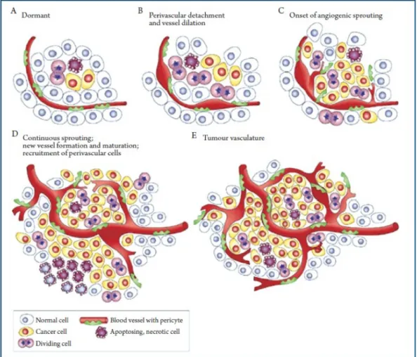
Due to the high proliferation rate of cancer cells, the tumor volume increases rapidly. When the tumor reaches a certain critical volume, the supply of oxygen and nutrients through original blood vessels becomes insufficient and endangers the survival of cancer cells. To overcome this problem, cancer cells activate the development of new blood vessels through angiogenesis (Figure 7). Figure 7: Tumor angiogenesis. After reaching a critical tumor mass, the surrounding blood vessels aren’t sufficient to support further tumor growth. Subsequently, tumor cells recruit perivascular cells and undergo EMT to create new blood vessels, thus developing their own tumor
The activation of angiogenesis by cancer cells involves several steps. First of all, the hypoxia status in fast growing tumors inhibits the degradation of the Hypoxia-Induced Factor 1 (HIF1). The increased half-life of this factor leads to the expression of genes involved in angiogenesis, such as the genes encoding proteins of the vascular endothelial growth factor (VEGF) family (VEGFA, VEGFB, VEGFC, VEGFD, VEGFE,) (Fraisl et al., 2009) and Stroma-Derived Factor 1 (SDF-1). While VEGFA induces angiogenesis, SDF-1 attracts pro-angiogenic myeloid cells to the tumor site (Grunewald et al., 2006). At some point, the balance between anti-angiogenic and pro-angiogenic factors switches in favor of the angiogenesis. This critical step is called “angiogenic switch” (Hanahan and Folkman, 1996). Under pressure of pro-angiogenic factors, endothelial cells (ECs) derived from tumor-proximal blood vessels migrate to the tumor to form new vessels. To control EC migration, an EC subtype, called “Tip cell” pilots the other ECs (called “stalk cells”) by following the pro-angiogenic factors gradient like JAGGED 1 (JAG1), Delta-Like 1 (DDL1) and Neuropilin-1 (Nrp1). Finally, tumor vessels are formed upon consolidation by pericytes recruited by the Platelet-Derived Growth Factor (PDGF) (Ribatti et al., 2011). Contrary to normal blood vessels, tumor blood vessels are generally leaky with more fenestration between ECs, less tight junctions between ECs and incomplete wrapping by pericytes (Ribatti et al., 2007).
In addition to EC recruitment, the formation of the new blood vessels to support tumor growth is also the consequence of cancer cells insertion in the newly formed vessels (Chang et al., 2000). This phenomenon has been particularly well described for brain tumors in which a subpopulation of cancer cells exhibiting stem cell like phenotypes are undergoing trans-differentiation to do so (Soda et al., 2011). Although still under debate, this feature of cancer stem cells is important in the context of anti-cancer drug design because these cells are largely the cause of tumor relapse. Previous work in the lab of D. Bagnard showed that inhibiting Plexin-A1 in glioblastoma inhibits its growth hence offering interesting therapeutic opportunities (Jacob et al., 2016). The leakage and permeability characteristics of these abnormal vessels facilitate the migration of cancer
associated vessel leakage is also the cause of passive drug targeting by penetrating the tumor bulk. It is important in the Enhanced Permeability and Retention (EPR) effect, a critical phenomenon for nanoparticles tumor accumulation in vivo. Consequently, the targeting of the tumor vasculature by nanoparticles is of great interest. For example, it has been shown that gold nanoparticles inhibit angiogenesis and lung metastases development in a mouse model of melanoma (Li et al., 2017). In their study, Li et al., had demonstrated that gold nanoparticles have no effect on cell viability but could decrease endothelial cell migration by the reduction of the metalloproteinase-2 which is known to promote endothelial cells angiogenesis and migration (Ma et al., 2014). Moreover, Li et al., also showed that gold nanoparticles could inhibit the Epithelial-Mesenchymal Transition (EMT) of B16F10 melanoma cells.
In addition to this direct effect of certain types of nanoparticle on tumor vasculature, the nanoparticles offer the possibility to take advantage of the EPR effect to accumulate therapeutic compounds at the tumor site.
1.6. Invasion and metastasis
Cancer cells have the ability to invade surrounding tissues and to migrate to distant sites including new organs. Whereas healthy cells are bound to each other through extracellular matrix (ECM) thus maintaining tissue integrity, cancer cells develop strategies to remove their adhesion to the ECM and to remodel it. The main class of adhesion molecules is the integrin family; consistently, integrins expression is often deregulated in cancer.
Carcinoma-associated fibroblasts (CAF) are activated by cancer cells to produce matrix metalloproteinases (MMPs). These MMPs degrade integrins to decrease ECM stiffness and allowing cancer cells to increase their migratory behavior.
A major phenomenon implicated in the development of pro-invasive behavior of cancer cells is the EMT (Figure 8). The EMT is a multistep dynamic process that occurs naturally during embryogenesis. It is involved in gastrulation, tissue morphogenesis (Nieto et al., 1994) and wound healing (Savagner et al., 2005). It leads to the transition of cells
exhibiting an epithelial state (polarized function, localization of E-cadherin at the cell membrane) to a mesenchymal state. Moreover, EMT has been linked to the acquisition of certain stem cell properties (like self-renewal and the ability to differentiate into various cell types) by some cancer cells in some type of carcinoma [e.g mammary carcinoma (Mani et al., 2008)]. However, cancer cells don’t undergo the entire transition to a complete mesenchymal state (Nieto et al., 2016). The number of intermediate phenotypes in cancer cells is still under debate and associated with various terms such as partial EMT, intermediate EMT, hybrid epithelial/mesenchymal, semi-mesenchymal or also incomplete EMT (Grigore et al., 2016). Cancer cells exhibiting intermediate EMT phenotypes are expected to show both epithelial (adhesion) and mesenchymal (migration) features. Figure 8: Epithelial-mesenchymal transition. Under stimulation from the environment, tumor cells undergo EMT and acquire metastasis and invasion ability thus promoting tumor spreading. Adapted from Tsubakihara and Moustakas (2018).
the EMT program occurs in probably all carcinoma types and is critical in cancer cell dissemination (Guo et al., 2012).
During carcinogenesis, the EMT is activated in cancer cells by EMT-inducing transcription factors (EMT-TFs). The EMT and EMT-TFs are also involved in cancer cell resistance to several chemotherapeutic drugs and kinase inhibitors. For example, EMT-TFs such as SNAI1, SMUG and ZEB1 are involved in resistance to platinum-based chemotherapeutic drugs in various cancer types such as breast cancer (Lim et al., 2013). TGFß induces the EMT by binding to TGFßR1 and TGFßR2 leading to the formation of SMAD1-SMAD5-SMAD4 and SMAD2-SMAD3-SMAD4 complexes. These complexes function as transcription factors for genes implicated in cell invasion, angiogenesis and cell growth among other functions (Xu et al., 2009). TGFß thus induces the expression of important proteins implicated in cell invasion and metastasis, among them, Tenascin-C (TNC) (Saupe et al., 2013). TNC is an extracellular matrix (stroma) protein overexpressed in many cancer types and associated with poor prognosis (Ni et al., 2017; Sundquist et al., 2017). It has been shown that TNC expression is associated with lung metastasis in breast cancer patients (Insua-Rodríguez et al., 2018). Strikingly, TNC is implicated in resistance to chemotherapeutic drugs (Wang et al., 2016). Moreover, as TNC is only poorly expressed in the adult organism (Chiquet-Ehrismann et al., 2014), it constitutes a suitable target for cancer therapy. Another protein involved in cancer invasion is the Nrp1. Nrp1 is a transmembrane protein expressed in glia cells as well as in some neurons and endothelial cells (Eichmann et al., 2005). It is overexpressed in various types of cancer cells (Bielenberg et al., 2006) and associated with a bad prognosis (Geretti and Klagsbrun, 2007). It has been shown that Nrp1 promotes EMT in oral squamous cell carcinoma (OSCC) (Chu et al., 2014) and several studies targeting Nrp1 demonstrated a decrease in cancer invasion and metastasis in gastric cancer (Peng et al., 2014), melanoma (Bai et al., 2015) and OSCC (Liu et al., 2015). The role of Nrp1 in cancer and its potential as a therapeutic target (Meyer et al., 2016) will be further discussed in the part II of this thesis.
Among the different hallmarks of cancer, the multiplicity of compensatory/redundant signaling pathways is a major hurdle for drug design. Strikingly, the nanoparticle technology offers the possibility to combine different treatments and thus target different pathways simultaneously, thereby reaching towards the Grail of anticancer drugs.
2.
Cancer therapeutic strategies
Current cancer treatments include surgery, radiotherapy and chemotherapy. However, during the last century, a number of new approaches to treat cancer have been investigated.
2.1. Classical treatment
2.1.1. Surgery
Whenever possible, tumors are removed by surgical resection. However, the surgical approach has several disadvantages. Due to the infiltrating behavior of cancer cells, it is often necessary to remove tissues at the border of the tumor, possibly including healthy tissues. In the case of tumors attacking major organs such as the liver, lungs or brain, this can lead to important damages decreasing the patient’s quality of life (Hatiboglu et al., 2018). Moreover, patient survival after surgery frequently depends on organs transplantation. To limit such problems as much as possible, surgeons need to be able to define the localization of the tumor as precisely as possible. Towards this aim, some new real-time tumor imaging techniques are under development. These new techniques allow the visualization of the tumor with cellular precision during surgery (Chi et al., 2014). However, such technique cannot be applied to tumors involving mobile tumor cells, such as leukemia.
them to be used as imaging agent. Moreover, metallic nanoparticles offer the possibility to be employed as a drug nanocarrier. The development of this type of nanoparticles and their application in cancer therapy will be discussed in the part III of this manuscript.
2.1.2. Radiotherapy
Radiotherapy (also called radiation therapy) is used in cancer treatment since more than a hundred years (Cosset, 2016). Although radiotherapy can be locally applied to cancer cells, healthy adjacent tissues are usually also exposed, thus leading to death of healthy cells (Taylor and Kirby, 2015). This causes numerous side effects usually including sore skin, tiredness, hair loss and vomiting. Additional side effects may occur depending on the treated zone (e.g. diarrhea when radiation is applied to abdominal or pelvic areas) (Lawrie et al., 2018).
Moreover, cancer cells are able to develop a resistance to radiation therapy through several mechanisms such as inhibition of reactive oxygen species (ROS) production, DNA damage repair, and inhibition of apoptotic pathways activated by radiation (Zhao et al., 2018).
Specific protocols to deliver radiation have been developed such as hypofractionated radiation therapy3. Two types of these protocols are used, the stereotactic body radiation
therapy (SBRT, non-surgical radiation therapy used to treat functional abnormalities and small tumors of the brain) and the stereotactic radiation surgery (SRS, to treat body tumors).
While the induction of DNA damage is a major mechanism explaining the therapeutic effect induced by conventional radiation therapy, hypofractionated radiation therapy is thought to rely also on the modification of the tumor microenvironment (TME) (Arnold et al., 2018).
Indeed, cancer cell death induced by radiation leads to the release of cytokines [i.e Interferon-y (IFN-y)] able to activate the immune system, especially when the hypo-
fractionated radiation protocol is applied. Indeed, several studies have shown that ablative doses of radiation [30 Gray (Gy)] induce a cytotoxic T-cell response through IFN-y release bablative doses of radiation [30 Gray (Gy)] induce a cytotoxic T-cell response through IFN-y cancer cells, which leads to a regression of the tumor (Filatenkov et al., 2015). However, the application of fractionated doses after the first large dose reduces the T-cell response. This could be explained by the death of the infiltrating lymphocytes due to the radiation. Thus, conventional radiation schedules may exert a negative effect on TME. To address this issue, the use of agents that block immunosuppressive signals has been investigated in combination with conventional radiation therapy. For example, treatment with a monoclonal antibody against the programmed cell death 1 protein (PD-1) in combination with fractionated radiation (5 x 2 Gy) in a dual-tumor mouse model led to a regression of both the irradiated tumor and distal tumors (termed abscopal effect) and this effect was correlated with the activation of a T-cell response (Dovedi et al., 2017). In addition to these effects on the immune system, radiation therapy has also an effect on the tumor vasculature (Song et al., 1974). Large doses (higher than 10 Gy per fraction) induce major damage to the tumor vasculature leading indirectly to the death of cancer cells (Park et al., 2012). Single low doses, in contrast, induce a transient increase in tumor blood flow (Wong et al., 1973). Moreover, it has been demonstrated that endothelial cells survive at a 2 Gy dose of radiation (Kuwahara et al., 2014). Consistently, low doses of radiation (< 5 Gy) are linked to a promotion, rather than inhibition, of tumor angiogenesis and neovascularization. Moreover, in vitro studies have shown that low dose radiation causes a stimulation of VEGF production by stroma cells and phosphorylation of VEGFR2 (Vala et al., 2010). Thus, therapeutic strategies combining radiation and anti-angiogenic treatments have been investigated. For example, the blockade of VEGF signaling using antibodies in animal models has been shown to potentiate radiation effect (Truman et al., 2010). Moreover, as aforementioned, cancer cells can develop a resistance against radiation therapy. In order to overcome this issue, efforts have been made to develop radiosensitizers. Particularly, gold nanoparticles have
been greatly investigated because they own a good ability to sensitize cancer cells towards irradiation (Her et al., 2017).
2.1.3. Chemotherapy
Chemotherapy relies on the use of chemical drugs presenting a cytotoxic activity. Some chemotherapeutic drugs target microtubules. Paclitaxel, for example, is a drug from the taxane family, which binds to the end of polymerized β-tubulin. This causes the stabilization of the microtubules, which leads to the inhibition of cell division and to cell death.The anthracyclin family is a class of antibiotics widely employed in cancer treatment as chemotherapeutic drugs. The main representative of this family is doxorubicin (DOX), which intercalates into DNA, thereby inhibits DNA synthesis, and cell division. Other chemotherapeutic drugs act as DNA damaging agents. One of these agents is Temozolomide, which is used in glioblastoma. The family of platinum anticancer agents, which includes cisplatin as the major representative, acts by crosslinking DNA, which inhibits DNA repair and synthesis.
Another class of chemotherapeutic drugs consist of topoisomerase I and II inhibitors. Topoisomerases are enzymes regulating DNA supercoiling by cleaving the DNA backbone. Thus, they play a critical role for DNA replication and repair. Their inhibition blocks the replication and repair of DNA in cells. Topoisomerase I inhibitors include irinotecan and camptothecin and topoisomerase II inhibitors include ectoposide and teniposide. It is known that also anthracyclin drugs exhibit topoisomerase II inhibition activity.
While clearly efficient on some tumor types, chemotherapies or combinations of chemotherapeutics show numerous side effects due to their lack of specific delivery and tumor targeting ability. For example, DOX is well known to produce cardiotoxicity. Several alternative approaches have been developed with aim to circumvent these
chlorotic stripe mosaic virus, have been used as a drug carrier to deliver DOX in a mouse model of TNBC (Alemzadeh et al., 2018). DOX was loaded inside the icosahedral nanoparticles, which were decorated with folic acid for targeting to cancer cells (FA-DOX-JgCSMV). The results show that the FA-DOX-JgCSMV particles were able to reduce tumor growth to the same extent as free DOX. Other carriers used for DOX delivery to tumors include cupper nanocubes (Li et al., 2019), co-polymer nanoparticles (Xu et al., 2018) and other virus-like nanoparticles (Finbloom et al., 2018). These different types of carriers are currently under further investigation and have the potential to increase the therapeutic effect of DOX.
2.2. Gene therapy
As mentioned above, cancer cells exhibit genomic alterations leading to an overexpression of genes promoting cell survival, proliferation, migration, avoidance of the immune system, and down regulation of genes controlling cell proliferation and invasion. One therapeutic strategy aims to re-establish a normal expression of these deregulated genes by strategies employing the expression of specific small interfering RNAs (siRNAs), short hairpin RNAs (shRNAs), microRNAs (miRNAs), or plasmid DNA (pDNA). miRNA are small non-coding RNA molecules (containing about 22 nucleotides) involved in post-transcriptional regulation of gene expression via base-pairing with complementary sequences within mRNA molecules. miRNA can act as a tumor suppressor genes or oncogenes. One important miRNA is miR-21, which regulates tumor suppressor genes, particularly PTEN. The expression of miR-21 is induced in several tumor types including glioblastoma, where it was discovered (Chan et al., 2005). miR.21 is overexpressed also in breast cancer and other tumor types (Volinia et al., 2006). miR-21 expression has been linked to the promotion of cancer cell proliferation, metastasis, and the inhibition of apoptosis (Pfeffer et al., 2015). Its overexpression was also linked to multi-drug resistance (Geretto et al., 2017) and radiation resistance in esophageal squamous cell carcinoma (Li et al., 2018a). Consequently, therapeutic strategies aim at the inhibition of miR-21. In a recent study, authors showed that renal carcinoma cells
and an increase in their chemosensitivity to paclitaxel and oxaliplatin (Gaudelot et al., 2017).
shRNA are short oligonucleotide sequences that can loop back on themselves and form a stretch of dsRNA that is cleaved by Dicer. In biotechnology, shRNAs are delivered to the cells via an expression vector for transcription in the nucleus. The resulting pre-mature shRNA is processed in the cytoplasm by Dicer and the resulting siRNA (short oligonucleotide sequence which is synthetically produced or processed from a stretch shRNA by the Dicer enzyme present in the target cells) is integrated into an RNA-induced silencing complex (RISC) that uses the siRNA as a guide for the sequence-specific cleavage or translational repression of target mRNA.
Delivery of genetic material to cancer cells relies on vectors which can be non-viral or viral. Non-viral vectors are synthetically produced chemical molecules such as lipofectamine or FuGene. Plasmid DNA or siRNAs are encapsulated or bound to these vectors (Chira et al., 2015). Non-viral vectors are easy to produce and rarely the cause of an inflammatory reaction (Kaminski et al., 2002). Viral vectors are mainly derived from mammalian viruses, however there is an emerging interest in the development of bacteriophages and plant viruses as vectors (Lam and Steinmetz, 2018). These vectors and their employment as delivery system are explained in part III of this manuscript.
Several nanoparticle types have been used to deliver genetic material (Xiao et al., 2019). For example, graphene oxide has been employed to deliver a siRNA targeting survivin, a protein inhibiting the apoptosis in cancer cells (Wang et al., 2018). The authors showed that the siRNA was able to inhibit tumor growth in vivo. In another study, authors used lipoprotein nanoparticles to co-deliver DOX and miR-21 inhibitor to MCF-7 breast cancer cells resistant to DOX (Rui et al., 2017). They showed that this formulation was able to reverse drug resistance of cancer cells and had a synergistic antiproliferative effect in vivo.
2.3. Immunotherapy
A promising approach is to modulate the immune system, which is often deregulated in cancer disease. For example, as already mentioned, cancer cells secrete factors recruiting immune cells to promote changes in the microenvironment which are favorable to tumor development.
2.3.1. Vaccines for cancer treatment
The idea behind cancer vaccine development is to educate the immune system to target cancer cells. To this end, different strategies are employed, a) injection of whole tumor lysate into the bloodstream, b) dendritic cell vaccines, c) presentation of Tumor Associated Antigens (TAA), and d) the use of DNA-vaccines. Dendritic cells (DCs) are Antigen Presenting Cells (APC). Vaccine-type dendritic cells are developed from blood isolated from the given patient followed by education of the immune system cells in this blood with antigens from tumor cells in vitro. This education induces the differentiation of the immune cells into DCs that are able to present the tumor antigens to other immune cells when injected back to the patient. In 2010, the Food and Drug Administration (FDA) approved the first therapeutic DC vaccine, which was named sipuleucel-T (Provenge®) and has been developed for the treatment of metastatic prostate cancer (Kantoff et al., 2010).
Although the normal immune system acts against cancer cells, its activity is reduced overtime if the stimulation by tumor antigens is insufficient. To circumvent this problem, DNA-vectors are employed as DNA vaccines that deliver bits of DNA coding for tumor-associated antigens (TAA) into cells. The transfected cells that produce the specific antigens are then capable of maintaining the cancer-controlling state of the immune system. Usually, adjuvants are required to support the recognition of the TAA by the immune system (Banday et al., 2015). Alternative approaches aim at methods to
been studied as TAA carriers including gold nanoparticles (Mocan et al., 2015), liposomes (Thomann et al., 2011), and virus-like nanoparticles (Yin et al., 2012). Such nanocarriers offer the possibility to load large quantities of TAA and to ensure their sustained release, thus increasing the uptake of antigens by DCs leading to a sustained T cells activation (Prasad et al., 2011). Moreover, it is possible to load the nanocarriers with TAAs in combinations with adjuvants (Hamdy et al., 2011).
2.3.2. Immune checkpoint inhibitors (ICIs)
Normal cells carry specific immune checkpoints, molecules that are recognized by T cells and able to keep T cells in an inactivate state in order to prevent the immune system from attacking healthy cells. Some cancer cells exploit this checkpoint to avoid the recognition by T cells. For example, the programmed death-ligand 1 (PD-L1) is expressed on some cancer cell types and interacts with its receptor PD-1 expressed on T cells. To interfere with this checkpoint interaction, certain immune checkpoint inhibitors (ICIs) are being developed. One of these agents called pembrolizumab consists of antibodies that specifically target PD-1 and has been approved for treatment of several cancer types including metastatic or non-operable melanoma, head and neck cancer squamous cell carcinoma and cervical cancer (Larkins et al., 2017; Pai-Scherf et al., 2017). Due to their ability to block immune cell inactivation, the application of ICIs can lead to auto-immune responses with numerous side effects (Spain et al., 2016).
2.3.3. Chimeric antigen receptor (CAR)-expressing T cells (CAR-T
cells)
A chimeric antigen receptor (CAR) is a genetically engineered chimeric receptor in which a part of a specific monoclonal antibody is combined with a signaling domain (Wilkins et al., 2017). To form CAR-T cells, T cells are extracted from the blood of the patient and then engineered to express CARs on their surface. The monoclonal antibody used to create each CAR determines its target. The second generation of CARs adds theexpression of co-stimulatory domain of receptor and the third generation includes an additional co-stimulatory domain to enhance T cell activation (Figure 9). Figure 9: Successive generations of CARs. (A) First generation of CARs associating antigen-binding domain with T-cell activation domain. (B) Second generation of CARs adds a costimulatory domain. (C) Third generation of CARs added a second costimulatory domain. From Chang and Chen (2017).
In 2017, the FDA approved the application of CAR-expressing T cells in advanced lymphoma for adults and acute lymphocytic leukemia for children (Liu et al., 2017). However, this approach requires further improvement before application to solid tumors. Indeed, the microenvironment of solid tumors exhibits several physical barriers that prevent infiltration by T cells. Moreover, immunosuppressive molecules are often expressed in high concentration within the tumor. To overcome these issues, one
microenvironment e.g. specific CARs T cells targeting VEGFR2 and thus, the tumor vasculature (Chinnasamy et al., 2010). This promising approach undergoes phase 1/2 clinical trials in liver cancer.
As described above, some cancer cells express PD-L1 to inhibit T cells activation. This mechanism of resistance is also effective against CAR-T cells (Tumeh et al., 2014). Therefore, CAR-expressing T cells have been engineered to secrete anti-PD-L1 antibodies thus restoring the function of these cells, at least in vitro (Suarez et al., 2016).
Interestingly, it is possible to couple the surface of CARs T cells to nanoparticles without impacting their function (Stephan et al., 2010). This opens the possibility to increase the specific delivery of therapeutic drugs loaded into nanoparticles using CARs T cells. The loaded therapeutic drug can be chosen to improve CARs T cells efficiency e.g. inhibiting immunosuppressive molecules (Siriwon et al., 2018).
Moreover, there is a new application area for nanoparticles targeting and remodeling the TME to increase immunotherapy (Gao et al., 2019). They have been used to target immunosuppressive enzymes (Zhu et al., 2012), the cytokines, the tumor extracellular matrix, and TAMs. This strategy aims to reverse the immunosuppressive environment instituted by the tumor in order to allow immunotherapeutic drugs to reach their targets.
2.4. Hormone therapy
Some cancers such as prostate cancer or Estrogen Receptor (ER) positive breast cancer, can be treated by hormone therapy (HT). HT is employed as neoadjuvant (treatment given before primary treatment which usually is surgery) or adjuvant therapy (treatment given after primary treatment). Prostate cancer cell growth is usually stimulated by androgens and, because androgens are mostly produced by testicles, one type of HT employed to treat prostate cancer consists of removing testicles by either surgery (orchiectomy) or by chemical castration using luteinizing hormone-releasing hormone agonists (Triptorelin, Leuprolide). ER positive breast cancer cells are stimulated by estrogen. Thus, HT in this case consists of approaches to reduce estrogeninhibitors (AI) are used in postmenopausal women to block the production of estrogen from androgen by the aromatase enzymes. There are three AI used for treatment of breast cancer, letrozole (Femara), anastrozole (Arimidex) and exemestane (Aromasin). Interestingly, a recent study shows that premenopausal women could benefit from AI if their ovarian functions are suppressed (SOFT, Suppression of Ovarian Function Study published in 2015). For premenopausal women, rather a Selective Estrogen Receptor Modulator (SERM) is used. The SERM binds to ER thus inhibiting estrogen signaling and also cancer growth. The major SERM prescribed to treat breast cancer in premenopausal women is tamoxifen (Nolvadex in pill form and Soltamox in liquid form). The side effects of SERMs includes headache, weakness, blood clots and stroke. However, another type of breast cancer characterized as ‘triple negative breast cancer’ (TNBC) is resistant to HT as it does not express ER, HER2 or progesterone receptor (PR), and this type of cancer represents 16% of all breast cancer types (Shah et al., 2012). This breast cancer is associated with a poor prognosis due to a high rate of relapse and a fast progression after relapse. The classical treatment protocol for TNBC involves neoadjuvant chemotherapy based on paclitaxel (Giordano et al., 2018). 15% of TNBC patients carry mutations in either the BRCA1 gene or the BRCA2 gene. These genes are tumor suppressor genes, both coding for proteins with DNA repair function. The BRCA1 mutation has been linked to a complete response (i.e tumor regression) when the patients were treated with cisplatin as a single-agent in a neoadjuvant protocol (Byrski et al., 2014). Considering that TNBC patients lack a specific target for therapy, others strategies have been developed. For example, inhibitors targeting poly ADP ribose polymerase (PARP) like veliparib have shown positive results in clinical trials (Rugo et al., 2016). However, TNBCs exhibit a high stromal and intratumoral level of TILs compared to other breast cancer types (García-Teijido et al., 2016) and high level of TILs is linked to a better clinical outcome for breast cancer patient (Denkert et al., 2010). TILs are also a prognostic factor and are correlated to an increased metastasis-free survival (Kreike et al., 2007), a decreased distant recurrence (Adams et al., 2014) and an improved overall
clinical outcome of TNBC has led to the employment of immunotherapeutic approaches. For example, pembrolizumab and atezolizumab (antibodies targeting PD-1 and PD-L1) are under investigation in both neoadjuvant and adjuvant settings.
2.5. Hyperthermia therapy
Hyperthermia therapy consists in the application of increased temperature (usually between 40°C and 45°C) to the whole or part of the body in order to kill cancer cells or to sensitize them to chemotherapy. Temperatures above 45°C are employed for thermal ablation. The main challenge encountered in this therapeutic approach is the destruction of cancer cells without damaging the healthy tissues. One strategy to achieve this goal consists in the use of metallic nanoparticles which can be excited by a magnetic field thus inducing an increased temperature of the particles. The coating of these nanoparticles with cancer cell targeting moieties allows the application for a more localized hyperthermia treatment. For example, ferric oxide nanoparticles carrying antibodies targeting the HER2 receptor accumulated at HER2 positive cells and caused cell death by hyperthermia upon application of a magnetic field in vitro (Zhang et al., 2011a).
Hyperthermia can support the efficiency of chemotherapeutic drugs by causing an elevation of tumor blood flow and vascular tumor permeability (Song et al., 2005). Interestingly, such synergistic effects between hyperthermia therapy and drug treatment can vary. For example, while hyperthermia therapy can have a synergistic with cisplatin there is no such affect if applied together with DOX (Lepock, 2005).
2.6. Photodynamic therapy (PDT)
The use of light as a therapeutic agent goes back to the ancient Greece, India and Egypt (Daniell and Hill, 1991) but was forgotten until the middle of 19th century. In 1903, Niels Finsen, a Danish physician, won the Nobel prize for the use of light to treat lupus vulgaris. However, this type of phototherapy wasn’t dynamic and didn’t introduce the use of a photosensitizer (PS). The first discovery of a photosensitizing agent was done by Prime, a French neurologist, who administrated eosin orally to his patient to treatepilepsy and noticed that lesions appeared on skin areas exposed to light (Prime, 1900). In modern photodynamic therapy, a PS is activated in the presence of oxygen and under specific light (usually a laser) condition (Figure 10). The wavelength used depends on the PS, with a longer wavelength allowing a deeper tissue penetration. Generally, the wavelength used in clinics are around 630 nm. The change of the PS state from inactivated to activated induces a production of reactive oxygen species (ROS) and other radicals thus leading to cell death by apoptosis or necrosis. PSs differ with respect to chemical structure, excitation wavelength, and efficiency to produce ROS. The first PS authorized for treatment was the ‘porfimer sodium’ (Photofrin®), which is composed of porphyrin derivatives oligomers that can be excited at 630 nm. It is used in the treatment of esophagus cancer since 1995 and for the treatment of non-small cell lung (NSCL) carcinoma since 1998. The three main families of PS are based on porphyrin, chlorin and the dyes (e.g 5-aminolevulinic acid, silicon phthalocyanine 4) (Ormond and Freeman, 2013). Figure 10: Mechanism of photodynamic therapy. A light source generates a light beam that excites a photosensitizer. The excited photosensitizer transfers electrons to oxygen, thus creating ROS. These ROS damages tumor cell DNA leading to cell death. As with many other treatments, also PS compounds can accumulate and cause damage in healthy organs (Mironov et al., 1992). Moreover, the requirement of light penetration
investigation. Indeed, like other drugs, PS can also be conjugated to a targeting moiety to increase their accumulation at the tumor site. For example, a chlorin PS conjugated to a peptide targeting Nrp1 exhibited a fast accumulation at the tumor site within a mouse glioma model (Thomas et al., 2008). Targeted delivery of PS compounds can also be achieved by combining them with cancer targeting agents on nanoparticles as a delivery platform (Lucky et al., 2015). Silica nanoplatform exhibiting Nrp1 targeting peptide on the surface and loaded with gadolinium for Magnetic resonance imaging (MRI) as well as chlorin for interstitial PDT (iPDT) was used for glioma treatment (Bechet et al., 2015). In this study, the authors used nanoparticles for MRI-guided implantation of an optic fiber for iPDT. In two different models of glioma tumor, nanoparticles loaded with gadolinium (with or without Nrp1 targeting peptide) were able to enhance the positive contrast in MRI enough to guide an optic fiber implantation for iPDT. Cerebral perfusion MRI was performed before and after iPDT to visualize modification in tumor perfusion after treatment. In both tumor type, a decrease in tumor perfusion was observed and even with nanoparticles not carrying Nrp1 targeting peptide. However, the perfusion was decreased up to 80% only in animals treated with nanoparticles carrying the peptide. Histological examination of brain tissue removed immediately after iPDT showed the presence of oedema and vascular disruption. Moreover, VEGF protein level was reduced after iPDT. These different results demonstrate the photodynamic activity in vivo induced by these functionalized nanoparticles.
In addition, two different types of polymer-lipid-PEG (polyethylene glycol) nanoparticles used as carriers of PS increased cellular uptake and caused stronger photocytotoxic activity as compared to the application of free PS (Pramual et al., 2017).
Unlike PDT, which is activated in the presence of oxygen and by specific light, photothermal therapy (PTT) depends on electromagnetic radiation, usually in the near infrared wavelength, to provoke an increase in temperature in the target tissue.