RESEARCH OUTPUTS / RÉSULTATS DE RECHERCHE
Author(s) - Auteur(s) :
Publication date - Date de publication :
Permanent link - Permalien :
Rights / License - Licence de droit d’auteur :
Institutional Repository - Research Portal
Dépôt Institutionnel - Portail de la Recherche
researchportal.unamur.be
University of Namur
Modification of the 1-phosphate group during biosynthesis of Capnocytophaga
canimorsus lipid A
Renzi, Francesco; Zaehringer, Ulrich; Chandler, C.E.; Ernst, R. K.; Cornelis, Guy; Ittig, Simon
Published in:Infection and Immunity
DOI: doi: 10.1128/IAI.01006-15 10.1128/IAI.01006-15 Publication date: 2015 Link to publication
Citation for pulished version (HARVARD):
Renzi, F, Zaehringer, U, Chandler, CE, Ernst, RK, Cornelis, G & Ittig, S 2015, 'Modification of the 1-phosphate group during biosynthesis of Capnocytophaga canimorsus lipid A', Infection and Immunity, vol. 84, no. 2, doi: 10.1128/IAI.01006-15, pp. 550-561. https://doi.org/doi: 10.1128/IAI.01006-15, https://doi.org/10.1128/IAI.01006-15
General rights
Copyright and moral rights for the publications made accessible in the public portal are retained by the authors and/or other copyright owners and it is a condition of accessing publications that users recognise and abide by the legal requirements associated with these rights. • Users may download and print one copy of any publication from the public portal for the purpose of private study or research. • You may not further distribute the material or use it for any profit-making activity or commercial gain
• You may freely distribute the URL identifying the publication in the public portal ? Take down policy
If you believe that this document breaches copyright please contact us providing details, and we will remove access to the work immediately and investigate your claim.
Modification of the 1-Phosphate Group during Biosynthesis of
Capnocytophaga canimorsus Lipid A
Francesco Renzi,aUlrich Zähringer,bCourtney E. Chandler,cRobert K. Ernst,cGuy R. Cornelis,a,dSimon J. Ittigd
Université de Namur, Namur, Belgiuma; Division of Immunochemistry, Research Center Borstel, Leibniz Center for Medicine and Biosciences, Borstel, Germanyb; Department of Microbial Pathogenesis, University of Maryland, Baltimore, Maryland, USAc; Biozentrum der Universität Basel, Basel, Switzerlandd
Capnocytophaga canimorsus, a commensal bacterium of dog’s mouth flora causing severe infections in humans after dog bites
or scratches, has a lipopolysaccharide (LPS) (endotoxin) with low-inflammatory lipid A. In particular, it contains a phosphoe-thanolamine (P-Etn) instead of a free phosphate group at the C-1 position of the lipid A backbone, usually present in highly toxic enterobacterial Gram-negative lipid A. Here we show that the C. canimorsus genome comprises a single operon encoding a lipid A 1-phosphatase (LpxE) and a lipid A 1 P-Etn transferase (EptA). This suggests that lipid A is modified during biosynthesis after completing acylation of the backbone by removal of the 1-phosphate and subsequent addition of an P-Etn group. As endotoxic-ity of lipid A is known to depend largely on the degree of unsubstituted or unmodified phosphate residues, deletion of lpxE or
eptA led to mutants lacking the P-Etn group, with consequently increased endotoxicity and decreased resistance to cationic
anti-microbial peptides (CAMP). Consistent with the proposed sequential biosynthetic mechanism, the endotoxicity and CAMP re-sistance of a double deletion mutant of lpxE-eptA was similar to that of a single lpxE mutant. Finally, the proposed enzymatic activities of LpxE and EptA based on sequence similarity could be successfully validated by mass spectrometry (MS)-based analy-sis of lipid A isolated from the corresponding deletion mutant strains.
S
ome Gram-negative bacteria have evolved different modifica-tions of their lipid A structure, leading to a reduced recogni-tion by the host and sensitivity to carecogni-tionic antimicrobial peptides (CAMP) (1–7). One of these modifications occurs on the 1- or 4=-phosphate of lipid A (1,4,7–10). 4=-Phosphatases (LpxF) have been reported in Rhizobium leguminosarum, Rhizobium etli,Por-phyromonas gingivalis, Francisella species, and Helicobacter pylori
(1,10–12). Deletion of lpxF and the resulting presence of the 4=-phosphate on lipid A leads to increased endotoxicity (1,12) and decreased resistance to CAMP (10,12). In the case of Francisella and H. pylori, virulence is reduced (11,12,13). 1-Phosphatases (LpxE) have been identified in H. pylori, P. gingivalis, R. etli, and others (1,6,10,12,14–16). Deletion of lpxE and the resulting presence of the 1-phosphate on lipid A leads to a slightly increased endotoxicity (1) and CAMP sensitivity (10). In H. pylori, position 1 is further modified by the addition of a phosphoethanolamine (P-Etn) (15,17,18), a modification known from other bacteria (15,17,18). This happens via a two-step mechanism, which first involves dephosphorylation of one phosphate residue located at position C-1 of the lipid A backbone by LpxE and subsequent
P-Etn transfer by a phosphoethanolamine transferase (EptA or
PmrC) (15,16). In H. pylori, lpxE and eptA are contained in one operon (Hp0021-Hp0022) (16).
We have previously characterized the lipid A structure of
Cap-nocytophaga canimorsus (19), a bacterial species that can cause rare but severe sepsis or meningitis in humans after dog bites or scratches (20–24). C. canimorsus belongs to the family
Flavobac-teriaceae in the phylum Bacteroidetes and is a usual member of
dog’s mouth flora (21,25–28). C. canimorsus lipid A consists of a 2,3-diamino-2,3-dideoxy-D-glucose (GlcN3N=)-(1=¡6)-linked to 2-amino-2-deoxy-D-glucose (GlcN) [-D -GlcpN3N=-(1¡6)-D-GlcpN lipid A hybrid backbone] containing an P-Etn group attached to the C-1 reducing end and lacking a 4=-phosphate (Fig. 1A). 3-Hydroxy-15-methylhexadecanoic acid [i17:0(3-OH)], 3-hydroxy-13-methyltetradecanoic acid [i15:0(3-OH)],
3-O-(13-methyltetradecanoyl)-15-methylhexadecanoic acid
[i17:0[3-O(i15:0)]], and 3-hydroxyhexadecanoic acid [16:0(3-OH)] are
at-tached to the backbone at positions 2, 3, 2=, and 3=, respectively (19). This structure differs from that of a potent Toll-like receptor 4 (TLR4) agonist like the Escherichia coli lipid A (Fig. 1B), consist-ing of a-(1=¡6)-linked GlcN disaccharide that is phosphory-lated at positions 1 and 4= and carries four (R)-3-hydroxymyr-istate chains [14:0(3-OH)] (at positions 2=, 3=, 2, and 3). The 2= and 3= 3-hydroxylated acyl groups in GlcN(II) are further esteri-fied with laurate and myristate, respectively (29).
We have identified lpxE and eptA genes in the genome of C.
canimorsus and found the overlapping genes to be organized in
one operon. We show that the deletion of lpxE or eptA leads to increased endotoxicity and decreased resistance to CAMP, where deletion of lpxE has a more severe effect. Interestingly, the endo-toxicity and CAMP resistance of a double deletion mutant of lpxE and eptA were the same as those of a single lpxE mutant. This suggests that the P-Etn-containing lipid A is synthesized by a sim-ilar two-step enzymatic process as in H. pylori, where dephosphor-ylation is necessary for substitution of 1-phosphate with P-Etn. Finally, we could successfully validate the proposed lipid A
struc-Received 9 August 2015 Returned for modification 22 September 2015 Accepted 27 November 2015
Accepted manuscript posted online 7 December 2015
Citation Renzi F, Zähringer U, Chandler CE, Ernst RK, Cornelis GR, Ittig SJ. 2016. Modification of the 1-phosphate group during biosynthesis of Capnocytophaga canimorsus lipid A. Infect Immun 84:550 –561.doi:10.1128/IAI.01006-15. Editor: S. M. Payne
Address correspondence to Simon J. Ittig, [email protected].
Supplemental material for this article may be found athttp://dx.doi.org/10.1128 /IAI.01006-15.
Copyright © 2016 Renzi et al. This is an open-access article distributed under the terms of theCreative Commons Attribution 4.0 International license.
on February 10, 2016 by BIBLIOTHEQUE UNIVERSITAIRE
http://iai.asm.org/
tures of the respective deletion mutants by mass spectrometry (MS) analysis, thus also further confirming, on a structural basis, the proposed enzymatic activities of LpxE and EptA as well as the two-step enzymatic mechanism in the lipid A biosynthesis.
MATERIALS AND METHODS
Bacterial strains and growth conditions. The bacterial strains used in this study are listed inTable 1. Escherichia coli strains were grown in LB broth at 37°C. Capnocytophaga canimorsus strain 5 (Cc5) (30) was routinely grown on heart infusion agar (HIA; Difco) supplemented with 5% sheep blood (Oxoid) for 2 days at 37°C in the presence of 5% CO2. Bacteria were
harvested by scraping colonies off the agar surface, washed, and resus-pended in phosphate-buffered saline (PBS). The following selective agents were added at the concentrations indicated: erythromycin, 10g/ ml; cefoxitin, 10g/ml; gentamicin, 20 g/ml; ampicillin, 100 g/ml; tetracycline, 10g/ml.
Genetic manipulations of C. canimorsus. Genetic manipulations of wild-type (wt) Cc5 have been described (31). Briefly, replacement cas-settes with flanking regions spanning approximately 500 bp homologous to regions directly adjacent to the lpxE or eptA gene (28) were constructed with a three-fragment overlapping PCR strategy. As the ATG codon of the
eptA gene is located within the coding region of lpxE, 106 bp upstream of
the eptA ATG codon was not deleted in an lpxE single knockout (⌬ 1833737-1833995). First, two PCRs were performed on 100 ng of Cc5 genomic DNA with primers A and B (Table 2) for the upstream flanking regions and with primers E and F for the downstream regions. Primers B and E contained an additional 5= 20-nucleotide extension homologous to the ermF or tetQ insertion cassette. The ermF and tetQ resistance cassettes were amplified from plasmids pMM13 and pMM104.A DNA, respec-tively, with primers C and D. All three PCR products were cleaned and then mixed in equal amounts for PCR using Phusion polymerase (Finnzymes). The initial denaturation was at 98°C for 2 min, followed by 12 cycles without primers to allow annealing and elongation of the over-lapping fragments (1 cycle consists of 98°C for 30 s, 50°C for 40 s, and 72°C for 2 min). After the addition of external primers (primers A and F), the
program was continued with 20 cycles (1 cycle consists of 98°C for 30 s, 50°C for 40 s, and 72°C for 2 min 30 s) and finally 10 min at 72°C. Final PCR products containing lpxE::ermF, eptA::ermF, lpxE-eptA::ermF, lpxE::
tetQ, eptA::tetQ, or lpxE-eptA::tetQ insertion cassettes were then digested
with PstI and SpeI for cloning into the appropriate sites of the C.
canimor-sus suicide vector pMM25 (31). The resulting plasmids were transferred by RP4-mediated conjugative DNA transfer from E. coli S17-1 to C.
cani-morsus strain 5 or C. canicani-morsus strain 5 Y1C12 mutant to allow
integra-tion of the inserintegra-tion cassette. Transconjugants were then selected for the presence of the ermF or tetQ cassette on erythromycin- or tetracycline-containing plates, respectively, and checked for sensitivity to cefoxitin. Deletion of the appropriate regions was verified by PCR.
Construction of complementation plasmids. Plasmid pMM47.A was used for expression of LpxE and EptA (31). Full-length lpxE, eptA, or
lpxE-eptA genes were amplified with the specific primers listed inTable 2 and cloned into plasmid pMM47.A using NcoI and XbaI or NcoI and XhoI restriction sites, leading to the insertion of a glycine at position 2. Ligated plasmids were cloned in E. coli TOP10.
Human TLR4 activation assay. HEK293 cells stably expressing hu-man Toll-like receptor 4 (hTLR4), myeloid differentiation factor 2 (MD-2), cluster of differentiation antigen 14 (CD14), and a NF-B-dependent reporter (secreted embryonic alkaline phosphatase) were from InvivoGen (HekBlue human TLR4). Growth conditions and endotoxicity assay were as recommended by the supplier (InvivoGen). Briefly, the desired amounts of heat-killed bacteria were placed in a total volume of 20l (diluted in PBS) and distributed in a flat-bottom 96-well plate (BD Fal-con). A total of 25,000 HekBlue human TLR4 cells in 180l were then added to each well, and the plate was incubated for 20 to 24 h at 37°C and 5% CO2. Detection of the secreted phosphatase followed the QUANTI-Blue protocol (InvivoGen). The challenged cells (20l) were incubated with 180l detection reagent (QUANTI-Blue; InvivoGen). The plates were incubated at 37°C and 5% CO2, and absorbance was measured at 655
nm using a spectrophotometer (Bio-Rad).
Polymyxin B sensitivity assay. Polymyxin B sulfate was obtained from Sigma-Aldrich. The MIC was determined by the agar dilution
FIG 1 Structures of C. canimorsus strain 5 and E. coli lipid A. (A) C. canimorsus strain 5 lipid A consists of a-(1=¡6)-linked GlcN3N=-GlcN disaccharide, to
which 3-hydroxy-15-methylhexadecanoic acid, 3-hydroxy-13-methyltetradecanoic acid, 3-O-(13-methyltetradecanoyl)-15-methylhexadecanoic acid, and 3-hydroxyhexadecanoic acid are attached at positions 2, 3, 2=, and 3=, respectively. The disaccharide carries a positively charged ethanolamine at the 1-phosphate and lacks a 4=-phosphate (19). (B) E. coli hexa-acylated lipid A consisting of a-(1=¡6)-linked GlcN disaccharide that is phosphorylated at positions 1 and 4= and carries four (R)-3-hydroxymyristate chains (at positions 2=, 3=, 2, and 3). The 2= and 3= 3-hydroxylated acyl groups in GlcN= are further esterified with laurate and myristate, respectively (29).
on February 10, 2016 by BIBLIOTHEQUE UNIVERSITAIRE
http://iai.asm.org/
method based on the CLSI/NCCLS recommendations (32). Briefly, 104
bacteria contained in 2l PBS were spotted on HIA plates containing 5% sheep blood and polymyxin B ranging from 0.5 mg/liter to 1,024 mg/liter (2-fold increase per step). The plates were incubated and examined for growth of visible colonies after 48 h and 72 h.
Genome annotation. The Blast-p search tool (33) against the C.
cani-morsus 5 genome (28) was used. Search sequences were obtained from the National Center for Biotechnology Information. All available
Bacte-roidetes group sequences were used in the search, but standard E. coli
sequences have also been included. The highest scoring subjects over all the searches have been annotated as corresponding enzymes. Difficulties in annotation were observed only for lpxE. The lpxE search was based on
lpxF and/or lpxE protein sequences from Porphyromonas gingivalis (1),
Francisella novicida (7), Rhizobium etli (10), Helicobacter pylori (12,16) and on all available Bacteroidetes group pgpB protein sequences.
Preparation of bacteria for LPS extraction. Compositional analysis of the lipopolysaccharide (LPS) from the wt C. canimorsus 5 strain previ-ously showed that it was highly contaminated with glucose from amyl-opectin, flavolipin, and capnin, known to be present in Capnocytophaga spp. and Flavobacteriaceae (34). In contrast, the LPS from the C.
canimor-sus 5 Y1C12 mutant (35) was devoid of such contaminating material. Since compositional analysis of the lipid A and LPS core obtained from the wt strain LPS and that of the Y1C12 mutant revealed no differences with respect to their sugars and fatty acids (19,34), the Y1C12 mutant was chosen as the background strain to isolate and analyze the lipid A of ⌬eptA, ⌬lpxE, and ⌬lpxE-eptA deletion mutants in detail by MS analysis. While the Y1C12 mutant was chosen as the background strain for MS analysis, please note that human TLR4 activation assays and polymyxin B sensitivity analysis are based on C. canimorsus 5⌬eptA, ⌬lpxE, and
⌬lpxE-eptA deletion mutants and complemented mutants based on these
dele-tion mutants. The C. canimorsus 5-based Y1C12 mutant has a transposon
insertion within a predicted glycosyltransferase-encoding gene, probably the N-acetyl fucosamine transferase WbuB, necessary for the formation of the O antigen (35). Endotoxicity of resulting C. canimorsus 5 Y1C12 ⌬eptA, ⌬lpxE, and ⌬lpxE-eptA deletion mutants was assessed, and the results confirmed results obtained with C. canimorsus 5⌬eptA, ⌬lpxE, and ⌬lpxE-eptA deletion mutants (data not shown). C. canimorsus bacteria were harvested from 25 blood plates in PBS, followed by centrifugation at 18,000⫻ g for 30 min. Bacteria were resuspended in cold acetone, incu-bated with shaking, resuspended in PBS containing 0.5% phenol for kill-ing, again harvested by centrifugation, washed with PBS, and resuspended in 1 ml water. One tenth of the volume was plated on appropriate growth plates to ensure complete bacterial killing. Bacteria were air dried prior to LPS extraction.
Purification and isolation of free lipid A suitable for MS analysis. Lipid A was isolated from lyophilized C. canimorsus cell pellets following the extraction method of El Hamidi et al. (36). Briefly, pellets were dis-solved in 70% isobutyric acid and 1 M ammonium hydroxide and incu-bated at 100°C for 1 h. Four hundred microliters of water was added to each sample, and the samples were snap-frozen on dry ice and lyophilized overnight. The samples were then washed twice with 1 ml methanol and reconstituted in 150l chloroform-methanol-water (3:1.5:0.25, vol/vol/ vol).
MS-based structural analysis. Lipid A structures were assessed by negative- and positive-ion matrix-assisted laser desorption ionization– time of flight mass spectrometry (MALDI-TOF MS). Lyophilized lipid A was extracted in chloroform-methanol, and then 1l was mixed with 1 l of norharmane MALDI matrix. All MALDI-TOF MS experiments were performed using a Bruker Microflex MALDI-TOF mass spectrometer (Bruker Daltonics, Billerica, MA). Each spectrum was an average of 300 shots. Electrospray (ES) tuning mix (Agilent, Palo Alto, CA) was used for TABLE 1 Bacterial strains and plasmids used in this study
Strain and genotype or
plasmid Origin, construction, description, or relevant genotype and/or phenotype Reference Strains
Cc5 Isolated from a case of human fatal septicemia after a dog bite in 1995 30
Cc5⌬lpxE Replacement of Ccan_16960 by ermF; Emr(primers 6493 to 6498) (⌬ 1833737-1833995) This study
Cc5⌬eptA Replacement of Ccan_16950 by ermF; Emr(primers 6499 to 6504) (⌬ 1831370-1832888) This study
Cc5⌬lpxE-eptA Replacement of Ccan_16960-16950 by ermF; Emr(primers 6493 to 6495 and 6502 to 6504)
(⌬ 1831370-1833995)
This study
Cc5 Y1C12 Tn4351 insertion in Ccan_23370, “wbuB”-like glycosyltransferase 35
Cc5 Y1C12⌬lpxE Replacement of Ccan_16960 by tetQ; Tcr(primers 7539 to 7544) (⌬ 1833737-1833995) This study
Cc5 Y1C12⌬eptA Replacement of Ccan_16950 by tetQ; Tcr(primers 7545 to 7550) (⌬ 1831370-1832888) This study
Cc5 Y1C12⌬lpxE-eptA Replacement of Ccan_16960-16950 by tetQ; Tcr(primers 7539, 7540, 7543, 7547, 7548, and 7550)
(⌬ 1831370-1833995)
This study
Plasmids
p-lpxE pMM47.AlpxE (expression plasmid carrying the complete lpxE gene from Cc5) This study p-lpxE-eptA pMM47.AlpxE-eptA (expression plasmid carrying the complete lpxE-eptA genes from Cc5) This study p-eptA pMM47.AeptA (expression plasmid carrying the complete eptA gene from Cc5) This study
pMM13 ColE1 ori; Apr (Emr); ermF from pEP4351 31
pMM25 ColE1 ori; Kmr(Cfr); suicide vector for C. canimorsus 31
pMM47.A ColE1 ori (pCC7 ori); Apr(Cfr); E. coli-C. canimorsus expression shuttle plasmid. C. canimorsus expression
is driven by an ermF promoter.
31 pMM104.A ColE1 ori (pCC7 ori); Apr(Tcr); E. coli-C. canimorsus shuttle plasmid, RP4 oriT. The PstI fragment of
pMM47.A containing repA was inserted into the PstI site of pLYL001.
31 pSI73 pMM25lpxE::ermF (suicide vector for deletion of lpxE [⌬ 1833737-1833995]) This study pSI74 pMM25eptA::ermF (suicide vector for deletion of eptA [⌬ 1831370-1832888]) This study pSI76 pMM25lpxE-eptA::ermF (suicide vector for deletion of lpxE-eptA [⌬ 1831370-1833995]) This study pFR28 pMM25lpxE::tetQ (suicide vector for deletion of lpxE [⌬ 1833737-1833995]) This study pFR29 pMM25eptA::tetQ (suicide vector for deletion of eptA [⌬ 1831370-1832888]) This study pFR30 pMM25lpxE-eptA::tetQ (suicide vector for deletion of lpxE-eptA [⌬ 1831370-1833995]) This study
on February 10, 2016 by BIBLIOTHEQUE UNIVERSITAIRE
http://iai.asm.org/
TABLE 2 Oligonucleotides used in this study Oligonucleotide reference no. Oligonucleotide name a Sequence (5 =-3 =) Restriction site Gene PCR primer order 6493 lpxE-A CCCTGCAGGGCACGTTCGTACCAGTTA PstI lpxE A 6494 lpxE-B GAGTAGATAAAAGCACTGTTATTTGCTTATTTTGAATATTTCGG lpxE B 6495 lpxE-C CTTATATTTGCCGCCGAAATATTCAAAATAAGCAAATAACAGTGCTTTTATCTACTCCGATAGCTTC ermF C 6496 lpxE-D CTTGCATTATCTTAACACTCATAAAAACAACACTCCCCTACGAAGGATGAAATTTTTCAGGGACAAC ermF D 6497 lpxE-E AAAAATTTCATCCTTCGTAGGGGAGTGTTGTTTTTATGAGTGTT lpxE E 6498 lpxE-F CAACTAGTAAACCGTTTCAGTTTGGGT SpeI lpxE F 6499 eptA-A CCCTGCAGTGTTCCTCGCCCTGTTAC PstI eptA A 6500 eptA-B GAGTAGATAAAAGCACTGTTTTATTGATTTTTTTTAACATAAAATTTTATC eptA B 6501 eptA-C GTTGTACTTAATGATAAAATTTTATGTTAAAAAAAATCAATAAAACAGTGCTTTTATCTACTCCGATAGCTTC ermF C 6502 eptA-D ATCTTGTAAATTACGGATTGGTCATTCAATAATTCTACGAAGGATGAAATTTTTCAGGGACAAC ermF D 6503 eptA-E AAAAATTTCATCCTTCGTAGAATTATTGAATGACCAATCCG eptA E 6504 eptA-F CAACTAGTTCCACCTCATTGAGATTCAC SpeI eptA F 6646 p-lpxE-fw CGTACCATGGTTTTTAAAGAATCAGCAAATAACC NcoI lpxE 6647 p-lpxE-rev CAGTTCTAGATTATTGATTTTTTTTAACATAAAATTTTATC XbaI lpxE 6648 p-eptA-fw CGTACCATGGGATTAAAAAAAATCAATAAATGGACTAACA NcoI eptA 6649 p-eptA_rev GCTTCTCGAGTTAGTCAAAAATGCTCATTTGC XhoI eptA 7539 lpxEtetKO-A GGCTGCAGTTTCCATTCCTTTGGCACGTTCG PstI lpxE A 7540 lpxEtetKO-B CAAAATCAAATGTTAAAAAAAAATTTGCTTATTTTGAATATTTCGGC lpxE B 7543 lpxEtetKO-C GCCGAAATATTCAAAATAAGCAAATTTTTTTTTAACATTTGATTTTG tetQ C 7544 lpxEtetKO-D GATTTTTTTTAACATAAAATTTTATCTTATTTTGATGACATTGATTTTTGG tetQ D 7541 lpxEtetKO-E CCAAAAATCAATGTCATCAAAATAAGATAAAATTTTATGTTAAAAAAAATC lpxE E 7542 eptAtetKO-F GGACTAGTCAAGGTAAAGCCAATGTTAAGC SpeI lpxE F 7545 eptAtetKO-A GGCTGCAGTATGGGGAGGAAAGCGTCAATATTG PstI eptA A 7546 eptAtetKO-B CAAAATCAAATGTTAAAAAAAAGCGGTACATTGTTAGTCCATTTATTG eptA B 7549 eptAtetKO-C CAATAAATGGACTAACAATGTACCGCTTTTTTTTAACATTTGATTTTG tetQ C 7550 eptAtetKO-D CGGATTGGTCATTCAATAATTTTATTTTGATGACATTGATTTTTGG tetQ D 7547 lpxEtetKO-E CCAAAAATCAATGTCATCAAAATAAAATTATTGAATGACCAATCCG eptA E 7548 eptAtetKO-F GGACTAGTCATTAAGTGCTACCCCTATCTTATC SpeI eptA F a In the oligonucleotide name (e.g., lpxE-A), the target gene is shown first and the PCR primer order (A, B, C, D, E, or F) is shown second. Forward and revers e orientations are indicated by fw and rev, respectively, at the end of the oligonucleotide name. tetKO refers to a knockout of the corresponding gene by introduction of a tetracycline resistance gene.
on February 10, 2016 by BIBLIOTHEQUE UNIVERSITAIRE
http://iai.asm.org/
calibration. Data were analyzed using Bruker Daltonik flexAnalysis soft-ware.
Immunoblotting of proteinase K-resistant structures. Bacteria were harvested from blood agar plates, washed once in 1 ml of PBS, and ad-justed to an optical density at 600 nm (OD600) of 1.5 in PBS. Five hundred microliters of bacterial suspension was pelleted and dissolved in 125l loading buffer (1% sodium dodecyl sulfate [SDS], 10% glycerol, 50 mM dithiothreitol, 0.02% bromophenol blue, 45 mM Tris [pH 6.8] in double-distilled water [ddH2O]). Samples were boiled at 99°C for 10 min. Protei-nase K (final concentration of 50g/ml) was added, and samples were incubated at 37°C overnight. After incubation, samples were boiled again for 10 min at 99°C, and a second volume of proteinase K (equal to the first) was added. The samples were incubated at 55°C for 3 h, boiled again for 5 min at 99°C, and loaded on a 15% SDS-polyacrylamide gel. The samples were analyzed by Western blotting using polyclonal, C. canimorsus 5 Y1C12-absorbed serum against C. canimorsus 5. This antibody was gen-erated from rabbits by immunization with heat-killed C. canimorsus 5 (Laboratoire d’Hormonologie, Marloie, Belgium). The C. canimorsus 5 Y1C12-absorbed serum was prepared by incubating twice an excess amount of Y1C12 mutant C. canimorsus 5 bacteria (harvested from blood agar plates and washed in PBS) with anti-C. canimorsus 5 serum at 4°C for 12 h. Bacteria were removed by repeated centrifugation. This results in an antiserum recognizing C. canimorsus 5 LPS (35).
RESULTS AND DISCUSSION
Identification of enzymes leading to the presence of 1 P-Etn on
lipid A. The genome of C. canimorsus 5 (28) (GenBank accession
no.CP002113.1) was analyzed for proteins with high sequence similarity to lipid A-modifying enzymes LpxE and EptA. Our search for a lipid A phosphatase was based on LpxE and/or LpxF sequences from P. gingivalis (1), F. novicida (11), R. etli (10), and
H. pylori (12, 16) and on all available Bacteroidetes group
pgpB sequences. Three lpxE or lpxF candidates were found
(Ccan_16960, Ccan_14540, and Ccan_06070) and individually deleted. Interestingly, the gene downstream of Ccan_16960 (GenBank accession no. YP_004740919.1), Ccan_16950 (Gen-Bank accession no.YP_004740918.1), was found to have high se-quence similarity to eptA, coding for a lipid A P-Etn transferase.
Ccan_16950 and Ccan_16960 form an operon, and the two genes
overlap by 20 bp (Fig. 2, bottom). Ccan_16960 has thus been
an-notated as lpxE, an annotation validated by mutagenesis, MS anal-ysis, and impact on endotoxicity and CAMP resistance, as de-scribed below. The association of lpxE and eptA genes reinforces the idea that the two gene products act in the same pathway and suggests that the modification of lipid A that they determine to-gether is essential for survival in the environment of a dog’s mouth.
Predicted lipid A structures in eptA, lpxE, and lpxE-eptA de-letion mutants. EptA has been proposed not to be active on lipid
A, in case the 1-phosphate has not been removed before by LpxE (15,16). Hence, deletion of the lipid A 1-phosphatase LpxE, the enzyme proposed to act first in this two-step mechanism, should result in a lipid A having a 1-phosphate (Fig. 2). Upon deletion of only the second enzyme acting in the pathway, the P-Etn trans-ferase EptA, a free hydroxy group at the “reducing end” of the lipid A backbone should result (Fig. 2). This would reflect the fact that LpxE is still active even in the absence of EptA (15,16). The result-ing lipid A in the eptA deletion mutant is thus predicted to lack both the 4= phosphate and the 1-phosphate. In the case of a lpxE and eptA double deletion mutant, the same 1-phospho lipid A is predicted as for the single deletion in lpxE (Fig. 2).
The 1-phospho lipid A variant predicted for ⌬lpxE and ⌬lpxE-eptA mutants should be the variant with the highest endotoxicity. In the case of an eptA deletion mutant, the free (hydroxy) 4= and 1 position should result in a very low endo-toxic lipid A, as is known from completely dephosphorylated synthetic lipid A analogues (37).
LpxE and EptA impact on endotoxicity. To study the
endo-toxic activity after the removal of the 1-phosphate or the addition of an P-Etn to the free position 1 of lipid A, we engineered⌬eptA and ⌬lpxE mutations and monitored endotoxicity using a HEK293 cell line overexpressing human TLR4/MD-2/CD14 and a secreted reporter protein (HEKBlue human TLR4 cell line). Acti-vation of this cell line essentially depends on TLR4, and other TLR stimuli may be neglected. Heat-killed bacteria from both mutant strains showed increased endotoxicity compared to wt bacteria, and mutation of lpxE had a more severe impact on endotoxicity
FIG 2 Schematic representation of the proposed enzymatic activity of LpxE, EptA, and LpxF in the biosynthesis of C. canimorsus lipid A (top) and illustration of the lpxE-eptA operon (drawn to scale) (bottom) corresponding to Ccan_16960 and Ccan_16950, respectively.
on February 10, 2016 by BIBLIOTHEQUE UNIVERSITAIRE
http://iai.asm.org/
(Fig. 3A). The endotoxicity of heat-killed C. canimorsus
⌬lpxE-eptA mutant was identical to that of the C. canimorsus⌬lpxE
mu-tant (Fig. 3A).
Complementation of the deleted genes with plasmid-borne genes expressed from the ermF promoter restored endotoxicity to the wt level, indicating that none of the mutation was polar (Fig. 3B,C, andD). The⌬lpxE strain could be complemented in trans with lpxE or lpxE-eptA, but not with eptA alone (Fig. 3B). A slight increase in TLR4 activation of the⌬lpxE strain complemented with eptA compared to that of the⌬lpxE strain was observed (Fig. 3B). This might be explained by transfer of P-Etn to other parts of the LPS molecule, as suggested by sequence similarity to the 3-de-oxy-D-manno-oct-2-ulosonic acid (Kdo) P-Etn transferase EptB
(38). Finally, the⌬eptA strain was complemented with eptA or
lpxE-eptA (Fig. 3C), and the lpxE-eptA deletion mutant was com-plemented with lpxE-eptA (Fig. 3D). We conclude from these complementation experiments that the eptA and lpxE mutations were nonpolar.
To exclude a strong impact on TLR4 activation upon mutation of lpxE, eptA, or lpxE and eptA due to various levels of LPS or LPS made accessible by heat killing, we determined the amount of LPS in all strains by Western blot experiments on equal amount of proteinase K-treated bacterial lysates with a C. canimorsus 5 LPS-specific antiserum (see Fig. S1 in the supplemental material). We
observed similar LPS band intensities for all strains tested, indi-cating that LPS amounts present in the bacteria and made acces-sible by heat treatment are not dramatically changed upon muta-tion of lpxE, eptA, or lpxE and eptA. Notably, a slight size shift of the LPS band was observed for all strains predicted not to exhibit a wt lipid A (⌬lpxE, ⌬eptA, and ⌬lpxE-eptA mutants and ⌬lpxE mutant complemented with a plasmid bearing eptA [⌬lpxE ⫹ p-eptA]). The migration pattern is altered for all strains predicted not to have the positively charged ethanolamine moiety present, which might explain this observation.
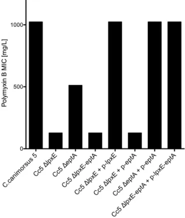
LpxE and EptA increase resistance to polymyxin B. Lipid A
modifications have been shown to not only affect endotoxicity but also to alter resistance to CAMP such as polymyxin B (10,29,39,
40). Hence, we monitored the MIC of polymyxin B for wt C.
canimorsus,⌬lpxE mutant, ⌬eptA mutant, and the
double-knock-out⌬lpxE-eptA strains. The wt C. canimorsus was highly resistant to polymyxin B, as it was still able to grow in the presence of 512 mg/liter polymyxin B (MICⱖ 1,024 mg/liter) (Fig. 4). The MIC decreased to 512 mg/liter for⌬eptA mutant bacteria and to 128 mg/liter for the⌬lpxE bacteria, showing an increased sensitivity to polymyxin B (Fig. 4). The MIC of the C. canimorsus lpxE-eptA double mutant was the same as that of the single⌬lpxE mutant (Fig. 4). The⌬lpxE strain could be complemented in trans with
lpxE, but not with eptA alone (Fig. 4). The⌬eptA strain was
com-FIG 3 Effect of lpxE or eptA deletion on endotoxicity. (A) Endotoxicity of heat-killed wild-type C. canimorsus strain 5 (Cc5),⌬lpxE, ⌬eptA, or ⌬lpxE-eptA
bacteria. The indicated multiplicity of infection (Moi) of heat-killed bacteria was assayed for TLR4-dependent NF-B activation with HekBlue human TLR4 cells. Data were combined from three independent experiments, and the error bars show the standard errors of the means. (B to D) As in panel A but the mutations were complemented in trans by the indicated plasmids (p-lpxE, plasmid bearing the lpxE gene). All mutations were shown to be nonpolar. Data were combined from three independent experiments, and the error bars show the standard errors of the means.
on February 10, 2016 by BIBLIOTHEQUE UNIVERSITAIRE
http://iai.asm.org/
plemented with eptA (Fig. 4), and the lpxE-eptA deletion mutant was complemented with lpxE-eptA (Fig. 4). We conclude from these complementation experiments that the lpxE, eptA, and
lpxE-eptA mutations were nonpolar.
The P-Etn modification at position C-1 thus contributed to the low endotoxicity and polymyxin B resistance of C. canimorsus, as was shown for H. pylori (12). The identical phenotype in endotox-icity and polymyxin B sensitivity of the single⌬lpxE and the dou-ble⌬lpxE-eptA mutants suggests that the P-Etn-containing lipid A is synthesized by a two-step enzymatic process similar to that de-scribed for H. pylori (15,16). In H. pylori, lipid A also carries an
P-Etn group at position C-1, generated in the course of the LPS
biosynthesis by removal of the lipid A 1-phosphate by LpxE, fol-lowed by transfer of an P-Etn residue by EptA from phosphati-dylethanolamine to the free reducing end of GlcN(I), where de-phosphorylation is necessary for substitution of 1-phosphate with
P-Etn (12,15,16). The nonpolar deletion of lpxE in C. canimorsus does not prevent the synthesis of EptA but likely leads to a lipid A with a 1-phosphate group, which would explain the high endotox-icity observed for this strain. Therefore, as in H. pylori, the C.
canimorsus EptA seems to accept only the free reducing end of the
lipid A backbone generated by the activity of LpxE as a substrate. The⌬lpxE mutation had a more severe effect than the ⌬eptA mutation, both with respect to endotoxicity and polymyxin B sen-sitivity. The difference between the two mutants can be explained by the fact that EptA adds a negative charge and a positive charge, whereas LpxE only removes a negative charge. In the two-step
mechanism, the⌬lpxE mutation would lead to an increase of a neg-ative charge (the unsubstituted 1-phosphate) compared to the wt, while the⌬eptA mutation would result in a free reducing end of lipid A compared to the P-Etn in the wt. As net negative charges are im-portant for interaction with CAMP as well as with TLR4/MD-2 (41), one would expect⌬lpxE to affect endotoxicity and CAMP sensitivity more than⌬eptA, which we found. This again supports the two-step enzymatic process of formation of the 1 P-Etn.
It is noteworthy that one would expect the mutation of eptA not to affect any charge-dependent mechanisms, as no net charge change is expected. C. canimorsus ⌬eptA bacteria showed in-creased endotoxicity and dein-creased CAMP resistance compared to bacteria with wt lipid A, while the lipid A variant predicted for a⌬eptA deletion mutant is lacking both the 1 and 4=-phosphate in the lipid A backbone. The C. canimorsus 1-dephospho lipid A in a ⌬eptA mutant is not expected to be endotoxic at all, as this lipid A species lacks both phosphates, and thus, the negative charges that are important for endotoxicity (41). Still the⌬eptA mutation re-sulted in a small change in polymyxin B sensitivity and a more pronounced change in endotoxicity. This hints at a heterogeneous lipid A population in the⌬eptA strain, which could result from a restricted activity of LpxE. Assuming a nonstoichiometric activity of LpxE in the⌬eptA strain, both the lipid A containing a free reducing end as well as the 1-phosphate at GlcN(I) should be present. In this case, the 1-phospho lipid A variant could account for the increase in endotoxicity, while its reduced amounts com-pared to the⌬lpxE mutant would explain the higher endotoxicity of the⌬lpxE deletion mutant over the ⌬eptA deletion mutant strain. It might thus be that the accumulation of
1-dephosphoryl-TABLE 3 MS analysis and interpretation of lipid A variants in wt and
lpxE, eptA, and lpxE-eptA deletion mutant strains
Component or mode and parameter Valuea for the component or parameter in the wt
Valueafor the component or
parameter in the following mutant:
⌬lpxE ⌬eptA ⌬lpxE-eptA
Components GlcN 1 1 1 1 GlcN3N 1 1 1 1 P 1 1 0 1 Etn 1 0 0 0 i15:0 1 1 1 1 i15:0(3-OH) 1 1 1 1 16:0(3-OH) 1 1 1 1 i17:0(3-OH) 1 1 1 1 Negative-ion mode
Calculated m/z for the [M-H⫹] ion
1,716.3 1,673.3 1,575.3 1,673.3
Found m/z for the [M-H⫹] ion 1,717 1,674 1,674b 1,674 Positive-ion mode Calculated m/z 1,764.3c 1,720.2c 1,603.3d 1,720.2c Found m/z 1,764c 1,722c 1,604d 1,722c a
0 indicates that the component is absent, and 1 indicates that the component is present once.
b
Ion [M-H⫹] detected in the negative-ion mode of lipid A from the⌬eptA mutant was raised from incomplete dephosphorylated lipid A. The major and representative lipid A molecule of this mutant lacks any charged group, and therefore, its pseudomolecular ion [M⫹Na⫹] could be analyzed only in the positive-ion mode.
c
Value for the [M-H⫹⫹2Na⫹] ion.
dValue for the [M⫹Na⫹] ion. FIG 4 Effect of lpxE or eptA deletion on resistance to polymyxin B. The MICs
of polymyxin B for wild-type C. canimorsus strain 5 (Cc5),⌬lpxE, ⌬eptA, or ⌬lpxE-eptA and complemented mutants are shown. Data were combined from three or four independent experiments, and the measured MICs were always identical.
on February 10, 2016 by BIBLIOTHEQUE UNIVERSITAIRE
http://iai.asm.org/
ated lipid A exerts a feedback regulatory effect on the activity of LpxE, preventing full dephosphorylation in the absence of EptA. The fraction of 1-phospho lipid A would thus increase, which would then be responsible for the observed increase in endotox-icity and sensitivity to polymyxin B.
MS-based structural analysis of eptA, lpxE, and lpxE-eptA de-letion mutants. In order to validate the enzymatic activities
pro-posed for LpxE and EptA and the predicted two-step enzymatic mechanism, we performed MS-based structural analysis of iso-lated lipid A species of the corresponding deletion mutants. One of the predicted lipid A structures, the 4=- and 1-hydroxy lipid A in ⌬eptA deletion mutant is devoid of any negative charge and thus not accessible to be analyzed in the negative-ion mode. Therefore, negative- and positive-ion mode MS was run to determine all lipid A variants expected based on the genetic analysis and endotoxic activity, respectively.
In the negative-ion mode, MS analysis confirmed the wt lipid A
(calculated m/z, 1,716.3; found m/z, 1,717) (Table 3andFig. 5). Observed mass differences of 14 m/z units (m/z of 1,731 or 1,703) were assigned to acyl chain heterogeneity. For all samples analyzed by MS, such peak “clusters” differing by ⌬14 m/z units were found, suggesting that acyl chain heterogeneity was independent of lpxE or eptA mutagenesis. This is in agreement with our previ-ous data on wt C. canimorsus lipid A (19,34).
In the negative-ion mode, all deletion mutant strains (⌬eptA, ⌬lpxE, and ⌬lpxE-eptA) showed a main peak at m/z of 1,674 (Table 3andFig. 5). The 1-phospho lipid A variant pre-dicted for⌬lpxE and ⌬lpxE-eptA mutant strains has a calcu-lated m/z of 1,673.3. Hence, ⌬eptA, ⌬lpxE, and ⌬lpxE-eptA deletion mutant strains feature 1-phospho lipid A. While this is the variant expected to occur for⌬lpxE and ⌬lpxE-eptA strains, ⌬eptA had been predicted to lack the 1-phosphate, thus having a free reducing end for GlcN(I) in the lipid A backbone (calcu-lated m/z, 1,575.3). However, due to the lack of a negative
FIG 5 Mass spectrometric analysis of lipid A of the indicated strains as analyzed by MALDI-TOF MS in the negative-ion mode.
on February 10, 2016 by BIBLIOTHEQUE UNIVERSITAIRE
http://iai.asm.org/
charged group, this dephospho lipid A variant cannot be ac-cessed by MS analysis in the negative-ion mode. Nevertheless, the detection of 1-phospho lipid A as well in the⌬eptA deletion mutant strain is in perfect agreement with the intermediary phenotype observed in endotoxicity and CAMP resistance (Fig. 3and4) and the proposed nonstoichiometric activity of LpxE in the⌬eptA strain.
In order to further confirm the postulated enzymatic mech-anisms, we performed negative-ion mode MS analysis also on complemented mutants (see Fig. S2 in the supplemental mate-rial). The⌬lpxE strain could be complemented in trans with
lpxE, as we confirmed the wt lipid A for the⌬lpxE strain
com-plemented with plasmid bearing lpxE (p-lpxE) (calculated m/z, 1,716.3; found m/z, 1,717). The⌬lpxE strain could not be
com-plemented in trans with eptA alone (Fig. S2), and the resulting strain showed a main peak at m/z of 1,674 (Fig. S2), matching the 1-phospho lipid A variant predicted for the⌬lpxE strain (calculated m/z, 1,673.3). The⌬eptA strain was complemented with eptA (Fig. S2), and the lpxE-eptA deletion mutant was complemented with lpxE-eptA (Fig. S2), as in both cases the wt lipid A for these strains was found (calculated m/z, 1,716.3; found m/z, 1,717).
Additional peaks measured at an m/z of 1,755 or 1,769 for the⌬lpxE ⫹ p-lpxE and the ⌬eptA ⫹ p-eptA strain are attrib-uted to a minor lipid A variant with two phosphates present and possibly with a classical GlcN-GlcN backbone, known to be present in C. canimorsus 5 (calculated m/z, 1,755.209; peak shift of ⌬14 m/z units due to acyl chain heterogeneity) (19). The two
phos-FIG 6 Mass spectrometric analysis of lipid A of the indicated strains as analyzed by MALDI-TOF MS in the positive-ion mode.
on February 10, 2016 by BIBLIOTHEQUE UNIVERSITAIRE
http://iai.asm.org/
phates might either be present as 1-phospho 4=-phospho lipid A or as 1-pyrophosphate lipid A. On the basis of the 1-pyrophosphate species detected in various species (42), we hypothesize that the peaks mea-sured at an m/z of 1,755 or 1,769 correspond to a 1-pyrophosphate variant with a classical E. coli type GlcN-GlcN lipid A backbone. The peak measured at an m/z of 1,725 for the⌬lpxE ⫹ p-eptA strain is similarly attributed to a bisphosphorylated species in combination with an exchange of 17:0(3-OH) by 15:0(3-OH) (calculated m/z, 1,725.186). The detection of these peaks exclusively in complemented mutants and the resulting changes in acyl chain preference and back-bone structure, however, should be investigated further.
We conclude from these complementation experiments that the eptA and lpxE mutations were nonpolar and that in the com-plemented mutant strains,⌬lpxE ⫹ p-lpxE, ⌬eptA ⫹ p-eptA, and ⌬lpxE-eptA ⫹ p-lpxE-eptA strains, the wt lipid A is reconstituted. In the positive-ion mode, MS analysis confirmed the wt lipid A (calculated m/z, 1,764.3; found m/z, 1,764) (Table 3andFig. 6). Again peak “clusters” differing by⌬14 m/z units were found for all samples. The 1-phospho lipid A variant has a calculated m/z of 1,720.2 [M-H⫹⫹2Na⫹] in the positive-ion mode. The main peaks for⌬lpxE and ⌬lpxE-eptA deletion mutants were both found at an m/z value of 1,722. This peak was absent in the ⌬eptA deletion mutant strain. The ⌬eptA deletion mutant’s main peak present at an m/z of 1,604 or 1,618 (peak shift of 14
m/z units due to acyl chain heterogeneity) corresponded to a
free hydroxy group that forms a reducing end in the lipid A backbone and has been calculated to an m/z of 1,603.3 or 1,617.3. It is noteworthy that a peak at an m/z of 1,601 is found in all samples in the positive-ion mode (and the corresponding peak shifted by 14 m/z units). The presence of 1-dephospho lipid A variants even in those mutants which contain the 1-phospho group has been assigned as artifacts well-known to appear from the wt strains due to the acid hydrolysis conditions necessary to liberate the lipid A from the phosphorylated Kdo found in the core of C. canimorsus LPS (19,34). These condi-tions obviously lead to a partial dephosphorylation at position 1 of lipid A. The main peak from the 1-hydroxy lipid A cluster for⌬lpxE and ⌬lpxE-eptA deletion mutants was found at an
m/z of 1,601 and not at an m/z of 1,604/1,618 as predicted. We
assume that the peak at 1,604 m/z found in the⌬eptA deletion mutant is identical to peaks at m/z of 1,601 found in⌬lpxE and ⌬lpxE-eptA deletion mutants.
According to the proposed model, the major and representa-tive lipid A molecule of the ⌬eptA mutant lacks any charged group, and therefore, its pseudomolecular ion [M⫹Na⫹] can be analyzed only in the positive-ion mode. In agreement with this, in the positive-ion (but not the negative-ion) mode of⌬eptA lipid A, the pseudomolecular ion [M⫹Na⫹] was detected. The ion
[M-H⫹] detected in the negative-ion mode of ⌬eptA lipid A was likely raised from incomplete dephosphorylated lipid A, resulting in 1-phospho lipid A. Notably, the [M-H⫹] ion was not detected for the⌬eptA mutant in the positive-ion mode, possibly reflecting its small proportion.
We further performed positive-ion mode MS analysis on com-plemented mutants (see Fig. S3 in the supplemental material). The ⌬lpxE strain could be complemented in trans with lpxE, as we confirmed the wt lipid A for this strain (calculated m/z, 1,764.3; found m/z, 1,764). The⌬lpxE strain could not be complemented in trans with eptA alone (Fig. S3), and the resulting strain showed a main peak at an m/z of 1,722 (Fig. S3), matching the 1-phospho
lipid A variant predicted for⌬lpxE (calculated m/z, 1,720.2). The ⌬eptA strain was complemented with eptA (Fig. S3), and the
lpxE-eptA deletion mutant was complemented with lpxE-lpxE-eptA (Fig. S3),
as in both cases, the wt lipid A for these strains was found (calcu-lated m/z, 1,764.3; found m/z, 1,764).
Additional peaks measured at m/z values of 1,803 and 1,817 for the⌬lpxE ⫹ p-lpxE and the ⌬eptA ⫹ p-eptA strain are attributed to a minor bisphosphorylated lipid A backbone variant, probably with a E. coli type classical GlcN-GlcN backbone known also to be present in small amounts in C. canimorsus 5 (calculated m/z, 1,802.196; peak shift of 14 m/z units due to acyl chain heterogene-ity) (19) and that correlates with additional peaks observed in the negative-ion mode MS analysis of these strains.
We conclude from these complementation experiments that the eptA and lpxE mutations were nonpolar and that in the com-plemented mutants,⌬lpxE ⫹ p-lpxE, ⌬eptA ⫹ p-eptA, and
⌬lpxE-eptA⫹ p-lpxE-eptA, the wt lipid A is reconstituted.
Overall, 1-phospho lipid A was validated as the main lipid A variant in⌬lpxE and ⌬lpxE-eptA deletion mutants. This confirms that LpxE acts as a lipid A 1-phosphatase and further corroborates the two-step enzymatic mechanism in which EptA is active only after LpxE-dependent removal of the 1-phosphate on lipid A. In agree-ment with endotoxicity and CAMP resistance, both 1-phospho and 1-dephospho lipid A variants were found present in the⌬eptA dele-tion mutant. This validates the funcdele-tion of EptA as lipid A phosphoe-thanolamine transferase and again supposes a two-step enzymatic activity in which LpxE can dephosphorylate lipid A even in the ab-sence of EptA. However, LpxE seems not to dephosphorylate every lipid A in the absence of EptA, which is reflected by the 1-phospho lipid A species identified in the⌬eptA deletion mutant.
The lipid A modification described in this work clearly repre-sents a virulence factor, since it dramatically reduces recognition and killing by the host’s innate immune system. However, human infections are rare events and dead ends for C. canimorsus. Thus, we can envision that the lipid A modification most likely evolved as a factor favoring the adaptation of C. canimorsus to its natural niche, the dog’s mouth.
ACKNOWLEDGMENTS
This work was supported by the Swiss National Science Foundation (grant 3100A0-128659) and the European Research Council (ERC) (AdG grant 2011-293605-CAPCAN) to G.R.C. F.R. is a postdoctoral research fellow (chargé de recherche) of the Belgian “Fonds National de la Recherche Scientifique” (FNRS). S.J.I. was supported by the Werner-Siemens Foun-dation.
The funders had no role in study design, data collection and interpre-tation, or the decision to submit the work for publication.
FUNDING INFORMATION
Werner Siemens Foundation provided funding to Simon Josef Ittig. EC | European Research Council (ERC) provided funding to Guy R. Cornelis under grant number 2011-293605-CAPCAN. Schweizerischer National-fonds zur Förderung der Wissenschaftlichen Forschung (SNF) provided funding to Guy R. Cornelis under grant number 3100A0-128659. Fonds De La Recherche Scientifique - FNRS (F.R.S. - FNRS) provided funding to Francesco Renzi.
The funders had no role in study design, data collection and interpreta-tion, or the decision to submit the work for publication.
REFERENCES
1. Coats SR, Jones JW, Do CT, Braham PH, Bainbridge BW, To TT, Goodlett DR, Ernst RK, Darveau RP. 2009. Human Toll-like receptor 4
on February 10, 2016 by BIBLIOTHEQUE UNIVERSITAIRE
http://iai.asm.org/
responses to P. gingivalis are regulated by lipid A 1- and 4=-phosphatase activities. Cell Microbiol 11:1587–1599.http://dx.doi.org/10.1111/j.1462 -5822.2009.01349.x.
2. Dixon DR, Darveau RP. 2005. Lipopolysaccharide heterogeneity: innate host responses to bacterial modification of lipid A structure. J Dent Res
84:584 –595.http://dx.doi.org/10.1177/154405910508400702.
3. Hajjar AM, Ernst RK, Tsai JH, Wilson CB, Miller SI. 2002. Human Toll-like receptor 4 recognizes host-specific LPS modifications. Nat Im-munol 3:354 –359.http://dx.doi.org/10.1038/ni777.
4. Mata-Haro V, Cekic C, Martin M, Chilton PM, Casella CR, Mitchell TC. 2007. The vaccine adjuvant monophosphoryl lipid A as a TRIF-biased agonist of TLR4. Science 316:1628 –1632. http://dx.doi.org/10.1126 /science.1138963.
5. Price NP, Jeyaretnam B, Carlson RW, Kadrmas JL, Raetz CR, Brozek KA. 1995. Lipid A biosynthesis in Rhizobium leguminosarum: role of a 2-keto-3-deoxyoctulosonate-activated 4= phosphatase. Proc Natl Acad Sci U S A 92:7352–7356.http://dx.doi.org/10.1073/pnas.92.16.7352. 6. Wang X, Karbaz MJ, McGrath SC, Cotter RJ, Raetz CRH. 2004. MsbA
transporter-dependent lipid A 1-dephosphorylation on the periplasmic surface of the inner membrane: topography of Francisella novicida LpxE expressed in Escherichia coli. J Biol Chem 279:49470 – 49478.http://dx.doi .org/10.1074/jbc.M409078200.
7. Wang X, McGrath SC, Cotter RJ, Raetz CRH. 2006. Expression cloning and periplasmic orientation of the Francisella novicida lipid A 4=-phosphatase LpxF. J Biol Chem 281:9321–9330.http://dx.doi.org/10.1074 /jbc.M600435200.
8. Curtis MA, Percival RS, Devine D, Darveau RP, Coats SR, Rangarajan M, Tarelli E, Marsh PD. 2011. Temperature-dependent modulation of
Porphyromonas gingivalis lipid A structure and interaction with the innate
host defenses. Infect Immun 79:1187–1193.http://dx.doi.org/10.1128/IAI .00900-10.
9. Herrera CM, Hankins JV, Trent MS. 2010. Activation of PmrA inhibits LpxT-dependent phosphorylation of lipid A promoting resistance to an-timicrobial peptides. Mol Microbiol 76:1444 –1460.http://dx.doi.org/10 .1111/j.1365-2958.2010.07150.x.
10. Ingram BO, Sohlenkamp C, Geiger O, Raetz CRH. 2010. Altered lipid A structures and polymyxin hypersensitivity of Rhizobium etli mutants lack-ing the LpxE and LpxF phosphatases. Biochim Biophys Acta 1801:593– 604.http://dx.doi.org/10.1016/j.bbalip.2010.02.001.
11. Wang X, Ribeiro AA, Guan Z, Abraham SN, Raetz CRH. 2007. Atten-uated virulence of Francisella mutant lacking the lipid A 4=-phosphatase. Proc Natl Acad Sci U S A 104:4136 – 4141.http://dx.doi.org/10.1073/pnas .0611606104.
12. Cullen TW, Giles DK, Wolf LN, Ecobichon C, Boneca IG, Trent MS. 2011. Helicobacter pylori versus the host: remodeling of the bacterial outer membrane is required for survival in the gastric mucosa. PLoS Pathog
7:e1002454.http://dx.doi.org/10.1371/journal.ppat.1002454.
13. Kanistanon D, Powell DA, Hajjar AM, Pelletier MR, Cohen IE, Way SS, Skerrett SJ, Wang X, Raetz CR, Ernst RK. 2012. Role of Francisella lipid A phosphate modification in virulence and long-term protective immune responses. Infect Immun 80:943–951. http://dx.doi.org/10.1128/IAI .06109-11.
14. Ingram BO, Masoudi A, Raetz CRH. 2010. Escherichia coli mutants that synthesize dephosphorylated lipid A molecules. Biochemistry (Mosc) 49: 8325– 8337.http://dx.doi.org/10.1021/bi101253s.
15. Tran AX, Karbaz MJ, Wang X, Raetz CRH, McGrath SC, Cotter RJ, Trent MS. 2004. Periplasmic cleavage and modification of the 1-phos-phate group of Helicobacter pylori lipid A. J Biol Chem 279:55780 –55791. http://dx.doi.org/10.1074/jbc.M406480200.
16. Tran AX, Whittimore JD, Wyrick PB, McGrath SC, Cotter RJ, Trent MS. 2006. The lipid A 1-phosphatase of Helicobacter pylori is required for resistance to the antimicrobial peptide polymyxin. J Bacteriol 188:4531– 4541.http://dx.doi.org/10.1128/JB.00146-06.
17. Cox AD, Wright JC, Hood DW, Moxon ER, Richards JC. 2003. Phos-phorylation of the lipid A region of meningococcal lipopolysaccharide: identification of a family of transferases that add phosphoethanolamine to lipopolysaccharide. J Bacteriol 185:3270 –3277.http://dx.doi.org/10.1128 /JB.185.11.3270-3277.2003.
18. Kim SH, Parreira VR, Bishop RE, Gyles CL. 2006. Phosphoethano-lamine substitution in the lipid A of Escherichia coli O157:H7 and its as-sociation with PmrC. Microbiology 152:657– 666.http://dx.doi.org/10 .1099/mic.0.28692-0.
19. Ittig S, Lindner B, Stenta M, Manfredi P, Zdorovenko E, Knirel YA, dal
Peraro M, Cornelis GR, Zähringer U. 2012. The lipopolysaccharide from
Capnocytophaga canimorsus reveals an unexpected role of the
core-oligosaccharide in MD-2 binding. PLoS Pathog 8:e1002667.http://dx.doi .org/10.1371/journal.ppat.1002667.
20. Bobo RA, Newton EJ. 1976. A previously undescribed gram-negative bacillus causing septicemia and meningitis. Am J Clin Pathol 65:564 –569. 21. Brenner DJ, Hollis DG, Fanning GR, Weaver RE. 1989. Capnocytophaga
canimorsus sp. nov. (formerly CDC group DF-2), a cause of septicemia
following dog bite, and C. cynodegmi sp. nov., a cause of localized wound infection following dog bite. J Clin Microbiol 27:231–235.
22. Butler T. 2015. Capnocytophaga canimorsus: an emerging cause of sepsis, meningitis, and post-splenectomy infection after dog bites. Eur J Clin Microbiol Infect Dis 34:1271–1280. http://dx.doi.org/10.1007/s10096 -015-2360-7.
23. Lion C, Escande F, Burdin JC. 1996. Capnocytophaga canimorsus infec-tions in human: review of the literature and cases report. Eur J Epidemiol
12:521–533.http://dx.doi.org/10.1007/BF00144007.
24. Pers C, Gahrn-Hansen B, Frederiksen W. 1996. Capnocytophaga
cani-morsus septicemia in Denmark, 1982-1995: review of 39 cases. Clin Infect
Dis 23:71–75.http://dx.doi.org/10.1093/clinids/23.1.71.
25. Bailie WE, Stowe EC, Schmitt AM. 1978. Aerobic bacterial flora of oral and nasal fluids of canines with reference to bacteria associated with bites. J Clin Microbiol 7:223–231.
26. Blanche P, Bloch E, Sicard D. 1998. Capnocytophaga canimorsus in the oral flora of dogs and cats. J Infect 36:134.http://dx.doi.org/10.1016 /S0163-4453(98)93918-4.
27. Mally M, Paroz C, Shin H, Meyer S, Soussoula LV, Schmiediger U, Saillen-Paroz C, Cornelis GR. 2009. Prevalence of Capnocytophaga
cani-morsus in dogs and occurrence of potential virulence factors. Microbes
Infect 11:509 –514.http://dx.doi.org/10.1016/j.micinf.2009.02.005. 28. Manfredi P, Pagni M, Cornelis GR. 2011. Complete genome sequence
of the dog commensal and human pathogen Capnocytophaga
canimor-sus strain 5. J Bacteriol 193:5558 –5559.http://dx.doi.org/10.1128/JB .05853-11.
29. Raetz CRH. 1990. Biochemistry of endotoxins. Annu Rev Biochem 59: 129 –170.http://dx.doi.org/10.1146/annurev.bi.59.070190.001021. 30. Shin H, Mally M, Kuhn M, Paroz C, Cornelis GR. 2007. Escape from
immune surveillance by Capnocytophaga canimorsus. J Infect Dis 195: 375–386.http://dx.doi.org/10.1086/510243.
31. Mally M, Cornelis GR. 2008. Genetic tools for studying Capnocytophaga
canimorsus. Appl Environ Microbiol 74:6369 – 6377.http://dx.doi.org/10 .1128/AEM.01218-08.
32. NCCLS/CLSI. 2003. Methods for dilution antimicrobial susceptibility tests for bacteria that grow aerobically: approved standard. National Com-mittee for Clinical Laboratory Standards/Clinical Laboratory and Stan-dards Institute, Wayne, PA.
33. Altschul SF, Madden TL, Schäffer AA, Zhang J, Zhang Z, Miller W, Lipman DJ. 1997. Gapped BLAST and PSI-BLAST: a new generation of protein database search programs. Nucleic Acids Res 25:3389 –3402.http: //dx.doi.org/10.1093/nar/25.17.3389.
34. Zähringer U, Ittig S, Lindner B, Moll H, Schombel U, Gisch N, Cornelis GR. 2014. NMR-based structural analysis of the complete rough-type lipopolysaccharide isolated from Capnocytophaga canimorsus. J Biol Chem 289:23963–23976.http://dx.doi.org/10.1074/jbc.M114.571489. 35. Shin H, Mally M, Meyer S, Fiechter C, Paroz C, Zähringer U, Cornelis
GR. 2009. Resistance of Capnocytophaga canimorsus to killing by human complement and polymorphonuclear leukocytes. Infect Immun 77:2262– 2271.http://dx.doi.org/10.1128/IAI.01324-08.
36. El Hamidi A, Tirsoaga A, Novikov A, Hussein A, Caroff M. 2005. Microextraction of bacterial lipid A: easy and rapid method for mass spec-trometric characterization. J Lipid Res 46:1773–1778.http://dx.doi.org/10 .1194/jlr.D500014-JLR200.
37. Rietschel ET, Kirikae T, Schade FU, Mamat U, Schmidt G, Loppnow H, Ulmer AJ, Zahringer U, Seydel U, Di Padova F, Schreier M, Brade H. 1994. Bacterial endotoxin: molecular relationships of structure to activity and function. FASEB J 8:217–225.
38. Reynolds CM, Kalb SR, Cotter RJ, Raetz CR. 2005. A phosphoethano-lamine transferase specific for the outer 3-deoxy-D-manno-octulosonic acid residue of Escherichia coli lipopolysaccharide. Identification of the eptB gene and Ca2⫹ hypersensitivity of an eptB deletion mutant. J Biol Chem 280:21202–21211.
39. Lee H, Hsu FF, Turk J, Groisman EA. 2004. The PmrA-regulated pmrC gene mediates phosphoethanolamine modification of lipid A and
on February 10, 2016 by BIBLIOTHEQUE UNIVERSITAIRE
http://iai.asm.org/
myxin resistance in Salmonella enterica. J Bacteriol 186:4124 – 4133.http: //dx.doi.org/10.1128/JB.186.13.4124-4133.2004.
40. Raetz CRH, Reynolds CM, Trent MS, Bishop RE. 2007. Lipid A modi-fication systems in Gram-negative bacteria. Annu Rev Biochem 76:295– 329.http://dx.doi.org/10.1146/annurev.biochem.76.010307.145803. 41. Park BS, Song DH, Kim HM, Choi B-S, Lee H, Lee J-O. 2009. The
structural basis of lipopolysaccharide recognition by the TLR4-MD-2 complex. Nature 458:1191–1195.http://dx.doi.org/10.1038/nature07830. 42. Jones JW, Shaffer SA, Ernst RK, Goodlett DR, Turecek F. 2008. Deter-mination of pyrophosphorylated forms of lipid A in Gram-negative bac-teria using a multivaried mass spectrometric approach. Proc Natl Acad Sci U S A 105:12742–12747.http://dx.doi.org/10.1073/pnas.0800445105.