HAL Id: tel-01980791
https://tel.archives-ouvertes.fr/tel-01980791
Submitted on 14 Jan 2019HAL is a multi-disciplinary open access archive for the deposit and dissemination of sci-entific research documents, whether they are pub-lished or not. The documents may come from teaching and research institutions in France or abroad, or from public or private research centers.
L’archive ouverte pluridisciplinaire HAL, est destinée au dépôt et à la diffusion de documents scientifiques de niveau recherche, publiés ou non, émanant des établissements d’enseignement et de recherche français ou étrangers, des laboratoires publics ou privés.
Recombinant C-type Lectin Receptors production and
selective ligand identification : new tools towards
immune system tailoring
Silvia Achilli
To cite this version:
Silvia Achilli. Recombinant C-type Lectin Receptors production and selective ligand identification : new tools towards immune system tailoring. Biomolecules [q-bio.BM]. Université Grenoble Alpes, 2018. English. �NNT : 2018GREAV014�. �tel-01980791�
5
Acknowledgements
First of all and foremost, my profound gratitude goes to Pr. Franck Fieschi and Dr. Corinne Vivès-Deniaud. They welcomed me in their team and gave me extremely valuable guidance and support. Also a big thank for translating the abstract of this thesis in French language.
My immense gratitude goes to Dr. Michel Thépaut. He showed me all the biochemistry tips and helped me immensely with the crystallography section.
I would like to express my deepest gratitude to the reviewers, Prof. Francesco Peri and Dr. Yann Guerardel, who kindly accepted to evaluate my dissertation and examine it.
My immense appreciation goes to Dr. Anne Imberty and Pr. Bernd Lepenies for being member of the jury and contribute to the examination of my work.
I own a great acknowledgement to the Biacore platform, particularly to Dr. Isabelle Bally and Jean Baptiste Reiser, and to the PAOL platform of the institute, specially to Pr. Christine Ebel and Aline le Roy for helping and teaching me to perform experiments.
ITC data were acquired at CIBB institute in Grenoble and Dr. Caroline Mass has my sincere gratitude for her help and advice.
A lot of thanks to Dr. Romain Vives and Pr. Olivier Renaudet for being part of the jury for my CST: they evaluated my work and guided me for two years.
I am indebted to all our collaborators: Dr. Niels Reichardt and Dr. Sonia Serna, who hosted me for two secondments and taught me how to perform glycan array screening, Pr. Bernd Lepenies and (not yet Dr.) Joao Monteiro, who hosted me for one secondment and showed me how to perform binding assays with cells, Dr. Ludovic Landemarre and (not yet Dr.) Blanka Didak for showing me their LECprofile assay, Pr. Anna Bernardi and Dr Laura Medve for all the glycomimetics provided and all the valuable help that you gave me. A big thank to Dr. Jeroen Codee and his PhD student Tim Hogervorst for their work on the multivalent glycoclusters synthesis and Pr. Yvette van Kooyk and her PhD student Eveline Li for the biological assays. I am deeply grateful for the IMMUNOSHAPE network for the great workshops and the memorable moments spent together.
Finally, I must acknowledge the financial support given by the Marie Sklodowska-Curie grant agreement No 642870 (ETN-Immunoshape), without which my work would not have been possible.
7
Ringraziamenti
Molte altre persone hanno contribuito, chi più chi meno, a questo mio successo. Questa parte dei ringraziamenti sarà scritta nella mia lingua madre.
Dedico questo a lavoro a:
I miei genitori e mia nonna Ceci i quali, pur non capendo assolutamente niente di quello che faccio, mi hanno guardato con orgoglio durante tutta la mia difesa.
Mio fratello Stefano, che mi considera più intelligente di quanto effettivamente io non sia. Gloria, la mia più cara amica e fervida sostenitrice.
Erika, che ha viaggiato per più di 7898 chilometri pur di starmi accanto. Clara, Adrica e Cinzia, the italian team in Grenoble.
Laura, Charles, Romain, Annelise, François, Muge e Sima, miei colleghi compagni di avventure e pause café.
Dulcis in fundo, Loreto. Mi ricorderò sempre di quando, alle 2 di notte, mi hai aiutato a rileggere questo manuscritto al contrario, dalla fine all’inizio, per trovare eventuali errori. Nessuna parola potrà mai esprimere tutta la gratitudine che meriti. Sei il mio porto sicuro nei momenti di sconforto.
11
Index
Introduction
331. The Immune System
351.1 Introduction
351.2 Innate Immune System
361.2.1 Dendritic cells and signals for adaptive immunity activation 37
1.2.2 Pattern recognition receptors 38
2. C-type lectin receptors
412.1 Introduction
412.2 C type lectin domains and glycan recognition: structural aspects
422.2.1 CLR signalling
452.2.2 CLR and TLR crosstalk
472.3 CLRs considered in the study
492.3.1 Blood Dendritic Antigen 2 (BDCA2)
492.3.2 DC-SIGN and DC-SIGNR (L-SIGN)
502.3.3 DC-Associated C-Type Lectin 1 (dectin-1)
522.3.4 Dendritic cell associated C type lectin 2 (Dectin-2)
532.3.5 Langerin
542.3.6 Liver and lymph node sinusoidal endothelial cell C-type lectin (LSECtin)
562.3.7 Macrophages C-type lectin (MCL)
572.3.8 Macrophage inducible Ca
2+- dependent lectin (Mincle)
593. Glycobiology
653.1 Introduction
653.2 Design of Mimetic
6812
3.2.2 Non-glycomimetics
703.3 Screening technique
713.3.1 Lectin microarray
713.3.1 Glycan/glycomimetic microarray
72 3.3.2.1 TETRALEC strategy 733.4 Multivalent Ligands: Targeting CLRs
744. Applications
784.1 Diagnosis
784.2 Imaging
804.3 Targeting
825. AIM OF THE THESIS
85Materials & Methods
916. Principles
936.1 Methods for protein characterization
936.1.1 Circular dichroism (CD)
936.1.2 Size Exclusion Chromatography Multi Angle Laser Light Scattering
SEC-MALLS (PAOL platform)
94
6.2 Methods for characterization of protein-ligand interaction
956.2.1 Lectin array (LectPROFILE)
956.2.2 Glycan array
966.2.3 Surface Plasmon Resonance (SPR)
966.2.4 Isothermal Titration Calorimetry (ITC)
1017. Methods
1037.1 Production of recombinant C-type lectin constructs
10313
7.1.2 Cloning of LSECtin His-CRD
1037.1.3 Preparation of bacterial preculture
1057.1.4 Preparation of bacterial culture samples for SDS-PAGE
1057.1.5 DC-SIGN and Langerin over-expression and purification
1057.1.6 Protein labelling
1057.1.7 Over-expression and inclusion body preparation of all CLR constructs 105
7.1.7.1 Refolding, purification and labelling of DC-SIGNR ECD 106
7.1.7.2 Refolding, purification and labelling of Dectin-2 Strep-ECD 107
7.1.7.3 Refolding, purification and labelling of Mincle His-ECD 107
7.1.7.4 Refolding and purification of His-GGG-CRD constructs 108
7.2 Biochemical and biophysical protein characterization and ligand
analysis
111
7.2.1 Protein samples for SDS-PAGE.
1117.2.2 Protein concentration determination
1117.2.3 Sample preparation for circular dichroism
1127.2.4 Sample preparation for SEC-MALS (PAOL platform)
1137.2.5 Sample preparation for lectin array analysis
1137.2.6 Sample preparation for glycan array analysis
1137.2.7 Sample preparation for flow cytometry
1147.2.8 Sample preparation for ITC
1147.2.9 SPR surface preparation for inhibition test
1147.2.10 SPR surface preparation for direct interaction test
115Results and Discussion
1198 Recombinant CLR Production and Functional Test
1218.1 Cloning
12214
8.2 Strategies for Recombinant Protein Expression
1268.2.1 Expression as soluble folded CLR 126
8.2.2 Expression as Inclusion Bodies 131
8.2.2.1 ECD constructs 131
8.2.2.1.1 DC-SIGNR ECD over-expression and purification results 132 8.2.2.1.2 Dectin-2 Strep-ECD over-expression and purification results 133 8.2.2.1.3 Mincle His-ECD over-expression and purification results 135
8.2.2.2 CRD constructs 137
8.2.2.2.1 DC-SIGNR His-CRD 138
8.2.2.2.2 BDCA2 and LSECtin His-CRD 140
8.2.2.2.3 Dectin-1, Dectin-2, Mincle, MCL His-CRD 143
8.3 TETRALEC, Artificial Tetrameric Lectins: a Tool to Screen Ligand and
Pathogen Interactions
151
9 Screening: identification of selective ligands towards human CLRs
1779.1 Glycan array
1779.1.1 Chemoenzymatic Synthesis of N-glycan Positional Isomers and Evidence for Branch Selective Binding by Monoclonal Antibodies and Human C-type Lectins Receptors
179
9.1.2 Other glycan array screening 195
9.2 Glycomimetic array
1979.2.1 On-chip screening of a glycomimetic library with C-type lectins reveals structural features responsible for preferential binding of dectin-2 over DC-SIGN/R and langerin
198
9.2.2 Other glycomimetic array screening 215
10 Characterization of new glycomimetics specific to DC-SIGN
21710.1 Robustness enhancement
21910.1.1 Facile access to pseudo-thio-1,2-dimannoside, a new glycomimetic DC-SIGN antagonist.
219
10.1.2 Additional data non-presented in the article: enzymatic assay 227
15
10.2 Development of glycomimetics selective towards DC-SIGN: the
“ammonium binding pocket” strategy
230
10.2.1 Ammonium binding pocket: amino derivatives 232
10.2.2 Ammonium binding pocket: triazole derivatives 234
11 Design of multivalent mannosylated ligands for CLR targeting
24711.1 Carbohydrate synthesis (Leiden)
24811.2 ELISA assays (Amsterdam)
24911.3 SPR Experiments (Grenoble)
25111.4 FACS Assays (Amsterdam)
255Conclusions and Perspectives
25912.1 Remarks on the production of non-commercially available lectins
26112.2 Exploiting glycan/glycomimetic array screenings
26512.3 Enhancement and development of new glycomimetics specific to
DC-SIGN
267
12.4 Study of mannose clusters targeting CLRs as a future tool to
trigger anticancer immune response
271
12.5 Final and general conclusions
273Bibliography
277Annexes
291Annexes Chapter 8
293Supporting Information Paper 1
325Annexes Chapter 9
331Supporting Information Paper 2
333Supporting Information Paper 3
35516
Supporting Information Paper 4
40717
List of Figures:
Fig.1 The immune system 35
Fig.2 Innate and Adaptive Immunity s 36
Fig.3 Activation of the adaptive immunity 38
Fig.4 PAMPs and DAMPs 39
Fig.5 The four different families of PRRs 39
Fig.6 CLRs and immunity 40
Fig.7 Examples of four animal lectin families 41
Fig.8 Schematic representation of a tetrameric CLR 42
Fig.9 Carbohydrate recognition domain 43
Fig.10 EPN and QPD motifs 43
Fig.11 C-type lectin subfamilies 44
Fig.12 Overview of the four motifs for signalisation 45
Fig.13 Dectin-2 and DCIR signalling 46
Fig.14 Cross talk between CLR and TLR 47
Fig.15 DC-SIGN and TLR4 during Mycobacterium infection 48
Fig.16 Portion of BDCA2 CRD structure 49
Fig.17 Portion of DC-SIGN and DC-SIGNR structure 50 Fig.18 Comparison of the CRD from DC-SIGN and DC-SIGNR, 51
Fig.19 The placenta, DC-SIGN. DC-SIGNR and HIV 52
Fig.20 Murine dectin-1 structure 53
Fig.21 Portion of Dectin-2 structure 54
Fig.22 Langerin structure 55
Fig.23 Birbeck granules 55
Fig.24 Genomic organization of DC-SIGNR, DC-SIGN and LSECtin 56
Fig.25 LSECtin 57
Fig.26 MCL and DC-SIGNR 58
Fig.27 Mincle expression 58
Fig.28 Portion of mincle structure 59
Fig.29 Mincle 60
Fig.30 Graphical overview of the nine CLRs investigated during my PhD 60 Fig.31 Potential information on content of DNA, RNA, protein and glycan 65
Fig.32 N-glycans 66
Fig.33 Linkage points for oligomer formatio 67
Fig.34 Example of calcium ion coordination 67
Fig.35 Cracking the glycocode 68
Fig.36 Schematic glycomimetic design 69
18
Fig.38 Glycomimetic against langerin 70
Fig.39 Antagonists developed in Laura Kiessling group 71
Fig.40 Glycan array 73
Fig.41 Multivalency 74
Fig.42 Binding mode 75
Fig.43 Multivalent ligands 76
Fig.44 Examples of multivalent inhibitors against DC-SIGN 77
Fig.45 Mutivalent ligand as vaccine 77
Fig.46 Exploitation of lectin-carbohydrate interaction in medical biology 78
Fig.47 Application of glycan array. 80
Fig.48 Example of “Smart” probe 81
Fig.49 Example of imaging 81
Fig.50 Targeting 82
Fig.51 TACAs 83
Fig.52 Vaccination using CLR targeting 84
Fig.53 The principal architecture of IMMUNOSHAPE ITN consortium and group involvement. 86
Fig.54 IMMUNOSHAPE network 88
Fig.55 Circular dichroism 93
Fig.56 SEC-MALLS 94
Fig.57 LectPROFILE schematic representation. 95
Fig.58 Glycan array glass slide 96
Fig.59 SPR principle 97
Fig.60 Plasmon resonance effect 97
Fig.61 Analyte interaction and bulk effet 98
Fig.62 Design of competition/inhibition SPR assay 99 Fig.63 SPR competition/inhibition sensorgram and curve 100
Fig.64 Oriented surface approach 101
Fig.65 Calorimetry principle 102
Fig.66 Bacterial cell wall section and location of the periplasm 121 Fig.67 Protein secretion pathway and signal peptides 122
Fig.68 Cloning strategies. 123
Fig.69 LSECtin His-CRD cloning 125
Fig.70 pTUM4 helper plasmid 126
Fig.71 Dectin-2 ompA Strep-ECD expression 127
Fig.72 Dectin-2 Strep-ECD expression 128
Fig.73 Dectin-2 pelB His-ECD expression 128
Fig.74 Dectin-2 pelB His-ECD Western Blot. 129
19
Fig.76 DC-SIGNR ECD expression 132
Fig.77 DC-SIGNR ECD purification 133
Fig.78 Dectin-2 Strep-ECD expression 133
Fig.79 Dectin-2 Strep-ECD purification 134
Fig.80 Dectin-2 Strep-ECD LECprofile analysis 135
Fig.81 Expression of mincle His-ECD 135
Fig.82 Mincle His-ECD purification 136
Fig.83 GAS recognition by Mincle His-ECD. 137
Fig.84 DC-SIGNR His-CRD expression and purification 138 Fig.85 DC-SIGNR His-CRD His tag cleavage and purification 139
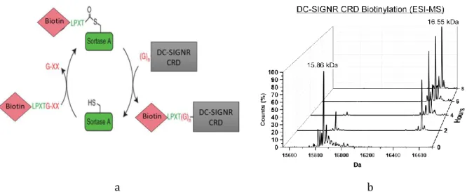
Fig.86 DC-SIGNR CRD biotinylation 139
Fig.87 BDCA2 and LSECtin His-CRD expression 140
Fig.88 BDCA2 biotynilation 141
Fig.89 BDCA2 His-CRD LECtprofile analysis 141
Fig.90 LSECtin His-CRD purification 142
Fig.91 LSECtin His-CRD LectPROFILE analysis 143
Fig.92 Dectin-1, Dectin-2, Mincle and MCL HIS-CRD expressions 143
Fig.93 Dectin-1 purification 144
Fig.94 GAS recognition by dectin-1 His-CRD 144
Fig.95 Mincle His-CRD purification 145
Fig.96 Dectin-2 His-CRD purification 146
Fig.97 Comparison between Dectin-2 His-CRD and Dectin-2 Strep-ECD LectPROFILE analysis 146
Fig.98 Dectin-2 His-CRD CD spectrum. 147
Fig.99 MCL His-CRD purification 147
Fig.100 MCL His-CRD CD spectrum. 148
Fig.101 MCL His-CRD His tag cleavage and purification 148
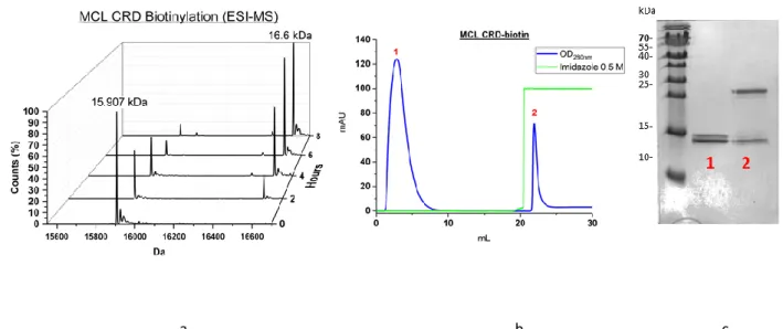
Fig.102 MCL CRD-biotinylation 149
Fig.103 Example of interaction detection by Agila Scanner 177
Fig.104 Glycan array 178
Fig.105 Glycan microarray incubation 196
Fig.106 Glycomimetic array 197
Fig.107 Other glycomimetic arrays 215
Fig.108 First and second generations of glycomimetics against DC-SIGN 217
Fig.109 6NH2-Man030 structure 218
Fig.110 psDi/DC-SIGN CRD X-ray structure 218
Fig.111 psDi and thio-psDi structures 219
Fig.112 thio-psdi enzymatic reaction 227
20
Fig.114 ID-246-4, ID-246-5, ID-246-11, ID-246-12 and JCH-423 competition assay against DC-SIGN 229 Fig.115 Best candidates from computational screening 230
Fig.116 The “ammonium binding pocket” 230
Fig.117 Synthetic strategies 231
Fig.118 Synthetic strategy and overall structure of amino derivatives 232
Fig.119 Amino derivatives structures. 232
Fig.120 IC50 comparison of the amino derivatives 233
Fig.121 Triazole derivatives strategy 234
Fig.122 Man062 competition assay 234
Fig.123 Man065 and Man064 structures 235
Fig.124 Man062, Man065, Man064 and psDI IC50 comparison. 235
Fig.125 Man066-67-68 structures. 236
Fig.126 Man062 corresponding methylated glycomimetics inhibition assay 236 Fig.127 List of triazole derivatives IC50 values and corresponding structures. 237
Fig.128 Man069 structure. 238
Fig.129 Man062, Man065 and Man089 competition assay 238
Fig.130 Examples of sensorgrams 239
Fig.131 Titrations of glycomimetic Man069 to DC-SIGN 240
Fig.132 Man069 direct interaction assay 241
Fig.133 One example of obtained crystal and diffraction pattern. 242 Fig.134 Crystal packing and evidence for ligand presence. 242
Fig.135 Mano69/DC-SIGN solved X-ray structure 244
Fig.136 Man069 canonical binding 244
Fig.137 Man069 non-canonical binding site 245
Fig.138 Cluster synthesis 248
Fig.139 20 mannosylated compounds 249
Fig.140 ELISA results 250
Fig.141 Inhibition curves 251
Fig.142 Sensorgrams obtained from B6 interaction 253
Fig.143 Kdapp values 254
Fig.144 Comparison of the Kdapp values 255
Fig.145 Interaction between biotinylated glycoclusters 255 Fig.146 BDCA2, dectin-1, MCL and dectin-2 His-ECD expressions 262
Fig.147 X-ray structure of dectin-2 262
Fig.148 The six asymmetric glycans and the two non-branched corresponding glycans 265 Fig.149 Structure of mannose based glycomimetics that specifically recognise dectin-2 266
Fig.150 Structures and IC50 comparison 267
21
List of Tables:
Table.1 Overview of the nine CLRs studied during my PhD 64 Table.2 List of lectins used in cancer biomarker research 79 Table.3 Digest assays of pUC57 LSECtin vector and pET30b vector. 103 Table.4 Preparation of NdeI/HindIII digestion mixtures for the test of positive DNA ligation. 104 Table.5 Refolding concentration, buffer for refolding and dialysis and protein yield/L culture for
the His-CRD considered.
109
Table.6 Required volumes for His tag cleavage of BDCA2, DC-SIGNR and MCL CRDs 110 Table.7 Required volumes for sortase reaction on BDCA2, DC-SIGNR and MCL CRDs. mg of
TETRALEC complex are also given.
110
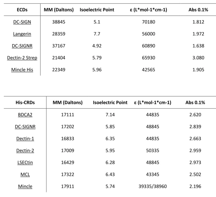
Table 8 and 9. Molecular weight, isoelectric points, molar extinction coefficient ε and Abs 0.1% values for the considered ECDs and CRDs.
112
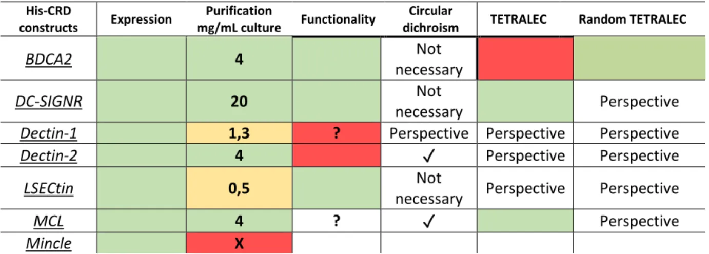
Table 10. List of analysed CLRs at the SEC-MALS and their specifications. 113 Table.11 Summary of all the attempts that have been made for soluble expression 130 Table.12 ECD expression and purification yield 149 Table.13 ECD expression, purification yield, functionality assay, CD assay and TETRALEC formation. 150
Table.14 Parameters of the crystal structure. 243
23
Abbreviations:
AAL : Aleuria Aurantia Lectin GalT : Galactosyltransferase
APCs : Antigen Presenting Cells GAS : Group A Streptococcus
ASGPR : Asialoglycoprotein Receptor GBPs : Glycan Binding Proteins
BDCA2 : Blood Dendritic Antigen 2 GI : Gastrointestinal
BSA-Man : Bovine Serum Albumin Mannosylated GL : Galectin
CD : Circular Dichroism GlucNAc : N-acetylglucosamine
cDCs : conventional DCs gp120 : GlycoProtein 120
CFG : Consortium for Functional Glycomics GPI : Glycosylphosphatidylinositol
CL : C-type Lectin hemiITAM : hemi-Immunoreceptor Tyrosine-based
Activation Motif
CLRs : C-type Lectin Receptors HIV : Human Immunodeficiency Virus
CM : Carboxymethyl HPLC : High Precision Liquid Cromatography
ConA : Concanavalin A HRP : Horseradish Peroxidase
CRAC : Cholesterol Recognition Amino acid
Consensus-like IFN : type I Interferon
CRD : Carbohydrate Recognition Domain IgG : Immunoglobulin G
CTLD : C-Type Lectin-like Domain IL : Interleuchin
CTLs : Cytotoxic T cells IL : I-type Lectin
DAMPs : Damage-Associated Molecular Patterns IPTG : Isopropyl 1-thio-D-galactopyranoside DAP12 : 12-kDa DNAX-Activating Protein IRAK : Interleukin Receptor-Associated Kinase DCIR : Dendritic Cell ImmunoReceptor ITC : Isothermal Titration Calorimetry
DCs : Dendritic Cells ITIM : Immunoreceptor Tyrosine-based Inhibition
Motif
DC-SIGN R : Dendritic Cell-Specific Intercellular
adhesion molecule-3-Grabbing Non-integrin Related LB : Luria Bertani
DC-SIGN : Dendritic Cell-Specific Intercellular
adhesion molecule-3-Grabbing Non-integrin LCs : Langerhans Cells
Dectin1/2 : DC-Associated C-Type Lectin 1/2 LDN : GalNAcβ1,4GlcNAc
DLS : Dinamic Light Scattering LDNF : GalNAcβ1,4[Fucα1,3]GlcNAc
DNA : Deoxyribonucleic Acid LN : N-acetyl lactosamine
DOL : Degree Of Labeling LPS : Lipopolysaccaride
dsbA : disulphide bond A LSECtin : Liver and lymph node Sinusoidal
Endothelial C-Type lectin
DSF : Differential Scanning Fluorimetry L-SIGN : Liver/Lymph node-Specific Intercellular
adhesion molecule-3-Grabbing Nonintegrin
ECD : Extracellular Domain mAbs : monoclonal Antibodies
EDC :
N-(3-dimethylaminopropyl)-N'-ethylcarbodiimide
MALDI-Tof : Matrix Assisted Laser Desorption
Ionisation - Time of Flight
EDTA : Ethylenediaminetetraacetic acid MALS : Multi Angle Light Scattering
ELISA : Enzyme-Linked ImmunoSorbent Assay manLAM : Mannosylated Lipoarabinomannan
ER : Endoplasmic Reticulum MAPK : Mitogen-Activated Protein Kinase
ESI-MS : Electrospray Ionization Mass Spectrometry MBP : Mannose-Binding Protein FACS : Fluorescence-Activated Cell Sorting MCL : Macrophage C-Type Lectin
FCRγ :Fc Receptor γ-chain MHC : Major Histocompatibility Complex
FucT : Fucosyltransferase Mincle : Macrophage Inducible Ca
2+-dependent
lectin
GalNAc :N-Acetylgalactosamine MMR : Macrophage-Mannose Receptor
24
MP : Mannose Posphate RNA : Ribonucleic Acid
MP3 : Multi step Protein Purification Platform ROS : Reactive Oxygen Species
MW : Molecular Weight RU : Resonance Unit
NADPH : Nicotinamide Adenine Dinucleotide
Phosphate SARS : Severe Acute Respiratory Sindrome
Nano ITC LV : Nano Isothermal Titration Calorimetry
Low Volume SAXS : Small Angle X-ray Scattering
NF-κB : Nuclear Factor – κB SEC : SecB Dependent
NHS : N-hydroxysuccinimide SEC : Size-Exclusion Chromatography
NK : Natural Killer SHP-1/2 : SH2-containing tyrosine Phosphatase-1/2
NLRs : Nucleotide-binding oligomerization domain
Like Receptors SLS : Static Light Scattering
NMR : Nuclear Magnetic Resonance SPAAC : Strain-Promoted Azide-Alkyne
Cycloaddition
ompA : outer membrane protein A SPR : Surface Plasmon Resonance
OVA : Ovoalbumin SRP : Signal Recognition Particle
PADS : Protecting group Aided Detection and
Separation of glycans SrtA : Sortase A
PAMPs : Pathogen-Associated Molecular Patterns Syk : spleen tyrosine kinase
PAOL : Protein Analysis On Line TACAs : Tumour-Associated Carbohydrate Antigens
PCR : Polymerase Chain Reaction TAT : twin-arginine translocation
pDCs : plasmacytoid DCs TCRs : T Cell Receptors
PEG : Polyethylene Glycol TDM : trehalose-6,6’-dimycolate
pelB : pectate lyase B Th : T helper
PMN : Polymorphonuclear TLC : Thin Layer Chromatography
PRRs : Pattern Recognition Receptors TLRs : Toll-Like Receptors
RA : Rheumatoid Arthritis TRAF : TNF Receptor Associated Factor
RFU : Relative Fluorescence Units UPLC-MS : Ultra performance liquid
chromatography - tandem mass spectrometer
RFI: Relative Fluorescence Intensity WC : Whole Cells RLRs : Retinoic acid-inducible gene-I-Like Receptors
25
Les résumés en Français
Chapitre 1. Le système immunitaire
Le contexte général de ce travail de thèse est le système immunitaire, source vitale de défense contre les agents pathogènes. L’immunité est divisée en deux systèmes communicants, l’immunité innée et l’immunité adaptative. Le paragraphe 1.2 décrit brièvement les acteurs cellulaires les plus importants de l’immunité innée. Ces cellules comprennent les macrophages, les neutrophiles, les basophiles, les éosinophiles, les mastocytes, les cellules tueuses (natural killer cells), et les cellules dendritiques (CD). Toutes ces cellules se développent sous la stimulation de certaines cytokines.
Les CDs sont spécialement importantes car elles constituent un lien entre l’immunité innée et adaptative et leur rôle principal est d’induire l’immunité adaptative (paragraphe 1.2.1). L’identification de micro-organismes étrangers par l’immunité innée est basée sur la reconnaissance de structures moléculaires conservés dans les micro-organismes et absentes chez l’hôte (paragraphe 1.2.2). Ces structures sont appelées “motifs moléculaires associés à des pathogènes” (PAMPs en anglais). Les récepteurs du système immunitaire inné qui reconnaissent les PAMPs sont appelés Pattern Recognition Receptors (PRR). Les PRRs peuvent être solubles, exprimés à la surface cellulaire ou intra-cellulaires. Les récepteurs lectine de type-C (CLRs) sont l’une des familles de PRRs.
Chapitre 2. Les récepteurs lectine de type-C
Les récepteurs lectine de type-C sont des lectines qui reconnaissent des groupements glucidiques spécifiques présents à la surface de leurs via un domaine structural appelé domaine de reconnaissance de carbohydrates (CRD). Cette reconnaissance est dépendante d’ions Ca2+ présents dans le site actif et ceci est expliqué dans le paragraphe 2.2. Apres contact avec le ligand, quatre voies peuvent être exploitées pour moduler le système immunitaire. La signalisation résultante dépend de plusieurs aspects, tel que la typologie du récepteur, son internalisation et la nature des ligands (paragraphe 2.2.1). Néanmoins, certaines constantes sont conservées parmi les différentes voies de signalisation : le motif basé sur la tyrosine, l’utilisation de SYK et SHP pour la modulation de la transcription et la coopération avec autre récepteur comme le récepteurs de type toll.
Enfin, la dernière partie du chapitre (paragraphe 2.3) décrit les neuf différentes CLRs étudiées pendant ce travail de thèse : BDCA2, DC-SIGN, DC-SIGNR, dectin-2, dectin-1, langerin, LSECtin, MCL et mincle. Pour chacun d'eux des informations sur la structure, la spécificité de reconnaissance et la voie de signalisation ont été donnés.
26
Chapitre 3. Glycobiologie
Les lectines décrites dans le chapitre précèdent peuvent déchiffrer le glycocode de glycans, porteur d’informations biologiques. Les glycans sont essentiels dans l’interaction cellule-cellule et la position de leurs groupements hydroxyle est cruciale pour la bio-reconnaissance.
Les sucres sont extrêmement complexes et une raison de cette complexité découle de la grande variété de liaisons possibles pour la formation d’oligomères. Outre leur complexité, les carbohydrates ont faible affinité et spécificité pour leurs partenaires protéiques. Afin de surmonter ces difficultés, des mimétique de carbohydrates doivent être développés. Trois différentes sections du glycan peuvent être optimisées. Ceci est développé dans des parties du paragraphe 3.2. Une fois que les glycomimétiques ont été synthétisés, leur interaction avec de lectines doit être validé par des expériences biochimique in vitro et, notamment, par des techniques de criblage (puce a sucres et puce a lectines, paragraphe 3.3).
L’optimisation et l’étude de ligand monovalent est la première étape vers le développement de inhibiteurs. Pour améliorer la faible affinité d’interaction, des ligands multivalentes sont ensuite envisagés afin d’atteindre l’effet d’avidité. Dans le paragraphe 3.4 différents exemples the ligands multivalents sont donnés.
Chapitre 4. Applications
Ce chapitre surligne la pertinence médicale de l’étude des interactions lectines-carbohydrates. Notamment, un focus a été fait sur le diagnostic (plusieurs maladies étant caractérisées par de changement de motif de glycosylation, sur l’imagerie pour pouvoir observer l’efficacité de l’internalisation au niveau cellulaire et sur le ciblage de lectines, fondamental pour la vaccination et le traitement du cancer.
Chapitre 5. Les objectifs de cette thèse
Le projet s’inscrit dans un contexte international et fait partie du réseau européen IMMUNOSHAPE. Le but du réseau est de combiner l’état de l’art de la synthèse chimique et des technologies de criblage pour développer des molécules immunothérapeutiques multivalentes basée sur des glycanes. Ma contribution au projet a été divisée en trois axes :
- La production de neuf récepteurs lectine de type-C. Différentes approches ont été testées pour optimiser l’expression et la production en bactérie. De plus, pour améliorer artificiellement la multivalence des protéines et l’affinité pour leurs ligands, une nouvelle stratégie visant à
27 multimeriser le construct CRD a été développée. Le complexe multimeric final comprenant quatre CRD biotinilés a été appelle TETRALEC.
- L’utilisation de méthodes de criblage d’interaction avec les ligands. Les techniques exploitées ont été : LectPROFILE assay, analyse par FACS, puce a sucre et a lectine.
- La caractérisation de l’interaction entre les ligands et les lectines par des études biophysiques. Cet axe est divisé en deux parties. La première est basées sur l’étude d’interaction avec des ligands monovalents et sélectifs pour DC-SIGN, tandis que la deuxième partie étudiée l’interaction avec des ligands multivalents.
Chapitre 6. Principes
Ce chapitre décrit la théorie des techniques utilisées dans ce travail de recherche.
Chapitre 7. Méthodes
Ce chapitre décrit en détail toutes les procédures ainsi que tous les produits chimiques, biologiques et les équipements utilisés dans ce travail de recherche.
Chapitre 8. Préparation de lectines recombinantes et test fonctionnel
Ce chapitre contient les résultats de la production des différentes lectines. En première intention, deux types de constructions ont été réalisées : soit le domaine extracellulaire complet (ECD) soit seulement le domaine de reconnaissance des sucres (CRD). Plusieurs constructions ciblant le periplasm de E.coli afin d’obtenir la protéine directement soluble ont été essayées. Malgré la large gamme de constructions testées, cette approche a conduit soit un faible niveau d’expression soit une expression insoluble. Pour cette raison, la stratégie d’expression periplasmique a été abandonnée. Les résultats d’expression et purification sont présentés au paragraphe 8.2.1.
Une stratégie alternative est présentée au paragraphe 8.2.2 et consiste en l’expression de protéines sans forme insoluble dans le cytoplasme bactérien, suivi par leur repliement in vitro. Trois constructions ECD ont été produites (chapitre 8.2.2.1). La production de DC-SIGNR-ECD a été réalisée sans obstacle majeur, et de rendements considérables de protéines pures et fonctionnelles ont été obtenus. Les rendements de
dectin-28 2 Strep-ECD et mincle His-ECD étaient inferieur. Néanmoins, la fonctionnalité a été confirmée et la spécificité de reconnaissance de dectin-2 a été étudiée plus en détails. La production des constructions CRD a été plus fructueuse (chapitre 8.2.2.2). DC-SIGNR, BDCA2 et LSECtin His-CRDs ont été produits comme protéines fonctionnelles. MCL His-CRD a été produit et son l’intégrité structurelle a été confirmée mais l’absence de ligands connus a empêché l’évaluation de sa fonctionnalité. Dectin-1 et Dectin-2 His CRD ont été produits mais sont non-fonctionnelles.
Enfin, la stratégie TETRALEC a été exploitée avec succès pour DC-SIGNR et MCL His-CRDs, tandis que des complexes TETRALEC avec biotynilation aléatoire ont été produits pour LSECtin et BDCA2 His-CRDs.
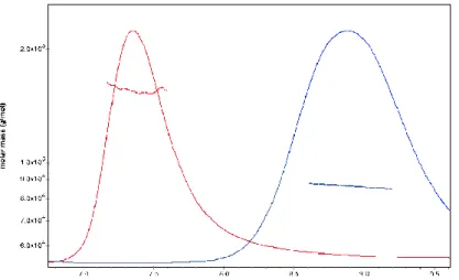
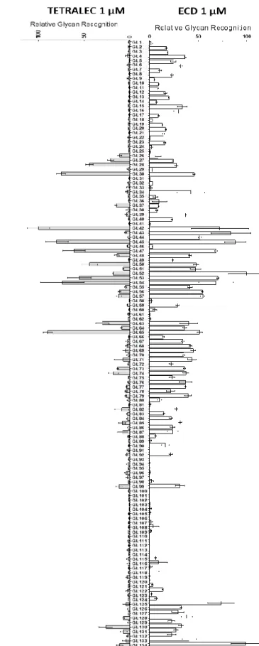
DC-SIGNR TETRALEC a été étudié plus en détails et les résultats sont présenté dans le papier numéro 1 (paragraphe 8.3). Le complexe a été caractérisé structuralement par SEC-MALS et la validation de sa fonctionnalité a été effectuée sur une puce à sucres dans le laboratoire de Dr. Niels Reichardt en Donostia-San Sebastian (Espagne) et in cellulo en utilisant la cytométrie en flux et montrant une interaction avec
Candida albicans dans le laboratoire de Pr. Bernd Lepenies en Hanover (Allemagne).
Chapitre 9. Criblage : identification de composés sélectifs des CLRs humains
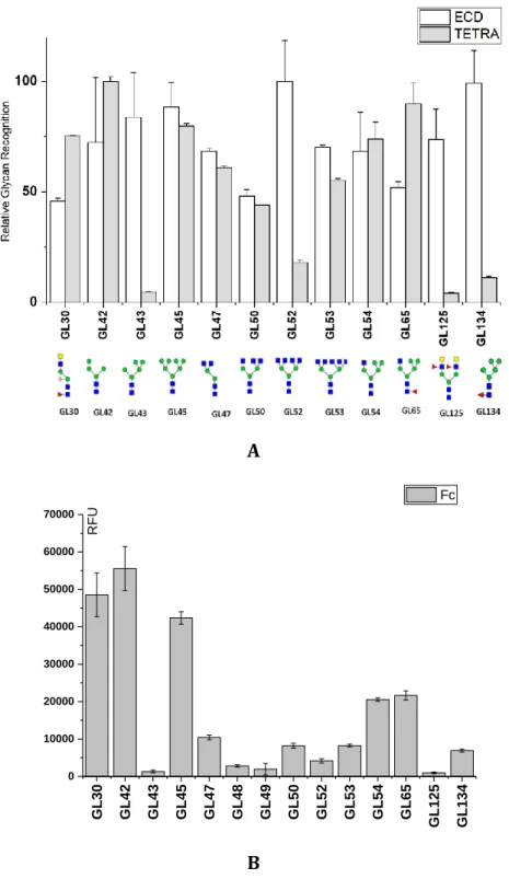
Ce chapitre décrit l’utilisation de puces à sucre pour cribler l’interaction entre des panels de glycanses (synthétisés par l’équipe de Dr. Niels Reichardt) et des glycomimétiques (synthétisés par l’équipe de Pr. Anna Bernardi) avec nos lectines marques par un fluorophore. Dans le paragraphe 9.1.1, le papier numéro 2 décrit une reconnaissance différentielle par trois lectines (DC-SIGN ECD, DC-SIGNR ECD et LSECtin CRD) de pairs de glycans qui sont des isomères de position.
La deuxième partie du chapitre décrit le criblage de glycomimétiques et les résultats sont présentés dans le papier numéro 3 (paragraphe 9.2.1). L’interaction obtenu par criblage sur puce a été a été confirmée aussi par SPR pour DC-SIGN et DC-SIGNR ECDs et de nouveaux glycomimétiques reconnus par dectin-2 ECD ont été identifiés.
Chapitre 10. Caractérisation de nouveau glycomimétiques spécifique vers DC-SIGN
Ce chapitre décrit la caractérisation par compétition en SPR de composés glycomimétiques spécifiques de DC-SIGN et est divisée en deux sous-parties principales. L’une décrit la stratégie utilisée pour augmenter la stabilité des glycomimétiques synthétisés et la seconde la recherche de composés encore plus spécifiques de DC-SIGN.
La première partie a été menée via deux collaborations différentes. Le papier numéro 4 (paragraphe 10.1.1) résulte d’une collaboration avec l’équipe de Pr. Anna Bernardi (Milano, Italie) pour caractériser l’effet du changement de liaisons glycosidiques par des liaisons avec un soufre. La deuxième collaboration a été
29 meée avec l’équipe du Pr. Jitka Moravcová pour le développement de C-glycosides et les résultats sont présentés au paragraphe 10.1.3.
La deuxième partie du chapitre (paragraphe 10.2) regroupe les travaux menés par l’équipe du Pr. Anna Bernardi sur le concept d’une « poche de liaison de ammonium » par des études de modélisations moléculaires. Le criblage par SPR des multiples glycomimétiques développés en ce sens nous ont permis d’identifier le composé Man069 qui présente une forte affinité et spécificité pour DC-SIGN. Les études biophysiques et structurales que nous avons réalisés sur le complexe DC-IGN/Man069 concluent le chapitre.
Chapitre 11. Conception de composées multivalents mannosylés pour le ciblage de CLR
Ce dernier chapitre de résultats est basé sur une collaboration avec deux autres équipes du réseau IMMUNOHAPE : l’équipe de Prof. Yvette van Kooyk (Amsterdam, Pays-Bas) et l’équipe de Prof. Jeroen Codee (Leiden, Pays-Bas). Le projet à la base de cette collaboration etait le développement d’une molécule multivalente hautement définit pour la vaccination contre le cancer, un des objectifs finals du réseau IMMUNOSHAPE. Notre contribution a consisté en la caractérisation par interaction directe en SPR de différents glycoclusters. Les résultats de cette évaluation sont présentés au paragraphe 11.3.
Chapitre 12. Conclusions et Perspectives
Tous les résultats sont résumés dans ce chapitre.
- Des remarques sur la production des lectines ont été faites dans le chapitre 9.1. Les moyens pour améliorer leur production sont aussi suggérés.
- Les résultats de criblage sont résumés et d'autres expériences sont aussi suggérées pour leur valider.
- Les résultats d’interaction pour l’ensemble des glycomimétiques monovalents testés contre DC-SIGN sont résumés et comparés dans le chapitre 9.3. Un composé optimal a été identifié et son interaction avec DC-SIGN a été de plus étudiée structuralement.
- Les résultats d’interaction pour l’ensemble des composés multivalents testés sont résumés et comparés dans le chapitre 9.4.
Enfin, dans le chapitre 9.5 des conclusions plus générales sont donnée, notamment par rapport à l’identification de candidats potentiels permettant d’adresser l’objectif initial à long terme du réseau IMMUNOSHAPE : la modulation du système immunitaire.
33
Part I.
35
1. The Immune System
1.1 Introduction
The immune system is our vital source of defense against infection and damage from external organism and toxin [1]. « Defense » and « immunity » appears as two crucial terms: while « defense » is used in a context of attack, implying defeat or victory, « immunity » is employed as synonym of resistance [2]. Immunity is a very complex network of soluble and cellular factors, with pro- and anti-inflammatory partners, and it is composed, in our body, by two communicating subgroups: the innate immune system and the adaptive immune system (Fig.1) [3].
Fig.1 The immune system. By sensing microbes (bacteria, parasites, fungi, and viruses), the innate immune system
awakes the adaptive one. Dendritic cells (DCs) link the two components of immunity. DCs are involved in antigen phagocytosis, processing and presenting to lymphocytes (T Cells). Adapted from [2].
The innate immune system is passed down from generation to generation [3] and it is characterized by the ability to sort out the self-molecular patterns from non-self or altered elements. The adaptive immune system, on the other hand, can develop memory and can adjust itself in response to pathogens. These two defensive systems can communicate through cells that are devoted to antigen processing, called Antigen-Presenting Cells (APCs).
The strong interaction between the innate and adaptive systems is allowed by the production of cytokines and chemokines [4] but this aspect of immune regulation will not be detailed within this manuscript.
36
1.2 Innate Immune System
Innate immunity, discovered by Elie Metchnikoff in 1916 during phagocytosis studies [5], describes the defense processes by which pathogens are recognized in an immediate yet nonspecific manner. Its typical timescale ranges from seconds to hours after antigen invasion [5]. The first physical defensive barrier is composed by cells of the epithelium, which is impenetrable by most external agents. Cells actively involved in immunity are divided into lymphocytes and phagocytes. Lymphocytes, which are the actors of the adaptive system, are T cells, B cells and Natural Killer (NK), while phagocytic [6] cells involved in the fast innate response include neutrophils and other types of granulocytes, macrophages, dendritic cells, and mast cells (Fig.2).
Fig.2 Innate and adaptive immunity. The players (and inter-players) of the innate and adaptive immune
systems.
The granulocytes family is composed by neutrophils, eosinophils and basophils, they are all involved in phagocytosis and they can also release granules in the extracellular space upon stimulation [7]. Neutrophils, in particular, can kill the infectious microorganism by using Reactive Oxygen Species (ROS), which are generated by the NADPH oxidase [8].
Macrophages are recruited to the inflammation site and are effective in ingesting microorganism as well as infected neutrophils. Once they have ingested the non-self-agent, macrophages migrate into lymph nodes or die [9].
37 present antigens to the lymphocytes (T cells) using Major Histocompatibility Complex (MHC) molecules and secrete cytokines that stimulate innate immune cells. Despite the fact that they are considered as part of the innate immunity, macrophages and DCs are executing effectors of the adaptive immune system [10].
1.2.1 Dendritic cells and signals for adaptive immunity activation
How fast the immune system responds to Pathogen-Associated (PAMPs) and Damage-Associated Molecules (DAMPs) is correlated to the availability of innate immune APCs [11]. Dendritic cells are the most potent APCs due to their ability to prime naïve T cells. DCs can be divided into plasmacytoid DCs (pDCs), monocyte–derived DCs (moDCs) and conventional DCs (cDCs) that all share the same hemapoietic cell progenitor. Although scarce [12], pDCs have important immunomodulation capabilities in T-cell mediated immune responses during viral infections and in the rapidly release type I interferon (IFN) [13]. moDCs are monocytes that have differentiated into DCs under inflammatory conditions and have a back-up role during acute inflammation [14]. cDCs, with a stellate morphology, which clearly distinguishes them from macrophages, have been shown to be the major cell type involved in migration into secondary lymphoid tissue and priming of naïve T cells[6]. By leading to an activation of the immune system or to its muffling, DCs play a pivotal and delicate role in balancing homeostasis as a down or over-regulation of the inflammation could, indeed, cause damage and disease [15].
How do DCs communicate with the adaptive immune system? When immature DCs encounter the antigen and receive other immune stimuli, they become mature. Endocytic receptors are then down regulated, CD40, CD80 and CD86 maturation markers are up-regulated, the level of MHC class II is raised and DCs migrate to the lymphoid organs where they communicate with T cells by three different signals (Fig.3a). The first one is based on the interaction between T cell receptors (TCRs) and MHC complexes loaded with antigenic peptide on DCs. The second signal involves the production of co-stimulatory signals and the interaction of the T cell co-stimulatory receptor CD28 with the ligand B7-1 expressed by DCs [16]. The third one is the production of inflammatory cytokines, IL-12 for instance, that helps T cell activation [6]. Without these three proper instructions from DCs, T cells would not be able to acquire effector functions and form memory cells [17]. The nature of the antigen presented and of the cytokine produced during DC maturation influences T cell differentiation. DCs can produce pro-inflammatory (IL-6) or anti-inflammatory (IL-10) cytokines and different chemokines, resulting in the recruitment of different T cell subsets at the infection site. CD4+T cell can differentiate into different T helper type cells Th1, Th2 or Th17.
Immunological synapses form between MHC class I or II from DCs and naïve T cells to allow the transfer of information about pathogen invasion. Several adhesion receptors mediate this cell-cell junction (Fig.3b).
38
A b
Fig.3 Activation of the adaptive immunity a) APC communication with T cells, adapted from [6], and b) electron
micrograph (courtesy of J. W. Uhr) showing a B cell and T cell bound to each other. The bar = 1 μm.
http://www.biology-pages.info/I/ImmSynapse.html.
1.2.2 Pattern recognition receptors
DCs, as professional sentinels of the innate immune cells, screen for pathogens by expressing Pattern Recognition Receptor (PRRs). They recognize non-self-elements and, thus, elicit the activation of immune response (inflammatory factor production). Two categories of patterns can be recognized by those receptors: Pathogen-Associated Molecular Patterns (PAMPs) and Damaged-Associated Molecular Patterns (DAMPs). Example of bacterial PAMPs are lipopolysaccharide (LPS), lipoproteins and peptidoglycan, while fungal PAMPs consist in carbohydrates of the cell wall [18]. PAMPs from viral origin are part of the glycoproteic envelope [19]. Once PAMPs are sensed by these receptors, DCs become activated with further production of chemokines and cytokines leading to inflammation (Fig.4). Inflammation could also be caused by damaged cells, e.g. by the above mentioned DAMPs, that results from tissue injury after bacterial infections. It is worth to note that DAMPs are also produced even in non-pathological conditions [20].
39 Fig.4 PAMPs and DAMPs. PAMPs and DAMPs interact with PRRs expressed by APCs with an effective cross
talk between innate and adaptive immunity (PMN=polymorphonuclear leukocytes, it refers to granulocytes[21]). Adapted from [22].
Those PRRs include the transmembrane C-type Lectin Receptors (CLRs) and Toll-Like Receptors (TLRs), together with the cytosolic receptors Nucleotide-binding oligomerization domain-Like Receptors (NLRs) and Retinoic acid-inducible gene-I-Like Receptors (RLRs) (Fig.5).
Fig.5 The four different families of PRRs. CLRs, TLRs, NLRs and RLRs. Adapted from [23].
TLRs, the first class of PPRs identified, are localized at the cell surface or at the endosome surface. TLR1,2,4,6 sense lipids, while TLR3,7,8 recognize viral RNA. TLR9 identifies bacterial DNA and it is strongly
40 involved in pro-inflammatory responses. Its deregulation could contribute to disease progression, e.g. sepsis [24]. Cross talk between PRRs signalling pathways can enhance the specificity of PAMP recognition and a focus on the synergistic activation of TLRs and CLRs will be given in chapter 2.2.2.
NLRs are involved in the regulation of inflammation and apoptosis during bacterial recognition. Finally, RLRs are helicases that sense viral RNA [25].
Depending on the antigen, on the PRR and on the APC involved, the immune system will be shaped towards an initiation of the immune response or towards the maintenance of self-tolerance (Fig.6).
This manuscript will focus on the specific PRR class of C-type lectin receptors, highlighting their structural and functional features and their contribution to the immune system response. C-type lectin receptors expressed by DCs are crucial for tailoring immune responses. They bind pathogens through the recognition of mannose, fucose, galactose and other carbohydrate structures. The combination of CLRs on APCs enables the recognition of most classes of human pathogens.
After pathogen uptake, several signalling pathways can induce the expression of specific cytokines and, consequently, trigger T cell differentiation. Some CLRs can directly induce activation of nuclear factor-κB (NF-κB), others affect signalling by Toll-like receptors (DC-SIGN) [26]. Therefore, CLRs represent an attractive target for immunotherapeutics.
Fig.6 CLRs and immunity. CLRs on DCs are the first step on the activation of the adaptive immune response. Adapted
41
2. C-type lectin receptors
2.1 Introduction
C-type lectin receptors are Glycan-Binding Proteins (GBPs) belonging to the large family of lectins. The term lectins derives from the Latin lectus, the past principle of legere, which means to choose or select [28]. In 1954 W.C. Boyd proposed this name in order to highlight their peculiar specificity [29]. The definition of lectin excludes both antibodies and enzymes, as glycans are not a substrate whose biochemical nature will be altered upon binding to the lectin. Lectins recognize glycan thanks to specific structural domain called Carbohydrate Recognition Domain (CRD). To date, 14 different CRD folds have been described and four examples of lectin family involved in different aspects of the immune responses are shown in the following figure (Fig.7).
Fig.7 Examples of four animal lectin families. (GL) galectin, (CL) C-type lectin, (MP) P-type lectin, (IL) I-type lectin [30].
The functions mediated by lectins are very diverse. Some lectins mediate interactions between cells and the extracellular matrix, while others are involved in the immune response. For example, L-Selectin is involved in lymphocytes homing [31] whereas serum mannose binding protein and ficolins activate the complement cascade [32]. Quality control is another important task of lectins. Calnexin and calreticulin in
42 the Endoplasmic Reticulum (ER) bind glucose on newly synthesized glycoprotein [33] and P-type lectins target lysosomial enzymes to endosomes by recognizing their mannose 6-phosphate [34].
In 1988, the group of Drickamer used for the first time the term « C-type lectin group » [35] to identify Ca2+-dependent lectins.
C-Type Lectins Receptors (CLR) bind to glycolipids and glycoproteins through a well-conserved globular domain CRD.
2.2 C type lectin domains and glycan recognition: structural aspects
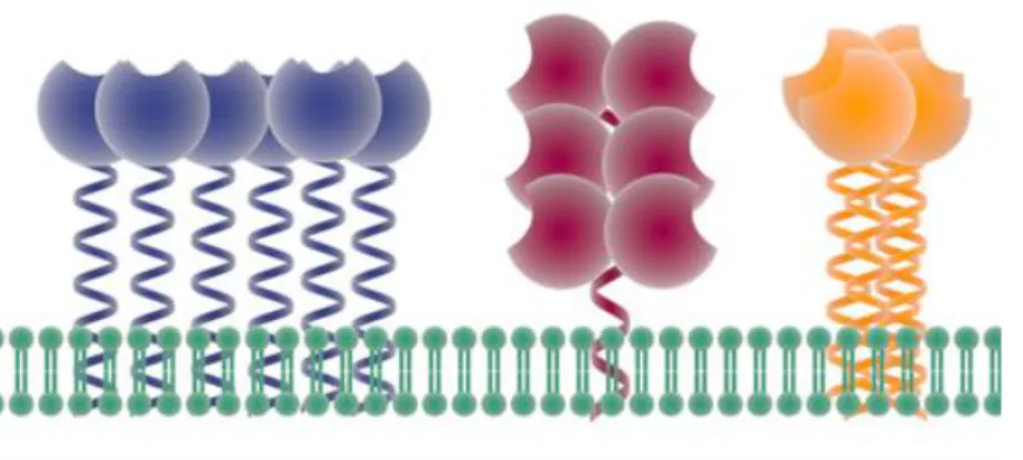
CLRs are divided into soluble or transmembrane proteins. In the latter, the CRD is connected to the cellular transmembrane region. In many cases, this connection occurs thanks to a coiled-coiled sequence termed neck domain. This neck domain is composed of a repeated sequence and, depending on the number of repetition and its length, the entire extracellular portion of the CLR, called ExtraCellular Domain (ECD), can oligomerize with a stoichiometry specific for the lectin for one lectin to another. Figure 8 schematises a tetrameric CLR with a neck oligomerization (Fig.8)
Fig.8 Schematic representation of a tetrameric CLR. The ECD is composed by the neck and the CRD.
To encompass proteins that contains a CRD domain with typical structural fold but that do not bind sugars [36] or lectins that conserve CLR characteristic properties but that are not Ca2+ dependent [37], the more general term « C-type Lectin-Like Domains » (CTLD) was introduced afterwards. However, for common usage, in this manuscript the term CRD will be used and not CTLD when referring to the carbohydrate recognition domain.
43 The CRD is, overall, a loop, with beta-strands at the N- and C-termini connected by two alfa helices and three antiparallel beta sheets (Fig.9).
a) b)
Fig.9 Carnohydrate recognition domain a) Generic CRD structure b) DC-SIGN CRD binding to
GlcNAc-Man3-GlcNAc http://www.imperial.ac.uk/research/animallectins/
Four cysteines are involved in disulphide bridges, crucial for the correct folding of the protein CRD. Up to four Ca2+ binding sites are found, but only one is involved in glycan recognition and is particularly conserved. The other Ca2+ binding sites play an important role in the CLR structure stability. The conserved Ca2+ binding site is characterized by specific motifs that lead to different specificities of the glycan recognition. The EPN (Gluc-Pro-Asn) motif leads to the interaction with mannose-type ligands (mannose, GlcNac, glucose), that contains 3-hydroxyl and 4-hydroxyl groups in equatorial position (Fig.10a), while the QPD (Gln-Pro-Asp) motif drives the recognition towards galactose-type glycans (galactose and GalNac), where the 4-hydroxyl group is axial (Fig.10b)[38]. In both cases the carbonyl side chains are involved in hydrogen bond formation with the specific monosaccharide and also coordinate two bonds with Ca2+. The proline is involved in the backbone conformation.
a b
Fig.10 EPN and QPD motif. a) EPN, mannose-type, motif b) QPD, galactose-type, motif. Coordination bond
are in green, H-bonds where hydroxyl acts as acceptor or donor are marked in pink or cyan dashed lines, respectively. Adapted from [35].
44 In addition to the primary binding site, secondary binding sites could be crucial in the glycan recognition and allow a broader interaction with the protein surface [39].
17 different groups of CRDs are described, depending on their domain architecture, phylogeny and function (Fig.11). Group III or Collectin group, for example, includes mannose-binding protein (MBP) with the typical collagen-like triple helical tail. E-Selectin belongs to the group IV and selectins are leukocyte adhesion molecules specific for sialyl-Lewisx [40]. macrophage mannose receptor (MMR) belongs to the group VI [41],[35].
All the protein studied during my PhD belong are involved in the activation of immunity. For example, Dendritic-cell-specific intercellular adhesion molecule-3-grapping non-integrin (DC-SIGN) and Mincle belong to type II receptors (group II) and they are characterized by a short cytoplasmic tail, a transmembrane domain and an extracellular domain ending with a Ca2+-dependent CRD. NK Receptors (Group V) include, as well, transmembrane proteins, e.g. Dectin1, with short cytoplasmic domain but with a CRD often lacking the Ca2+ dependency.
45
2.2.1 CLR signalling
Once a CLR encounter a pathogenic ligand, four different ways could be exploited to warn the immune system (Fig.12).
Fig.12 Overview of the four motifs for signalisation a) direct CLRs coupling to Syk b)
Indirect CLRs coupling to Syk c) CLRs with ITIM domain d) CLRs without ITAM or ITIM domain adapted from [43]
- a) Direct CLRs coupling to Syk: CLR cytoplasmic tail possesses a tyrosine-based motif called hemiITAM (hemi-Immunoreceptor Tyrosine-based Activation Motif). Dimerization of two phosphorylated CLR leads to recruitment of the tyrosine kinase Syk via its tandem SH2 domains. This path leads to myeloid cell activation [43]. An example is dectin-1.
- b) Indirect CLRs coupling to Syk: CLR cytoplasmic tail possesses a tyrosine-based motif and requires an ITAM containing adaptor molecule, such as Fc Receptor γ-chain (FCRγ). Again the phosphorylation events trigger Syk activation [43]. Examples are dectin-2 (Fig.13a), BDCA2 and mincle.
- c) CLRs with Immunoreceptor Tyrosine-based Inhibition Motif (ITIM) domain recruits phosphatases leading to a subsequent down regulation of immune response [43]. An example is the Dendritic Cell ImmunoReceptor DCIR.
- d) CLRs without ITAM or ITIM domain: these CLRs can signal after antigen capture of by other signalisation motives and modulate the signalling of other receptors. Examples are DC-SIGN are langerin [41]. DC-SIGN signalisation will be detailed afterwards.
46 The intracellular signalling pathways activated by dectin-2 and DCIR will be detailed below (Fig.13).
Dectin-2 is a CLR that indirectly couples with Syk and the association with FCRγ via an arginine residue is required for Dectin-2 surface expression. When dectin-2 recognizes fungi, the activation of Syk regulates the production of ROS [44], used as microbicidal agents, and the activation of complex involved in gene transcription regulation (Fig.13a).[43]
A b
Fig.13 Dectin-2 and DCIR signalling. a) Dectin-2 indirect coupling to SYK and intracellular signalling pathway,
adapted from [43] b) DCIR ITIM domain andphosphatase recruitment, adapted from [45].
DCIR, in contrast, bears an ITIM domain believed to mediate inhibitory signals in DCs (fig 13b). Phosphorylated ITIMs mediate recruitment of the SH2-containing tyrosine phosphatase-1 and 2 (SHP-1/2) which negatively controls NF-κB signalling to antigen response[45].
DC-SIGN do not possess any ITAM or ITIM domain. Nevertheless, it possesses in the cytoplasmic tail a tyrosine motif, necessary for the intracellular signalling [46], and a di-leucine motif involved in endosomal/lysosomal pathway [47]. This motif was shown by Engering et al [48] to be involved in the internalization of DC-SIGN-ligand complex.
The outcome of the signalling depends on multiple aspects, such as the type of receptor, the ligand (nature, architecture, density) and the internalization of the receptor involved. Nevertheless, some constants are
47 kept among the different CLR signalling: the tyrosine-based motif, the use of SYK and SHP for the modulation of gene transcription. Another common point is the ability to cooperate with other receptors to regulate myeloid cell functions [43].
2.2.2 CLR and TLR crosstalk
CLRs expressed at the surface of APCs are not only involved in pathogen recognition but also in their internalisation and a simultaneous activation of CLRs and TLRs can occur to trigger the appropriate immune response. Both PRRs lead to the activation of both nuclear factor-kappa B (NF-κB) and the mitogen-activated protein kinase (MAPK), resulting in an overlapping production of cytokines and chemokines. This net effect strikes, for example, during Candida infections, as shown in Figure 14, with a cooperation between TLR4/2 and dectin-1/2. MtD88 adaptor molecule is used by TLRs for the activation of the Interleukin Receptor-Associated Kinases IRAK1, IRAK2 and IRAK4 and subsequent ubiquitination of TNF Receptor Associated Factor TRAF6. Finally, MAPK and NF-κB are activated downstream. CLRs interaction with Syk triggers the recruitment of the Card9/Bcl10/Malt1 protein complex, leading as well to the MAPK and NF-κB activation (Fig.14) [49]. The final outcome is inflammation and the production of cytokines.
48 In 2018 S. Gringhuis et al. [50] have identified the mechanism by which DC-SIGN modulates TLR-dependent responses in human DCs (Fig.15). Mannosyl caps on the terminal D-arabinan (manLAM), found in pathogenic Mycobacterium, interact with DC-SIGN, leading to the activation of Raf-1. Activation of Raf-1, in turn, allows the acetylation of p65, the activating subunits of NF-kB, but only after TLR signalling had activated NF-κB. Indeed, TLR enables p65 translocation to the nucleus where the latter can then get activated by DC-SIGN downstream pathway. The acetylationof p65 extends the transcriptional activity of NF-kB and boostthe transcription rate of anti-inflammatory IL10 gene.
Fig.15 DC-SIGN and TLR4 during Mycobacterium infection. p65 and p50 subunits form NF-kB complex. Adapted from
49
2.3 CLRs considered in the study
This chapter will focus on the nine different CLRs investigated during my PhD: BDCA2, DC-SIGN, DC-SIGNR (L-SIGN), dectin2, dectin1, langerin, LSECtin, MCL and mincle. For each of them some information will be given, from structure features to binding specificities, from their contribution to the signalling cascade to the pathologies in which they are involved.
2.3.1 Blood Dendritic Antigen 2 (BDCA2)
BDCA2 is the only CLR considered in this study exclusively expressed by pDC BDCA2 CRD has a typical CLR with an EPN sequence at the Ca2+ binding site that should lead to interaction with mannose-type glycans. However, Glu178 of the EPN motif is positioned outside the calcium binding site, which is partially occupied by the side chain of Arg179 [52]. A first study [53] in 2011 by glycan array indicated an unusual binding of BDCA2 towards galactose-terminated biantennary glycans. In 2015 S. Jegouzo et al. [54] identified that BDCA2 binds in a very selective way glycans containing the epitope Galβ1-3/4GlcNAcβ1-2Man. Resolution of the CRD structure in the presence of this trisaccharide revealed that the mannose residue interacts with the primary binding site, while the other two sugars contribute to the interaction by occupying « a shallow groove » (Fig.16).
Fig.16 Portion of BDCA2 CRD structure. Complex of BDCA2 CRD with Galβ1–4GlcNAcβ1–2Man adapted from [54]
BDCA2 does not have an endocytic activity, being exclusively involved in intracellular signalling pathway. Its cytoplasmic domain does not contain any known signalling motif, therefore it must associate to the transmembrane adaptor FCRγ, which interferes with TLR9-induced activation of pDCs and leads, eventually, to the inhibition of type I IFN secretion[55]. This is an attractive feature to evade type I IFN responses that is used by Hepatitis B virus to facilitate its spreading [56]. Finally, it was also shown that colorectal cancer cells express BDCA2 ligands [55].
50
2.3.2 DC-SIGN and DC-SIGNR (L-SIGN)
A lot could be said on DC-SIGN (by typing “DC-SIGN” on the research tool of PubMed, one would find 1500 hits). Many thesis in our group have indeed focused on the study and the targeting of DC-SIGN1. However, for the sake of simplicity, in this chapter I will mainly focus on the similarities and differences between DC-SIGN and its related CLR, DC-DC-SIGNR, both studied during my PhD. For relevant publications based exclusively on DC-SIGN please refer to [57], [58], [59],[60],[61].
The names themselves indicate their importance in the initiation of T cell immunity by interacting with ICAM-3. Dendritic Cell Specific Intracellular adhesion molecule–3 (ICAM-3) Grabbing Nonintegrin (DC-SIGN) and DC-SIGN Related (DC-SIGNR) also termed Liver/Lymph node-Specific Intercellular adhesion molecule-3-Grabbing integrin (L-SIGN) appear to be the product of a gene duplication and share 77% of homology [62]. The EPN motif is present in both proteins and enables mannose binding.
Their binding to mannose derived glycans was studied by H. Feinberg et al [63],[64]. As typical feature of mannose binding mode to CLR, equatorial 3- and 4-OHs of the internal sugar form both coordination bonds with the Ca2+ (Fig.17). In addition, they form hydrogen bonds with amino acids that also serve as Ca2+ ligands. Moreover, the 6-OH forms a water-mediated contact with Asp367 (Asn379 in DC-SIGNR).
a b
Fig.17 Portion of DC-SIGN and DC-SIGNR CRD structure a) DC-SIGN and b) DC-SIGNR X-ray structure in complex
with GlcNAc2- Man3. Adapted from [63].
One primarily difference between the two CLRs lays in the binding sites: Val351 of DC-SIGN is replaced by Ser363 in DC-SIGNR. This is the main reason why DC-SIGNR does not interact with fucose containing Lewisx antigens, while DC-SIGN enables the van der Walls interactions necessary for fucose interaction [37].
51 For both DC-SIGN and DC-SIGNR, the neck repetitions allow protein oligomerization into a tetramer with an affinity increase for glycan recognition. The final coil-coil unit of their neck domain is the most divergent in term of residues and confer different CRD presentations [65],[66]. SAXS analysis performed by Franck Fieschi group [57] revealed different coiled-coil neck arrangements that lead to structural differences : the « closed flower » conformation of DC-SIGN is characterized by the CRD alignment with the neck domain, while the « opened flower » organization of DC-SIGNR prevents DC-SIGNR to have the suitable oligomeric orientation for the recognition of some DC-SIGN ligand (Fig.18).
Fig.18 Comparison of the CRD from SIGN and SIGNR. Closed flower for SIGN and open flower for
DC-SIGNR, adapted from [57].
The different CRD orientation of the two lectins is responsible for different binding efficiency towards multivalent ligand. This has been recently confirmed by glycan functionalized quantum dots that showed that the spatial orientation of DC-SIGNR does not favour binding to multiple mannose residues [67].
The two receptors are also characterized by different expression patterns. DC-SIGN is highly found at the surface of monocytes and in subsets of immature and mature DCs in dermis, mucosa, spleen and placenta, while the name “L-SIGN” comes from the fact that it is expressed in endothelia cells of lymph nodes and liver but not on DCs. Similarly to DC-SIGN, DC-SIGNR is involved in HIV-1 virus infection by binding to gp120 [60]. Moreover, being found in placenta, it is implicated in the HIV-1 virus mother to foetus transmission (Fig.19) [68].
52 Fig.19 The placenta, DC-SIGN/DC-SIGNR and HIV. The placenta plays an important role in the transmission of HIV-1
infection from mother to foetus. Released viral particles may become adsorbed onto DC-SIGNR on the placental capillary endothelium [68].
DC-SIGNR can establish interactions with ICAM‐3‐expressing T cells and this may enable activated T cells to recirculate to the liver and to the lymph nodes [69]. DC-SIGNR do not possess ITAM-ITIM domains and shares potential internalization di‐leucine motif with DC-SIGN. Nevertheless, it is still under investigation whether it is an endocytic receptor, while DC-SIGN is confirmed to be involved in pathogen uptake.
2.3.3 DC-Associated C-Type Lectin 1 (dectin-1)
Dectin-1 is a natural killer (NK)-cell-receptor-like and it is found on the surface of peripheral blood leukocytes and DCs in muscle, stomach [70] and lung [71]. It is also called β-Glucan receptor because it recognises β-1-3 linked and β-1-6 linked glucan (laminarin and zymosan) from bacteria and fungi (A.
fumigatus, C. neoformans, C. albicans). Its presumed binding site is a « shallow groove » [37] defined by
Trp221 and His223, and modelling studies suggest that the interaction with laminarin is mostly driven by hydrophobic forces. Unfortunately, all the attempts to crystalize the CRD with long β-glucan chain have failed so far. One peculiarity of this lectin is that it does not require metallic ions for binding. Nevertheless, a Ca2+ binding site was found and a structural role was attributed to it. Figure 20 presents the X-ray structure of murine dectin-1 binding to β-glucan chain [37].
Gatner et al. [72] observed that during the recognition of zymosan, both dectin-1 and TLR2 were recruited