HAL Id: tel-01556313
https://tel.archives-ouvertes.fr/tel-01556313
Submitted on 5 Jul 2017
HAL is a multi-disciplinary open access
archive for the deposit and dissemination of sci-entific research documents, whether they are pub-lished or not. The documents may come from teaching and research institutions in France or abroad, or from public or private research centers.
L’archive ouverte pluridisciplinaire HAL, est destinée au dépôt et à la diffusion de documents scientifiques de niveau recherche, publiés ou non, émanant des établissements d’enseignement et de recherche français ou étrangers, des laboratoires publics ou privés.
Interaction of the nucleocapsid domain of the Human
lmmunodeficiency Virus type-1 with the cellular protein
Unr : implication in viral IRES dependent translation
Nedal Taha
To cite this version:
Nedal Taha. Interaction of the nucleocapsid domain of the Human lmmunodeficiency Virus type-1 with the cellular protein Unr : implication in viral IRES dependent translation. Virology. Université de Strasbourg, 2015. English. �NNT : 2015STRAJ028�. �tel-01556313�
UNIVERSITÉ DE STRASBOURG
Ecole Doctorale des Sciences de la Vie et de la Santé
UMR CNRS 7213, Faculté de pharmacie, ILLKIRCH
THÈSE
présentée par :Nedal TAHA
Soutenue le : 03 juillet 2015Pour obtenir le grade de :
!"#$%&'($')*%+,-$&.,#/'
de Strasbourg
Discipline/ Spécialité: Sciences du Vivant/Virologie
Interaction du domaine nucléocapside de la polyprotéine
Gag du VIH-1 avec la protéine cellulaire Unr: implication
sur la traduction IRES-dépendante du virus
THÈSE dirigée par :
Dr. MELY Yves Professeur, Université de Strasbourg
RAPPORTEURS :
Dr. JACQUEMIN-SABLON Hélène Chargée de recherche, INSERM
Dr. OHLMANN Théophile Directeur de recherche, INSERM
AUTRES MEMBRES DU JURY :
Dr. PAILLART Jean-Christophe Directeur de recherche, CNRS
I dedicate this thesis:
To my beloved parents
To my brothers Ahmad and Rawad
Acknowledgments
First of all, I would like to express my sincere gratitude for my thesis director Professor Yves MELY for giving me the opportunity to pursue my PhD in the laboratory. Thank you Professor Mely for your effort spent in advising me, correcting my thesis, and supporting me. A very special thanks goes to Dr. Eleonore REAL for her continuous supervision, encouragement, and motivation throughout the four years. She, more than anyone else, has contributed to my professional growth. Thank you Eleonore for all your time, patience, and support. Thank you for giving as much as you could from your expertise and for pushing me always to the best. You have set the standards high. I am grateful that I was your student and I owe you a lot.
I am honored by the kind acceptance of Dr. Jean Christophe PAILLART, Dr. Theophile OHLMANN, and Dr. Helene JACQUEMIN-SABLON to be jury members for my thesis. I would like to express my gratitude for Dr. Hugues DE ROCQUINY and Dr. Emmanuel BOUTANT for their beneficial scientific discussions and their availability to answer all my questions. I would like to thank Dr. Ludovic RICHERT and Dr. Halina ANTON for their assistance with the imaging platform and analysis. An additional thanks for Romain VAUCHELLES for the help with Confocal microscopy. Kamal Kant SHARMA, thank you for all your help in protein purification. A special thanks goes to Dr. Serena BERNACCHI at the Institute of Biologie Moleculaire et Cellulaire (IBMC) for her help with RNA transcription. ! "#$%&! $''! ()(*)+,! -.! "#)! /0$*-+$"-1+)! 2)! 31-4#-"-%156) )"! 7#$+($8-'-9:;! ),4)81$'':! Manuel PIRES, Salah EL MESHRI, Hala El MEKDAD, Noémie KEMPF, Avisek GHOSE, Hassan KARNIB, Liliana ZAITER, and Wassem ASHRAF.
I would not have survived this phase without the support of my dear friends Israa DANDACHE, Zeinab DARWICH, and Sarwat ZGHEIB. Thank you ladies for being there throughout the good and the bad times.
<%! $! 4)+,-%$'! ')=)'>! ! 8$%?"! *6"! 2))4':! "#$%&! @ABCD! .-+! #1,! 1%.1%1")! ,644-+">! '-=)>! $%2! cheerful encouragement especially during the last two years of my thesis. Thank you my cheerleader for always lifting me up when I am down. Thank you for bearing my bad temper and mood swings!!
Finally, no words can ever express my deepest gratitude to my family. Your unconditional encouragement and infinite support made it possible for me to pursue and finish my PhD. I would have never made it without you: MOM, DAD, AHMAD, and RAWAD. Thanks for believing in me, pushing me, and supporting me. Thanks for everything. I am what I am because of you.
1
Table of Contents
Abbreviations ... 5
Introduction ... 11
Chapter I-Human Immunodeficiency Virus type 1 (HIV1) ... 13
1.1 HIV Classification ... 13
1.2 History of HIV infection ... 14
1.3 HIV Origin and Phylogeny ... 14
1.4 Epidemiology of HIV infection ... 17
1.5 HIV transmission ... 19
1.6 Progression of HIV infection ... 20
1.7 HIV treatment ... 22
Chapter II- Organization of the HIV-1 Particle ... 24
2.1 Structure of the Viral Particle ... 24
2.2 Genomic Organization of HIV ... 25
2.2.1 Proviral DNA ... 25
2.2.2 Viral RNA (gRNA) ... 28
2.2.3 Other Non-coding sequences ... 31
2.3 Viral Proteins ... 32
2.3.1 Envelope proteins ... 33
2.3.2 Gag and its Cleavage Products ... 34
2.3.3 Viral Enzymes ... 39
2.3.4 Regulatory and auxillary proteins ... 40
Chapter III- HIV-1 Life Cycle ... 47
3.1 The early phase or the pre-integrative phase ... 48
3.1.1 Virus Entry ... 48
3.1.2 Reverse Transcription and uncoating ... 50
3.1.3 Nuclear Import and DNA integration ... 54
3.2 The Late Phase ... 58
3.2.1 Proviral cDNA expression ... 58
2
3.2.3 Translation of Viral Proteins ... 63
3.2.4 Viral Assembly, Budding, and Maturation ... 63
3.2.5 Budding and Maturation ... 66
Chapter IV- The HIV-1 Nucleocapsid protein ... 68
4.1 NCp7: a cleavage product of Gag ... 68
4.2 Nucleocapsid Structure ... 69
4.3 Nucleic Acid Binding ... 71
4.4 RNA chaperone ... 72
4.5 Roles of NC in the viral life cycle ... 73
4.5.1 NCp7 roles in the early phase ... 73
4.5.2 NC roles in the late phase ... 77
Chapter V- HIV-1 Translation ... 85
5.1 Overview of Eukaryotic Translation ... 85
5.1.1 Cap-Dependent Translation Initiation ... 86
5.1.2 Internal Ribosome Entry Site (IRES) Dependent Translation Initiation ... 88
5.2 HIV-1 Translation ... 92
5.2.1 HIV-1 Cap-dependent Translation ... 93
5.2.2 HIV-1 IRES-Dependent Translation ... 95
Chapter VI- Unr (Up-stream of the N-ras) ... 102
6.1 Unr gene: Discovery and Evolutionary Considerations ... 102
6.2 Unr expression ... 103
6.3 Unr cellular localization ... 104
6.4 Unr Domain Structure ... 104
6.5 RNA binding properties of Unr ... 107
6.6 Functions of Unr ... 109
6.6.1 Unr Regulates IRES-dependent translation ... 109
6.6.2 Unr regulates Cap-Dependent Translation ... 112
6.6.3 Unr regulates the stability and translation of mRNA ... 114
6.6.4 Unr regulates cell differentiation, proliferation, and death ... 115
Aim ... 117
Materials and Methods ... 119
3 A- Cell lines ... 121 B- DNA constructs ... 121 C- Primary Antibodies ... 123 D- Secondary Antibodies ... 124 E- Primers ... 124 II-Methods ... 125 A- Cell Biology ... 125 B- Biochemistry ... 126 C- Cell Assay ... 129
D- Virus production, infection, quantification ... 129
E- Imaging Protocols ... 131
F- Molecular Biology ... 134
Results ... 139
Part I: The IRES transacting factor Upstream of N-Ras interacts with HIV-1 Gag/NCp7: functional implication on the IRES driven translation and infection. ... 141
Manuscript 1 ... 145
Supplementary data ... 179
Part II: Investigating the Cellular Distribution and Interactions of the HIV-1 Nucleocapsid Protein by Quantitative Fluorescence Microscopy ... 189
Manuscript 2 ... 193
General Conclusions and Future Perspectives ... 218
Bibliography ... 226
5
ABBREVIATIONS
Aa Amino Acid
AIDS Acquired immune deficiency syndrome
ALIX Apoptosis-Linked gene 2 Interacting protein X
Apaf-1 Apoptotic protease-activating factor 1
APCs Antigen Presenting Cells
APOBEC3G/F Apolipoprotein B mRNA-Editing enzyme-Catalytic polypeptide-like 3G/F
ARE AU-rich instability element
AUF1 AU-rich binding factor 1
AZT Azidothymidine
BAF Barrier-to-autointegration
BMH Branched Multiple Hairpin
CA Capsid
CBC Cap Binding Complex
CBP p300/CREB Binding Protein
CDC Center for Disease Control and Prevention
CPSF6 Cleavage and Polyadenylation Specific Factor 6
CRFs Circulating Recombinant Forms
cPPT Central PolyPurine Tract
Crm1 Chromosome maintenance 1
CrPV Cricket Paralysis Virus
CSDs Cold Shock Domains
6
CTD C-Terminal Domain
CTS Central Termination Sequence
CypA Cyclophillin A
DIS Dimer Initiation Site
DCC Dosage Compensation Complex
EMCV EncephaloMyoCarditis Virus
ER Endoplasmic Reticulum
ESCRT-1 Endosomal Sorting Complex Required for Transport-1
ESE Exonic Splicing Enhancers
ESS/ISS Exonic/Intronic Splicing Silencers
FDA Federal Drug Administration
FMDV Foot-and-Mouth Disease Virus
Gag Group specific antigen
GDP Guanosine DiPhosphate
GTP Guanosine TriPhosphate
GRID Gay Related Immune Deficiency
gRNA Genomic RNA
HAART Highly Active Anti-Retroviral Therapy
HAT Histone Acetyl Transferase
HAV Hepatitis A Virus
HCV Hepatitis C Virus
HIV Human Immunodeficiency Virus
7
HRV Human Rhinovirus
HuR Human antigen R
ICTV International Committee on Taxonomy of Viruses
IDUs Injection Drug Users
INSTIs Integrase Strand Transfer Inhibitors
IN Integrase
IRES Internal Ribosome Entry Site
ITAF IRES-Trans-Acting Factors
La Lupus autoantigen
(L) domains Late domains
LDI Long Distance Interaction
LEDGF/p75 Lens Epithelium-derived Growth Factor -75kDa
LTRs Long Terminal Repeats
MA Matrix
mCRD Major Coding Region Determinant of instability
MHR Major Homology Region
mRNP Messenger RiboNucleoProtein complex
Msl-2 Male-Sex lethal 2
MSM Men who have Sex with Men
Myr Myristyl
Myr (e) Myristyl-exposed
Myr (s) Myristyl-sequestered
8
NCp7 Nucleocapsid protein
Nef Negative Regulatory Factor
NES Nuclear Export Signal
NLS Nuclear Localization Signal
NNRTIs Non-Nucleoside Reverse Transcriptase Inhibitors
NRTIs Nucleoside/Nucleotide analogue Reverse Transcriptase Inhibitors
NPC Nuclear Pore Complex
nPTB Neuronal isoform of PTB
NTD N-terminal Domain
NUPs NucleoPorins
ORFs Open Reading Frames
PABP Poly(A)-Binding Protein
PBS Primer Binding Site
PAIP-1 PABP Interacting Protein
PCAF P300/CBP-Associated Factor
PDI Protein Disulfide Isomerase
PIC Pre-Integration Complex
PIs Protease Inhibitors
PPT PolyPurine Tract
PR Protease
PTB Polypurine Tract Binding protein
PTH ParaThyroid Hormone
9
R Redundant
RanGTP Ran bound to Guanosine TriPhosphate
Rev Regulatory of Virion expression
RRE Rev Response Element
RT Reverse Transcriptase
SD Splicing Domain
SIV Simian Immune deficiency Virus
SU Surface glycoprotein
TAR Transactivation response Region
TRBP TAR RNA Binding Protein
Tat Transactivator of transcription
TM Transmembrane protein
TNPO3 Transportin 3
Unr Upstream of N-ras
UNG2 Uracil-DNA glycosylase 2
UTR UnTranslated Region
U3 !"#$%&"!&'(
U5 !"#$%&"!&)(
VCC Virus Containing Compartement
vDNA Viral DNA
Vif Viral infectivity factor
VLPs Virus Like Particles
10
Vpu Viral protein U
11
13
Chapter I-Human Immunodeficiency Virus type 1 (HIV1)
1.1 HIV Classification
Human Immunodeficiency Virus (HIV) is the causative agent of acquired immune deficiency syndrome (AIDS) (Barré-Sinoussi et al. 1983). Human Immunodeficiency Virus is a member of the Lentivirus genus in the subfamily Orthoretrovirinae of the family Retroviridae. Retroviruses are part of a large diverse family of enveloped RNA viruses sharing common structures, compositions, and replicative properties. The family name Retroviridae is derived from the fact that its members reverse transcribe their genomic RNA (gRNA) into a linear double stranded DNA which is integrated into the host genome (Coffin et al. 1997). Lentivirus (Latin: Lenti meaning slow) genus includes non-oncogenic retroviruses that produce multi-
organ diseases characterized by long incubation periods, long-term illnesses, and persistent infections with latency periods (Campbell & Robinson 1998). Lentiviruses are also distinguished from the oncogenic retroviruses by their ability to infect non dividing cells such as macrophages and monocytes (Vodicka 2001). According to the recent classification of the International Committee on Taxonomy of Viruses (ICTV), the Lentivirus genus comprises nine species (Figure 1): six non-primate and three primate viruses which are Human Immunodeficiency Virus type 1 and 2 (HIV-1, HIV-2) and Simian Immunodeficiency Virus type (SIV).
Figure 1: HIV taxonomy as classified by the International Committee on Taxonomy of Viruses (ICTV) 2013).
14
1.2 History of HIV infection
In 1981, the first clinical case of AIDS was observed in the United States of America after outbreaks of Pneumocystis carinii pneumonia (Gottlieb et al. 1981) and *+,-."(.& .+/0-1+ (Friedman-Kein 1982) among young homosexual men. At that time, the disease did not have a name and it was thought to be restricted to the gay community so it was given different names including Gay Related Immune Deficiency (GRID) or gay cancer since it was often accompanied by cancer. Afterwards, it was found that the disease is not restricted to homosexuals and was thus defined properly by the Center for Disease Control and Prevention (CDC) as AIDS (CDC 1982, current trend updates). However, at that time, the causing agent of the disease was still unknown.
Later in 1983, the team of Professor Luc Montagnier at the Pasteur Institute in Paris isolated the virus from the lymph nodes of a patient with generalized lymphadenopathy corresponding to a prodromal phase of HIV infection (Barré-Sinoussi et al. 1983; Montagnier et al. 1984). Françoise Barré-Sinoussi and Luc Montagnier were awarded the Nobel Prize for Medicine in 2008 for the discovery of HIV (Lever & Berkhout 2008).
Few years after HIV discovery, a new lentivirus was isolated from West African AIDS patients in 1986. This new virus was related to the Simian Immunodeficiency Virus and in general associated with a lower viral load, a slower decrease of CD4 and clinical progression than HIV-1 (Marlink et al. 1994; Clavel et al. 1986). Since it was shown to cause AIDS, the virus was named HIV-2 (Le Guenno et al. 1987).
1.3 HIV Origin and Phylogeny
Genome sequence alignment of HIV-1 and HIV-2 isolates indicated that they share only 40% sequence homology (Guyader et al. 1987). Phylogenetic studies have showed that HIV-1 is closest to Simian Immunodeficiency Virus of chimpanzee (SIVcpz) while HIV-2 is closer to the Simian Immunodeficiency Virus of mangabeys (SIVsm) (
15 Figure 2). Thus, it was suggested that HIV-1 was derived from the SIVcpz by intraspecies transmission in West Africa (Gao et al. 1999). This could have occurred in West Equatorial Africa as a result of exposure to animal blood while hunting, butchering, or consumption of raw meat (Buonaguro et al. 2007). On the other hand, HIV-2 originated as zoonosis from the sooty mangabey (Reeves & Doms 2002).
Figure 2: HIV origin (A) HIV-1 originates from a single subspecies of the chimpanzee, Pan troglodytes
troglodytes (B) HIV-2 originates from the sooty mangabey, Cercocebus atys (C) the radial phenogram demonstrates the close relationship between HIV and SIV env sequences (distances indicates the relative degree of evolutionary relatedness) (Stebbing et al. 2004).
Due to differences in transmission rates and virulence, HIV-1 is pandemic with a worldwide distribution, while HIV-2 is more endemic, with stable prevalence rates in most countries (Remy 1998; Coffin et al. 1997). For instance, even if HIV-2 is transmitted by the same routes as HIV-1, it is at a lower rate, probably because of a very low virus load in many asymptomatic individuals (Berry et al. 1998; Kanki et al. 1994).
Globally circulating HIV-1 strains are classified into four phylogenic groups: M, N, O, and P. The three groups M, N, and O are the result of independent cross species transmission events from SIV strains naturally infecting African primates to humans (Sharp & Hahn 2011).
Group M (Major) includes the pandemic strains of HIV and is globally distributed in almost every country. Since group M is globally distributed, this led to the predominance of different M subtypes in different areas. Group M is divided into nine subtypes (A-D, F-H, J, K) in addition to more than 40 different circulating recombinant forms (CRFs) generated when a population is co-infected with multiple subtypes (Sharp & Hahn 2011) (Figure 3). Group O (Outlier) regroups viruses isolated from individuals living in Gabon, Cameroon and Equatorial Guinea and represents less than 1% of HIV-1 infections (Peeters et al. 1997;
16 Mauclère et al. 1997). The group has a 30-50% sequence divergence from group M depending on the compared gene (Yamaguchi et al. 2003). A new HIV strain, distinct from group M and Group O, was isolated from Cameroon in 1998 and called Group N (New or nonM/nonO). To date, only 14 cases have been documented (Simon et al. 1998; Vallari et al. 2010; Delaugerre et al. 2011). Recently, in 2009, group P was discovered in a Cameroonian woman living in France (Plantier et al. 2009). Group P has been identified so far only in one other person also in Cameroon (Vallari et al. 2010; Mourez et al. 2013).
HIV-2 is divided into seven subtypes (A- G) with subtype A accounting for the majority of HIV-2 infections (Reeves & Doms 2002).
Figure 3: A comparison of the genetic similarity between different HIV and SIV strains by aligning the full genome sequences of 87 human and simian lentiviruses. HIV-2 and HIV-1 share around 50260%
sequence identity. The origins of the HIV-1 groups indicate two probable jumps from chimpanzee (groups M and N) and gorilla (group O) species. HIV-1 M subtypes probably evolved from a single introduction into the human population before diverging into different subtypes (Ariën et al. 2007).
17
1.4 Epidemiology of HIV infection
According to the World Health Organization (WHO), an estimated 35 million people were living with HIV in 2013 of which 3.2 million were children below the age of 15 (Figure 4).The increase of the number of people living with AIDS from previous years is due to the fact that more people are receiving the life-saving antiretroviral therapy. There were 2.1 million new HIV infections globally showing a 38% decline in the number of new infections from 3.4 million in 2001. In parallel, the number of AIDS deaths is also declining with 1.5 million AIDS deaths in 2013, down from 2.3 million in 2005. The highest mortality rate is in Sub-Saharan Africa where there is the highest HIV prevalence (Figure 5).
18
Figure 5: Geographic distribution of adult and children deaths due to AIDS in 2013 as estimated by WHO. The highest mortality rate is in Sub-Saharan Africa where there is the highest HIV prevalence
Molecular epidemiology studies showed that there is a specific geographic distribution pattern of HIV-1 subtypes. Globally, the most prevalent genetic subtypes are A, B, and C with subtype C accounting for more than half of all HIV-1 infections. Subtype C viruses are prevalent in the countries accounting for more than 80% of the global HIV-1 infections. Subtype A viruses are predominant in central and eastern Africa as well as Eastern Europe. Subtype B is dominant in Western and Central Europe, America, Australia, Northern Africa, and Middle East. In addition, CRFs are gaining more importance in the HIV pandemic constituting around 18% of infections (Buonaguro et al. 2007).
Subtype distribution was highly associated with demographic parameters suggesting highly compartmentalized epidemics, determined by social and behavioral characteristics of the patients (Abecasis et al. 2013). In France, viral diversity has increased in all risk groups over the last 15 years and although subtype B is the major subtype in France, the number of non-B virus infected patients is increasing (Chaix et al. 2013; Brand et al. 2014).
19
1.5 HIV transmission
HIV transmission occurs via direct sexual contact, vertical transmission from mother to child, or exposure to contaminated blood, or blood products (Sleasman & Goodenow 2003). The CDC reported that more than half of the new infections occur among men who have sex with men (MSMs). However, heterosexuals and injection drug users (IDUs) are also significantly affected by HIV (Figure 6). The mother to child transmission rate during pregnancy, labor, delivery, or breast feeding is estimated by the WHO to range from 15 to 45% in the absence of intervention.
Figure 6: Estimated new HIV infection by route of transmission. Men who have sex with Men (MSM)
remain the group most heavily affected by HIV in the United States accounting for more than 63% of new infections in 2010 (CDC 2010).
20
1.6 Progression of HIV infection
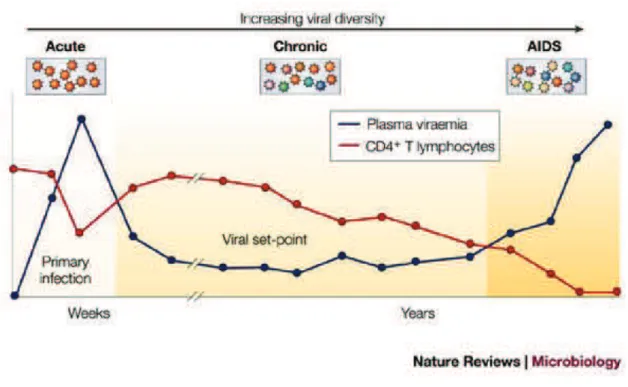
Although the course of HIV infection may vary among different patients, a common pattern is recognized. HIV-1 infection progresses in three phases: acute infection, chronic infection, and AIDS (Figure 7).
Acute infection
The early stage of HIV infection is the acute or primary infection. It corresponds to the period between the infection and the moment when HIV antibodies are detected in blood. This usually occurs 4 to 6 weeks after exposure although in some cases it can take up to 12 weeks.
Shortly after a person becomes infected with HIV, the virus replicates causing a huge rise of viremia (concentration of virus in blood) and rapid loss of CD4+ cells. This phase of intense viral replication promotes the dissemination of the virus throughout the body. This is +00-1,+!"%3& 45& 61-!-!$07%-.".& 7"8%(& .51,9-1.& .$0:& +.& ;%<%/=& .-/%& 9:/-+9=& +!3& 0%/<"0+7& adenopathy. Rash and fever are the most characteristic symptoms of primary HIV infection. Eventually, the immune system develops a response against the infection bringing back the virus down to a steady-state level which varies depending on each individual.
Since HIV antibodies are not present in blood yet, diagnosis cannot be performed with standard serological tests. However, diagnosis is done based on the detection of viral RNA or p24 (HIV capsid protein) antigens in the plasma of infected individuals. Seroconversion, the onset of anti HIV-antibodies in blood, is the hallmark of transition from this phase to chronic infection (Pantaleo et al. 1993; Kahn & Walker 1998).
Chronic (latent) HIV infection is also referred to as the asymptomatic phase since no
clinical symptoms are usually observed. During this phase, the infection is latent through a balance between CD4+ cell production and virus production. In some cases, the virus may be "!+09"<%&"!&>/%.%/<-"/&0%77.?&.$0:&+.&1+0/-,:+@%.&-/&ABCD&1%1-/5&0%77.E&F-G%<%/=&9:%&9%/1& 67+9%!05(& ".& 1".7%+3"!@& ."!0%& "!& 9:".& ,%/"-3 all patients have a gradual deterioration of the immune system manifested by the depletion of CD4+ cells. The inevitable outcome of this
21 progressive depletion of CD4 and immune system deterioration is the AIDS phase (Ho et al. 1995; Pantaleo et al. 1993).
AIDS
AIDS refers to Acquired Immunodeficiency syndrome. It is also called symptomatic phase. Progression to AIDS occurs when CD4+ T cells count falls below 200 cells per mm3. At this point, the immune system failure leads to the emergence of opportunistic infections HA59-1%@+7-<"/$.=& 9-I-,7+.1-.".EEEJ& +!3& 9$1-/.& H*+,-."(.& .+/0-1+=& 751,:-1+EEEJ& G:"0:& would lead to death (Pantaleo et al. 1993).
Figure 7: Schematic representation of the course of HIV infection defined by the level of viral replication.
Plasma viremia is represented by the blue line whereas CD4+ T cells level is represented by the red line. Patterns of CD4 decline and virus load increase over the average course of untreated HIV infections (Simon & Ho 2003).
22
1.7 HIV treatment
Up to this moment, there is no drug that can eradicate HIV infection. However, antiretroviral treatment allows viral suppression and reduction of morbidity and mortality (Reviewed in Arts & Hazuda 2012; Este & Cihlar 2010). The Food and Drug Administration (FDA) approved till today 34 single commercial drugs to treat HIV infection. Those drugs are classified into six classes:
- Nucleoside/nucleotide analogue reverse transcriptase inhibitors (NRTIs): This was the first class of drugs approved by FDA in 1987 when the therapeutic activity of azidothymidine (AZT) was discovered. These analogues compete with the natural nucleotides for "!0-/,-/+9"-!& "!9-& 9:%& !%G75& .5!9:%."K%3& BLME& N"!0%& 9:%5& 7+08& +& '(& :53/-I57& @/-$,& +9& 9:%& deoxyribose moiety, the formation of a phosphodiester bond is thus inhibited resulting in termination of the growing viral DNA chain.
- Protease inhibitors (PIs), first introduced in 1995, inactivate the viral protease (PR) preventing the cleavage of the viral polyproteins Gag and Pol and hence interfering in virion maturation and infectivity.
- Non nucleoside reverse transcriptase inhibitors (NNRTIs): In 1996, the first NNRTI drug nevirapine was approved. This drug class binds to a hydrophobic pocket near the reverse transcriptase enzyme active sites thus modifying the active site conformation and reducing polymerase activity.
- Fusion inhibitors: In 2003, the first peptide fusion inhibitor (enfuvirtide) was approved by the FDA, it targets the viral gp41 preventing the fusion of the viral and cellular membranes.
-CCR5 antagonists: are allosteric inhibitors which block the interaction of the virus with one of its receptors (CCR5) on the cell surface membrane. The first CCR5 antagonist (maraviroc) was approved in 2007.
23 -integrase inhibitors also known as integrase strand transfer inhibitors (INSTIs) are the most recent antiretroviral drugs approved by the FDA. The leading drug of this class, Raltegravir, rapidly became a powerful tool in the anti-HIV strategy.
After the first HIV-1 drugs were given as monotherapy, HIV care evolved to include a >0-089+"7?& -;& +!9"/%9/-<"/+7& +@%!9.E& O:". was referred to as highly active retroviral therapy (HAART). Combinational therapy was found to dramatically suppress viral replication and reduce the plasma HIV-1 viral load. HAART regimes combine three drugs belonging to at least two classes of the commercially available drugs. Currently, there is three FDA approved drug cocktails (Arts & Hazuda 2012; de Béthune & Béthune 2010; Este & Cihlar 2010).
Despite the efficiency of HAART and the reduction in morbidity and mortality; HIV continues to spread in many parts of the world. This is because, even with extended antiretroviral treatment, the HIV reservoir is not eradicated because treatment does not affect 9:%&,--7&-;&7+9%!9&,/-<"/$.%.&"!&>/%.%/<-"/&0%77.?&.$0:&+.&1+0rophages and CD4 T lymphocytes (Ho et al. 2013). In addition, the genetic variability of HIV strains confers drug resistance by the apparition of escaping mutants. Thus, a safe and effective vaccine would be a promising tool to stop the spread of HIV.
Despite all the attempts, development of such a candidate vaccine remains elusive (Wang et al. 2015; Ensoli et al. 2014). Trials using recombinant forms of the HIV envelope, the target of neutralizing antibodies in HIV infected persons, were unsuccessful due to the high variability of HIV antigens and also due to trimerization, glycosylation, or conformational changes. Attempts to induce T cell mediated immune response also failed since latently infected cells survive cytotoxic T lymphocyte surveillance. When those latent cells are activated, they have the ability to infect new cells before being cleared (Johnston & Fauci 2007). Virus like particles (VLPs), non-infectious particles capable of assembly, have been demonstrated to be potent and efficient stimulators of both humor and cell mediated immune response. Therefore, VLPs are considered as possible HIV vaccines (Yao et al. 2003; Jaffray et al. 2004).
24
Chapter II- Organization of the HIV-1 Particle
2.1 Structure of the Viral Particle
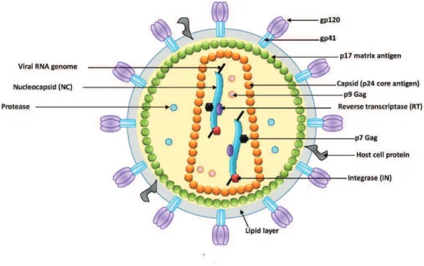
The structure of mature HIV-1 virions was determined by electron microscopy in 1987. HIV forms spherical, membrane enveloped virions with a diameter around 100-120 nm (Gelderblom et al. 1987). The viral envelope is composed of a lipid bilayer. From the HIV-1 envelope around 10 protein projections called spikes can be observed. Spikes are trimers of dimers of the envelope surface (SU, gp120) and transmembrane (TM, gp41) glycoproteins (Aloia et al. 1993; Checkley et al. 2011). During the virus budding a large array of cellular proteins are also incorporated inside the virus including actin, major histocompatibility complex proteins MHC I and MHC II, or intracellular adhesion molecules as ICAM1, ABPQ=R& (Arthur et al. 1992; Ott 2008; Linde et al. 2013). Some of them like cyclophilinA were shown to play a role in the HIV infection process (Thali et al. 1994).
Directly, below the envelope, a matrix shell formed by the oligomerization of ~ 2000 copies of the matrix protein (MA, p17) lines the inner surface of the envelope (Figure 8 for a schematic representation of a mature virion). The conical shaped capsid assembled from the homo-oligomerization of ~ 2000 copies of the capsid protein (CA, p24) is located in the core of the viral particle. The capsid encloses two copies of the viral RNA genome covered by nucleocapsid proteins (NCp7) forming a ribonucleoprotein complex which also contains the viral enzymes protease, reverse transcriptase, and integrase (Frankel & Young 1998; Turner & Summers 1999). Additionally the viral accessory proteins Vpr (around 270 copies per virion (Müller et al. 2000), Vif, and Nef (10 copies/virion, (Welker et al. 1998) are enclosed within the virion in addition to a number of molecules of cellular origin implicated in the virus infection cycle. Among them are found: the reverse transcription primer tRNALys3 (Kleiman et al. 2004), Cyclophillin A (Braaten et al. 1996), actin (Wilk et al. 1999), tRNA synthetase (Cen et al. 2001), and APOBEC3G (Zennou et al. 2004).
25
Figure 8: Schematic structure of the HIV-1 mature viral particle. The HIV virion is represented as a sphere
containing two copies of the RNA genome associated to the nucleocapsid (NC) protein. The ribonucleocapsid is enclosed within a core made of capsid (p24) protein, which is surrounded by a shell of matrix (p17) proteins that associates to the virion envelope. The external glycoprotein is formed of a dimer of the subunits SU (gp120) and TM (gp41). The virus enzymes integrase (IN) and reverse transcriptase (RT), and protease are also indicated. Adapted from (Sherman & Greene 2002).
2.2 Genomic Organization of HIV
2.2.1 Proviral DNA
In order to replicate its genome, HIV needs to integrate a DNA copy (also called proviral DNA or vDNA) of its genome into the host chromosome. This genomic DNA comprises a central region coding for the structural, enzymatic, and accessory proteins flanked on both extremities by non-coding regions responsible for the control of integration, encapsidation and expression of the genome.
26 2.2.1.1 Coding Region
The viral genome codes for fifteen mature proteins. Nine of them, called the enzymatic and structural proteins, are obtained from the cleavage of the three polyproteins Gag, Gag-pol, and Env (Figure 9). The other ones are the regulatory (Tat, Rev) and auxiliary proteins (Nef, Vif, Vpr, Vpu) (Freed 2001; Frankel & Young 1998). HIV proteins and their functions will be further developed within this chapter.
Figure 9: HIV-1 DNA genome scheme. All structural, accessory and enzymatic genes are indicated in addition
to the two LTRs as present in an integrated provirus.
2.2.1.2 Non coding regions
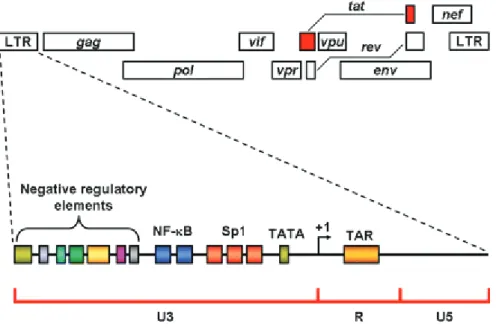
All retroviruses possess at both ends sequences of nucleotides called Long Terminal Repeats (LTRs) derived f/-1&9:%&)(&+!3&'(&Untranslated Region (UTR) at the extremities of the viral genomic RNA (gRNA) (Temin 1982; Piekna-Przybylska et al. 2011). These sequences are important for the integration of the proviral DNA into the host genome, its transcription into viral RNA, as well as the reverse transcription of the gRNA into viral DNA (vDNA) (Pereira et al. 2000; Reicin et al. 1995).
S+0:&TOU&".&3"<"3%3&"!9-&9:/%%&/%@"-!.V& '&H !"#$%&"!&'(J=&U&HU%3$!3+!9J=&and U5 (Unique in )(J.
27
Figure 10: Detailed structure of the LTR. The LTR is divided into three regions U3, R, and U5 (Romani et al. 2010). It contains many binding sites for cellular factors implicated in transcription. Some of these sites such
as the negative regulatory elements down regulate HIV-1 transcription when bound to regulatory proteins. The HIV-1 LTR contains a TATA box, two repeats of the transcription factor NF-W4&4"!3ing site, and three repeats of Sp1 binding site. The start of transcription occurs at the beginning of the R region indicated by (+1).
The U3 region (positions -454 to 0 in HXb2 strain vDNA) is a G- rich proviral DNA sequence which behaves as a promoter for the viral transcription of the integrated viral DNA by the cellular RNA polymerase II. The U3 region is further divided into three region: a core promoter harboring the TATA box and three binding sites for the transcription factor Sp1, an activator domain with two NF-XY&4"!3"!@&."9%., and a modulator domain which binds several cellular and transcriptional factors (Gaynor 1992).
M79:-$@:&4-9:&TOU.&+/%&"3%!9"0+7&"!&.%#$%!0%=&9:%5&+/%&3";;%/%!9&"!&9%/1.&-;&;$!09"-!E&O:%&)(- TOU&3"/%09.&9:%&"!"9"+9"-!&-;&9/+!.0/",9"-!&G:%/%+.&9:%&'(&%!3&".&"!<-7<%3&"!&9:%&07%+<+@%&+!3& polyadenylation of viral transcripts (Klaver & Berkhout 1994; Brown et al. 1991). Transcription of the HIV-1 provirus starts by the binding of cellular factors, including NF-XY=& N,Z=&9:%&OMOM&4-I&4"!3"!@&,/-9%"!=&+!3&ULM&,-751%/+.%&[[=&9-&9:%&,/-1-9%/&/%@"-!&"!&9:%&)\- LTR. Transcription starts at the beginning of the R reg"-!&"!&9:%&)(-LTR and it ends in the R /%@"-!& -;& 9:%& '(-LTR (Romani et al. 2010) resulting in the so-called 5(-and 3(-UTR in the gRNA (Figure 11).
28
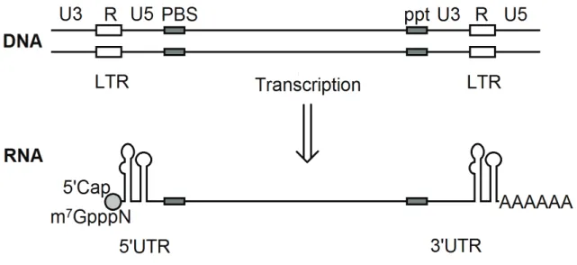
Figure 11: HIV proviral DNA (vDNA) and RNA (gRNA) transcription product. The vDNA is flanked by
long terminal repeats (LTR) containing U3, R and U5 regions in addition to the reverse transcription signals: the primer binding site (PBS) and polypurine tract (PPT). Tra!.0/",9"-!&-;&F[]&BLM&,/-3$0%.&ULM&0+,,%3&+9& )(& %!3&+!3&,-75+3%!57+9%3&+9&'(&%!3&(Das et al. 1998).
2.2.2 Viral RNA (gRNA)
The HIV-1 genome consists of a dimer of two (+) identical RNA strands of 9.2 kb held together noncovalently with interactions involving a limited number of base pairs in the DIS loop of the LTR (Paillart, Shehu-Xhilaga, et al. 2004). These RNA molecules are capped at 9:%"/&)(-%!3&+!3&,-75+3%!57+9%3&+9&9:%"/&'(-end (Figure 11). Each strand of the viral genome has three major coding regions corresponding to the polyproteins Gag, Pol, and Env. The genome also encodes for the regulator (Tat, Rev) and the auxiliary proteins (Nef, Rev, Vif, Vpr, Vpu, and Tat). The coding regions are flanked by noncoding regions or Untranslated regions (UTRs) at both ends -;& 9:%& @%!-1%V& )(- OU& +!3& '(-UTR (Coffin et al. 1997). The !"#$%&.%#$%!0%&"!&)( H )J&".&,/%.%!9&-!75&"!&9:%&)(-UTR whereas the unique sequence in '( H 'J&".&,/%.%!9&-!75&"!&9:%&'(-UTR.
29 2.2.2.1 !"-UTR
O:%& :"@:75& .9/$09$/%3& )(-UTR region is the most conserved part of the HIV genome and is involved in several steps of the viral life cycle including reverse transcription, RNA transcription, genomic packaging, translation, and dimerization (Berkhout 1996; Brasey et al. 2003).
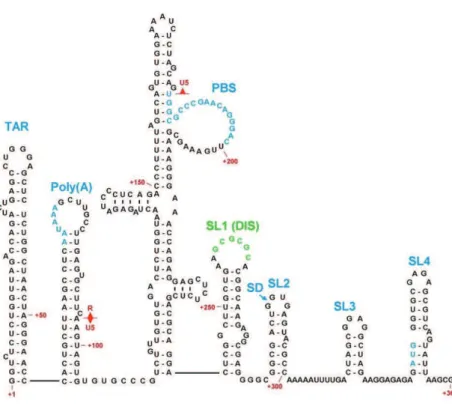
O:%&)(-UTR of the full length gRNA contains in this order (Figure 12): the R region, the U5 /%@"-!=&9:%&,/"1%/&4"!3"!@&."9%&H^YNJ=&+!3&9:%&,+08+@"!@&."@!+7&_E
The repetitive sequence R: O:%&)(-UTR starts with the redundant R region (positions +1 to
95 in the HXb2 strain vDNA), a 96 nucleotide sequence, present at both ends of the viral @%!-1%E& O:%& DZ& ."9%& 0-//%.,-!3.& 9-& OMU& 4%@"!!"!@& +9& 9:%& )(& %!3& where transcription starts and to the cap site. This R region plays an important role in reverse transcription and is divided into two hairpins with distinct functions: TAR and poly(A).
The TAR (Transactivation Responsive Region) hairpin in the R region corresponds to the
first 58 nucleotides of the viral RNA (positions +1 to +58 in the HXb2 strain vDNA). TAR plays a significant role in the Tat-mediated transcription activation of the viral genome from t:%& ,/-1-9%/& 7-0+7"K%3& "!& 9:%& )(-LTR of the proviral genome. When the viral protein Tat is bound to TAR, it promotes the binding of a transcription elongation factor P-TEFb, composed of cyclin T1 and cdk9. Interaction of Tat with TAR and P-TEFb causes the hypophosphorylation of the C-terminal domain of RNA polymerase II hence increasing transcription (Karn & Stoltzfus 2012). Additionally, virus harboring mutations in the TAR region show a reduced ability in reverse transcription and packaging thus reflecting a role of TAR in both processes. However, TAR requirement is conformation dependent (Harrich et al. 2000). Additionally, TAR is important in the dimerization of the RNA genome since opening of the TAR hairpin leads to aberrant RNA dimerization and packaging (Das et al. 2012).
Moreover, the TAR stem-loop has been shown to inhibit translatio!&3"/%09%3&45&9:%&)(-UTR. However, the La autoantigen protein, an RNA binding protein implicated in cap-independent translation initiation of poliovirus RNA (Meerovitch et al. 1989), was shown to bind to the TAR stem-loop and to alleviate the transl+9"-!&/%,/%.."-!&-;&9:%&)(-UTR (Svitkin et al. 1994).
30
The poly(A) hairpin is localized downstream of TAR in the R region (positions +72 to +77
in the HXb2 strain vDNA); it contains the polyadenylation signal (AAUAAA) responsible for 9:%& +33"9"-!& -;& 9:%& ,-75HMJ& 9+"7& +9& 9:%& '(-end of the mRNA. Despite the presence of this sequence on both extremities of the HIV RNA genome; it is functional -!75& +9& 9:%& '(-end G:"7%&"9&".&/%,/%..%3&+9&9:%&)(-end (Brown et al. 1991).
The Unique #$" ! (U5), an 84 nucleotide sequence (positions +96 to +179 in the HXb2 strain
vDNA), is the first sequence to be transcribed during reverse transcription. It is involved in the formation of the LTR required for the integration of the proviral DNA. The U5 region is also implicated in a long-distance base-pairing interaction with a region encompassing the start codon of the Gag open reading frame (Abbink & Berkhout 2003) suggested to regulate the translation of Gag. This structure is seen in one of the HIV-1 leader conformations thought to be implicated in the riboswitch which regulates multiple processes like gRNA dimerization and packaging (Abbink et al. 2005; Ooms et al. 2004).
The Primer Binding site (PBS) region ".& +!& ZQ& !$07%-9"3%& .%#$%!0%& "!& '(& -;& )& HDZQZ& 9-&
+198 in HXb2 strain vDNA). It plays an important role in reverse transcription by serving as a primer binding site for the human primer of reverse transcription: tRNALys3 (Kleiman 2002; Mak & Kleiman 1997).
The leader region or psi region: It is the region located between the PBS and the gag
initiation codon (+236 to +336 HXb2 strain vDNA) and its secondary structure is composed of four hairpins called SL1 to SL4 connected by short linkers (Clever & Parslow 1997; Harrison & Lever 1992). SL1 contains the DIS sequence (Dimer Initiation site, +256 to +261), a 6 nucleotide stem loop, involved in the dimerization of the viral genome through formation of a kissing dimer intermediate with the DIS of another RNA molecule (Skripkin et al. 1994). SL2 contains the Splicing Domain (SD, +281 to +299) with a major splice donor site where all HIV-1 transcripts, with the exception of the full length mRNA, are cleaved at this site generating different viral mRNAs H`(U%"775&%9&+7E&Zaa)J. SL3 is the major packaging ."9%&:+/4-/"!@&9:%&_&:+"/,"!&+!3&".&-;&,+/9"0$7+/&"!9%/%.9&."!0%&"9.&.%#$%!0%&".&:"@:75&0-!.%rved among different strains of HIV-1 (Hayashi & Iwakura 1993). SL4 harbors the gag start codon for the initiation of Gag translation.
31
Figure 12: HIV-1 5' RNA Structural Elements. These are the TAR element, the poly(A) hairpin, the U5-PBS
complex, and stem-loops 124. Nucleotides and numbering correspond to the HIV-1 HXB2 sequence (Russell et al. 2004).
2.2.3 Other Non-coding sequences
Other non-coding sequences exist within the viral genome outside the LTRs.
Polypurine tract (PPT, +8614 to +8638) and central polypurine tract (cPPT, +4318 to
+4350) are purine rich sequences resistant to the RNase H activity of the reverse transcriptase. ^^O&".& +9&9:%&'(&%!3&-;&9:%&<"/+7&@%!-1% upstream of the U3 region, whereas cPPT is at the center of the genome in the open reading frame of the pol gene. During reverse transcription, the genomic viral RNA is degraded with the exception of these two sequences which serve as primers for the synthesis of the (+) DNA strand (Charneau & Clavel 1991; Charneau et al. 1992). In addition to this property, the main role of PPT and cPPT may be to protect the viral genome from DNA editing by the cytidine deaminase of the APOBEC family and other
32 factors (Hu et al. 2010). Both PPTs may counteract these enzymes, which are incorporated into the virions and act as natural defense barriers against HIV infection by causing lethal mutations within the viral genome (Wurtzer et al. 2006). cPPT is responsible during reverse transcription (RT) of the formation of a triple stranded DNA structure called DNA flap. A number of papers underlined the role of cPPT in the nuclear import and uncoating of the pre-integration complex (PIC) since HIV mutants lacking the central DNA flap were found defective in nuclear import which could be restored by re-insertion of the DNA flap sequence (Zennou et al. 2000; Sirven et al. 2000; Rivière et al. 2010; Arhel et al. 2007). However, other works showed that the cPPT is not absolutely necessary for nuclear import (Dvorin et al. 2002; Limón et al. 2002; Marsden & Zack 2007).
The Rev-Responsive Element (RRE, +7710 to +8061): is a 352 nucleotide highly structured
RNA element located in the env gene. Being present on all partially spliced and unspliced viral RNA transcripts, it serves as a docking site for the viral protein Rev associated to nuclear %I,-/9& 0%77& ;+09-/.& HA/1Zb& U+!cO^RJ& /%.$79"!@& "! the nuclear export of these transcripts (Fernandes et al. 2012; Malim et al. 1989; Farjot et al. 1999; Taura et al. 1998).
2.3 Viral Proteins
The viral genome contains three main open reading frames (ORFs) gag, gag-pol, and env which enable the synthesis of the structural proteins, viral enzymes, and envelope proteins respectively. The genome also encodes for six regulatory and auxiliary proteins Nef, Rev, Vif, Vpr, Vpu, and Tat (Freed 2001; Frankel & Young 1998).
The unspliced gRNA can be translated to provide a 55 kDa myristoylated Gag precursor protein (Pr55Gag) which is cleaved according to a highly ordered and controlled mechanism by the viral protease (PR) during the maturation step to yield the matrix p17 (MA), capsid p24 (CA), nucleocapsid p7 (NC) and p6 proteins. In addition, two peptides, SP1 and SP2, are also generated from sequences on both sides of the NCp7 coding region (Bell & Lever 2013).
33 Alternatively, a (-1) ribosomal frameshift can occur at the end of Pr55Gag translation resulting in the extension of the gag ORF and continued translation into the pol ORF generating Pr160 (Gag-Pol), a Gag-Pol fusion protein. This protein is cleaved concomitantly or right after the virus release (Kohl et al. 1988) leading to the viral enzymes protease (PRp12), reverse transcriptase (RTp66/p51), and integrase (INp32) in addition to the Gag cleavage products (Waheed & Freed 2012; Ganser-Pornillos et al. 2008), as a result of a highly complex and controlled mechanism H^%99"9&%9&+7E&Pdd)b&*e!!5f&%9&+7E&PdZ'J.
The Env, auxiliary and regulatory proteins are the translation products of mono-spliced (Env, Vpu, Vpr, Vif) and multi-spliced mRNA (Tat, Rev, Nef). The env ORF encodes for the glycosylated gp160 Env polyprotein precursor which is cleaved later to yield the surface glycoprotein (SU) gp120 and transmembrane protein (TM) gp41 (Decroly et al. 1994; Moulard & Decroly 2000).
The auxiliary and regulatory proteins are generated as a result of alternative splicing of the viral mRNA due to the presence of several donor and acceptor splicing sites. Regulatory proteins Tat and Rev are essential for virus propagation. In contrast, the auxiliary proteins Vif, Vpr, Vpu , and Nef are not necessary for virus propagation in cell cultures, but they seem to play a key role in viral pathogenesis in vivo (Freed 2001; Frankel & Young 1998).
2.3.1 Envelope proteins
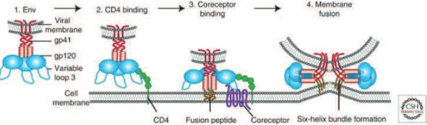
The HIV env gene is expressed from a singly spliced mRNA coding also for Vpu (Hunter & Swanstrom 1990; Freed & Martin 1995). Translation of env to produce envelope proteins takes place on endoplasmic reticulum-bound ribosomes. Concomitantly to its translation on the rough endoplasmic reticulum, the envelope glycoprotein precursor (gp160) is N- and O-glycosylated with oligosaccharide side chains before its oligomerization (mainly as trimers) and trafficking to the Golgi apparatus (Schawaller et al. 1989; Bernstein et al. 1994; Leonard et al. 1989). In the Golgi, cellular furin and furin-like proteases cleave gp160 to yield the mature SU glycoprotein (gp120) and the TM glycoprotein (gp41) (Hallenberger et al. 1992). After cleavage, the two envelope proteins gp120 and gp41 stay associated by non-covalent
34 interactions to form a heterotrimeric complex composed of three copies of gp120 and gp41. The complexes of gp120/gp41 are trafficked to the plasma membrane where they are incorporated as trimeric spikes projecting from the viral envelope (Zhu et al. 2003). The surface glycoprotein gp120 is highly glycosylated (20-35 N-glycosylation sites) and is responsible for virion attachment to the cell surface by binding to CD4 receptors on the surface of infected cells. The transmembrane protein gp41 is less glycosylated (3-5 glycosylation sites) and mediates the anchoring of the envelope protein into the host plasma membrane after receptor binding (Checkley et al. 2011; Hunter & Swanstrom 1990; Freed & Martin 1995).
2.3.2 Gag and its Cleavage Products
Gag (group specific antigen) is a 55 kDa myristoylated multidomain protein. As it was mentioned earlier, during virion maturation, Gag is cleaved by the viral protease (Figure 13) into the matrix protein p17 (MA), capsid protein p24 (CA), nucleocapsid protein p7 (NC) and p6 protein in addition to the spacer peptides SP1 and SP2 (Ganser-Pornillos et al. 2008; Waheed & Freed 2012). The Gag protein is the central actor in particle formation. The role of Gag in assembly will be elucidated within the chapter 3.2.4.
Figure 13: Model of the HIV-1 Gag polyprotein showing MA, CA with its N-terminal and C-terminal
domains (NTD/CTD) and NC domain. Protease cleavage sites are indicated by arrow heads (Ganser-Pornillos et al. 2008).
35 2.3.2.1 Matrix (MA)
The Matrix protein is the N-terminal component of the Gag and Gag-Pol polyproteins. It is a multifunctional protein with several functions in both the early and late phases of viral replication. In a mature virus particle, the 132 amino acids MA protein lines the inner surface of the viral envelope (Frankel & Young 1998).
The MA domain targets Gag to its assembly site at the plasma membrane. A myristyl (myr) group cotranslationally attached to the N-terminus of the protein (Bryant & Ratner 1990) is responsible for anchoring Gag to the inner leaflet by its insertion into the plasma membrane. However, myristoylation alone is not sufficient for stable membrane binding (Yu et al. 1992). The myr group synergizes with a highly basic patch of amino acid residues to facilitate Gag anchoring to the plasma membrane through electrostatic interactions with the acidic phospholipids in the plasma membrane (Saad et al. 2006). Mutations blocking myristoylation or affecting the basic patch lead to inefficient Gag targeting to the plasma membrane (Ghanam et al. 2012; Bukrinskaya 2004).
Gag membrane binding is thought to be /%@$7+9%3& 45& +& >15/".957& .G"90:& 1%0:+!".1?& G:%/%& the myristate shifts between two conformations (Figure 14): the myristyl-exposed (myr e) and myristyl- sequestered (myr s) states (Spearman et al. 1997). The exposure of this myr is dependent on protein trimerization and factors causing protein self-association such as increasing Gag concentration or RNA binding. Thus, a MA monomer is in the myr (s) state. On the other hand, MA trimerization triggers the exposure of the myristyl group (Zhou & Resh 1996; Tang et al. 2004). Therefore, HIV-1 Gag multimerization is coupled to efficient membrane binding through mediating the exposure of the myristyl group (Resh 2004; Ono et al. 2005; Dalton et al. 2005). Moreover, it was shown that MA binds preferentially to phosphatidylinositol-4,5-biphosphate (Ptdlns(4,5)P2), a phospholipid enriched in the inner leaflet in the plasma membrane (Ono et al. 2004). In fact, depletion of Ptdlns (4,5)P2 from the plasma membrane leads to the accumulation of Gag at the late endosomes rather than at the plasma membrane. Moreover, this interaction also triggers the exposure of the myristyl switch suggesting a possible mechanism for Gag targeting to lipid domains where Ptdlns (4,5)P2 is enriched (Saad et al. 2006).
36
Figure 14: Myristyl switch as proposed by Tang et al. In monomeric Gag, the myristyl is sequestered inside
the matrix domain of Gag; however, upon trimerization the myristyl group is exposed (Resh 2004).
Additionally, in most but not all cell lines, the MA domain of Gag is required for the incorporation of Env proteins into the viral particles. This cell dependent requirement of MA for gp41 incorporation suggests that host factors are involved in the interaction between MA and gp41 cytoplasmic tail and thus, required for Env incorporation into the virus (Dorfman et al. 1994; Freed & Martin 1996a; Murakami & Freed 2000).
Although not clearly understood, MA as a domain of the polyprotein Gag seems also to play a role in viral gRNA packaging. This is attributed to its basic RNA binding domain (aa 26-30) able in the absence of the NC domain, to drive Gag binding to viral and cellular RNAs (Burniston et al. 1999; Ott et al. 2005). The MA domain of Gag serves thus as one of the scaffolds that bring together Gag, RNAs, and Env to the plasma membrane.
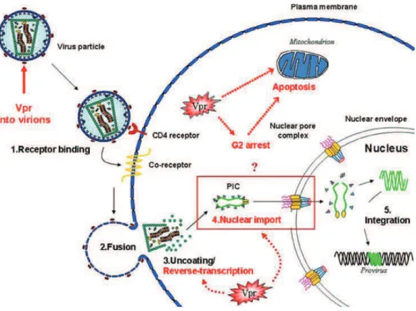
As a part of the preintegration complex, the mature MA protein is also involved in the early steps of the life cycle. Since MA bears two nuclear localization signals (NLS) (Bukrinsky, Haggerty, et al. 1993; Haffar et al. 2000), it may contribute to the import of the preintegration complex (Gallay et al. 1996).
2.3.2.2 Capsid (CA)
Capsid is the second domain of the Gag polyprotein (mentioned CA as a domain of Gag or CAp24 as the mature form). Around 2000 CAp24 molecules arranged in hexamers form the
37 viral capsid, the virion core. CAp24 is a 24 kDa protein structurally divided into two independently folded domains: the N-terminal domain (NTD) and the C- terminal domain (CTD) connected by a short flexible linker. The capsid plays roles both in the early and late steps of the viral life cycle (Frankel & Young 1998; Waheed & Freed 2012).
A loop in the NTD interacts with the cellular protein cyclophilin A (CypA) leading to incorporation of the latter into HIV virions (Thali et al. 1994; Luban et al. 1993; Franke et al. 1994). Knocking down CypA or inhibiting its incorporation into virions caused a decrease in infection indicating that CypA is important in the early infection steps (Braaten et al. 1996; Braaten & Luban 2001). It is hypothesized that CypA could stabilize the trans conformation of the G89-P90 bond in CA and thus, affect the viral core stability (Bosco et al. 2002). It is also believed that CypA, at high CypA:CA ratio, could modulate CA-CA interactions within the lattice of the core, thereby causing its destabilization (Grättinger et al. 1999; Gamble et al. 1996). CAp24 is also a target of the 0%77$7+/& /%.9/"09"-!& ;+09-/& OU[g)h& G:ich binds to and targets the core to the proteasome early in the infection (Malim & Bieniasz 2012).
The CTD of CA bears a stretch of 20 amino acids labeled as the Major Homology Region (MHR) due to its conservation across unrelated viruses (Wills & Craven 1991). The CTD plays a role mainly in assembly and is significant for capsid dimerization and Gag oligomerization (Lingappa et al. 2014).
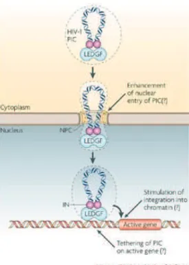
Furthermore, CA plays also a positive role in the viral genome nuclear import. Several genome-wide siRNA studies identified Nup358, Nup 153, and the nuclear transport factor (TNPO3) as required for HIV-1 infection (Brass et al. 2008; König et al. 2008). This requirement was linked to CA where a specific mutation in CA determined the HIV-1 ability to use Nup 153, TNPO3, and to some extent Nup 358 (Lee et al. 2010). It was proposed by Price et al. that CA binding to the cellular protein cleavage and polyadenylation specific factor 6 (CPSF6) facilitates its use of nuclear import factors such as TNPO3 and nucleoporins. Inaddition, supporting evidence indicates a role of CA in integration efficiency, and chromatin targeting (Zhou et al. 2011; Valle-Casuso et al. 2012; Lee et al. 2010; Schaller et al. 2011).
38 2.3.2.3 Nucleocapsid (NC)
NC is a 55 amino acid basic protein characterized by two highly conserved zinc finger motifs connected by a basic sequence (Thomas & Gorelick 2008; B. P. Roques et al. 1997). NC exists in the cell and the virus as a domain of the Gag polyprotein (NC) and as a mature protein (NCp7). By binding to nucleic acids, NC and NCp7 are involved in many processes including reverse transcription, genomic RNA encapsidation and assembly of the viral particle by promoting Gag oligomerization. It coats the viral RNA protecting it from nucleases (Darlix 1995, Berkowitz 1993, Dorfmann 1993, Gorelick 1988). NC/ NCp7 will be discussed in more details in Chapter IV.
2.3.2.4 P6
The p6 domain comprises the 51 C-terminal amino acids of Gag and is important for the incorporation of the viral protein Vpr into the virion. It harbors two late (L) domains PTAP and YPXnL (Meng & Lever 2013), whose roles will be described in the next chapter.
2.3.2.5 Spacer peptides SP1 and SP2
SP1 and SP2 are two cleavage maturation products of Gag. SP1 (also called p2) is located between the CA and NC domains whereas SP2 (also called p1) separates the NC region from p6. The precise role of these spacers is still not clarified; however, they seem to regulate the protease cleavage rate (Pettit et al. 1994; Lee et al. 2012). Furthermore, SP1 has been shown to be essential for Gag polymerization and infectivity (Kräusslich et al. 1995) since any mutation in the first residues of SP1 destroys Gag ability to assemble. In link with these two properties, this spacer is supposed to stabilize the immature-like hexamer and its proteolysis is presumed to destabilize the Gag lattice leading to the formation of conical shaped mature capsid lattice (Gross et al. 2000; Datta et al. 2011; Ganser-Pornillos et al. 2008).
39
2.3.3 Viral Enzymes
2.3.3.1 Protease (PR)
The HIV-1 protease belongs to the family of aspartyl proteases with an active site formed at the interface of the two subunits of the active homo dimer (Navia et al. 1989; Wlodawer et al. 1989). It is released from the Gag-pol precursor by an autocatalytic mechanism. As the virus buds from the plasma membrane, immature particles are noninfectious. Gag and Gag-pol must be cleaved by the viral PR leading to conformational rearrangements within the particles producing mature infectious viruses. The action of PR is detailed in the maturation section of this manuscript (chapter 3.2.5).
Because PR activity regulation is crucial for virus infectivity, PR has been a prime target for the development of anti-HIV drugs. Nowadays several drugs are commercialized and included into HAART. However, drug resistance has emerged due to the genetic variability of the virus leading to selection of strains which can replicate in the presence of those drugs (Sayer et al. 2010; Broder 2010; Doherty et al. 2011).
2.3.3.2 Reverse Transcriptase (RT)
The name of the HIV-1 virus family is due to this enzyme since these viruses need to integrate their genome into the host chromosome and thus to reverse transcribe their gRNA into double DNA. RT is a heterodimer composed of the subunits p66 (560 aa) and p51 (450 aa), both derived from Gag-pol. p66 and p51 share the same amino terminus sequence. The larger subunit, p66 contains the active sites for both the DNA polymerization and RNase H activities. The p51 subunit is considered to mainly play a structural role (Wang et al. 1994). Reverse transcriptase is a multifunctional enzyme which presents RNA-dependent and DNA-dependent DNA-polymerase activities. It presents also a polymerase DNA-dependent and independent ribonuclease H activity that degrades the RNA strand from the RNA:DNA heteroduplex during and after reverse transcription, respectively (Peliska & Benkovic 1992; Gopalakrishnan et al. 1992; Peliska et al. 1994; Goff 1990). Reverse transcription is presented in details in part 3.1.2 of this manuscript.
40 RT has been the first target for anti-HIV drug design. Two major classes of drugs target RT: nucleoside/nucleotide analogue reverse transcriptase inhibitors (NRTIS) such as AZT and non-nucleoside reverse transcriptase inhibitors (NNRTIs) such as nevirapine (Frankel & Young 1998). At least two molecules from these classes are combined to a third antiviral drug targeting another HIV protein in the classical anti-HIV cocktails.
2.3.3.3 Integrase (IN)
HIV integrase is a 32 kDa protein (288 residues) encoded by Gag-pol, like the other viral enzymes. IN is active probably as a tetramer composed of two IN dimers bound to DNA (Ellison et al. 1995). IN is composed of three structural and functional domains: an N-terminal zinc binding domain (HHCC type) that facilitates oligomerization (Bushman et al. 1993), a catalytic central domain which adopts an RNase H fold and a C-terminal DNA binding domain involved in IN and DNA contact (Chiu & Davies 2004). IN is essential for the incorporation of vDNA into the chromosomal DNA of the target cell. The mode of action of integrase in presented in the integration in chapter 3.1.3 of this manuscript.
In addition to integration, IN was reported to be implicated in reverse transcription (Wu et al. 1999; Zhu et al. 2004) where it increases the processivity of RT and suppresses the formation of pause products (Dobard et al. 2007). It also enhances reverse transcription via its interaction with cellular proteins such as SIP2/Gemin 2 and cellular dynein light chain (Jayappa et al. 2015). IN may play also a role in processes nuclear import of the PIC to the nucleus of the host cell, and chromatin targeting (Ao et al. 2010; Hendrix et al. 2011). As the other enzymes, IN is also a target for antiretroviral drugs with several molecules approved by the FDA, as for example raltegravir in 2007.
2.3.4 Regulatory and auxillary proteins
2.3.4.1 Nef (Negative Regulatory Factor)
Nef gene encodes a 27 kDa (206 residues) N-terminally myristoylated protein associated with the cytoplasmic face of cellular membranes (Niederman et al. 1993). Nef is abundantly produced early in the infection when its mRNA represents three quarter of the total viral load mRNA of the cell (Klotman et al. 1991). Despite its name as negative factor, Nef is defined as
41 a pathogenic factor since a deletion in nef gene greatly reduced the severity of infection in humans or rhesus macaques infected by HIV-1 or SIV (Kestler et al. 1991; Kirchhoff et al. 1995). Nef plays multiple roles in the viral life cycle:
A) Nef downregulates CD4 receptors at the surface of infected cells by three pathways. First it redirects some CD4 from the trans-Golgi network to the endosomal compartment, second it triggers the endocytosis of CD4 which are at the membrane and third, it targets CD4 from the endosome to the lysosome for degradation (Kim et al. 1999; Piguet et al. 1998; Mangasarian et al. 1997). CD4 down regulation by Nef i) counteracts the inhibitory effect induced by high CD4 expression level in HIV-1 producer cells (Ross et al. 1999), ii) liberates Lck protein kinase bound to CD4 altering T-cell ability to respond to antigens and cytokines (Chrobak et al. 2010) and iii) prevent potentially lethal superinfection (Michel et al. 2005).
B) Nef downregulates MHCI molecules on the cell surface by a still uncertain mechanism, therefore blunting cytotoxic T cell (CTL) recognition of infected cells and escaping the immune response (Collins et al. 1998).
C) Nef delays the endocytosis and recycling of the T cell receptor (TCR-CD3). TCR is an important component of the immunological synapse formed between antigen presenting cells (APCs) and T cells for antigen recognition. Retarding endocytosis of TCR-CD3 results in less efficient synapse formation (Iafrate et al. 1997).
D) Nef enhances HIV infectivity by facilitating the penetration of the viral core into the cell cortical actin network known as a barrier for intracellular parasitic organisms (Campbell et al. 2004).
2.3.4.2 Vpr (Viral Protein R)
Vpr is a 14 kDa (96 residues) multitask protein packaged into mature virions through the interaction with the p6 domain of Gag. Despite its small size, many functions have been attributed to Vpr (Figure 15), including cell cycle arrest, apoptosis, nuclear import of the PIC, and accuracy of reverse transcription (Romani & Engelbrecht 2009; Guenzel et al. 2014). The HIV-1 genome encodes Vpr whereas the HIV-2 genome encodes both Vpr/Vpx proteins. Both