HAL Id: hal-01743531
https://hal.archives-ouvertes.fr/hal-01743531
Submitted on 5 Dec 2020
HAL is a multi-disciplinary open access
archive for the deposit and dissemination of sci-entific research documents, whether they are pub-lished or not. The documents may come from teaching and research institutions in France or abroad, or from public or private research centers.
L’archive ouverte pluridisciplinaire HAL, est destinée au dépôt et à la diffusion de documents scientifiques de niveau recherche, publiés ou non, émanant des établissements d’enseignement et de recherche français ou étrangers, des laboratoires publics ou privés.
Testing the application of sea temperature proxies for
the Ordovician
Carys Bennett, Mark Williams, Melanie Leng, Martin Lee, Magali Bonifacie,
Damien Calmels, Richard Fortey, John Laurie, Alan Owen, Alex Page, et al.
To cite this version:
Carys Bennett, Mark Williams, Melanie Leng, Martin Lee, Magali Bonifacie, et al.. Oxygen isotope analysis of the eyes of pelagic trilobites: Testing the application of sea temperature proxies for the Ordovician. Gondwana Research, Elsevier, 2018, 57, pp.157 - 169. �10.1016/j.gr.2018.01.006�. �hal-01743531�
Bennett, C. E. et al. (2018) Oxygen isotope analysis of the eyes of pelagic
trilobites: testing the application of sea temperature proxies for the
Ordovician. Gondwana Research, 57, pp. 157-169.
(doi:
10.1016/j.gr.2018.01.006
)
This is the author’s final accepted version.
There may be differences between this version and the published version.
You are advised to consult the publisher’s version if you wish to cite from
it.
http://eprints.gla.ac.uk/159426/
Deposited on: 26 March 2018
Enlighten – Research publications by members of the University of Glasgow http://eprints.gla.ac.uk
1
Oxygen isotope analysis of the eyes of pelagic trilobites: testing the application of sea temperature
1
proxies for the Ordovician
2
Bennett, Carys E. 1,2*, Williams, Mark2, Leng, Melanie J.3, Lee, Martin, R.4, Bonifacie, Magali5,6, Calmels, 3
Damien5,7, Fortey, Richard A.8, Laurie, John R.9, Owen, Alan W.4, Page, Alex A.2, Munnecke, Axel10, 4
Vandenbroucke, Thijs R. A.1,11 5
1 Evo-Eco-Paleo, UMR 8198 du CNRS, Université Lille 1, Avenue Paul Langevin, bâtiment SN5, 59655 Villeneuve
6
d'Ascq cedex, France.
7
2School of Geography, Geology and the Environment, University of Leicester, University Road, Leicester, LE1 7RH,
8
UK.
9
3 NERC Isotope Geosciences Laboratory, British Geological Survey, Keyworth, Nottingham, NG12 5GG, UK and
10
Centre for Environmental Geochemistry, School of Biosciences, Sutton Bonington Campus, University of
11
Nottingham, Loughborough, LE12 5RD, UK.
12
4School of Geographical & Earth Sciences, University of Glasgow, Gregory Building, Lilybank Gardens, Glasgow,
13
G12 8QQ, UK.
14
5Institut de Physique du Globe de Paris, Equipe Géochimie des Isotopes Stables, Sorbonne Paris Cité, Univ Paris
15
Diderot, UMR 7154 CNRS, F-75005 Paris, France.
16
6Institut de Physique du Globe de Paris, Observatoire Volcanologique et Sismologique de Guadeloupe, Le Houëlmont,
17
97113 Gourbeyre Guadeloupe, France.
18
7Université Paris-Sud, Laboratoire GEOPS, UMR 8148 CNRS, F-91405 Orsay, France.
19
8The Natural History Museum, Cromwell Road, London SW7 5BD, UK.
20
9Department of Biological Sciences, Macquarie University, NSW 2109, Australia.
21
10Universität Erlangen-Nürnberg, GeoZentrum Nordbayern, Fachgruppe Paläoumwelt, Loewenichstrasse 28, D –
22
91054, Erlangen, Germany.
23
11Department of Geology, Ghent University, Krijgslaan 281/S8, 9000 Ghent, Belgium.
3
Research Highlights
1
1. Preservation assessment shows some well-preserved Ordovician trilobite eyes 2
2. Ordovician trilobite eyes yield 18O values similar to Ordovician brachiopods 3
3. SIMS and clumped isotope results indicate diagenetic alteration of trilobites 4
4. Classic protocols to assess preservation may be inapt for most ancient carbonates 5
6
Abstract
7
The oxygen isotope composition of well-preserved trilobite eye calcite, retaining its original optical 8
properties, represents a possible source of information on Paleozoic sea temperatures. Species of the 9
epipelagic telephinid genera Carolinites and Opipeuterella from strata of Early to Middle Ordovician age in 10
Spitsbergen and Australia were analyzed, and compared with benthic asaphid species. Scanning electron 11
microscope (SEM), cathodoluminescence (CL), electron microprobe and Electron Backscatter Diffraction 12
(EBSD) techniques were used to assess eye preservation prior to isotope analysis. Some apparently well-13
preserved eyes are identified from the Valhallfonna (Spitsbergen) and Emanuel (Australia) formations. The 14
eyes show a wide variation in 18O values: –6.2‰ to –9.8‰ for the Valhallfonna Formation, –3.2‰ to – 15
10.4‰ for the Emanuel Formation, and –3.6‰ to –7.4‰ for the Horn Valley Siltstone (Australia). Intra-eye 16
Secondary Ion Mass Spectrometry (SIMS) isotope results reveal an even larger range in 18O in some 17
specimens (18O of –2.4‰ to –10.4‰), suggesting that the trilobite eyes have undergone cryptic 18
recrystallization. A sub-set of trilobite cuticle from the three formations were analyzed for their carbonate 19
clumped isotope compositions (47), and yielded crystallisation temperatures above 50oC, consistent with 20
diagenetic alteration. The SIMS and 47 results suggest that classic preservation assessment protocols for the 21
stable isotope study of deep-time carbonate samples may be insufficient, especially for these techniques. 22
There is a need for extensive microstructural characterisation of lower Paleozoic biogenic carbonates, by 23
techniques including EBSD, SIMS and 47, before their stable isotope signatures can be used with certainty 24
in paleoclimate studies. 25
4
Index terms: Stable isotope geochemistry; marine geochemistry; instruments and techniques;
1
biomineralization; petrography, microstructures, and textures 2
Keywords: Trilobite, oxygen isotopes, Ordovician, paleotemperature, microstructure
3
1. Introduction
4
Telephinid trilobites are amongst the few unequivocally pelagic organisms preserved in Ordovician rocks 5
that have a carbonate biomineralized skeleton. The most common telephinid, Carolinites genacinaca, had a 6
global paleoequatorial distribution during the Early Ordovician (Floian) and the morphology of the 7
Carolinites body plan, eye shape and eye position suggest that species were epipelagic, living near the sea
8
surface in the mixed layer (McCormick and Fortey, 1998, 1999). The holochroal eyes of telephinid trilobites 9
are composed of hundreds of interlocking lenses of calcite and are sufficiently large to provide sufficient 10
material for stable isotope analysis, which in turn might conceivably yield an estimate of Ordovician sea 11
temperature. 12
Trilobite cuticle has been used in Ordovician isotope paleoclimate studies in the same way as brachiopod 13
calcite (Brand, 2004; Finnegan et al., 2011). Studies on the ultrastructure of the biomineralized trilobite 14
exoskeleton (the cuticle) show that original features such as horizontal lamination, relict organic material, 15
and pore canals can be preserved (Dalingwater, 1973; Dalingwater et al., 1991), although evidence for 16
diagenetic alteration is also common (Wilmot, 1990; Budil and Hörbinger, 2007). Investigations into the 17
original chemical and isotope compositions of trilobite cuticles indicate that they were formed from low-Mg 18
calcite (Wilmot and Fallick, 1989; Lee et al., 2012; McRoberts et al., 2013; Teigler and Towe, 1975). Some 19
specimens have been recorded with an intermediate-Mg calcite composition (Brand, 2004; McAlister and 20
Brand, 1989). However, as the preservation of the cuticle ultrastructure in these latter specimens was not 21
examined in detail, the possibility remains that the chemical composition of these cuticles may reflect 22
diagenetic recrystallization in Mg-rich fluid. The lenses in the schizochroal eyes of phacopine trilobites have 23
5 been identified as being originally composed of high-Mg calcite of ~7.5 mol% MgCO3, while the cuticle 1
comprises low-Mg calcite of ~1.4 to 2.4 mol% MgCO3 (Lee et al., 2012). 2
As the physical properties of a functional calcite lens are well understood, even if trilobite eyes do not have 3
modern analogues, it is possible to assess their degree of preservation (Torney et al., 2014). Previous work 4
on the ultrastructure of schizochroal trilobite eyes using EBSD has revealed that the original structure of the 5
eye lenses can be identified (Lee et al., 2007, 2012; Torney et al., 2014). This finding suggests the 6
possibility that the original chemical and isotope composition of eye calcite may also be retained. High-7
resolution SEM analysis of the lenses of phacopine trilobite eyes show that the intralensar bowl (present 8
only in schizochroal eyes) was composed of high-Mg calcite with micro-crystals of dolomite indicating 9
diagenetic alteration (Lee et al., 2007; 2012). SIMS analysis can be used to target small (less than 20 m 10
diameter) areas of a fossil in situ, such as trilobite eye lenses, and SIMS analysis of 18O has been 11
successfully applied to the analysis of conodont microfossils (Wheeley et al., 2012). 12
Biogenic proxies that have been hitherto used to provide Early and Middle Ordovician marine 13
paleotemperatures are the oxygen isotopic compositions of brachiopod calcite and conodont apatite (Shields 14
et al., 2003; Trotter et al., 2008; Wadleigh and Veizer, 1992), along with carbonate clumped isotope 15
compositions (47) from trilobite, brachiopod and coral calcite for the Late Ordovician (Finnegan et al., 16
2011). However, some of these results are controversial. For instance, brachiopod isotope data from the 17
Lower Ordovician can have very low 18O (down to –10‰). This result implies very high (up to + 60oC) 18
seawater temperatures [if it is assumed that seawater 18O was similar to today], or diagenetic alteration at 19
higher temperatures than that of the ocean, diagenetic alteration by a different fluid, or that seawater 18O 20
was substantially different from present (Veizer et al., 1999; Shields et al., 2003), or a combination of those 21
alternatives. In contrast, 18O from conodont apatite from the Ordovician suggests that seawater 22
temperatures were between 30-40oC if a value similar to modern 18Owater of –1‰VSMOW is used (Trotter et 23
al., 2008). Oxygen isotope data can also reflect other environmental parameters; for example, Bickert et al. 24
6 (1997) showed that the 18O values of well-preserved Silurian brachiopods reflect salinity differences due to 1
varying fresh-water input rather than temperatures. Nevertheless, the development of proxies that can 2
provide robust and reasonable estimates of seawater temperature for the early Paleozoic is important for 3
many reasons. For example, such information can help constrain the environmental feedbacks or triggers of 4
the Great Ordovician Biodiversification Event (Trotter et al., 2008; Amberg et al., 2016), and it also 5
represents one of the few available proxies for ground-truthing General Circulation Models of early 6
Paleozoic climate (Vandenbroucke et al., 2009; Pohl et al., 2014; 2016). Changes in seawater temperatures 7
may have had significant effects on the ability of organisms to biomineralize (Pruss et al., 2010). The 47 8
paleothermometer has great potential for more accurate paleotemperature reconstruction because it is 9
independent of the 18O of mineralising fluids (Ghosh et al., 2006). Usefully, the recrystallization 10
temperature of diagenetic calcite recorded by 47 is thought to remain stable over hundreds of millions of 11
years, at temperature histories below ~250oC (Ghosh et al., 2006). However, in deeply buried sedimentary 12
deposits or carbonates having a moderate-temperature burial history (~100°C on timescales of hundreds of 13
million years) the closed-system solid-state diffusive reordering of atoms can modify 47 towards lower 14
values and higher apparent equilibrium temperatures (Passey and Henkes, 2012; Henkes et al., 2014; Stolper
15
and Eiler, 2015). 16
Here we test whether: (i) the unique functional morphology and microstructure of trilobite eyes allows for 17
the accurate assessment of the preservation of their calcite lenses; and (ii) the subsequent 18O analyses of 18
well-preserved eyes of epipelagic trilobites could serve as a sea temperature proxy. Trilobite eye 19
microstructure and composition were compared to those of the trilobite cuticle. Two isotope methods were 20
used on trilobite material from three sedimentary formations considered to have experienced relatively 21
limited diagenesis: (i) conventional determination of oxygen isotope compositions, and (ii) carbonate 22
clumped isotope 47 analysis. 23
2. Materials Utilised
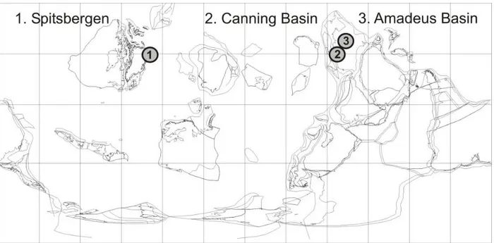
7 Trilobite eyes were analyzed from three broadly coeval Ordovician formations from separate, equatorial 1
basins: 1) Valhallfonna Formation, Spitsbergen; 2) Emanuel Formation, Canning Basin, Australia; and 3) 2
Horn Valley Siltstone, Amadeus Basin, Australia (Figure 1). For all formations, the following materials 3
were analyzed for 18O, 13C and clumped isotope 47 compositions, where possible: pelagic and benthic 4
trilobite eyes and cuticle, sedimentary carbonate (host rock) and coarsely crystalline diagenetic calcite 5
cement (spar). For comparison, additional trilobite cuticle was analyzed from the Upper Member of the 6
Dalby Limestone of Västergötland, Sweden, as this basin has experienced significantly higher burial 7
temperatures. The Supplementary Material (extended version) details further the type and number of 8
analyses from each formation. 9
10
Figure 1. Paleogeographical map of trilobite sample sites: 1) Spitsbergen, Norway (Valhallfonna 11
Formation); 2) Canning Basin, Western Australia (Emanuel Formation); and 3) Amadeus Basin, Northern 12
Territory, Australia (Horn Valley Siltstone). Paleogeographic reconstruction for the Middle Ordovician, 13
470Ma, Galls Projection, cropped at latitude 30oN, BugPlates: www.geodynamics.no. 14
15
2.1 Valhallfonna Formation
8 Forty-three trilobite eyes were examined from eleven samples collected from the Floian (Arenigian)
1
Valhallfonna Formation of Ny Friesland, Spitsbergen (Fortey, 1975; Fortey and Bruton, 1973). Four species 2
of the telephinid Carolinites were studied (C. angustagena, C. genacinaca, C. nevadensis and C. sibiricus), 3
along with cuticle (but no eyes) from benthic olenids (species indeterminate). The depositional environment 4
is interpreted to be a marine shelf setting (Fortey and Barnes, 1977). The Ny Friesland succession is thought 5
to have undergone shallow burial, with Conodont Alteration Index (CAI) values of 1 suggesting maximum 6
temperatures of 90oC (Bergström, 1980). Calcite veins and calcite spar cements within the cavities of 7
ostracod fossils and in pore spaces in the limestone indicate evidence for some diagenetic alteration. 8
2.2 Emanuel Formation
9
Trilobites were studied from the Floian (Bendigonian) Emanuel Formation of the Canning Basin, Western 10
Australia (Laurie and Shergold, 1996). Fifty-one trilobite eyes were examined from the telephinid 11
Opipeuterella sp. and benthic asaphids (species indeterminate), occurring in five samples from the type
12
section of the Emanuel Formation. The burial history of the Ordovician Canning Basin indicates low 13
thermal maturation, with a CAI index of 1 (Nicoll et al., 1993) and Apatite Fission Track Analysis (AFTA) 14
indicating temperatures of ~100oC during the Late Devonian/Early Carboniferous (Arne et al., 1989). 15
2.3 Horn Valley Siltstone
16
Trilobites were studied from the Floian to Dapignian (Bendigonian to Yapeenian) Horn Valley Siltstone of 17
the Amadeus Basin, Northern Territory, Australia, which can be partly correlated to the upper part of the 18
Emanuel Formation from the Canning Basin (Laurie, 2006). Over 100 trilobite eyes from Carolinites 19
genacinaca and benthic asaphids (species indeterminate) were examined from three samples collected from
20
a field section at Mt Olifent (fig. 6 in Laurie, 2006). The burial history for the Amadeus Basin has been 21
determined by AFTA and organic maturity data that indicate maximum burial during the Late 22
Carboniferous, with Ordovician strata subjected to maximum temperatures of 140oC (Gibson et al., 2007). 23
9
2.4 Västergötland
1
Two test samples of Telephina cuticle and its host rock were analyzed for 47 from the Upper Member of the 2
Dalby Limestone, Sandbian (Late Ordovician), in Västergötland, Sweden, introducing contrasting data from 3
the opposite end of the preservation spectrum. The Västergötland rocks have been heated by local Permian 4
intrusions, giving CAI values of 6 to 7 and temperatures of over 300oC (Bergström, 1980). 5
3. Methods
6
3.1 Preservation Assessment Protocol
7
In total, 198 trilobite eyes were examined under reflected light using a binocular microscope and by SEM, 8
which allowed an initial assessment of eye lens integrity and preservation. Of these, 34 specimens 9
representing a range of preservation states were selected for thin section analysis, based on the eye size 10
(greater than 1.8 mm length), to enable a detailed preservation assessment. Polished thin sections were 11
examined by cathodoluminescence (CL) microscopy and the geochemical variation in eye specimens was 12
quantified using electron microprobe facilities at the universities of Lille (France) and Leicester (UK). 13
Trilobite eye specimens were imaged by SEM under high vacuum using Secondary Electron and Back 14
Scattered Electron detectors. Electron Backscatter Diffraction (EBSD) analysis of polished thin sections was 15
undertaken at the University of Glasgow to examine intra-lens variations in crystallographic orientations 16
(Torney et al., 2014). 17
3.2 Isotope Analyses
18
A total of 182 isotope analyses were undertaken on trilobite eyes, cuticle and host rock from all three 19
formations (Appendix C). 18O, 13C and 47 analyses were performed using three methods: 1. Conventional 20
isotope analysis of carbonate powder, with lens/cuticle extraction using both a hand-held dental drill and 21
automated micro-mill, at the British Geological Survey and at the University of Erlangen, Germany; 2. 22
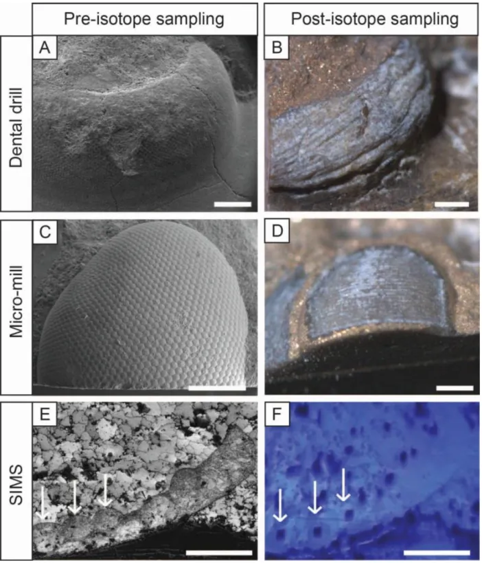
Secondary Ion Mass Spectrometry (SIMS) analysis of 15 m diameter areas of thin sections (Figure 2) at 23
10 the Centre de Recherches Pétrographiques et Géochimiques (CRPG-CNRS) facility in Nancy, France; 3. 1
Clumped isotope analysis (47) and 18O and 13C analysis of carbonate powder at the Institut de Physique 2
du Globe de Paris (IPGP, Stable Isotope team). The extraction of material by dental drill is subject to human 3
error and samples likely contain some underlying sediment matrix material. Micro-mill extraction is 4
accurate to an error of ~ 5 m, however it is possible that underlying sediment is sometimes sampled. SIMS 5
analysis is the preferred technique in this context because it can accurately sample intra-lens material from 6
thin sections. For a more detailed account of the methods, refer to the Supplementary Methods (extended 7
version) section. 8
11 1
Figure 2. Lens extraction methods for isotope analysis. A-B) Hand drilling of lenses using a dentist drill for 2
conventional isotope analysis (specimen HV_sp2, SE and light microscope images, respectively); C-D) 3
Micro-mill automated drilling for conventional isotope analysis (specimen T_178_sp10, SE and light 4
microscope images, respectively); E-F) Secondary Ion Mass Spectrometry (SIMS) analysis of a well-5
preserved eye (specimen A_178_sp1, EBSD image quality map and light microscope image, respectively). 6
12 Arrows indicate examples of lenses that were analyzed for oxygen isotopes. Scale bars 500 m (A-D) and 1 100 m (E-F). 2 4. Results 3 4.1 Preservation 4
The majority of the trilobite eyes examined had intact lenses that look superficially well-preserved. The 5
hexagonal lenses are arranged in a single layer, which is on average 70 m in thickness. Individual eye 6
lenses are on average 50 m in diameter, with no mineral partitions between them. Lens size varies between 7
species, with telephinids having significantly larger lenses than asaphids, and moreover, a different eye 8
morphology. Here we describe the results of the preservation assessment protocol from 34 specimens 9
examined in thin section. Specimens are classified as ‘well-preserved’ or ‘poorly preserved’ based on their 10
microstructural and geochemical properties (Table 1). 11
12 13
13 Table 1. Trilobite eye preservation
1
Specimen Trilobite Species CL SEM Observations EBSD Observations Preservation
Valhallfonna Formation, Spitsbergen
1.1A_sp1 Carolinites sibiricus n/a lenses fractured, silicified n/a poor
1.1B_sp1 Carolinites sibiricus L pervasive porosity, recrystallized, lens boundary overgrowths lenses undefined, major recrystallization poor 1.1B_sp2 Carolinites sibiricus L pervasive porosity, recrystallized, fractured lenses no lens boundaries preserved, major recrystallization poor 1.2C_sp1 Carolinites genacinaca L pervasive porosity, recrystallized, zoned calcite, micro-dolomite lenses defined, major recrystallization poor 1.2C_sp2 Carolinites genacinaca L pervasive porosity, recrystallized, zoned calcite, micro-dolomite lenses undefined, major recrystallization poor 2.1B_sp1 Carolinites genacinaca NL lenses defined, no apparent recrystallization lenses defined, minor recrystallization, trabeculae? good 2.1B_sp2 Carolinites genacinaca SL-L pervasive porosity, recrystallized, micro-dolomite crystals lenses defined, recrystallized internally, trabeculae poor 2.6_sp1 Carolinites angustagena NL recrystallized, large crystals of dolomite, euhedral pyrite n/a poor
2.6_sp2 Carolinites angustagena NL-L recrystallized, zoned calcite-dolomite n/a poor
2.6_sp3 Carolinites angustagena L recrystallized, zoned calcite-dolomite, euhedral pyrite n/a poor 2.6_sp4 Carolinites angustagena NL-L recrystallized, calcite-dolomite, euhedral pyrite n/a poor
Emanuel Formation, Canning Basin, Australia
T_178_sp1 Opipeuterella sp. NL lenses defined, no apparent recrystallization lenses defined, minor recrystallization at base of lenses good T_178_sp3 Opipeuterella sp. L lenses defined, crystal zoning in centre of lenses lenses defined, minor recrystallization at lens boundaries poor T_178_sp4 Opipeuterella sp. L lenses defined, crystal zoning in centre of lenses lenses defined, minor recrystallization poor T_178_sp10 Opipeuterella sp. NL lenses defined, pervasive porosity, micro-dolomite crystals lenses defined, minor recrystallization, trabeculae good T_205_sp3 Opipeuterella sp. NL lenses defined, pervasive porosity, micro-dolomite crystals lenses defined, minor recrystallization at cornea, trabeculae good
T_205_sp5 Opipeuterella sp. L recrystallized, fractured lenses n/a poor
A_159_sp1 Asaphid indet. NL lenses defined, pervasive porosity, micro-dolomite crystals lenses defined, minor recrystallization, trabeculae good
A_159_sp4 Asaphid indet. SL lenses defined, no apparent recrystallization n/a good
A_178_sp1 Asaphid indet. NL lenses poorly defined, internal porosity lenses defined, trabeculae, cuticle recrystallized good A_205_sp1 Asaphid indet. NL lenses defined, pervasive porosity, micro-dolomite crystals lenses defined, minor recrystallization, trabeculae good A_205_sp3 Asaphid indet. NL recrystallized, lens boundaries poorly defined lenses undefined, recrystallized poor A_205_sp5 Asaphid indet. NL lenses defined, pervasive porosity, micro-dolomite crystals lenses defined, minor recrystallization, trabeculae good A_205_sp10 Asaphid indet. NL lenses defined, pervasive porosity, micro-dolomite crystals lenses defined, minor recrystallization good
Horn Valley Siltstone, Amadeus Basin, Australia
HV_sp2 Asaphid indet. L lenses distinct, recrystallized lenses defined, calcite twinning across all lenses poor HV_sp4 Carolinites genacinaca SL-L lenses undefined, recrystallized, euhedral pyrite within lenses lenses defined, major recrystallization poor HV_sp5 Carolinites genacinaca SL lenses undefined, some recrystallization, pyrite within lenses lenses defined, major recrystallization poor HV_sp6 Asaphid indet. L lenses fractured, partly recrystallized, pyrite within lenses lenses undefined, recrystallization, calcite twinning poor HV_sp20 Asaphid indet. L lenses defined, partly recrystallized, pyrite within lenses lenses defined, calcite twinning across all lenses poor HV_sp27 Asaphid indet. L lenses undefined, recrystallized, internal porosity no lens boundaries preserved, major recrystallization poor HV_sp37 Carolinites genacinaca NL lenses undefined, recrystallization, euhedral pyrite within lenses lenses defined, recrystallization, calcite twinning poor
14 HV_sp43 Carolinites genacinaca SL-L lenses defined, porosity and crystal zoning within lenses lenses defined, recrystallization, calcite twinning poor
HV_sp94 Carolinites genacinaca SL-L lenses defined, crystal zoning and pyrite within lenses lenses undefined, major recrystallization poor
Trilobite eye preservation assessment data table, for specimens examined in polished thin section. Some specimens were not observed under EBSD due to their 1
lenses being heavily dolomitised, silicified, or fractured/broken. Note that only specimens from the Valhallfonna Formation were analyzed on the electron 2
microprobe (see Appendix 1). Abbreviations: CL = cathodoluminescence; L = luminescent; NL = non-luminescent, SL = slightly luminescent; SEM = 3
Scanning Electron Microscope; EBSD = Electron back scatter diffraction. 4
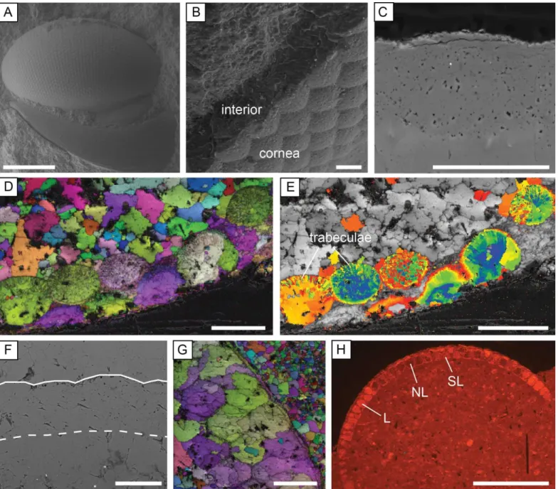
15 Well-preserved lenses are composed of a single calcite crystal, which is non-luminescent. The outer 1-2 m 1
of the lens surface consists of the cornea, which is micro-crystalline (Figure 3B) and luminescent. EBSD 2
maps show that calcite had a uniform crystallographic orientation within each lens, with the c-axis of the 3
calcite parallel to the lens axis (Figure 3D). Micro-scale pitting (Figure 3C) and sub-crystal boundaries 4
(Figure 3D) are observed in some lenses. In some specimens the lenses contain trabeculae, which are 5
microcrystallites oriented perpendicular to the lens surface and are thought to be an original structure 6
(Clarkson et al., 2006; Schoenemann and Clarkson 2011). The trabeculae can also be seen in SEM images of 7
broken lens sections, and can be identified in EBSD maps by slight differences in the crystallographic 8
orientations of the constituent sub-crystals (Figure 3E). Apparently syntaxial calcite cements occur next to 9
the interior lens surface of some specimens, but EBSD shows these cements have a different 10
crystallographic orientation to the lens calcite. 11
16 1
Figure 3. Trilobite eye preservation. A: Opipeuterella sp., with intact lenses and libriginal cuticle (specimen 2
T_205_sp4, SE image). B: The recrystallized cornea and interior calcite of the eye lenses (specimen 2.6P, 3
SE image). C: A highly polished telephinid eye lens that contains micro-crystalline dolomite crystals and 4
micro-pitting (specimen T_178_sp10, BSE image). D-E: EBSD images of an asaphid specimen in thin 5
section showing crystallographic continuity within the lenses (D) and the preservation of radial trabeculae 6
structures (E) (specimen A_178_sp1). Image D is an inverse pole figure map overlain on an image quality 7
map, and image E is an orientation tolerance map overlain on an image quality map. F: Lenses that are 8
completely recrystallized, top and base of lenses marked by white lines, the basal boundary is unclear due to 9
17 crystal overgrowths (specimen 1.1B_sp1, BSE image, thin section). G: EBSD inverse pole figure map 1
(overlain on an image quality map) of recrystallized lenses, with calcite crystals overlapping the lens 2
boundaries (specimen 1.2C_sp2, thin section). H: Eye specimen with varying luminescence of the lenses 3
under CL; L = luminescent; NL = non-luminescent, SL = slightly luminescent (specimen HV_sp43, thin 4
section). Scale bars 500 m (A, H), 50 m (B-G). 5
6
In those specimens classified as poorly preserved, the eye lenses exhibit major alteration and lack pristine 7
microstructures such as trabeculae. The microstructural evidence for extensive alteration of calcite lenses is 8
as follows: 1. indistinct lens boundaries where the calcite crystals cross the lens-sediment boundary, form 9
sub-crystals, or exhibit calcite twinning; 2. extensive pitting, resulting from the loss of small crystals and the 10
presence of micropores, indicating that the lenses are recrystallized (Figure 3F); 3. a wide range of 11
crystallographic orientations within a lens, indicating large-scale recrystallization (Fig. 3G); 4. The lens 12
calcite is luminescent (Figure 3H). Minor alteration of lens calcite is evident by variable or partial 13
luminescence due to trace amount variations in Mn and Fe, which can be detected by SEM and electron 14
microprobe analysis. Under CL, recrystallization can be recognised by zoning in the lens calcite crystals and 15
a luminescence intensity that is high, and similar to calcite spar cements in the host rock. Lenses that are 16
more significantly altered can contain diagenetic quartz, dolomite and pyrite crystals. 17
Micro-dolomite crystals are present within the lenses of nine eye specimens (some well-preserved and 18
others poorly preserved), and are associated with a pervasive microporosity/micro-pitting (Figure 3C). In 19
some specimens, celestine can be recognised in BSE images as small white (i.e., high mean atomic number) 20
crystals. Micro-crystalline dolomite is absent from the eyes of the Horn Valley Siltstone Formation 21
trilobites, which are the most highly altered, and also does not occur in the cuticle. Micro-crystalline 22
dolomite of a similar crystal size, celestine and a microporous texture are also features of the eyes of 23
phacopine trilobites (Lee et al., 2007, 2012). 24
18 Trilobite eyes from the Valhallfonna Formation were analyzed using the electron microprobe to test for 1
geochemical variation. The range in trace elements for calcite eyes is 0.6 - 1.9 wt% MgCO3, 0 - 0.9 wt% 2
FeCO3 and 0 - 0.5 wt% MnCO3. Trilobite eyes that are luminescent (and classified as poorly preserved) 3
have slightly elevated Mn concentrations relative to non-luminescent lenses. There is no significant 4
difference in the composition of eye calcite compared to that of the associated trilobite cuticle, or carbonate 5
in the host sedimentary deposit (micrite and calcite spar) (Appendix B). Eyes containing micro-crystalline 6
dolomite do not exhibit higher levels of Mg compared to those without micro-crystalline dolomite. 7
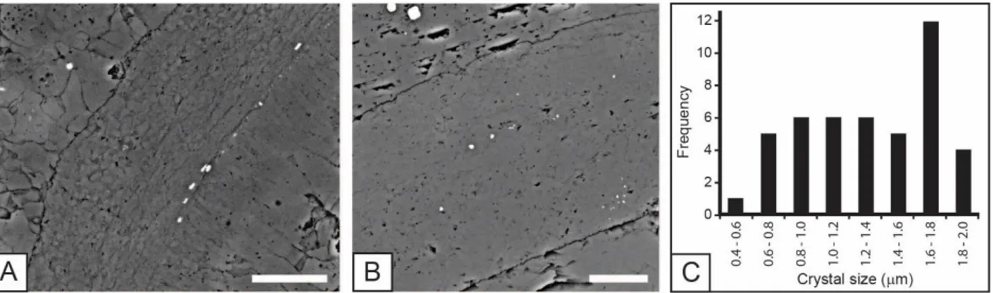
Specimens of trilobite cuticle from the Valhallfonna Formation were examined (Appendix A and B). Eight 8
of the 44 studied had an internal structure comprising two layers of aligned crystals (Figure 4A), although 9
most specimens lacked any internal structure. The cuticle is composed of prismatic, interlocking calcite 10
micro-spar, with a consistent crystal size (Figure 4B), and no evidence of an outer prismatic layer of coarser 11
crystals. EBSD of cuticle associated with trilobite eyes showed that in all specimens the cuticle crystals are 12
randomly oriented. Some specimens have small euhedral pyrite crystals on the margins of the cuticle 13
fragment, or in the interior. Most specimens are non-luminescent, but others exhibit partial or bright orange 14
luminescence. Cuticle from six samples was analyzed by electron microprobe, with a maximum 15
concentration of 2.3 wt% MgCO3, 0.2 wt% FeCO3, 0.4 wt% MnCO3 and 0.6 wt% SrCO3. In comparison to 16
the trilobite eyes, the cuticles contain relatively pure calcite with low trace element concentrations. The size 17
of the calcite crystals within the cuticle was measured from SEM images (Figure 4C), and average size does 18
not correlate with the size of the cuticle fragment examined or its chemical composition (Appendix A and 19
B). 20
19 1
Figure 4. Trilobite cuticle preservation. A-B: Polished thin section, BSE images. A: Carolinites sibiricus 2
cuticle with an internal structure of aligned crystals on the inner region (to the right), and larger crystals on 3
the outer region. The crystal size is relatively large and small pyrite crystals are present on the internal 4
margin of the specimen (sample 1.1B). B: Carolinites genacinaca cuticle with no internal structure and a 5
relatively small crystal size (sample 1.2C_N_TS2). C: Plot of cuticle crystal size frequency. Scale bars for A 6
and B are 25 m. 7
In summary, specimens with the best-preserved eyes are from the Emanuel Formation, with nine out of 8
thirteen eyes classified as well-preserved. Apart from one eye, all from the Valhallfonna Formation are 9
poorly preserved. All eyes from the Horn Valley Siltstone are poorly preserved. Trilobite cuticles from the 10
Valhallfonna Formation have variable microstructure, and EBSD shows that they lack any consistent 11
crystallographic orientation. Well-preserved eyes have an integral preservation of lens calcite with a single 12
crystallographic orientation and a composition of low-Mg calcite. In contrast, poorly preserved specimens 13
exhibit multiple smaller crystals within a lens, unclear lens boundaries, luminescence, and varied chemical 14
composition. 15
4.2 Stable Isotope Data
16
In total, 50 trilobite eyes were analyzed for isotope composition from the three formations, and where 17
possible, the preservation state of each specimen was assessed prior to isotope analyses using the protocols 18
described above (Appendix C). There is no difference in 13C and 18O between trilobite eye calcite 19
extracted by a hand-held dental drill and that extracted by micro-mill (conventional isotope analysis 20
20 methods), or from clumped isotope analysis. For 18O, there is a significant difference between conventional 1
isotope analysis (Figure 5) and SIMS isotope results (Figure 6). 2
3
Figure 5. Dental drill and micro-mill 18O and 13C conventional isotope results for the three formations 4
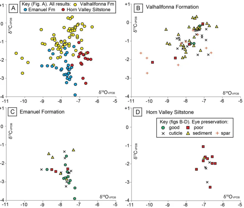
studied. A: All results (trilobite eye, cuticle and host rock), plotted by individual formation. B: Results from 5
the Valhallfonna Formation, including trilobite eyes and other material. C: Results from the Emanuel 6
Formation. D: Results from the Horn Valley Siltstone, note that all the eyes from this formation are 7
interpreted to be poorly preserved. The key to symbols for graphs B-D is illustrated within D. 8
21 1
Figure 6. 18O results from SIMS analysis plotted against conventional isotope results, for individual 2
specimens, including results from trilobite eye calcite, cuticle and rock. 3
4 5
22
4.2.1 Conventional Isotope Results
1
The total ranges in isotope composition for trilobite eyes (independent of preservation state), cuticle, and 2
host rock (sediment and spar) analyzed are 18O –6.3‰ to –10.5‰ and 13C +0.3‰ to –3.9‰. Each 3
formation plots in a different, but overlapping, 13C and 18O field (Figure 5A), and within each formation 4
the trilobite eye isotope values are within the range of the results from the trilobite cuticle and host rock 5
(Figure 5B-D). 6
With regards to the Valhallfonna Formation, the combined isotope results from all material (trilobite eyes, 7
cuticle and host rock) show a low covariance between 13C and 18O (R2 = 0.28) with values ranging from 8
13C +0.3‰ to –2.7‰ and 18O –6.3‰ to –10.5‰. This result is different from the other formations, which 9
lack any covariance between 13C and 18O (R2 = 0.13 Emanuel Formation; R2 = 0.01 Horn Valley 10
Siltstone). Well-preserved trilobite eyes yield 18O that are on average higher than those of poorly preserved 11
eyes, ranging from 18O –7.2‰ to –7.7‰, although there is some overlap (Figure 5B). There is no 12
significant difference in isotope composition between Carolinites species, stratigraphic units, or between the 13
cuticle of Carolinites and olenid trilobites (Appendix C). The 13C or 18O variation between cuticle 14
specimens that were identified as having relatively small or large calcite crystals is also negligible. 15
Results for all materials from the Emanuel Formation range from 18O of –7.3‰ to –9.1‰ and 13C of – 16
1.3‰ to –3.9‰. Well-preserved trilobite eyes have a similar range in 13C and 18O to that of poorly 17
preserved eyes (Figure 5C). There is no significant difference in isotope composition between the planktonic 18
Opipeuterella sp. and benthic asaphid trilobites, or between different samples (Appendix C). It is possible
19
that the outlier well-preserved eye result of 18O –8.8‰ is lower than the other well-preserved eye results 20
due to the presence of diagenetically altered matrix material underlying the calcite lenses. Post-sampling 21
photographs after the micro-mill had powdered the lenses do not indicate that this is the case, however the 22
23 imprecision of the micro-mill technique compared to intra-lens sampling by SIMS means that all micro-mill 1
samples must be considered to be subject to some sediment contamination. 2
Results for all materials from the Horn Valley Siltstone have a fairly narrow range of 18O –6.4‰ to –7.4‰ 3
and a broader range of 13C –1.1‰ to –3.3‰. All eyes examined were poorly preserved and there is no 4
difference in isotope composition between the planktonic Carolinites genacinaca and benthic asaphid 5
trilobites, or between different samples (Appendix C). 6
The isotope composition of lenses containing micro-crystalline dolomite does not differ from those 7
composed purely of calcite. All samples were analyzed as calcite and it is uncertain whether dolomite 8
crystals even contributed to the signal due to the small quantity of dolomite present (<1%). The presence of 9
dolomite does not correspond to the preservation state of the eyes (Table 1). 10
Four larger well-preserved specimens were analyzed to test the average isotope variation across the eye. For 11
each specimen, two sub-samples were taken for isotope analysis using the micro-mill or dental drill. Two of 12
the specimens had a consistent composition in both sub-samples, while the other two showed a different 13
composition, with a variability of up to circa 1‰ in 18O and 13C (Appendix C). 14
Twenty-one specimens with cuticle material attached to the eye (such as the librigena or glabella) were 15
analyzed to compare the composition of the cuticle and adjacent eye lenses. In well-preserved specimens 16
from the Valhallfonna Formation, the cuticle is similar in composition to that of the eyes (with a maximum 17
difference in 18O of 0.6‰) while in poorly preserved specimens the difference is greater (increasing to 18
18O 2.4‰) (Appendix C). In the Emanuel Formation, the composition of the cuticle is similar to that of the 19
eyes (with a difference in 18O of 0.8‰) for asaphids and Opipeuterella sp., with no significant difference 20
between poorly preserved and well-preserved specimens (Appendix C). However, the difference in 18O 21
between eyes and cuticle from the Horn Valley Siltstone, which contains only poorly preserved specimens, 22
is less than 0.2‰. 23
24
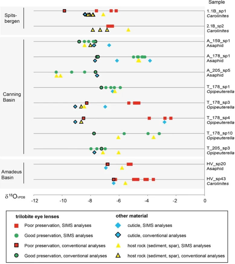
4.2.2 SIMS Oxygen Isotope Results
1
SIMS analysis of individual lenses across trilobite eyes in thin section reveals a wide range in 18O, from – 2
2.4‰ to –10.4‰ (Table 2). The range can dramatically vary even within specimens identified as well-3
preserved, and is often different from the conventional isotope analysis results of the same specimen 4
extracted using the dental drill or micro-mill (Fig. 6). For most of the 12 specimens analyzed, the lens 5
composition is within the general range of 18O as that of cuticle and host rock. However, many of the SIMS 6
results for trilobite eyes, cuticle and host rock yield 18O values that are substantially more positive than the 7
conventional isotope analysis results. 8
Specimens identified as well-preserved and poorly preserved have SIMS 18O results within the same range, 9
and there is no correlation with specimens that have micro-crystalline dolomite within the lenses. The 10
average 18O, and the range of 18O within a single eye, does not correlate with the state of preservation of 11
the eyes. There is no difference between the results from telephinids and asaphid specimens from the same 12
formation, even in well-preserved eyes. 13
Multiple lenses of individual specimens were analyzed using SIMS, giving an intra-eye 18O range, with the 14
greatest range seen within a single eye specimen of 2.7‰ and the lowest range of 0.3‰. Eight specimens 15
have a relatively high 18O range (1‰ or greater), while four have a low range in values (less than 1‰) 16
(Figure 6). 17
The SIMS and conventional isotope analysis results for individual specimens show some variations. All 18
trilobite cuticle analyses of specimens from the two methods show a high disparity (with 18O difference 19
greater than 1‰) (Figure 6). Eleven trilobite eye specimens were assessed by SIMS and conventional 20
isotope analysis, and of these, six specimens have a high disparity between the SIMS and conventional 21
methods, while five specimens had similar results (18O <1‰ difference). There is no apparent link between 22
the analysis conditions (beam intensity, chamber pressure, measurement error, run order) and those trilobite 23
25 eye samples with a large range in 18O (see Supplementary Information). In general, 18O results obtained 1
by SIMS are more positive (by as much as 6‰) in comparison to those obtained by conventional isotope 2
analysis. 3
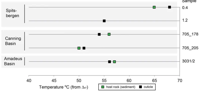
4.2.3. Clumped Isotope Results
4
Of the 12 samples examined for their 47 compositions, the cuticle and limestone analyzed from 5
Västergötland had markedly low 47 values of 47CDES25 = +0.383 and +0.396‰, respectively [reported 6
against the absolute Carbon Dioxide Equilibrated Scale - hereafter CDES - reference frame described by 7
Dennis et al. (2011) and referring to CO2 extracted by phosphoric acid digestion at 25°C]. These values 8
correspond to very high “apparent equilibrium temperature” (over 200oC, see Appendix D) representing the 9
“blocking temperature” with respect to diffusional resetting of the calcite clumped isotope thermometer 10
(e.g., Passey and Henkes, 2012; Bonifacie et al., 2013). 11
In contrast, the range in 47CDES25 values for the three other basins investigated in this study is much higher 12
(with 47CDES25 from 0.581 to 0.610‰ for Spitsbergen, 0.608 to 0.623‰ for the Canning Basin and 0.606 to 13
0.608‰ for the Amadeus Basin; Appendix D). These 47 values correspond to temperature ranges of 55-14
68oC (average 63oC) for the Valhallfonna Formation, 50-56oC (average 52oC) for the Emanuel Formation 15
and 56-57oC for the Horn Valley Siltstone Formation, respectively (Figure 7). These temperature estimates 16
were calculated from the 47-T universal calibration published by Bonifacie et al. (2016) defined on all (Ca, 17
Mg, Fe)CO3 carbonates. Only small differences in 18O are observed between cuticle and limestone (up to 18
0.45 ‰: Appendix D) and there is no difference in temperatures (derived from 47 data) between those of 19
the host rock and cuticle from the same sample. These results indicate that all fossil calcite materials 20
analyzed have experienced (and imprinted a 47 signature characteristic of) higher temperatures than those 21
at which they originally precipitated in Ordovician seawater. 22
26 1
Figure 7. Clumped isotope temperature estimates from 47 for the three main formations studied. For further 2
details including conventional isotope 18O and 13C results, 47 values and reconstructed water 18Owater, 3
see Appendix D. 4
5
The 18Owater from which the calcite in these three formations was precipitated was calculated using the 6
temperature estimated from 47 and the 18O of the carbonate, and referenced to published Ordovician 7
18OVSMOW estimates. They correspond to 18Owater of –0.4 to +1.0‰ (average 0.2‰) for the Valhallfonna 8
Formation, –1.6 to –0.2‰ (average –0.9‰) for the Emanuel Formation and 0.0 to +0.6‰ (average +0.3‰) 9
for the Horn Valley Siltstone (Appendix D). Because the 47 signatures found here do not represent those 10
acquired over original carbonate crystallisation from seawater, the calculated 18Owater of the mineralizing 11
fluid cannot be interpreted in terms of the original seawater 18O, but rather likely reflects the properties of 12
diagenetic fluids. However because the 18Owater results are fairly comparable to that estimated for 13
Ordovician seawater (–1‰ 18OVSMOW: Trotter et al., 2008) it is probable that diagenesis may have occurred 14
due to the flow of seawater through the rocks during burial, down to a maximum depth of two kilometres. 15
5. Discussion
27 The eye preservation assessment using a combined methodology (SEM, CL, EBSD and electron
1
microprobe) indicates a range in degrees of alteration. The preservation of the trabeculae has been described 2
as an indicator of good eye preservation (Schoenemann and Clarkson, 2011), but it has also been noted that 3
trabeculae can be preserved in diagenetically altered specimens (Torney et al., 2014). The cornea (outermost 4
layer of the trilobite eye) is likely to have been a transparent sheet in vivo (Clarkson et al., 2006), and its 5
present micro-crystalline state indicates that it was recrystallized during diagenesis. The loss of in vivo 6
microstructures from trilobite cuticle indicates that the majority have been recrystallized: the outer prismatic 7
layer (Dalingwater, 1973; Dalingwater et al., 1991) is absent, the presence of pyrite within the cuticle is 8
consistent with diagenetic alteration (Wilmot, 1990), as is the homogeneous microstructure of the calcite 9
(Budil and Hörbinger, 2007). As the cuticle is thought to mineralize in an organic mesh-like framework, 10
similar to that of an ostracod carapace (Teigler and Towe, 1975), the lack of correspondence between crystal 11
size and size of the exoskeleton may indicate diagenesis. Large cuticle crystals may be expected to have 12
incorporated trace-elements or have a different isotope composition to microcrystalline cuticle calcite. 13
Moreover, the crystallographic orientation of the cuticle crystals (observed to be random under EBSD) has 14
changed, with crystal c-axis perpendicular to the cuticle surface (Teigler and Towe, 1975). 15
In terms of the visual protocols used to assess preservation, the most useful method was EBSD analysis, due 16
to the crystallographic orientation information it shows. SEM microstructural observations were also 17
valuable and identified porosity and micro-crystals (Table 1). While CL generally corresponded to other 18
indicators, with luminescent lenses showing an elevated trace-element profile and evidence for 19
recrystallization, lenses that were recrystallized were sometimes non-luminescent. Thus, CL is the least 20
reliable technique, however it does generate visual data that can be used as a starting point for more detailed 21
preservation analysis via other techniques. 22
The SIMS results differ significantly from the corresponding conventional isotope analysis results in over 23
half the trilobite eye specimens analyzed. This discrepancy may be due to presence of a fine-scale mixture 24
of primary and secondary calcite within the lenses. In addition, little is known about the composition of 25
28 trilobite cuticle, or vital effects, in comparison to brachiopods where studies on modern specimens can be 1
used to determine 18O paleotemperature equations (Brand et al., 2013). However, it must also be 2
considered that the inconsistency between analytical datasets could reflect a systematic analytical error in 3
the SIMS data in this study. Previous work has found differences in the trace element content (Mg/Ca, Sr/Ca 4
and Mn/Ca ratios) of marine bivalves analyzed using conventional bulk dissolution and SIMS. These 5
differences have been interpreted as reflecting variable shell organic content, crystal structures and small 6
inter-crystalline heterogeneities in trace element concentrations (Freitas et al., 2009). It is therefore possible 7
that the presence of organic matter within trilobite eyes, sub-crystal boundaries, or pitted surfaces could 8
have affected our SIMS data. However, the cross-comparison of the SIMS results with the micro-crystalline 9
characteristics of each specimen (Table 1) does not reveal a correspondence between the presence of micro-10
crystalline features (porosity, crystal zoning, micro-dolomite) and wide ranging or positive 18O values. The 11
size of the ion beam used for the SIMS analysis (15 m) may have resulted in the crossing of crystal 12
boundaries, although most of the spots sampled the centre region of the eye lens and were verified by 13
photographs. It is more likely that the results represent genuine intra-eye variation rather than contamination 14
from the host rock. Further detailed work is needed to attempt to correct the SIMS data for the variables of 15
inter-crystalline heterogeneities. The SIMS analysis has revealed large intra-eye variations of lens 16
geochemistry that would otherwise remain undetected. Despite the limitations of the method, for example 17
micro-scale pitting and sub-crystal boundaries that may cause analytical error, SIMS is still a useful tool for 18
assessing diagenesis. 19
The range of isotope compositions within a single eye specimen (as revealed by both SIMS and 20
conventional isotope analysis) indicates cryptic recrystallization. This means that recrystallization has likely 21
occurred in specimens that were identified as well-preserved using the SEM, CL, EBSD and electron 22
microprobe protocols outlined above. Ten out of the sixteen specimens examined had ranges in 18O of 23
greater than 1‰ within the same eye. If, as seems reasonable, all the lenses in an eye were biomineralized 24
(or indeed recrystallized) at the same time, in the same conditions, then they should have the same isotope 25
29 composition. For each formation, there is no significant difference in 18O or 13C between trilobite eye 1
calcite or cuticle and the host rock. In addition: 1. all trilobite cuticle examined is interpreted as 2
diagenetically altered, with a complete loss of crystallographic structure, indicating recrystallization was 3
pervasive; 2. calcitic spar within the host rock, typically resulting from diagenetic growth, is within the same 4
isotope range as the trilobite eye and cuticle results (Fig. 5B); and 3. the counterintuitive finding that pelagic 5
trilobite species, which lived in relatively warm waters near the sea surface, have the same 18O as benthic 6
species, indicates that all 18O has been reset. Therefore, most, if not all, of the material analyzed is 7
interpreted to have been diagenetically altered, despite our original assessment of good preservation using 8
microstructural criteria. In terms of eye preservation, there is little geochemical difference between eyes 9
identified as well-preserved or poorly preserved, both in terms of conventional isotope analysis and SIMS 10
results. In addition, the degree of recrystallization (complete or partial) has no significant correspondence 11
with the isotope results. 12
Detailed SIMS analysis shows a large range in 18O values within a single eye specimen, even those that on 13
microstructural criteria are well-preserved. Our preservation assessment can therefore be criticised for not 14
identifying subtle preservation features. For example, the presence of trabeculae and lens crystallographic 15
continuity as identified by EBSD mapping was interpreted to indicate well-preserved eyes, but the 16
boundaries of the trabeculae were not examined in detail using TEM as they were by Torney et al. (2014). 17
Micro-crystalline dolomite was observed in well-preserved and poorly preserved specimens, and further 18
analysis using TEM is needed to further understand the origin of this mineral. Despite these limitations, the 19
present study has made the most extensive preservation assessment of any trilobite material prior to isotope 20
analysis. 21
The presence of micro-crystalline dolomite within the holochroal eyes examined here may indicate an 22
original high-Mg calcite composition (as interpreted for the eyes of phacopine trilobites by Lee et al., 2012), 23
with high-Mg calcite being diagenetically altered to low-Mg calcite. The presence of micro-crystalline 24
30 dolomite in both poorly- and well-preserved eyes, supports this interpretation. The absence of
micro-1
crystalline dolomite in the Horn Valley Siltstone samples may be due to their more extensive diagenetic 2
alteration. However, the lenses containing micro-crystalline dolomite do not have an elevated Mg content, as 3
recorded in phacopine eyes, of up to 6 mol% MgCO3 (Lee et al., 2007, 2012). This could be due to the 4
diagenetic loss of magnesium over time. However, the sporadic occurrence of microcrystalline-dolomite - it 5
is not present in all eyes identified as well-preserved - means that at present we cannot conclude that the 6
holochroal lenses were originally composed of high-Mg calcite. An alternative explanation might be that the 7
dolomite grew during diagenetic alteration and micro-recrystallisation of low-Mg calcite. Further detailed 8
investigation of the micro-crystalline dolomite at the nanometre scale is needed to determine an original 9
high-Mg calcite composition of the lenses. 10
The host rock and fossil material from each of the three basins have a distinctive range in 13C and 18O 11
values, which may reflect their different diagenetic histories. The 47 data reveal a minimum temperature of 12
trilobite cuticle (re)crystallisation of 50oC, which is well in excess of the threshold for most organisms living 13
in the surface layers of the modern oceans (Brock, 1985) and must be interpreted as resulting from the 14
diagenetic alteration of the original cuticle. The Västergötland cuticle clumped isotope results are at the 15
extreme temperature end (200°C), where the original 47 values (i.e. the original 13C–18O bonding 16
distribution acquired over crystallisation) have been reset due to closed-system solid-state diffusion 17
alteration. This conclusion is consistent with independent Västergötland CAI values of 6 to 7, indicative of 18
peak temperatures exceeding 300oC resulting from heating by Permian igneous intrusions (Bergström, 19
1980). In contrast, the Australian and Spitsbergen samples have lower temperatures calculated from the 47 20
(from 50-68oC; Figure 7), which reflect early recrystallization temperatures. 21
The thermal history of the sedimentary formations can be linked to the fidelity of eye preservation. The 22
Emanuel Formation was subject to the lowest burial temperatures (maximum temperatures of 70-80oC: 23
Nicoll et al., 1993) and its sample set contains the best-preserved trilobite eyes. The Valhallfonna Formation 24
31 was subject to a slightly higher burial temperature (up to 90oC: Bergström, 1980) and its sample set contains 1
just one well-preserved specimen. While the Horn Valley Siltstone experienced a much higher burial 2
temperature (up to 140oC: Gibson et al., 2007) and yielded no well-preserved specimens. 3
It could be argued that the conventional 18O and 13C results for the trilobite cuticle, eyes and host rock 4
reflect original compositions and were not affected by partial recrystallization and only the 47 composition 5
changed. Indeed, solid-state re-ordering can alter the C-O bonds within a shell while retaining original shell 6
microstructures and trace-element concentrations (Henkes et al., 2014). Experimental studies on brachiopod 7
shells by Henkes et al. (2014) have shown that solid-state reordering can start to reset the original C-O 8
bonds (and thus 47 compositions) of calcite if it experienced temperatures above 100oC for more than 9
hundreds of millions of years. Stolper and Eiler (2015) argue that reordering can start to occur at
10
temperatures of 75oC, and becomes more significant over 120oC. To explore this possibility further, the 11
thermal history of each basin must be examined in terms of the duration spent at higher temperatures during 12
burial. The detailed burial history of Ny Friesland, Spitsbergen has not yet been reconstructed. The Canning 13
Basin experienced temperatures of 70-80oC for approximately 200 Myr in the Mesozoic, but only underwent 14
a brief higher temperature interval of ~100oC (Arne et al., 1989) or <90oC (Wallace et al., 2002) in the early 15
Carboniferous, making solid state diffusion unlikely. The Stairway Sandstone, which overlies the Horn 16
Valley Siltstone in the Amadeus Basin, experienced burial temperatures of over 100oC in the Permian-17
Triassic (for approximately 80 million years), and Cretaceous (for approximately 110 million years) (Gibson 18
et al., 2007). It is possible that solid-state diffusion may have affected the specimens from the Horn Valley 19
Siltsone, although the time that the rocks experienced temperatures over 100oC is fairly short. Overall, low-20
temperature recrystallization, over solid-state diffusion, is the most likely mechanism responsible for 21
altering the trilobite eyes and cuticle. 22
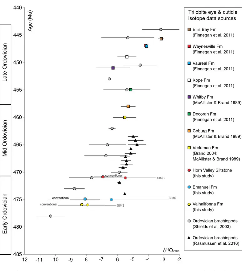
Figure 8 shows trilobite 18O data from the present study plotted against published geochemical data from 23
trilobite cuticle and brachiopods through the Ordovician. There are no published data available for Lower 24
32 Ordovician trilobites, but those from the Middle and Upper Ordovician range from 18O of –3‰ to –7‰ 1
(McAlister and Brand, 1989; Brand, 2004; Finnegan et al., 2011), which is within the range of that recorded 2
in the present study. Ordovician-Silurian studies comparing brachiopods and trilobites found them to have a 3
similar 18O to each other (McAlister and Brand, 1998; Wilmot and Fallick, 1989). While Finnegan et al. 4
(2011) found Upper Ordovician trilobites to have a similar 18O composition to that of contemporaneous 5
brachiopods and corals. Of all the published trilobite isotope studies, that by Finnegan et al. (2011) is the 6
only one where the preservation of the trilobite cuticle has been examined via trace element and 7
microstructural studies, prior to isotope analysis. 8
33 1
Figure 8. Trilobite eye and cuticle data from this study (conventional isotope analysis and SIMS), plotted 2
with trilobite cuticle data from the literature, and brachiopod data (Shields et al., 2003; Rasmussen et al., 3
2016). Grey and black bars are isotope ranges, with the mean isotopic value indicated by the symbol in the 4
centre. 5
34 The 18O data from trilobites in the present study are within the range of contemporaneous brachiopods, or 1
more positive (Figure 8). The exceptions are new data from Baltica which have higher 18O values 2
(Rasmussen et al., 2016). Given our results, this raises questions about the possibility of brachiopod calcite 3
recrystallization from some earlier studies. Brachiopod specimens from the Lower to Middle Ordovician 4
that were analyzed by conventional isotope techniques were assessed for preservation using trace element 5
geochemistry and SEM and CL characterization (Veizer et al., 1999; Shields et al., 2003), but not by EBSD 6
or SIMS. Significant cryptic diagenesis may have altered the isotope composition of these specimens: in 7
general it is thought that only the secondary layer of the brachiopod shell is reliably unaltered and the 8
preservation state of this layer can vary between taxonomic groups (Garbelli et al., 2012); In addition, SIMS 9
analysis of brachiopods reveals that within certain groups, the secondary layer is only precipitated in isotope 10
equilibrium with seawater towards the innermost part of the shell (Cusack et al., 2012). This fact implies 11
that isotope studies where the entire secondary layer was analyzed (for example Shields et al., 2003) may 12
reflect non-equilibrium fractionation. This is variable between brachiopod groups, for example in spiriferid 13
brachiopods the prismatic tertiary layer is more resistant to diagenesis than the secondary layer (Grossman et 14
al., 1993). Even in samples that have experienced low burial temperatures, recrystallisation can occur: in a 15
study of Silurian brachiopods, Cummins et al. (2014) argued that elevated clumped isotope temperatures (up 16
to 56oC) found in samples with CAI = 1 were likely resulting from diagenetic alteration due to 17
recrystallization. This result cautions the use of apparently pristine biogenic specimens to interpret ancient 18
paleotemperatures, without prior carbonate clumped isotope analysis. Due to the effects of sold-state 19
reordering of clumped isotopes, which may not alter 13C and 18O values, analysis using the range of 20
methods utilised in this study (SIMS, CL, SEM and EBSD) should be combined to jointly assess fossil 21
preservation and isotope geochemistry. 22
Brachiopod data have been used to suggest that the 18O of Ordovician seawater was lower than at the 23
present-day (–3‰ 18OVSMOW) in order to account for a reasonable paleotemperature calculation for the 24
most negative 18O results (Veizer et al., 1999; Shields et al., 2003). Modelling of long-term seawater 25
35 geochemical composition estimates of Ordovician 18OVSMOW at approximately –6‰ (Jaffrés et al., 2007), 1
and a revised calibration of the brachiopod data gives a value of –5‰ 18OVSMOW for the Early Ordovician at 2
475 Ma (Veizer and Prokoph, 2015). However, Trotter et al. (2008) used a value of –1‰ to interpret the 3
temperature of formation of Ordovician conodont apatite, giving similar ocean temperatures for the Early 4
Ordovician to that interpreted from the brachiopod data. The Ordovician brachiopod calcite, trilobite calcite 5
and conodont apatite (Trotter et al., 2008; Veizer and Prokoph, 2015) isotope data show an increase in 6
carbonate or phosphate 18O values over the Ordovician, which indicates either cooling oceans, changing 7
seawater chemistry, or increasing diagenetic imprints at higher temperature with time. Conodont apatite has 8
been demonstrated to be well-preserved (Wheeley et al., 2012) and is therefore useful as a paleotemperature 9
proxy in the Paleozoic (Joachimski and Buggisch 2002). The results of Trotter et al. (2008) indicate that 10
conodont apatite may be a much more reliable proxy than brachiopod calcite, as it yields more reasonable 11
ocean paleotemperature calculations. How do the trilobite results influence this debate? The diagenetic 12
alteration of trilobites demonstrated here reveals that the most negative 18O values in the Early to Middle 13
Ordovician (those less than 18O –7‰) are probably the result of diagenesis rather than a distinctive 14
seawater composition. Results of the present study therefore bring into question the fidelity of some Early-15
Middle Ordovician brachiopod records and call for a re-appraisal. The new positive 18O brachiopod data by 16
Rasmussen et al. (2016) for the Early Ordovician highlight the need for a review of all brachiopod data from 17
this time. As no EBSD or SIMS work has been undertaken on Lower to Middle Ordovician trilobite or 18
brachiopod specimens analyzed for their isotope composition prior to this study, the possibility remains that 19
significant cryptic diagenesis may have altered these specimens. We therefore caution the interpretation of 20
isotope results without rigorous preservation assessment and advise that SIMS analysis should be added to 21
the tool-box of preservation studies. 22
6. Conclusions 23
The holochroal eyes of species of the pelagic trilobite genera Carolinites and Opipeuterella from the 24
Floian (Lower Ordovician) Valhallfonna Formation, Spitsbergen, and the Floian-Dapingian (Lower-25
36 Middle Ordovician) Emanuel and Horn Valley Siltstone formations of Australia were examined to 1
assess their preservation state. 2
The use of trilobite eyes for vision places strict constraints on the in vivo microstructure of their 3
constituent calcite lenses, and as such is a unique means of guiding our preservation assessment. 4
The microstructure and chemical composition of the eyes was assessed using SEM, CL, EBSD and 5
electron microprobe. The Valhallfonna and Emanuel formations contain the best-preserved 6
specimens, based on these assessment protocols. Well-preserved eyes are composed of low-Mg 7
calcite, are non-luminescent, have clear crystallographic boundaries and retain their original optical 8
structures such as trabeculae. 9
Trilobite cuticle is composed of low-Mg calcite, but the wide range of crystal size within the cuticle, 10
random crystal orientation and lack of original structures indicates that all specimens have been 11
recrystallized. 12
The 18O and 13C results show a different range for each formation. However, intra-eye isotope 13
analyses using SIMS reveals a large range in 18O in some specimens, of up to 2.7‰. There is also 14
no systematic isotope difference between rock, cuticle, eyes and diagenetic cements, or between 15
benthic and pelagic trilobite eyes or cuticle. 16
A sub-set of trilobite cuticles from the three formations was analyzed with carbonate clumped 17
isotope thermometry, generating a temperature range of 50-68oC and so indicating low-temperature 18
diagenetic alteration of the trilobite calcite. 19
Despite rigorous preservation assessment protocols, the SIMS and 47 data both show that all 20
trilobite eye and cuticle specimens analyzed here have, at least in part, been diagenetically altered, 21
including those that were interpreted to be well-preserved using microstructural criteria. The 22
presence of sparse micro-crystalline dolomite within some eye specimens hints at cryptic 23
recrystallization processes, although the impact of the presence of dolomite on the isotope results 24
remains unclear. 25