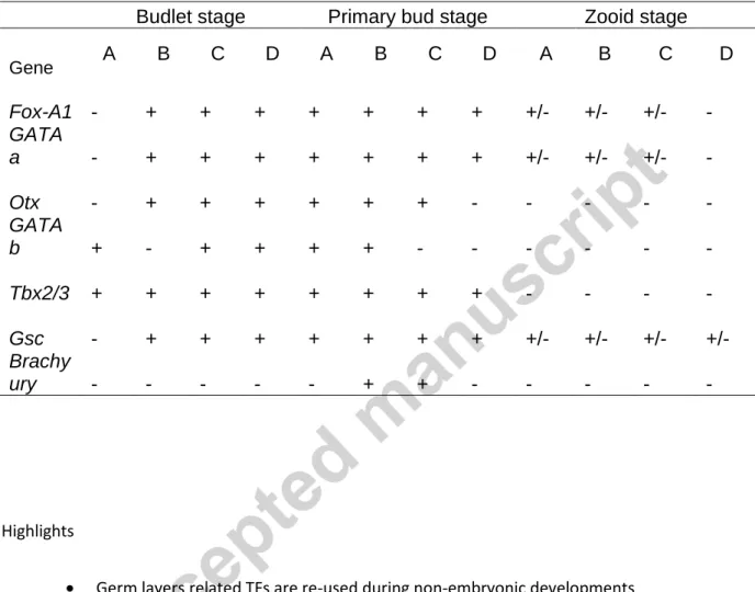
Re-deployment of germ layers related TFs shows regionalized expression during two non-embryonic developments
Texte intégral
Figure
Documents relatifs
l’utilisation d’un remède autre que le médicament, le mélange de miel et citron était le remède le plus utilisé, ce remède était efficace dans 75% des cas, le
This research has been carried out in order to introduce the biotization to the synthetic seed technology of ‘Carrizo’ citrange (C. trifoliata), one of the most widespread
We have developed a novel biosensor that integrates in an unique platform two different sensing techniques: Love mode acoustic wave and surface plasmon resonance [1].. The
I will consider h ow individuals make sense of concepts like surrendering to their disease and reaching out to a 'higher power' as well as their experiences with performing each of
Consider Streeck’s claim that soaring public indebtedness following the Great Recession “reflected the fact that no democratic state dared to impose on its society another
In this study of lagoon sediment during a cyanobacterial bloom (Microcystis aeruginosa) we found a homogeneous group of MTB (LR-1) that are probably sulfate reducing
Third generation, from now on: Services evolution and global convergence. Network and
Turkey pointed out that the second paragraph of the preamble and Article 2.1 of the Convention for the Safeguarding Intangible Cultural Heritage, and
