doi: 10.3389/fphar.2019.00133
Edited by: Peter Vee Sin Lee, The University of Melbourne, Australia Reviewed by: Zhiping Liu, Augusta University, United States Connie Wong, Monash University, Australia *Correspondence: Frédéric Antonio Carvalho [email protected]; [email protected]
Specialty section: This article was submitted to Translational Pharmacology, a section of the journal Frontiers in Pharmacology Received: 27 September 2018 Accepted: 05 February 2019 Published: 26 February 2019 Citation: Boudieu L, Mountadem S, Lashermes A, Meleine M, Ulmann L, Rassendren F, Aissouni Y, Sion B, Carvalho FA and Ardid D (2019) Blockingα2δ-1 Subunit Reduces
Bladder Hypersensitivity and Inflammation in a Cystitis Mouse Model by Decreasing NF-kB Pathway Activation. Front. Pharmacol. 10:133. doi: 10.3389/fphar.2019.00133
Blocking
α
2
δ-1 Subunit Reduces
Bladder Hypersensitivity and
Inflammation in a Cystitis Mouse
Model by Decreasing NF-kB Pathway
Activation
Ludivine Boudieu1,2, Sarah Mountadem1,2, Amandine Lashermes1,2, Mathieu Meleine1,2, Lauriane Ulmann3,4, François Rassendren3,4, Youssef Aissouni1,2, Benoit Sion1,2, Frédéric Antonio Carvalho1,2* and Denis Ardid1,2
1NEURO-DOL, Université Clermont Auvergne, Clermont-Ferrand, France,2U1107, Inserm, Clermont-Ferrand, France,3IGF, CNRS, INSERM, Univ Montpellier, Montpellier, France,4Labex ICST, Montpellier, France
Bladder pain is frequently associated with bladder inflammation, as in conditions like interstitial cystitis (IC), for which current analgesic therapies have limited efficacy. The antinociceptive effect of alpha-2-delta (α2δ) ligands on inflammation-associated visceral
pain like that experienced in cystitis has been poorly investigated. To investigate the effect of pregabalin (PGB), an α2δ ligand, we evaluated its impact on mechanical
hyperalgesia in a mouse model of cystitis induced by cyclophosphamide (CYP). We further studied its effect on inflammation and NF-kB pathway activation. Acute cystitis was induced by intraperitoneal injection of 150 mg kg−1of CYP in C57Bl/6J male mice. PGB was subcutaneously injected (30 mg kg−1) 3 h after CYP injection. The effect of PGB on CYP-induced mechanical referred hyperalgesia (abdominal Von Frey test), inflammation (organ weight, cytokine production, α2δ subunit level, NF-kB pathway
activation) were assessed 1 h after its injection. In parallel, its effect on cytokine production, α2δ subunit level and NF-kB pathway activation was assessed in vitro
on peritoneal exudate cells (PECs) stimulated with LPS. PGB treatment decreased mechanical referred hyperalgesia. Interestingly, it had an anti-inflammatory effect in the cystitis model by reducing pro-inflammatory cytokine production. PGB also inhibited NF-kB pathway activation in the cystitis model and in macrophages stimulated with LPS, in which it blocked the increase in intracellular calcium. This study shows the efficacy of PGB in hypersensitivity and inflammation associated with cystitis. It is therefore of great interest in assessing the benefit ofα2δ ligands in patients suffering from cystitis.
INTRODUCTION
Bladder pain is frequently associated with bladder inflammation, as in interstitial cystitis (IC) (Ogawa et al., 2015). The mechanisms include an increase in mucosal bladder permeability
(Buffington and Woodworth, 1997), leading to sensitization
of bladder afferent pathways and inflammation (Yoshimura
et al., 2002), and sensitization of peripheral and/or central
pain pathways (Ogawa et al., 2015). At the periphery,
inflammatory processes could be involved. Increased levels of pro-inflammatory cytokines (Erickson et al., 2002) and a decrease in those of the anti-inflammatory IL-4 cytokine (Ueda
et al., 2000) have been observed in IC patients. The efficacy of
anti-nerve growth factor (NGF) therapy in humans (Evans et al., 2011) confirms the involvement of NGF in the pathophysiology of bladder pain. All these peripheral mediators can sensitize the mechanosensitive afferent fibers or increase their recruitment
(Sengupta and Gebhart, 1994).
The existing treatments of IC involve non-pharmacological
approaches such as behavioral modifications, bladder
hydrodistention, neurostimulation, and surgery.
Pharmacological treatments are divided into two categories, peripheral (i.e., intravesical) and systemic. The intravesical treatments include dimethyl sulfoxide, heparin, lidocaine, and onabotulinum toxin A (Kuo and Chancellor, 2009;Ogawa et al., 2015). Systemic pharmacological therapies include amitriptyline, a tricyclic antidepressant used in the first line therapy for neuropathic pain to improve symptoms associated with IC such as pain and urgency (van Ophoven et al., 2004).
Anticonvulsants are used in the treatment of several chronic pain syndromes, targeting neuronal excitability (Johannessen
Landmark, 2008). Among these, the α2δ ligands, gabapentin
(GBP) and pregabalin (PGB) act on voltage-gated calcium (Ca2+) channels (VGCCs) to inhibit pre-synaptic glutamate release
(Johannessen Landmark, 2008). In addition, previous studies
have demonstrated potent and selective binding of PGB toα2
δ-1 andα2δ-2 subunits (Li et al., 2011). These molecules are now
proposed in first line with antidepressant drugs for treatment
of neuropathic pain (Johannessen Landmark, 2008). Their
beneficial effect has been observed in cystitis patients (Hansen,
2000;Sasaki et al., 2001). However, some preclinical studies failed
to find any effect of GBP in rodent cystitis models (Rudick et al., 2009). Our study aimed to investigate the potential beneficial effects of PGB, an α2δ ligand, on a mouse model of cystitis
induced by cyclophosphamide (CYP). It has been reported that α2δ ligands reduce the activation of the nuclear factor kB
(NF-kB) in neuroblastoma and glioma cells (Park et al., 2008). In light of these findings, we further investigated the mechanism of action ofα2δ ligands by studying their interaction with NF-kB
pathway activation.
MATERIALS AND METHODS
Acute Cystitis Induction
All experiments were performed according to the ethical guidelines set out in the Guide for the Care and Use of
Laboratory Animals and with approval of the “Comité d’Ethique pour l’Expérimentation Animale Auvergne” (C2E2A), the local ethics committee (Reference number: EU0116-5330). All experiments were performed on C57Bl/6J male mice weighing 20–24 g (JANVIER LABS, Le Genest Saint Isle, France). Animals
were given access to food and water ad libitum and housed
with a 12-h light-dark cycle. Acute cystitis was induced by intraperitoneal injection of 150 mg kg−1of CYP. Control mice
received saline injection.
Pregabalin Treatment
Pregabalin ((S)-((+)-3-(aminomethyl)-5-methylhexanoic acid; Dochem lot PRE20110601) was dissolved in 0.9% saline. 3 h after CYP injection, mice were subcutaneously injected with PGB (30 mg kg−1) or saline. Tests were performed 1 h later.
Mechanical Referred Hyperalgesia
Testing
Mechanical cutaneous abdominal sensitivity was assessed with von Frey filaments (Biosed, Vitrolles, France) before the animals were injected and 4 h after CYP injection. The filaments were applied to the lower abdominal area close to the urinary bladder and the median 50% threshold (T50) was determined by the up-and-down method (Chaplan et al., 1994). Briefly, this method is based on the use of the 3.22 filament size (0.16 g), which corresponds to the intermediate size of the filament range. The filament is applied perpendicular to the lower abdominal area close to the urinary bladder, exerting sufficient force to flex it for a period of 5 s. Two answers were possible: (i) animal reacts (abdominal contraction), this response is marked “X” and the test continues with the smaller filament on the range (2.83/0.07 g); (ii) animal does not respond, this response is marked “O” and the test continues with the larger filament in the range (3.61/0.4 g). This scheme continues until the objectivized response of the animal changes compared to the first reaction observed with the 3.22 filament size. From that moment, four filaments are applied (always according to the same method) and the test is finished. The pattern thus obtained corresponds to a score available in the appendix of the Chaplan publication (Chaplan et al., 1994). Finally, thanks to the Dixon formula (Dixon, 1980), it possible to calculate the median 50% threshold (T50): (10[Xf +κδ]
) / 10000, with Xf, size of the last applied filament,κ, score and δ, average difference between stimuli.
Bladder Culture
Following euthanasia, the bladder was removed, cut open longitudinally, washed in PBS and cultured in RPMI1640 medium containing penicillin and streptomycin. After 24 h incubation at 37◦
C with 5% CO2, supernatants were centrifuged
at 4◦
C and used for assaying cytokines by ELISA.
Enzyme-Linked Immunosorbent Assay
All ELISA kits are DuoSet kits from R&D Systems, and assays were performed according to the manufacturer’s protocol.
Tissue Myeloperoxidase Assay
One-third of the bladder was homogenized (50 mg mL−1)
in 0.5% hexadecyltrimethylammonium bromide (Sigma) in 50 mM PBS, (pH 6.0), freeze-thawed three times, sonicated and centrifuged. Myeloperoxydase (MPO) was assayed in
the supernatant by adding 1 mg mL−1 of O-dianisidine
dihydrochloride (Sigma) and 5 × 10⁻⁴% H2O2. One unit of MPO
activity was defined as the amount that degraded 1.0µmol of peroxide/min at 25◦
C.
Histology
Bladder domes were fixed for 24 h in 4% buffered formalin at 4◦
C and then subjected to Hematoxylin and Eosin staining on 5µm thick tissue sections. The mucosal thickness was measured with Gimp 2.8 software. Four measured per sections and three sections per animal were assessed.
Western Blotting
Four hours after CYP injection, mice were euthanized and their bladders collected. For IκBα, phospho-p65 and α2δ-1 total expression study, bladders were homogenized in ice-cold lysis buffer containing stop buffer and protease inhibitor cocktail.
For determination of α2δ-1 membrane/cytoplasmic expression,
proteins were extracted according to the manufacturer’s protocol for use of the Compartmental Protein Extraction Kit (Merck Millipore). Blotting was performed at 4◦
C overnight with the following antibodies: IκBα (1:500; Santa Cruz Biotechnology), phospho-p65 (Ser276; 1:500; Santa Cruz Biotechnology), α2δ-1 (1:500; Santa Cruz Biotechnology), phospho-ERK1/2 (T202/Y204; 1:1000; Cell signaling Technology), ERK1/2 (1:1000; Cell Signaling Technology), EGFR (1:1000; Santa
Cruz Biotechnology), and β-actin (Sigma-Aldrich). Protein
quantification was performed by densitometry using ChemiDoc
MP imager and Image LabTMsoftware (Bio-Rad).
In vitro Peritoneal Exudate Cell Studies
Resident mouse peritoneal exudate cells (PECs) were collected from euthanized animals. The abdominal skin was incised for reveal the abdominal muscle. Two abdominals lavage were successively realized with 5 ml of PBS+0.5% of fetal bovine serum (FBS). The cells thus collected were count on Malassez cell, centrifuged (300 g, 8 min, 4◦
C) and resuspended in
DMEM (+10% FBS and seeded at the density of 4 × 10⁵
cells/well). After overnight incubation, cells were co-incubated
with LPS (100 ng mL−1
; Sigma-Aldrich Chimie,
Saint-Quentin-Fallavier, France) and PGB (11.3 µM) or saline in
serum-free media.
Intracellular Calcium Imaging and
Measurement
Cells (PECs) were loaded with 2 µM of Fura-2-acetoxymethyl
ester (Fura-2/AM, Life Technologies), 0.5% BSA in the recording saline solution. After 1 h, cells were stimulated for 2 min with LPS (1 µg/mL). When indicated, cells were also pre-incubated with PGB (11.3µM) for 15 min. Intracellular Ca2+concentration
([Ca2+]) was assessed by recording the changes in cytoplasmic
[Ca2+] with the ratiometric fluorescent probe Fura-2 in PECs. The MetaFluor Imaging System (Molecular Devices) was used for fluorescence acquisition and analysis of individual cells. Pairs of images were acquired every 2 s. A single PEC was considered as a responder if the F340/F380 ratio for a single PEC increased by 0.05.
Statistical Analysis
All data were expressed as mean ± SEM and analyzed with GraphPad Prism5 software. Differences in T50 and the effects
of PGB on ex vivo parameters were analyzed by a 1-way
ANOVA (Treatment) followed by Tukey post hoc test for
multiple comparisons. The effects of PGB on IκBα,
phospho-p65 and α2δ-1 expression in PECs culture were analyzed by
a 2-way ANOVA (Model, Treatment) followed by Bonferroni post hoc test for multiple comparisons. For calcium imaging experiments, statistical differences were elicited by a Mann–
Whitney U-test. A p-value less than 0.05 was considered
statistically significant.
RESULTS
Effect of Pregabalin on
Cutaneous-Referred Bladder
Hypersensitivity in Acute
Cyclophosphamide-Induced Cystitis
Before CYP injection, the von Frey test showed no difference between the different groups (saline: 0.37 ± 0.14 g, PGB: 0.34 ± 0.08 g, CYP: 0.48 ± 0.16 g, CYP/PGB: 0.38 ± 0.10 g, data not shown). In the group receiving adjuvant and saline injection
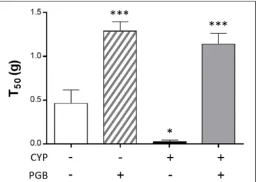
FIGURE 1 | Effect of pregabalin (PGB; 30 mg kg−1, s.c.) on cystitis-induced
mechanical referred hyperalgesia assessed by the abdominal von Frey test. The von Frey filaments were applied to the lower abdominal area close to the urinary bladder. Median 50% threshold (T50) was determined by the up-and-down method. The values are expressed as a mean ±S.E.M. and compared by a 1-way ANOVA (Treatment) followed by Tukey post hoc test for multiple comparisons. N = 8/group.∗
p< 0.05,∗ ∗
p< 0.01,∗ ∗ ∗
p< 0.001 vs. control group.
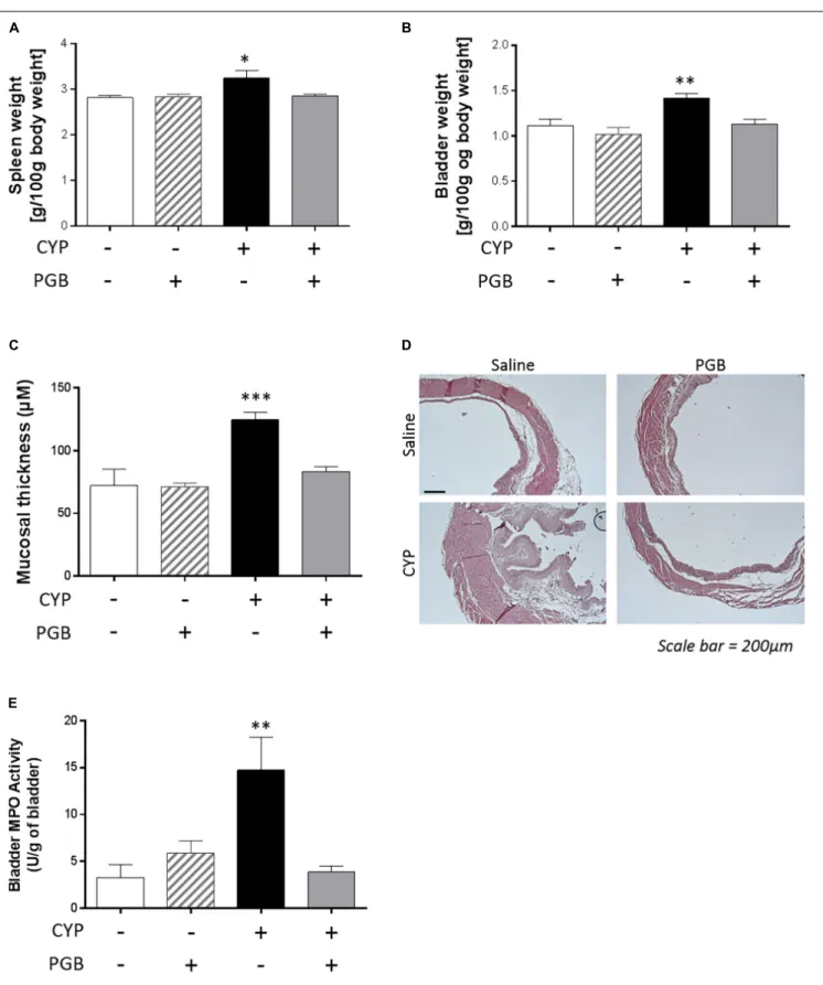
FIGURE 2 | Effect of PGB (30 mg kg−1, s.c.) on spleen (A) and bladder (B) weight, mucosal thickness (C,D) and bladder MPO activity (E) in cyclophosphamide
(CYP)-induced cystitis in mice. The values are expressed as a mean ±S.E.M. and compared by a 1-way ANOVA (Treatment) followed by Tukey post hoc test for multiple comparisons. For panels A, B, E, the result represented n = 8 animals / group and for panel C, 4 measured per section and 3 sections per animals were analyzed.∗p< 0.05,∗ ∗p< 0.01,∗ ∗ ∗p< 0.001 vs. control group.
(control group), T50 scores (0.43 ± 0.16 g) were comparable to those obtained before animals were injected. Cyclophosphamide treatment induced a significant decrease in the von Frey scores (0.006 ± 0.001 g,p< 0.05 vs. control group). Subcutaneous acute PGB administration led to a marked significantly increase in the T50 score in healthy mice (1.29 ± 0.11 g,p< 0.001 vs. control group) and in CYP-treated animals (1.14 ± 0.12 g,p< 0.001 vs. control group) (Figure 1).
Effect of Pregabalin on Inflammatory
Parameters in Acute
Cyclophosphamide-Induced Cystitis
Cyclophosphamide induced a significant increase in the weight of the spleen (3.24 ± 0.17 g/100 g body weight,p< 0.05 vs. control group) and bladder (1.42 ± 0.05 g/100 g body weight,p< 0.01 vs. control group). This increase was not reproduced when animals were treated with PGB (spleen weight: 2.85 ± 0.04 g/100 g body weight; bladder weight: 1.13 ± 0.06 g/100 g body weight, ns, vs. control group) (Figures 2A,B).
The cyclophosphamide treatment induced a significant increase in mucosal thickness of the bladder (124.50 ± 6.21µM,
p < 0.001, vs. control group). In contrast, PGB acute
treatment prevented bladder thickening (83.55 ± 3.60 µM,
ns, vs. control group) (Figure 2C). The deleterious impact of CYP on bladder structure and the beneficial effect of PGB treatment were confirmed in a morpho-anatomical observation (Figure 2D).
Cyclophosphamide treatment induced an increase in bladder MPO activity (14.76 ± 3.49 U/g of bladder,p< 0.01, vs. control group). In contrast, PGB-treated animals had normal levels of MPO activity (3.85 ± 0.64 U/g of bladder, ns, vs. control group) (Figure 2E).
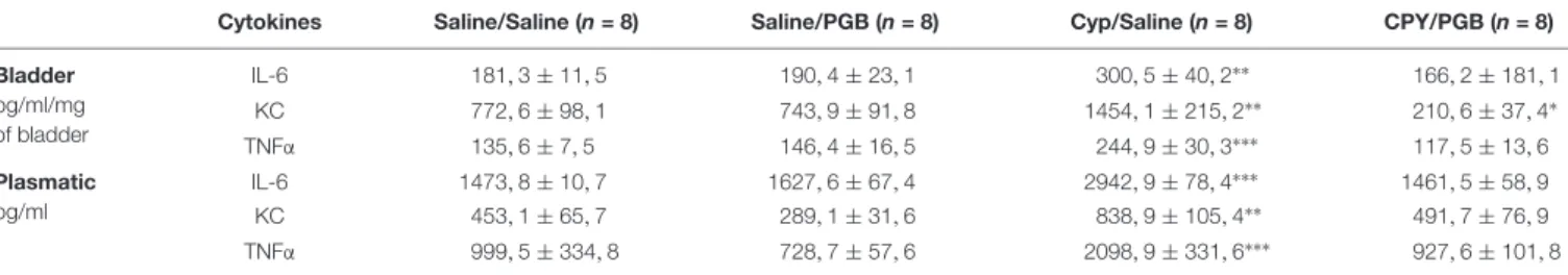
Local (bladder) and systemic (plasma) markers of
inflammation (IL-6, KC, TNFα cytokines) presented the same profile with an increased level in animals receiving CYP and treated with saline, and normal levels in animals receiving CYP and treated with PGB (Table 1).
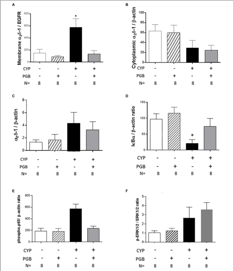
Effect of Pregabalin on Membrane
Addressing
α
2δ-1 Subunit
Cyclophosphamide induced a significant increase in
membrane expression of α2δ-1 subunit in the bladder of
the CYP-treated animals (575.50 ± 141.80, p < 0.05, vs.
control group) (Figure 3A). The membrane expression of
α2δ-1 subunit in animals receiving CYP and treated with
PGB was similar to that in control animals (131.80 ± 58.44, ns, vs. control group) (Figure 3A). No differences were
observed between the groups in α2δ-1 subunit cytoplasmic
expression (Figure 3B) and in total α2δ-1 subunit
expression (Figure 3C).
Effect of Pregabalin on NF-kB Pathway
Activation in Bladder
Cyclophosphamide induced a strong and significant decrease in IkBα expression in the bladder of saline-treated animals
(20.63 ± 12.45, p < 0.05, vs. control group) but not in
that of PGB-treated mice (74.29 ± 25.05, ns, vs. control group) (Figure 3D). Concomitantly, PGB treatment blocked the significant increase in phospho-p65 expression in the bladder of mice receiving cyclophosphamide (234.20 ± 39.07, ns, vs. control group) (Figure 3E). Since a lot of other signaling pathways are involved in inflammation, we have checked the phospho-ERK1/2 pathway. Similarly, as for the phospho-p65, cyclophosphamide induced an increase in phospho-ERK1/2 expression in the bladder of saline-treated animals, and a PGB treatment did not change the level of
phospho-ERK1/2 expression in cyclophosphamide-treated
mice (Figure 3F).
Effect of Pregabalin on LPS-Induced
α
2δ-1 Expression, NF-kB Pathway
Activation, Cytokine Production and
Intracellular Calcium Increase on
Peritoneal Exudate Cell Culture
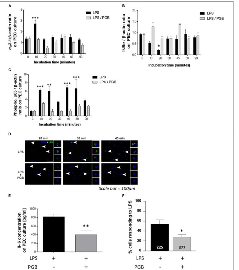
In PEC culture, LPS treatment induced a significant increase in α2δ-1 expression blocked by PGB treatment (Figure 4A).
LPS induced a decrease in IkBα expression (Figure 4B) and an increase in phospho-p65 expression (Figure 4C) in LPS-stimulated PECs blocked by PGB treatment.
The increase in p65 subunit was located in the nucleus of the cells, as shown by detection of the p65 subunit by nucleus marker (DAPI). This localization was not observed following PGB treatment (Figure 4D).
TABLE 1 | Effect of pregabalin (30 mg kg−1, s.c) on IL-6, KC and TNFα concentration in bladder and plasmatic level in cyclophosphamide (CYP)-induced cystitis mice
model.
Cytokines Saline/Saline (n = 8) Saline/PGB (n = 8) Cyp/Saline (n = 8) CPY/PGB (n = 8)
Bladder pg/ml/mg of bladder IL-6 181, 3 ± 11, 5 190, 4 ± 23, 1 300, 5 ± 40, 2∗∗ 166, 2 ± 181, 1 KC 772, 6 ± 98, 1 743, 9 ± 91, 8 1454, 1 ± 215, 2∗∗ 210, 6 ± 37, 4∗ TNFα 135, 6 ± 7, 5 146, 4 ± 16, 5 244, 9 ± 30, 3∗∗∗ 117, 5 ± 13, 6 Plasmatic pg/ml IL-6 1473, 8 ± 10, 7 1627, 6 ± 67, 4 2942, 9 ± 78, 4∗∗∗ 1461, 5 ± 58, 9 KC 453, 1 ± 65, 7 289, 1 ± 31, 6 838, 9 ± 105, 4∗∗ 491, 7 ± 76, 9 TNFα 999, 5 ± 334, 8 728, 7 ± 57, 6 2098, 9 ± 331, 6∗∗∗ 927, 6 ± 101, 8
The values are expressed as a mean l’S.E.M. and compared by a 1-way ANOVA (Treatment) followed by Tukey post hoc test for multiple comparisons. N = 8/group.
∗
p< 0.05,∗ ∗
p< 0.01,∗ ∗ ∗
FIGURE 3 | Effect of PGB (30 mg kg−1, s.c.) on membrane addressingα
2δ-1 subunit and on NF-κB pathway activation in cyclophosphamide (CYP)-induced cystitis
in mice. The expression ofα2δ-1 subunit was evaluated by western blot on cytoplasmic membrane (A), cytoplasm (B), and total cell (C) level. The NF-κB pathway
activation was evaluated by the semi quantification of lkBα (D) and phospho-p65 (E). The phospho-ERK1/2 pathway activation was also evaluated by the semi quantification ERK1/2 and phosphor-ERK1/2 and the ratio between these two forms was calculated (F). The values are expressed as a mean ±S.E.M. and compared by a 1-way ANOVA (Treatment) followed by Tukey post hoc test for multiple comparisons. N = 8/group.∗p< 0.05,∗ ∗p< 0.01,∗ ∗ ∗p< 0.001 vs. control
FIGURE 4 | Effect of PGB (11.3µM) on α2δ-1 subunit (A) IκBα (B), and phospho-p65 (C) expression in resident peritoneal exudate cells (PECs) stimulated with LPS
(100 ng ml−1). The expression was measured 10, 20, 30, 45, 60, and 90 min after LPS with or without PGB treatment by western blot analyses (n = 4/condition).
Intracellular localization of p65 in LPS-stimulated (100 ng ml−1) PEC treated or not with PGB (11.3µM) was visualized by immunohistochemistry. Arrowheads
indicate cells which are magnified in side panels (scale bar: 100µm). (D) Effect of PGB on IL-6 level in resident PECs stimulated with LPS 24 h after these treatments (Continued)
FIGURE 4 | Continued
(n = 4/group). (E) Percentage of LPS-stimulated PECs having [Ca2 +]
iincrease when incubated with saline solution or PGB (11.3µM). (F) A single PEC was
considered as a responder if the F340/F380 ratio for a single PEC increased by 0.05. The number of analyzed cells is shown at the bottom of the histograms (225 cells for the LPS condition and 377 cells for the LPS + PGB condition). The values are expressed as a mean ±S.E.M. and compared by a 2-way ANOVA (Model, Treatment) followed by Bonferroni post hoc test for multiple comparisons forα2δ-1, IκBα and phospho-p65∗p< 0.05,∗ ∗p< 0.01,∗ ∗ ∗p< 0.001 vs. t0 and by a
1-way ANOVA (Treatment) followed by Tukey post hoc test for multiple comparisons for IL-6 and intracellular calcium concentration analyses∗p< 0.05,∗ ∗p< 0.01, ∗ ∗ ∗p< 0.001 vs. LPS treated group.
The increased production of IL-6 cytokine by PECs stimulated with LPS was significantly reduced after PGB treatment (Figure 4E) as was KC cytokine production (data not shown).
Asα2δ-1 ligands inhibit activation of VGCCs, we decided to
further investigate the cellular mechanisms of PGB by measuring intracellular [Ca2+] in cultivated PECs with a Fura-2 probe. In
three independent experiments, stimulation with LPS induced an increase in intracellular Ca2+concentrations in a total of 126 out of 225 PECs (53.3 ± 9.3%). Pre-incubation with PGB significantly reduced the number of PECs having an intracellular [Ca2+] rise
in response to LPS to 105 out of 377 (21.2 ± 3.6%,p< 0.05 vs. LPS group) (Figure 4F).
DISCUSSION
Alpha 2 delta ligands, developed as anticonvulsants and used to treat neuropathic pain, exerted a potent anti-hypersensitive and anti-inflammatory effect in a murine cystitis model. We clearly show that CYP treatment induced an increase in bladder sensitivity and inflammation that is blocked by PGB treatment. The marked decrease in cytokine overexpression induced by PGB could be due to reduced activation of the NF-kB pathway.
Cyclophosphamide-induced cystitis is a widely used model to assess bladder inflammation and related pain (Lantéri-Minet
et al., 1995). Using this model,Boudes et al. (2011)performed von
Frey stimulation and, as in our study, found increased sensitivity in the lower abdominal area. We first showed that PGB treatment greatly increases von Frey scores in CYP-treated animal, evidence that this anticonvulsant drug has an antinociceptive effect. To our knowledge, this is the first time that preclinical data show a potential benefit of using α2δ ligands in an experimental IC
model. One previous preclinical study failed to find any effect of GBP in rat or mice cystitis models (Rudick et al., 2009). However, these preclinical results were surprising according the reports of its efficacy in treatment of IC (Hansen, 2000;
Sasaki et al., 2001). Another clinical study reported the beneficial
effects of a mixed treatment using GBP, amitryptiline and non-steroidal anti-inflammatory agents on bladder pain syndrome
(Kwon et al., 2013). The antinociceptive effect of α2δ ligands
is not surprising given that these drugs are frequently used in the treatment of chronic pain, mainly neuropathic (Verma
et al., 2014) but also in fibromyalgia (Traynor et al., 2011).
There is some evidence of their efficacy in the treatment of visceral pain, notably in IBS patients (Gale and Houghton, 2011). Their effect does not seem to involve a GABAergic mechanism but could result from their blockage of VGCC activation. In
fact, α2δ ligands bind to the α2δ subunit exclusively expressed
by high-voltage gated channels (Cheng and Chiou, 2006). Of the four isoforms, PGB seems to exhibit greater affinity for α2δ-1 subunits expressed in the peripheral and central nervous
systems (Cheng and Chiou, 2006). PGB has a better affinity for these units than GBP, another α2δ ligand (Taylor, 2009)
several findings suggest an involvement of these subunits in the context of visceral pain at a peripheral level in intestinal primary afferent fibers (Needham et al., 2010), or at a central level (Liao
et al., 2010). Blockage of α2δ subunits at both peripheral and
central levels could explain their antinociceptive effect in our cystitis model. However, another peripheral mechanism could be involved. Our study clearly showed that PGB is able to decrease several signs of bladder inflammation, a property that, to our knowledge, has never been mentioned in any of the preclinical models of inflammatory pain in which α2δ ligands were tested
(Hurley et al., 2002).
The key mediator of pro-inflammatory mediator production and inflammation triggering is NF-kB (p65) (Hayden et al., 2006). Given that GBP or PGB binding onα2δ subunits is able to inhibit
this factor in neuroblastoma and glioma cells (Park et al., 2008), the anti-inflammatory effect of PGB could be due to the blockade of NF-kB pathway activation. In the CYP model or in anin vitro model of PEC culture acute administration of PGB can prevent IkBα (main negative regulator of NF-kB pathway) degradation, and p65 phosphorylation (the mark of nuclear translocation of this transcription factor). A decrease in NF-kB pathway activation could result from a decrease in intracellular calcium [Ca2+]i induced by blockade of α2δ subunits. An increase in
[Ca2+]i is important for the activation of intracellular function as transcriptional control (Mandeville and Maxfield, 1996) to promote NF-kB activation and subsequently the expression of genes involved in inflammatory responses (Martin et al., 2006).
While the major Ca2+ entry pathway in non-excitable cells
involves store-operated calcium channels, recent works also suggest that functional VGCCs are expressed in cells such as dendritic cells and macrophages (Gupta et al., 2009). Here, we propose that PGB induces a blockage inα2δ-1 subunit membrane
expression thereby reducing the number of PECs that respond to LPS by increasing [Ca2+]i.
CONCLUSION
To conclude, our study shows that α2δ ligands can reduce
bladder hypersensitivity in a cystitis model and therefore is of potential benefit in patients with bladder pain. Our findings also suggest that α2δ ligands possess anti-inflammatory properties
the mechanism of this anti-inflammatory effect involves a decrease inα2δ-1 membrane expression, in [Ca2+]i and in
NF-kB pathway activation. Clinical studies in patients with bladder pain syndrome are needed to confirm these preclinical data.
AUTHOR CONTRIBUTIONS
LB was involved in protocol and project development, collected and analyzed the data, and wrote the manuscript. SM collected and analyzed the data. MM, YA, and BS were involved in protocol
and project development. AL, LU, and FR collected the data. FC and DA were involved in protocol/project development, the analysis of data, and wrote the manuscript.
FUNDING
This work was supported by ANR (French National
Research Agency) VISCERALGY (ANR-09-MNPS-037-01), the Auvergne Region and FEDER (European fund for regional economic development).
REFERENCES
Boudes, M., Uvin, P., Kerselaers, S., Vennekens, R., Voets, T., De Ridder, D., et al. (2011). Functional characterization of a chronic cyclophosphamide-induced overactive bladder model in mice.Neurourol. Urodyn. 30, 1659–1665. doi: 10.1002/nau.21180
Buffington, C. A., and Woodworth, B. E. (1997). Excretion of fluorescein in the urine of women with interstitial cystitis.J. Urol. 158, 786–789. doi: 10.1016/ S0022-5347(01)64316-7
Chaplan, S. R., Bach, F. W., Pogrel, J. W., Chung, J. M., and Yaksh, T. L. (1994). Quantitative assessment of tactile allodynia in the rat paw.J. Neurosci. Methods 53, 55–63. doi: 10.1016/0165-0270(94)90144-9
Cheng, J.-K., and Chiou, L.-C. (2006). Mechanisms of the antinociceptive action of gabapentin.J. Pharmacol. Sci. 100, 471–486. doi: 10.1254/jphs.CR0050020 Dixon, W. J. (1980). Efficient analysis of experimental observations. Annu.
Rev. Pharmacol. Toxicol. 20, 441–462. doi: 10.1146/annurev.pa.20.040180. 002301
Erickson, D. R., Xie, S. X., Bhavanandan, V. P., Wheeler, M. A., Hurst, R. E., Demers, L. M., et al. (2002). A comparison of multiple urine markers for interstitial cystitis.J. Urol. 167, 2461–2469. doi: 10.1016/S0022-5347(05) 65005-7
Evans, R. J., Moldwin, R. M., Cossons, N., Darekarc, A., Millsc, I. W., Scholfieldc, D., et al. (2011). Proof of concept trial of tanezumab for the treatment of symptoms associated with interstitial cystitis. J. Urol. 185, 1716–1721. doi: 10.1016/j.juro.2010.12.088
Gale, J. D., and Houghton, L. A. (2011). Alpha 2 delta (α(2)δ) ligands, gabapentin and pregabalin: what is the evidence for potential use of these ligands in irritable bowel syndrome.Front. Pharmacol. 2:28. doi: 10.3389/fphar.2011. 00028
Gupta, S., Salam, N., Srivastava, V., Singla, R., Behera, D., Khayyam, K. U., et al. (2009). Voltage gated calcium channels negatively regulate protective immunity toMycobacterium tuberculosis. PLoS One 4:e5305. doi: 10.1371/journal.pone. 0005305
Hansen, H. C. (2000). Interstitial cystitis and the potential role of gabapentin. South. Med. J. 93, 238–242. doi: 10.1097/00007611-200093020-00021 Hayden, M. S., West, A. P., and Ghosh, S. (2006). NF-kappaB and the immune
response.Oncogene 25, 6758–6780. doi: 10.1038/sj.onc.1209943
Hurley, R. W., Chatterjea, D., Rose Feng, M., Taylor, C. P., and Hammond, D. L. (2002). Gabapentin and pregabalin can interact synergistically with naproxen to produce antihyperalgesia.Anesthesiology 97, 1263–1273. doi: 10. 1097/00000542-200211000-00033
Johannessen Landmark, C. (2008). Antiepileptic drugs in non-epilepsy disorders: relations between mechanisms of action and clinical efficacy.CNS Drugs 22, 27–47. doi: 10.2165/00023210-200822010-00003
Kuo, H.-C., and Chancellor, M. B. (2009). Comparison of intravesical botulinum toxin type A injections plus hydrodistention with hydrodistention alone for the treatment of refractory interstitial cystitis/painful bladder syndrome.BJU Int. 104, 657–661. doi: 10.1111/j.1464-410X.2009.08495.x
Kwon, W.-A., Ahn, S. H., Oh, T. H., Lee, J. W., Han, D. Y., Jeong, J. H., et al. (2013). Effect of low-dose triple therapy using gabapentin, amitriptyline, and a nonsteroidal anti-inflammatory drug for overactive bladder symptoms in patients with bladder pain syndrome.Int. Neurourol. J. 17, 78–82. doi: 10.5213/ inj.2013.17.2.78
Lantéri-Minet, M., Bon, K., de Pommery, J., Michiels, J. F., and Menétrey, D. (1995). Cyclophosphamide cystitis as a model of visceral pain in rats: model elaboration and spinal structures involved as revealed by the expression of c-Fos and Krox-24 proteins.Exp. Brain Res. 105, 220–232. doi: 10.1007/BF00240958 Li, Z., Taylor, C. P., Weber, M., Piechan, J., Prior, F., Bian, F., et al. (2011).
Pregabalin is a potent and selective ligand forα(2)δ-1 and α(2)δ-2 calcium channel subunits.Eur. J. Pharmacol. 667, 80–90. doi: 10.1016/j.ejphar.2011. 05.054
Liao, X., Zhou, M.-T., Mao, Y.-F., Xu, H., Chen, H., Sun, J. H., et al. (2010). Analgesic effects of gabapentin on mechanical hypersensitivity in a rat model of chronic pancreatitis.Brain Res. 1337, 104–112. doi: 10.1016/j.brainres.2010. 04.035
Mandeville, J. T., and Maxfield, F. R. (1996). Calcium and signal transduction in granulocytes. Curr. Opin. Hematol. 3, 63–70. doi: 10.1097/00062752-199603010-00010
Martin, L., Pingle, S. C., Hallam, D. M., Rybak, L. P., and Ramkumar, V. (2006). Activation of the adenosine A3 receptor in RAW 264.7 cells inhibits lipopolysaccharide-stimulated tumor necrosis factor-alpha release by reducing calcium-dependent activation of nuclear factor-kappaB and extracellular signal-regulated kinase 1/2.J. Pharmacol. Exp. Ther. 316, 71–78. doi: 10.1124/jpet.105. 091868
Needham, K., Bron, R., Hunne, B., Nguyen, T. V., Turner, K., Nash, M., et al. (2010). Identification of subunits of voltage-gated calcium channels and actions of pregabalin on intrinsic primary afferent neurons in the guinea-pig ileum.Neurogastroenterol. Motil. 22, e301–e308. doi: 10.1111/j.1365-2982.2010. 01567.x
Ogawa, T., Ishizuka, O., Ueda, T., Tyagi, P., Chancellor, M. B., and Yoshimura, N. (2015). Current and emerging drugs for interstitial cystitis/bladder pain syndrome (IC/BPS).Expert Opin. Emerg. Drugs 20, 555–570. doi: 10.1517/ 14728214.2015.1105216
Park, S., Ahn, E. S., Han, D. W., Lee, J. H., Min, K. T., Kim, H., et al. (2008). Pregabalin and gabapentin inhibit substance P-induced NF-kappaB activation in neuroblastoma and glioma cells.J. Cell. Biochem. 105, 414–423. doi: 10.1002/ jcb.21837
Rudick, C. N., Schaeffer, A. J., and Klumpp, D. J. (2009). Pharmacologic attenuation of pelvic pain in a murine model of interstitial cystitis.BMC Urol. 9:16. doi: 10.1186/1471-2490-9-16
Sasaki, K., Smith, C. P., Chuang, Y. C., Lee, J. Y., Kim, J. C., Chancellor, M. B., et al. (2001). Oral gabapentin (neurontin) treatment of refractory genitourinary tract pain.Tech. Urol. 7, 47–49.
Sengupta, J. N., and Gebhart, G. F. (1994). Mechanosensitive properties of pelvic nerve afferent fibers innervating the urinary bladder of the rat.J. Neurophysiol. 72, 2420–2430. doi: 10.1152/jn.1994.72.5.2420
Taylor, C. P. (2009). Mechanisms of analgesia by gabapentin and pregabalin– calcium channel alpha2-delta [Cavalpha2-delta] ligands.Pain 142, 13–16. doi: 10.1016/j.pain.2008.11.019
Traynor, L. M., Thiessen, C. N., and Traynor, A. P. (2011). Pharmacotherapy of fibromyalgia. Am. J. Health Syst. Pharm. 68, 1307–1319. doi: 10.2146/ ajhp100322
Ueda, T., Tamaki, M., Ogawa, O., Yamauchi, T., and Yoshimura, N. (2000). Improvement of interstitial cystitis symptoms and problems that developed during treatment with oral IPD-1151T.J. Urol. 164, 1917–1920. doi: 10.1016/ S0022-5347(05)66917-0
van Ophoven, A., Pokupic, S., Heinecke, A., and Hertle, L. (2004). A prospective, randomized, placebo controlled, double-blind study of amitriptyline for the treatment of interstitial cystitis. J. Urol. 172, 533–536. doi: 10.1097/01.ju. 0000132388.54703.4d
Verma, V., Singh, N., and Singh Jaggi, A. (2014). Pregabalin in neuropathic pain: evidences and possible mechanisms.Curr. Neuropharmacol. 12, 44–56. doi: 10.2174/1570159X1201140117162802
Yoshimura, N., Seki, S., Chancellor, M. B., de Groat, W. C., and Ueda, T. (2002). Targeting afferent hyperexcitability for therapy of the painful bladder syndrome. Urology 59, 61–67. doi: 10.1016/S0090-4295(01) 01639-9
Conflict of Interest Statement: The authors declare that the research was conducted in the absence of any commercial or financial relationships that could be construed as a potential conflict of interest.
Copyright © 2019 Boudieu, Mountadem, Lashermes, Meleine, Ulmann, Rassendren, Aissouni, Sion, Carvalho and Ardid. This is an open-access article distributed under the terms of the Creative Commons Attribution License (CC BY). The use, distribution or reproduction in other forums is permitted, provided the original author(s) and the copyright owner(s) are credited and that the original publication in this journal is cited, in accordance with accepted academic practice. No use, distribution or reproduction is permitted which does not comply with these terms.