Publisher’s version / Version de l'éditeur:
ACS Omega, 2, 11, pp. 7621-7636, 2017-11-07
READ THESE TERMS AND CONDITIONS CAREFULLY BEFORE USING THIS WEBSITE. https://nrc-publications.canada.ca/eng/copyright
Vous avez des questions? Nous pouvons vous aider. Pour communiquer directement avec un auteur, consultez la
première page de la revue dans laquelle son article a été publié afin de trouver ses coordonnées. Si vous n’arrivez pas à les repérer, communiquez avec nous à [email protected].
Questions? Contact the NRC Publications Archive team at
[email protected]. If you wish to email the authors directly, please see the first page of the publication for their contact information.
NRC Publications Archive
Archives des publications du CNRC
This publication could be one of several versions: author’s original, accepted manuscript or the publisher’s version. / La version de cette publication peut être l’une des suivantes : la version prépublication de l’auteur, la version acceptée du manuscrit ou la version de l’éditeur.
For the publisher’s version, please access the DOI link below./ Pour consulter la version de l’éditeur, utilisez le lien DOI ci-dessous.
https://doi.org/10.1021/acsomega.7b00805
Access and use of this website and the material on it are subject to the Terms and Conditions set forth at
Initial structural models of the Aβ42 dimer from replica exchange
molecular dynamics simulations
Blinov, Nikolay; Khorvash, Massih; Wishart, David S.; Cashman, Neil R.;
Kovalenko, Andriy
https://publications-cnrc.canada.ca/fra/droits
L’accès à ce site Web et l’utilisation de son contenu sont assujettis aux conditions présentées dans le site LISEZ CES CONDITIONS ATTENTIVEMENT AVANT D’UTILISER CE SITE WEB.
NRC Publications Record / Notice d'Archives des publications de CNRC:
https://nrc-publications.canada.ca/eng/view/object/?id=f6d6252e-1bbf-45c4-b84a-764106b323d4 https://publications-cnrc.canada.ca/fra/voir/objet/?id=f6d6252e-1bbf-45c4-b84a-764106b323d4
Initial Structural Models of the Aβ42 Dimer from Replica Exchange
Molecular Dynamics Simulations
Nikolay Blinov,
*
,†,‡Massih Khorvash,
§David S. Wishart,
∥Neil R. Cashman,
§and Andriy Kovalenko
‡,† †Department of Mechanical Engineering, University of Alberta, Edmonton, Alberta T6G 1H9, Canada
‡
National Institute for Nanotechnology, National Research Council of Canada, Edmonton, Alberta T6G 2M9, Canada
§
Department of Medicine, University of British Columbia, Vancouver, British Columbia V6T 2B5, Canada
∥
Departments of Computing Science and Biological Sciences, University of Alberta, Edmonton, Alberta T6G 2E8, Canada
*
S Supporting InformationABSTRACT: Experimental characterization of the molecular structure of small amyloid (A)β oligomers that are currently considered as toxic agents in Alzheimer’s disease is a formidably difficult task due to their transient nature and tendency to aggregate. Such structural information is of importance because it can help in developing diagnostics and an effective therapy for the disease. In this study, molecular simulations and protein−protein docking are employed to explore a possible connection between the structure of Aβ monomers and the properties of the intermonomer interface in the Aβ42 dimer. A structurally diverse ensemble of conformations of the monomer was sampled in microsecond timescale implicit solvent replica exchange molecular dynamics simulations. Representative structures with different solvent exposure of hydrophobic residues and secondary structure content were selected to build structural models of the dimer. Analysis of these models reveals that formation of an intramonomer salt bridge (SB) between Asp23 and Lys28 residues can prevent the building of a hydrophobic interface between the central hydrophobic clusters (CHCs) of monomers upon dimerization. This structural feature of
the Aβ42 dimer is related to the difference in packing of hydrophobic residues in monomers with the Asp23−Lys28 SB in on and off states, in particular, to a lower propensity to form hydrophobic contacts between the CHC domain and C-terminal residues in monomers with a formed SB. These findings could have important implications for understanding the difference between aggregation pathways of Aβ monomers leading to neurotoxic oligomers or inert fibrillar structures.
1. INTRODUCTION
Soluble oligomers composed of amyloid-β (Aβ) peptides are currently considered as neurotoxic agents in Alzheimer’s disease (AD).1−7
Aβ peptides of different length (mostly made of 37− 43 residues) originate from proteolytic cleavage of the amyloid precursor protein (APP). Under certain conditions, they can aggregate into amyloid fibrils, a pathological hallmark of AD, and neurotoxic Aβ oligomers are formed as intermediates or off-path products of fibrillization.8 The high resolution molecular structure of Aβ oligomers remains mostly unknown and there is no consensus on the mechanisms of oligomeriza-tion. Inhibition of initial misfolding and aggregation of Aβ peptides is considered as one of the most promising therapeutic targets for AD.8−13
In this context, resolving the atomic structure of aggregation-prone states of Aβ peptides and small oligomers, as well as identification of initial events in oligomerization pathways is of great importance.
Although high resolution molecular models of mono-meric14−27 and fibrillar28−30 Aβ species have been resolved
under different conditions, no such information is available for Aβ oligomers, mostly because of their transient nature. Also, there is no agreement on the conformational properties of monomeric species involved in initial aggregation. Solution
NMR experiments revealed that Aβ monomers in aqueous solutions adopt mostly random/collapsed/extended coil conformations,14,16,19,24,25,31whereas in nonpolar environments
(such as mimicking lipid membranes), their structures are characterized by substantial α-helical content.18,20,26,27 This suggests that Aβ monomers in coiled or α-helical conforma-tions may be involved in the initial stages of oligomerization (e.g., dimerization), with following structural conversion into β-rich structures in larger Aβ species.32−34
Experimental data confirm that structural changes occur in initially unstructured Aβ1−40 (Aβ40) monomers upon oligomerization, with a substantial increase in β-sheet content already in dimers and its gradual increase in larger oligomers.33The possibility of β-sheet structural organization of Aβ oligomers, including antiparallel cross-β sheet arrangements, was demonstrated experimentally for different oligomer preparations.33,35Although it is not clear
whether soluble Aβ monomers with β-sheet structure are involved in the formation of oligomers in vivo, such a possibility was explored in theoretical studies.36−38Structural
Received: June 16, 2017
Accepted: October 26, 2017
Published: November 7, 2017
Article
http://pubs.acs.org/journal/acsodf Cite This: ACS Omega 2017, 2, 7621-7636
models of Aβ dimers or larger oligomers were built with β-sheet monomers extracted from the experimental models of amyloid fibrils38or obtained with molecular dynamics (MD) conforma-tional sampling.36,37Experimentally, the presence of Aβ dimers in the brain was first reported a few years ago,39 and their possible toxicity has been confirmed recently.40,41Although it still remains controversial whether the Aβ dimer exists in vivo and is a smallest neurotoxic agent in AD, resolving its molecular structure remains an important (and challenging) task because such information can provide molecular details of the oligomerization pathway and can be further utilized in the rational development of anti-AD drugs targeting the initial stages of aggregation of Aβ peptides.
Molecular simulations and modeling complement experi-ment, and can provide important insight into dynamical and structural properties of different Aβ species in situations where experimental information is not readily available, such as in the case of Aβ oligomers (see, for example, a recent review42). Aβ dimers were studied in all-atom explicit and implicit solvent MD simulations with and without ligands (such as inhibitors of aggregation or metal ions).36−38,43−49 Larger oligomeric
species, as well as different stages of aggregation/fibrillization of Aβ monomers and other amyloidogenic proteins, were mostly investigated with coarse-grained MD and Monte Carlo simulations.45,50−54
Most of the computational studies of Aβ dimers were focused on prediction of their structural properties,36−38,48,49,54−56
dimerization pathways under differ-ent conditions, including in the presence of metal ions,36,46and
on possible inhibitory mechanisms of various ligands.43,44,47In most of these works, initial structures of the Aβ dimer were modeled based on the experimental solution or solid state NMR data,43,44,49 or were built from Aβ monomer con-formations with different structural features (such as extended β-sheet structural content) sampled in MD simulations.36,37,54 Because of the intrinsically disordered nature of Aβ peptides7,14 characterized by conformational diversity, it is very likely that multiple distinct conformations with an elevated propensity for aggregation will be involved in the formation of initial dimeric constructs (or larger oligomers), which will possibly further undergo major conformation changes along different aggregation pathways to form metastable (e.g., oligomeric) or thermodynamically stable (e.g., fibrillar) Aβ species. This is in line with the “conformational selection” paradigm for molecular recognition57,58recently used to model
Aβ dimers48and ring-shaped Aβ1−42 (Aβ42) oligomers.48,59 In the current study, we build structural models of the Aβ42 dimer based on the hypothesis of the initial hydrophobic association of monomers and conformational selection mechanism of the first step of dimerization. The Aβ42 constructs were chosen because they constitute a major component of amyloid plaques in AD and are characterized by a higher propensity for aggregation compared to the more abundant Aβ40 monomeric species. This could make misfolding and aggregation of Aβ42 monomers a better target for therapeutic intervention in AD. Conformational space of the Aβ42 monomer was sampled with generalized Born/solvent area (GBSA) implicit solvent replica exchange molecular dynamics (REMD) simulations, and then representative conformations with different propensity for aggregation were selected by clustering time series of a solvent exposed surface area (SASA) of hydrophobic residues. Initial structural models of the Aβ42 dimer were built with ZDOCK protein−protein docking software60,61from the representative conformations of
Aβ42 monomers characterized by different SASA of hydro-phobic residues and secondary structure content. We also consider other structural features of monomers, such as propensity to form the Asp(D)23−Lys(K)28 salt bridge (SB). This (intra- or interpeptide) SB was observed in most of the structural models of Aβ40 and Aβ42 amyloid fibrils,62
and it may be disrupted in neurotoxic oligomers, as follows from experimental data on 5E3 conformational antibody specificity toward oligomeric Aβ species.63,64It is worth noting that the antioligomer conformational antibodies do not recognize dimeric Aβ species, possibly because the antibody-specific epitopes of the toxic Aβ oligomers have some distinctive structural features not present in the dimeric structures, in addition to solvent exposed Lys28.
Analysis of structural models of the Aβ42 dimer proposed in this study suggests possible correlations between formation of the interpeptide interface between the central hydrophobic clusters (CHCs) and such structural characteristics of monomers as secondary structure content and propensity to form the D23−K28 SB. In particular, the CHC−CHC interface in α-helical rich models of the Aβ dimer is formed only for monomers with a disrupted D23−K28 SB and thus solvent exposed Lys28. No such interface is observed for most of the dimer models with a formed D23−K28 SB or in the dimers with β-sheet structural content. Molecular dynamics simu-lations were carried out to demonstrate the stability of the interpeptide CHC−CHC interface at the microsecond (μs) timescale.
2. RESULTS
2.1. Structural Ensemble of the Aβ42 Monomer.In the course of MD simulations, the initial fully extended conformation of the Aβ42 monomer collapses toward compact structures on the timescale of a few nanoseconds. The resulting structural ensemble has a narrow distribution of the radius of gyration peaked at approximately 9.7 Å (Figure S1 of Supporting Information (SI)), compared to the 43.52 Å value of the extended conformation. The structural ensemble of the Aβ42 monomer is characterized by a high structural diversity, as expected for this intrinsically disordered system. The monomer samples a range of conformational states with different secondary structure contents and extent of solvent exposure of hydrophilic and hydrophobic residues (Figures S2 and S3of SI). There is also a difference in propensity to form intrapeptide salt bridges between charged residues for different parts of the MD trajectory. In the context of the current study, formation of a salt bridge between Asp(D)23 and Lys(K)28 residues is of special interest (Figure S4 of SI). The experimental data on recognition of toxic Aβ oligomers by a conformational antibody suggest that Lys28 is solvent exposed in oligomers.63,64This indicates that the D23−K28 SB may be
disrupted in the toxic oligomeric species. In contrast, Lys28 forms an interpeptide SB with Asp2330or intrapeptide SB with
Ala42 in the experimental models of the Aβ42 amyloid fibril.28,29Because the conformational antibody specific to toxic oligomers does not recognize both the fibrillar and monomeric Aβ species, Lys28 may form favorable electrostatic contacts with other residues in the Aβ42 monomer, similar to what is observed in the fibrillar Aβ structures.
The analysis of time evolution of the distance between Asp23 and Lys28 residues reveals that the MD trajectory has segments characterized by a different propensity to form the D23−K28 SB (Figure 1). In particular, the last part of the production run
(>2.5 μs) has a higher propensity compared to that of the rest of the trajectory. Because the solvent exposure of Lys28 may be one of the structural signatures of the toxic Aβ oligomers as discussed above, two segments of the MD trajectory with different propensity to form the D23−K28 SB are used in the next section to identify representative conformations of the Aβ42 monomer for building initial structural models of the dimer.
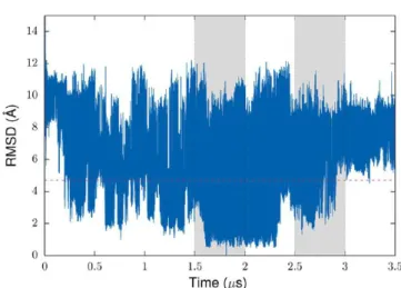
Despite the fact that the radius of gyration quickly relaxes to a quasi-stationary state, a root-mean-square deviation (RMSD) of atomic positions is characterized by relatively large fluctuations at the microsecond timescale (Figure 2). The time series of RMSD features two distinctive domains separated by a 4.7 Å threshold (indicated by the red dashed line inFigure 2; see also Figure S5 of SI), with a “large RMSD” domain dominating in a long run (Figure S6of SI). The ensemble of conformations from this domain is characterized by a broader distribution of SASA for different residue groups. The SASA distribution of the hydrophobic residues, for example, is shifted toward a higher exposure compared to that of the “small RMSD” domain (Figure S7 of SI). Because of the large-scale fluctuations of RMSD at the microsecond timescale, it is not obvious whether the small RMSD domain features will reoccur in a longer simulation. On the other hand, there is a much better agreement between calculated and experimental J-coupling values if only the conformations from the large RMSD domain are used (see the discussion in theMaterial and Methodssection). This suggests that this domain may describe the conformational properties of the Aβ42 monomer more accurately, and thus it is used for selection of representative structures of the monomer in the next section.
It is important to emphasize that using MD sampling based on the implicit solvent GBSA model may provide a conformational bias due to overestimation of specific electro-static interactions and a higher tendency (compared to explicit solvent simulations) to form α-helical structures.65,66Thus, the conformational ensemble generated in this study is
charac-terized by an elevated α-helical and a reduced random coil content (Figure S2of SI), compared to what was observed in previous all-atom explicit solvent MD studies of different Aβ monomeric species (see, for example, a recent review67). At the same time, the current REMD protocol predicts regions with a higher propensity to form different elements of secondary structures, including α-helices, turns, and β-strands (Figure S8
of SI), in agreement with some previous MD simulations of Aβ peptides that sampled the conformational properties of Aβ peptides in agreement with experimental data.68 Along with reasonable J-coupling data, this suggests that monomeric constructs selected for building initial models of the Aβ42 dimer may represent physiologically relevant conformations of the Aβ42 monomer. This is further supported by the fact that implicit solvent models can perform well in predicting structural properties of biomolecules,66,69,70 including intrinsi-cally disordered peptides and folded small peptides.66
2.2. Selection of Representative Conformations of the Aβ42 Monomer.Solvent exposure of hydrophobic residues is one of the most important factors defining aggregation propensity of amyloidogenic proteins and peptides. An increase in the extent of exposure due to changes in solvent environmental conditions (such as the level of pH, salt concentration, solvent composition, or intra-cellular crowding) may affect conformational equilibrium between different Aβ structural states by shifting the balance toward oligomeric species. Initial hydrophobic association of Aβ peptides can be followed by structural rearrangement caused by formation of specific interactions, including hydrogen bonds and salt bridges, between constituting monomers. Formation of such inter-actions at the earlier stages of an aggregation pathway may be impeded by a high cost of desolvation of polar and charged residues, which can be reduced after initial hydrophobic association.
In the current study, conformations of the Aβ42 monomer with a high solvent exposure of hydrophobic residues and their potential interaction partners are used to build initial models of
Figure 1.Time evolution of the distance between CG atomic site of Asp23 and NZ site of Lys28 in the REMD simulation of the Aβ42 monomer. The gray areas show two production segments of the trajectory used for selection of representative conformations for modeling the Aβ42 dimer. A segment on the left is characterized by the D23−K28 SB mostly in the off state. The right segment features transitions between on and off states, with a relatively high population of the D23−K28 SB on state. Here and below, the data are shown for the REMD replica corresponding to 25 °C.
Figure 2.Time evolution of the Cα RMSD in the REMD simulation of the Aβ42 monomer. As a reference structure for the RMSD calculation, a structure closest to the centroid of the most populated cluster from clustering of time series of the radius of gyration was used. The radius of gyration of this structure is 9.72 Å, which is close to the value corresponding to the maximum of the radius of gyration distribution calculated for different parts of the trajectory (Figure S1of SI). The gray areas show two segments of the trajectory used for selection of representative conformations for modeling the Aβ42 dimer.
the Aβ42 dimer. These conformations were identified by clustering time series of the hydrophobic SASA (Figure 3) from
the two production segments of the MD trajectory. Clustering was performed with the hierarchical agglomerative (bottom-up) technique71 implemented in the cpptraj program from the
Amber molecular dynamics package.72Five structural clusters were identified for each production segment of the MD trajectory. Representative structures with the smallest RMSDs of Cα atomic positions relative to the centroids of clusters with the largest hydrophobic SASA and the most populated clusters were selected for building initial structural models of the Aβ42 dimer. These structures are characterized by a substantial α-helical content. This is a consequence of a relatively high population of such structures in the structural ensemble of the Aβ42 monomer sampled with the implicit solvent GBSA model. To include in the analysis conformations with other structural features, clustering of hydrophobic SASA was performed for a subset of MD frames with β-strand content. Selected representative structures were used to build initial models of the Aβ42 dimer, along with the α-helical structures. It is worth noting that due to the substantial memory allocation required for calculation of the matrix of pairwise distances in the parameter space, clustering could not be performed for the entire production trajectory without skipping a substantial portion of MD frames. With every second frame
omitted, clustering with the protocol described above results in identification of five representative structures with none of them having a formed D23−K28 SB. With a modified protocol set to identify 10 structural clusters, only 2 out of 10 representative structures belong to the second production section, and both of them are characterized by a formed D23− K28 SB. Among the other eight structures, there are no structures with an elevated hydrophobic SASA and a disrupted SB. Clustering of the entire trajectory may provide a more accurate representation of the population distribution of different structures in the conformational ensemble, compared to a separate clustering of two production segments of the trajectory adopted here. At the same time, using trajectory segments with different propensity to form the D23−K28 SB guarantees a structural diversity of representative conformations that are of high relevance in the context of the current study. In particular, the approach allows us to select representative structures with a high solvent exposure of hydrophobic residues with the SB both in the off and on states.
2.2.1. Representative Conformations of the Aβ42 Mono-mer with α-Helical Structure Content.Conformations of the Aβ42 monomer representing the structural cluster with a large hydrophobic SASA and the most populated cluster from the first production segment of the trajectory with a low propensity to form the D23−K28 SB are shown in the top panel ofFigure 4. A distinctive feature of these structures is a hydrophobic interface between the central hydrophobic cluster (residues Leu17, Val18, Phe19, and Phe20) and C-terminal hydrophobic Val40 and Ile41. It is worth noting that such contacts are absent in the Aβ40 monomer, which may partially explain differences in conformational properties and propensity for aggregation between Aβ42 and Aβ40 peptides.
The large hydrophobic SASA of the structure in the top-left panel ofFigure 4(1942 Å2compared to 1134 Å2value for the
structure from the most populated cluster shown in the right panel) can be attributed to a partial solvent exposure of residues in the central hydrophobic cluster (CHC) as well as to solvent accessibility of hydrophobic Ile32 and Val36. For the structure representing the most populated cluster (the top right panel ofFigure 4), residues from the CHC form hydrophobic contacts with Ile32 and Val36, which results in a decreased hydrophobic SASA. Despite a relatively small exposure of hydrophobic residues, this conformation can be considered as a good candidate to model initial hydrophobic association because of a possible reduction of the entropic penalty toward association. This a consequence of a relatively good packing of the hydrophobic residues, which may result in reduced flexibility of this structure. Also, hydrophobic patches formed by the CHC and Ile32−Val36 domains, which are disconnected in the structure with the large hydrophobic SASA, are now merged in a large continuous patch, creating favorable conditions for hydrophobic association of monomers.
We also note that Lys28 residue, which may form an intra- or interpeptide SB in experimental models of the Aβ42 amyloid fibril and is implicated in the conformational antibody recognition of toxic oligomers,63,64 is solvent exposed in all
representative structures from the first production segment of the trajectory. Asp23 residue, a SB partner of Lys28 in some fibrillar structures, is solvent exposed in the most populated cluster, and is buried inside the peptide fold for the structure with the large solvent exposure of hydrophobic residues where it forms a SB with Arg5. The latter is characterized by a high propensity to form SBs with multiple residues, including Asp1,
Figure 3.Time evolution of solvent exposed surface area (SASA) of hydrophobic (the upper panel) and hydrophilic (the bottom panel) residues of the Aβ42 monomer. The gray areas show two production segments of the trajectory.
Glu3, Glu11, Glu22, and Asp23, in the representative structures from both production segments of the trajectory.
In contrast to the case of the first production segment of the trajectory where none of the representative structures had the D23−K28 SB, two out of five representative conformations
from the second segment are characterized by a formed SB. These two structures belong to the least populated structural clusters and have a relatively high solvent exposure of hydrophobic residues. In all other structures, the D23−K28 SB is disrupted and Lys28 is mostly solvent exposed. Asp23 residue is partially buried in these structures and may form a SB with Arg5, similar to the case of the first segment of the trajectory.
The representative structures from the cluster characterized by a large hydrophobic SASA and the most populated cluster from the second production part of the trajectory are shown in the middle panel ofFigure 4. Their hydrophobic SASA is 2150 and 1462 Å2 , respectively. Similar to the first segment, the
structure with the D23−K28 SB off state from the most populated cluster has the hydrophobic contacts between the CHC domain and the C-terminal hydrophobic residues (the middle right panel ofFigure 4). What is new is that there is no hydrophobic interface between the CHC domain and C-terminal Val40 and Ile41 for the structure with the largest hydrophobic SASA. This structure is characterized by a formed D23−K28 SB (the middle left panel ofFigure 4). Inspection of all representative structures from both production segments of the trajectory suggests that formation of the D23−K28 SB correlates with disruption of the hydrophobic interface between the CHC domain and C-terminal residues of the monomer.
It is worth noting that the structures with the largest hydrophobic SASA belong to the least populated structural clusters. This agrees with the notion that in aqueous environments, burying of hydrophobic residues is a thermody-namically favorable process resulting in optimization of the solvation part of a system’s free energy.
2.2.2. Representative Conformations of the Aβ42 Mono-mer with β-Sheet Structure Content.The structural models of the Aβ42 monomer discussed above are characterized by a substantial α-helical content. Most probably, this is a consequence of under-sampling of the conformational ensemble of the monomer with the implicit solvent GBSA model, resulting in a reduced population of random coil and β-strand structures. To address this problem in the context of probing possible dimerization pathways, and to further explore the diversity of the conformational space of the Aβ42 monomer, representative structures were also selected from a subset of MD frames characterized by an elevated β-strand structure content. As in the previous section, these structures were identified with clustering of time series of the hydrophobic SASA. Most of the selected conformations are characterized by a relatively high solvent exposure of hydrophobic residues, with the exception of one structure with a large α-helical content. This structure was excluded from the following analysis because of its similarity to the structures discussed in the previous section.
The representative conformations of the Aβ42 monomer with β-strand content have a different pattern in packing of hydrophobic residues compared to that of most of the α-helical structures characterized by a disrupted D23−K28 SB. In particular, there is no hydrophobic interface between the CHC domain and C-terminal hydrophobic residues, which was observed only for the α-helical structures with the intrapeptide D23−K28 SB. It is worth noting that in all structures with β-strand content, Lys28 is solvent exposed and it does not establish a SB with Asp23. Similar to what was observed for some α-helical structures, Asp23 can form a SB with Arg5.
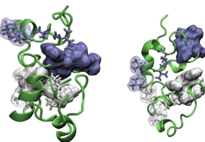
Figure 4.Representative conformations of the Aβ42 monomer with a large solvent exposure of hydrophobic residues (the left column) and from the most populated structural clusters (the right column). Top panel: structures from the first production segment of the MD trajectory characterized by a low propensity to form the D23−K28 salt bridge. Middle panel: structures from the second production segment of the trajectory characterized by a high propensity to form the salt bridge. Bottom panel: structural models of the Aβ42 monomer with β-sheet content. Asp23 and Lys28 residues are drawn in stick representation and colored according to their atom names. Hydro-phobic residues from the central hydroHydro-phobic cluster (CHC), and hydrophobic Ile32, Val36, Val40, and Ile41 residues from the C-terminal part of the monomer are shown in stick representation. Transparent molecular surfaces of residues from the CHC (white color), as well as of Val40 and Ile41 residues (blue color) are also shown.
All β-strand representative structures are characterized by the antiparallel β-sheet structural motif. The structure with the largest hydrophobic SASA (the bottom-left panel ofFigure 4) has a β-hairpin formed by Ser26, Asn27 and Ala30, Ile31 residues. In the structure representing the most populated cluster, β-strands are formed by His14, Gln15 and Met35, Val36 residues (the bottom-right panel of Figure 4). The conformation with the largest exposure of hydrophobic residues (with the hydrophobic SASA of 1887 Å2compared to 1777 Å2
for the structure from the most populated cluster) belongs to the third (out of five) most populated structural cluster, not to the least populated one as was observed for the α-helical conformations in the previous section. This can be explained by the fact that MD frames with a particular structural feature (β-strand content in this case) were preselected for clustering analysis, which may affect a population balance linked to the system’s free energy in unbiased ensembles.
2.3. Structural Models of the Aβ42 Dimer.All models of the Aβ42 dimer discussed below were generated with the ZDOCK protein−protein docking software.60,61 The top 10 structures from docking simulations for each dimer model were optimized and rescored with the Amber molecular dynamics package,72as discussed in theMaterial and Methodssection.
2.3.1. Models of the Aβ42 Dimer Built from Monomeric Constructs with a High Solvent Exposure of Hydrophobic Residues. By utilizing the hypothesis of initial hydrophobic association, in this section, structural models of the Aβ42 dimer were built from the monomeric constructs representing the structural clusters with a large hydrophobic SASA. For both production segments of the MD trajectory, monomers with a high solvent exposure of hydrophobic residues belong to the least populated clusters. This indicates that these monomeric constructs are unlikely to form initial aggregates at low physiological concentrations, but probability of dimerization may increase with increasing monomer concentration. The equilibrium balance between different monomeric structures can also be shifted toward constructs with a higher exposure of hydrophobic residues as a consequence of a change in the environmental conditions. This can make initial hydrophobic association of monomers a more likely event, as supported by some experimental data. In another scenario, monomers with a large hydrophobic SASA interact in the process of dimerization with more abundant conformations representing highly populated structural clusters characterized by a lower solvent exposure of hydrophobic residues. This possibility will be discussed in the next sections.
The top-ranked structural models of the Aβ42 dimer constructed from the monomers with a high solvent exposure of hydrophobic residues from both production segments of the trajectory are shown inFigure 5(see alsoFigures S9 and S10of SI where three top-ranked models of the dimer for each trajectory segment are shown). The interpeptide interface in all top-ranked models built with the monomer from the first segment characterized by a disrupted D23−K28 SB includes hydrophobic contacts between the CHC domains and contacts between the CHC domain of one monomer and the C-terminal hydrophobic residues of the other (the left panel of Figure 5
andFigure S9of SI). For the second-ranked structure, there is an additional hydrophobic contact between C-terminal Ile41 residues of the monomers (the middle panel ofFigure S9 of SI). The three top-ranked models represent structural proper-ties of 8 out of 10 top-ranked dimer models built with the ZDOCK software, with the third-ranked structure (the right
panel of Figure S9 of SI) representing the largest structural cluster (4 out of 10 structural models).
Along with the hydrophobic interactions, there are favorable electrostatic intermonomer contacts between Asp7 and Lys16 residues for all three top-ranked structures. This may result in the formation of SBs between these residues. For the first-ranked structure, all other ionizable residues with the exception of Asp23, which may form an intrapeptide SB with Arg5, are solvent exposed and are not involved in the formation of favorable electrostatic intra- or interpeptide contacts. For the third-ranked structure, Lys16 from both monomers in the dimer can alternatively form favorable electrostatic contacts with either Glu3 or Asp1. For the second-ranked structure, there is also unfavorable electrostatic contact between Asp1 and Glu22. This indicates that this model may undergo structural changes after initial association to optimize the electrostatic interactions.
It is worth noting that the hydrophobic residues from the CHC domains and C-termini are only partially buried in the interpeptide interface in the dimer models discussed above. Residues that are not involved in formation of the interpeptide interface may form relatively large continuous hydrophobic patches. This can potentially result in further association of dimers with formation of Aβ42 tetrameric constructs and other larger oligomers.
The representative structure of the Aβ42 monomer with a high solvent exposure of hydrophobic residues from the last production segment of the trajectory is characterized by a formed D23−K28 SB. Dimer models built with this monomeric construct do not have the hydrophobic interpeptide CHC− CHC interface (the right panel ofFigure 5andFigure S10of SI). This is different from the case of the models without the intrapeptide D23−K28 SB discussed above. Still, the top-ranked models have multiple interpeptide hydrophobic contacts. The first-ranked model, for example, has a relatively good hydrophobic interface (Figure S11 of SI) formed by a
Figure 5. First-ranked models of the Aβ42 dimer built from the monomeric constructs with a high solvent exposure of hydrophobic residues from the first (the left panel) and the second (the right panel) production segments of the trajectory. Residues in the central hydrophobic clusters are shown by their molecular surface, hydro-phobic Ile32 and Val36 and C-terminal Val40 and Ile41 are shown in stick representation, with white and blue colors used for different monomers. For the Ile32 and Val36 residues, transparent molecular surfaces are also shown.
Phe20−Val24−Val24−Phe20 “sandwich” of hydrophobic resi-dues, as well as a hydrophobic interface between Val12, Ile31, Ile32, and Leu34 residues from both monomers.
To quantify contributions of individual residues to the interpeptide interface in dimeric constructs, we calculated average desolvation profiles of the monomers for all dimer models obtained with the ZDOCK software. A desolvation profile is defined as the difference between normalized SASAs of residues in constituting monomers and a dimer, averaged over 10 top-ranked structures for each dimer model. The dimer models with the intrapeptide D23−K28 SB in the off state (the top panel ofFigure 6) are characterized by a larger decrease in solvent exposure of hydrophobic residues from the CHC domain compared to that of the models with the formed D23− K28 SB (the bottom panel of Figure 6). This suggests that there is a correlation between formation of the D23−K28 SB and a propensity to form the interpeptide CHC−CHC interface not only for the top-ranked models, but also for a
structurally diverse ensemble of dimeric structures identified in docking experiments.
2.3.2. Models of the Aβ42 Dimer Built with Monomeric Constructs from the Most Populated Structural Clusters.To verify whether a hydrophobic interface between the CHC domains can form upon aggregation of monomers with a relatively low exposure of hydrophobic residues, structural models of the Aβ42 dimer were built with the monomeric constructs representing the most populated structural clusters. For these monomers with a disrupted D23−K28 SB, all top-ranked dimer models have interpeptide contacts between the CHC domains (Figure 7andFigures S12 and S13of SI). This
is consistent with the previous observation that monomers with a disrupted D23−K28 SB have a high propensity to form the interpeptide CHC−CHC interface. This is further confirmed by inspection of the corresponding desolvation profiles (Figure S14of SI).
Dimers originating from the first segment of the trajectory are characterized by additional interactions between hydro-phobic residues from the CHC domain of one monomer and the C-terminal domain of the other (the left panel ofFigure 7
andFigure S12of SI), which is similar to what was observed for the dimer models built from the monomer with a large hydrophobic SASA. The interpeptide interface of the first-ranked structure can be further stabilized by π-stacking interactions between Phe19 residues from the CHC domains. For the second-ranked structure, there is an additional hydrophobic contact between C-terminal Ile41 residues of the monomers. All of these models are characterized by a relatively high solvent exposure of hydrophilic residues and reduced exposure of hydrophobic residues, and, in contrast to the case of the dimer models built with the monomer structure with a large hydrophobic SASA, they do not have favorable interpeptide SBs.
For the final production segment of the trajectory, most of the top-ranked dimer models belong to two distinctive structural groups covering 7 out of 10 top-ranked structures (the right panel ofFigure 7andFigure S13of SI). In addition to the hydrophobic interpeptide CHC−CHC interface, the first-ranked structure may have hydrophobic contacts between Val36 residues, and the second-ranked one between the CHC domain of one monomer and the C-terminal hydrophobic
Figure 6. Changes in solvent accessible surface area (SASA) of individual residues upon dimerization of the Aβ42 monomer with a high solvent exposure of hydrophobic residues. The top and bottom panels are for the dimer models built with the monomers from the first and second production segments of the trajectory with the D23−K28 SB in the off and on states, respectively. The SASAs of individual residues are normalized relative to their values in the Gly−X−Gly tripeptide structure in a fully extended conformation. Vertical lines give standard error of means for the ten top-ranked dimer models. Contributions of residues from the CHC domain are shown with a lighter color.
Figure 7.Same as inFigure 5, but for the Aβ42 dimer models built with the monomers representing the most populated structural clusters.
residues of the other. The latter structure can be also stabilized by π-stacking interactions between Phe19 and Phe20 for both monomers.
2.3.3. “Heterogeneous” Models of the Aβ42 Dimer. Aβ monomers interacting at the initial stage of dimerization most probably have different structural properties. In a likely scenario, one of the monomers with an elevated propensity for aggregation (such as resulting from a high solvent exposure of hydrophobic residues) may belong to a low-populated structural cluster, whereas its interaction partner from a highly populated cluster will have a reduced propensity for aggregation. To describe such a situation, models of the Aβ42 dimer were built with monomers having a different degree of solvent exposure of hydrophobic residues, such as that characterized by a large hydrophobic SASA and representing the most populated structural clusters. The monomers with the large hydrophobic SASA from both production segments of the trajectory belong to the least populated structural clusters, and the monomers from the most populated clusters have a reduced hydrophobic SASA.
The dimer models originating from the first and second segments of the trajectory are compared inFigure 8 (see also
Figures S15 and S16of SI). In the case of the first segment, both constituting monomers do not have the D23−K28 SB. Most of the corresponding dimer models (9 out of 10 top-ranked models, with the exception of the structure top-ranked second) are characterized by the interpeptide CHC−CHC interface. The first-ranked model has additional hydrophobic contacts between C-terminal hydrophobic residues of one monomer and the CHC domain of the other (the left panel of
Figure 8). The model ranked third has a good packing of hydrophobic residues, with the hydrophobic interface formed between the CHC domains, and the CHC domain and C-terminal residues for both monomers (the right panel ofFigure S15of SI). The structure ranked second has a few hydrophobic contacts, such as between the terminal domains, and the C-terminal residues of one monomer and the CHC domain of the
other (the middle panel of Figure S15 of SI). Most of the models, including all three top-ranked ones, do not have favorable interpeptide electrostatic contacts.
The representative structures of the monomer from the last segment of the trajectory corresponding to the most populated cluster and having a high solvent exposure of hydrophobic residues are characterized by the D23−K28 SB in the off and on state, respectively. Most of the dimer models built with these monomeric constructs (8 out of 10, including all three top-ranked models) do not have hydrophobic contacts between the CHC domains of the monomers, in agreement with the assumption that the D23−K28 SB protects from formation of the interpeptide CHC−CHC interface. The above confirms this hypothesis for dimers in which only one of the constituting monomers has the D23−K28 SB. Even without CHC−CHC interactions, the top-ranked dimer models have multiple hydrophobic contacts between residues from other parts of the monomers. For the first-ranked model (Figure S16 of SI), for example, there are hydrophobic interactions between Leu31, Val36, and Val39 residues of the monomer from the most populated cluster, and Leu34, Met35, Val36, and Val40 residues from the monomer with a high solvent exposure of hydro-phobic residues from the least populated cluster. All three top-ranked structures belong to the same structural cluster and thus show a similar pattern in hydrophobic interactions.
2.3.4. Structural Models of the Aβ42 Dimer Built from Monomeric Constructs with β-Sheet Content. To explore possible structural features of the interpeptide interface in dimers made of monomers with β-sheet content, corresponding models were built by docking both identical and distinctive monomeric β-sheet constructs, similar to what was done in the previous sections for the α-helical structures. Most of the top-ranked dimeric constructs with β-sheet content do not have the interpeptide CHC−CHC interface (Figure 9and Figures S17
and S18of SI). This is different from the case of the models with a high α-helical content and the intrapeptide D23−K28 SB off state. It is worth noting that the β-sheet monomers are characterized by a disrupted D23−K28 SB.
For the dimer model built from the monomeric construct with a large SASA of hydrophobic residues, all three top-ranked structures belong to the same structural cluster. These structures are characterized by multiple hydrophobic contacts
Figure 8. First-ranked models of the Aβ42 dimer built with the monomeric constructs representing the most and the least populated structural clusters from the first (the left panel) and the second (the right panel) production segments of the trajectory. The monomers from the least populated clusters are characterized by a highest solvent exposure of hydrophobic residues among all the representative structures. These monomers have the D23−K28 SB in the off and on states for the dimer models shown in the left and right panels of the figure, respectively. Notations are the same as inFigure 5.
Figure 9.First-ranked models of the Aβ42 dimer built from the β-sheet monomers representing the structural cluster characterized by a large hydrophobic SASA (the left panel) and the most populated cluster (the right panel). Notations are the same as inFigure 5.
(such as between Ile31 and Leu34 residues of one monomer and Phe20, Val24, and Ile32 residues of the other), and they can be further stabilized by the interpeptide SB between Asp1 and Lys28. The dimer model built with the monomer from the most populated cluster is probably the least stable construct among all the structures considered in the current study. Although some hydrophobic contacts are observed for low ranking structures, all three top-ranked structures do not have any hydrophobic or favorable electrostatic contacts. Moreover, some acidic residues from both constituting monomers are located close enough to provide an electrostatic penalty for aggregation.
In the case of “heterogeneous” dimer models built with the monomers characterized by a high solvent exposure of hydrophobic residues and representing the most populated cluster, all three top-ranked structures do not have the interpeptide CHC−CHC interface. A structure ranked first (Figure S18of SI) has a relatively good hydrophobic contact between multiple residues, such as between Phe4 and Val18 of one monomer and C-terminal Val40−Ala42 of the other, and between Val12 and Val36 of one monomer, and Phe20, Val24, Ile32, Met35, and Val36 of the other. There are no interpeptide SBs formed for this structure. The other two top-ranked structures belong to the same structural cluster. These structures do not have a good hydrophobic interface, but they can be stabilized by interpeptide SBs between Asp1 and Lys28, and between Lys16 and Asp1.
2.4. Structural Relaxation and Dynamic Stability of Initial Dimer Models. In solution, Aβ peptides exist in dynamic equilibrium between different conformational states, as well as between monomeric and aggregated species. Because of the long timescales involved, this cannot be directly modeled with all-atom explicit solvent simulations. In a simplified picture, formation of low-order oligomers, such as dimers, can be described as a two stage process that includes initial conformational selection with following structural relaxation and reorganization of stable oligomeric species (induced fit), and possible dissociation of less stable structures. The models of the Aβ42 dimer from the previous section provide structural information on the initial interpeptide interface that is consistent with the conformational selection mechanism of peptide association. To model structural relaxation of initial aggregates and assess their dynamic stability, we carried out MD simulations of selected dimeric constructs (the protocol is explained in theMaterial and Methodssection).
Simulations were performed for all three top-ranked models for each of the following systems: (i) the dimer built from the monomers with a large hydrophobic SASA and the D23−K28 SB off state (the left panel ofFigure 5andFigure S9of SI); (ii) the dimer built from the monomers with a large hydrophobic SASA and the D23−K28 SB on state (the right panel ofFigure 5 and Figure S10 of SI); (iii) the dimer built from the monomers with β-sheet content characterized by a large hydrophobic SASA (the left panel ofFigure 9). In total, nine simulations were carried out. In the case of the dimer with the intrapeptide D23−K28 SB and the β-sheet dimer, there is no hydrophobic interface between the CHC domains of the monomers, which is formed in the α-helical models without the SB.
In all MD simulations, no dissociation events were observed at the microsecond timescale. The arrangement of monomers in the dimer models is mostly preserved in the course of simulations, however a significant relaxation of the initial
structures and large fluctuations of the secondary structure content occur, which is indicative of a high level of structural flexibility of the Aβ dimer. Interestingly, the extent of changes (relative to the initial structure) in propensity to form different secondary structure elements depends on a particular model of the dimer (Figures S19−S21of SI). Thus, there is a significant increase in the β-strand/β-bridge content for the dimers with the CHC−CHC interpeptide interface (the α-helical models with a large hydrophobic SASA and no D23−K28 SB) (Figure 10and the top-left panel ofFigure S19of SI). In contrast, there
is no such increase for the other two dimeric models: in the case of the α-helical dimer with the D23−K28 SB, the content remains negligible during the simulation (the top-left panel of
Figure S20of SI), and for the β-sheet dimer, it mostly fluctuates around its initial value (the top-left panel ofFigure S21of SI). Experimental data suggest that even low-order Aβ aggregates, including dimers, are characterized by an elevated β-sheet structural content.33The above finding may indicate that initial
association of Aβ peptides involves α-helical constructs with a disrupted D23−K28 SB, with following relaxation to β-sheet rich structures. Alternatively, there could be an equilibrium between different dimeric species under physiological con-ditions with the structural balance shifted (compared to the monomeric ensemble) toward β-sheet structures.
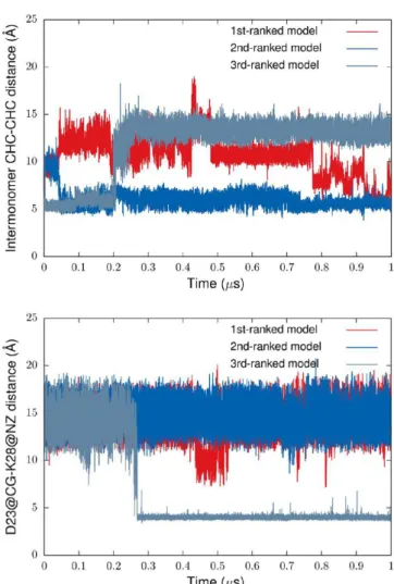
For two out of three simulated dimer models with the initial interpeptide CHC−CHC interface (the α-helical dimer with the D23−K28 SB off state), the interface was present by the end of the simulations. Interestingly, disruption of the interface in the model ranked third correlates with establishing the intrapeptide D23−K28 SB in one of the constituting monomers (Figure 11). This suggests that the SB not only prevents formation of the initial interface between the CHC domains as discussed in the previous section, but may also control its stability in dimers after initial relaxation. It is worth noting that for the dimer models with an initially disrupted D23−K28 SB, the SB was formed only for one monomer from the third-ranked α-helical dimer model.
For the first-ranked model with the initial CHC−CHC contacts between the monomers, this interface was disrupted and then re-formed at the timescale of a few hundreds of
Figure 10. Number of residues involved in the β-strand/β-bridge structural content as a function of simulation time. The results are for the Aβ42 dimer model built from the monomers with a large hydrophobic SASA and a disrupted D23−K28 SB. The initial structure does not have any β-strand/β-bridge content.
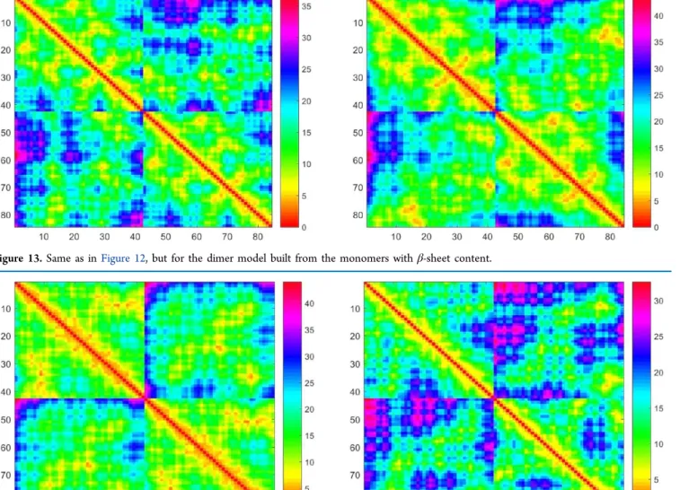
nanoseconds. Analysis of the inter-residue distance map for this model indicates optimization of the interpeptide CHC−CHC interface in the course of simulation (Figure 12). Also, the CHC domain and adjacent residues from one of the monomers establishes better contacts with most parts of the other. Some of the initial interpeptide contacts are lost by the end of simulation, mostly between monomer termini. At the same time, constituting monomers are surprisingly stable as seen from the contact map. Thus, one of the two monomers show only minor changes in “native” contacts (the right bottom block of the distance map fromFigure 12) with better packing of residues in the final structure. For the second monomer, some contacts are lost predominately between residues in termini segments and between N-terminal residues and residues 28−32. The dimer built with the β-sheet monomers is also characterized by a stability of the initial interpeptide interface and optimization of both inter- and intrapeptide interactions in the course of simulation (Figure 13). In contrast, for the top-ranked α-helical dimer model without the CHC− CHC interface, there is a significant decrease in the initial intra-and interpeptide contacts (Figure 14). The interpeptide D23− K28 SB in these models of the α-helical dimer without the CHC−CHC interface is quite stable. Its disruption was observed only for two constituting monomers from the dimer models ranked second and third. These simulations provide insight into the relative stability of structural models of the Aβ42 dimer with and without the initial interpeptide CHC− CHC interface and also characterized by different secondary structure contents.
3. DISCUSSION
After being released from a lipid membrane as a product of proteolytic cleavage of APP, Aβ peptides most probably lose their α-helical transmembrane conformation, as supported by solution NMR experimental data for different monomeric Aβ species in aqueous and nonpolar solutions.14−27 The
experimental studies have also demonstrated that soluble Aβ monomers can be classified as intrinsically disordered proteins7,14 and their structural properties are characterized by transitions between multiple conformational states. It is very
Figure 11.Time evolution of the interpeptide CHC−CHC distance (the upper panel) and the distance between Asp23 and Lys28 residues for one of the constituting monomers (the bottom panel) in the course of MD simulation of the Aβ42 dimer. As an initial structure, the dimer model built from the monomers with a large hydrophobic SASA and the D23−K28 SB off state was used. The distances are measured between geometric centers of the CHC domains, and between the CG site of Asp23 and NZ site of Lys28.
Figure 12.Inter-residue distance map for the first-ranked dimer model with the interpeptide CHC−CHC interface. The constituent monomers for this model characterized by a disrupted D23−K28 SB are from the first production segment of the trajectory. Left panel: the distance map for the initial structure. Right panel: the distance map averaged over the last 5 ns of 1 μs simulation. Distances are measured between Cα atoms for each residue. Diagonal and off diagonal 42 × 42 residue blocks give the intra- and interpeptide maps, respectively.
unlikely that under normal physiological conditions, the equilibrium in the conformational ensemble of Aβ peptides is shifted toward neurotoxic oligomeric species. In a more plausible scenario, the pathological conversion is triggered by Aβ peptide overproduction, impaired clearance, or changes in environmental conditions (such as pH level, salt concentration, or presence of lipids or other substrates) caused by currently unknown pathological conditions, or a combination of these factors. The conformational equilibrium of the structural ensemble of Aβ peptides may first be shifted from mostly unstructured to α-helical and/or β-sheet rich structures, which will further nucleate oligomerization. Such structures can be more aggregation-prone because of multiple factors, including reduced (compared to unstructured conformations) entropic penalty, a higher solvent exposure of hydrophobic residues, a favorable charge state, among others.
In this study, monomeric constructs with a potentially high propensity for aggregation were selected from the structural ensemble of the Aβ42 monomer sampled in implicit solvent REMD simulations. These structures are characterized by different secondary structure content, state of the D23−K28 SB, and solvent exposure of hydrophobic residues. Structural analysis suggests that there may be a correlation between
secondary structure content and propensity to form the D23− K28 SB on the one hand, and a pattern in packing of hydrophobic residues in the Aβ42 monomer in the other. In particular, a contact between the CHC domain and C-terminal Val40 and Ile41 residues was observed in α-helical rich structures only for a disrupted D23−K28 SB. Also, there is a reduced propensity to form such contacts for structures with β-sheet content. These structural features of the monomer are reflected in a propensity to form the interpeptide interface between the CHC domains in models of the Aβ42 dimer generated with the ZDOCK protein−protein docking soft-ware.60,61There is a higher tendency to form such an interface for α-helical monomers with a disrupted D23−K28 SB, compared to that of monomers with a formed SB or monomers with β-sheet structure content. The results of this study suggest that if the dimerization pathway involves initial association of monomers with a substantial α-helical content, such monomers may have a disrupted D23−K28 SB. In this case, relaxation of initial aggregates will be accompanied by an increase in β-sheet content of dimers, in agreement with experimental data. Thus, formation of the intrapeptide D23−K28 SB in Aβ monomers may affect both the propensity to establish a hydrophobic
Figure 13.Same as inFigure 12, but for the dimer model built from the monomers with β-sheet content.
Figure 14.Same as inFigure 12, but for the dimer model built with the monomers from the second production segment of the trajectory. These monomers do not have the interpeptide CHC−CHC interface and are characterized by a formed D23−K28 SB.
interface between CHC domains of monomers and the propensity to adopt a β-sheet rich conformation in dimers.
The interpeptide CHC−CHC and CHC−C-terminal domain interfaces as well as interactions between C-terminal hydrophobic residues in Aβ dimers have been observed in previous modeling studies.37,43,44,51,54 What is new is that
formation of such structural patterns may correlate with a propensity to form an intrapeptide D23−K28 SB and secondary structure of a monomer. This could have potential implications for better understanding of oligomerization pathways of Aβ peptides. Interactions between the CHC domain and hydrophobic Val40−Ala42 in the Aβ42 monomer with the D23−K28 SB off state can facilitate formation of a large continuous hydrophobic patch, which could result in an elevated propensity for aggregation compared to that of Aβ40 constructs and Aβ42 monomers with a formed D23−K28 SB, or monomers with a substantial β-sheet structural content. Further, the difference in packing of hydrophobic residues between monomeric constructs with solvent exposed and protected Lys28 (such as in the case of a formed D23−K28 SB) could be a factor discriminating between aggregation pathways leading to toxic aggregates or inert fibrillar structures, as supported by the fact that solvent exposure of Lys28 is an important structural feature of the epitope recognized by the conformational antibody in the toxic oligomeric Aβ species.63,64
4. CONCLUSIONS
The interplay between structural features of the Aβ42 monomer, such as secondary structure content and a propensity to form the D23−K28 SB, and properties of the interpeptide interface in the Aβ42 dimer identified in the current study provides a new insight into the possible oligomerization pathways of Aβ peptides. These findings are also of potential practical importance because they reveal structural features of Aβ constructs that can be utilized in development of drug candidates for inhibition of formation of toxic Aβ oligomers. In particular, they could help to identify monomeric constructs characterized by both an elevated tendency for aggregation (such as with a high solvent exposure of hydrophobic residues) and specific structural features that may be responsible for formation of toxic oligomers. These constructs can be used as receptor models for structure-based drug design or virtual screening of large libraries of druglike compounds to target pathological aggregation of Aβ peptides.
For practical applications, the above structural hypotheses need to be verified with conformational sampling covering a larger conformational space of the Aβ42 monomer (for example, with all-atom explicit solvent MD simulations and multiple force fields to produce statistically meaningful results) combined with a combinatorial analysis of a larger number of diverse monomeric structures to build initial models of the dimer, with following assessment of dynamic and thermody-namic stability of suggested dimer models. This work is currently underway and results will be reported in forthcoming publications.
5. MATERIALS AND METHODS
5.1. Molecular Dynamics Simulations.A conformational ensemble of the Aβ42 monomer was sampled in GBSA implicit solvent REMD simulations. A fully extended conformation of the monomer was used as input for MD simulations to avoid a possible bias due to an arbitrary choice of initial structure. This
structure was prepared with the tleap program from the Amber14 molecular dynamics package.72 Charge states of
ionizable residues were assigned with the PROPKA pro-gram.73−75
Residues with basic side chains have pKa values
greater than 10 and thus are protonated at the neutral pH conditions modeled in this study. All acidic residues are considered deprotonated because their predicted pKavalues are
less than 5. PROPKA assigned the pKavalue of 6.35 to histidine
residues, which provides some flexibility in assignment of their protonation states. It is very unlikely that peptides with a substantial noncompensated charge will aggregate to form a dimer. Thus, in this study focusing on building initial molecular models of the Aβ42 dimer, all histidine residues were single-protonated at Nϵ sites, which guaranteed electrical neutrality of monomers.
In the REMD simulations, 12 replicas spanning the temperature range 298.15−579.90 K were used. The number of replicas and temperature series were selected with the temperature generator for REMD-simulations server (http:// folding.bmc.uu.se/remd/)76 to set the replica exchange probability to 0.2. Simulations were performed with the Amber12 molecular dynamics package77 and the Amber ff12SB force field.78,79A modified version of the GB solvation model80(igb = 5) was used with the mbondi2 set of van der Waals radii.80The ionic strength of solvent was set to 0.2 M (saltcon = 0.2) to mimic physiological conditions. To control temperature, Langevin dynamics was used with the collision frequency of 5 ps−1. All bonds that include hydrogen atoms
were constrained with the SHAKE algorithm.81 The cutoff parameters for non-bonded interactions and pairwise summa-tion for calculating the effective Born radii were set to 999.0 Å. The REMD simulations were preceded by structure optimization to relax possible steric clashes, followed by thermalization and equilibration runs. Minimization was performed with 250 steepest descent steps followed by 250 conjugate gradient minimization steps. In the thermalization run, temperature of the system was gradually increased to target temperatures for each replica during 50 000 MD steps. Thermalization was followed by 50 000 MD equilibration steps. In the equilibration and thermalization runs, the same MD parameters were used as in the production simulations with the exception of the time step which was set to 1 fs in the thermalization and equilibration runs, and to 2 fs in the production runs. In the production REMD simulations, the number of MD steps between attempted replica exchanges was 2500. To avoid possible chirality inversions at high temper-atures, chirality restraints generated with the makeCHIR_RST program from the Amber molecular dynamics package were imposed in the REMD simulations. All other parameters were set to their default values.72
A similar MD setup was used to assess dynamical stability of select models of the Aβ42 dimer. These models were built and optimized as explained in the next section. To avoid dissociation of the dimers at high temperatures accessible in REMD simulations, their stability was tested with conventional MD at a temperature of 25 °C.
Before it was used to sample a conformational ensemble of the Aβ42 monomer, the REMD protocol described above was validated by folding the Trp-cage miniprotein82starting from a fully extended initial conformation. The miniprotein was successfully folded on the nanosecond timescale (Figures S22 and S23of SI) with the same set of REMD parameters as that used for simulation of the Aβ monomer (with the exception of
the number of replicas, which was set to 10 to insure the same exchange rates for the 20-residue long miniprotein).
In silico conformational sampling of intrinsically disordered peptides is a challenging problem. Sampling results are very sensitive to the choice of a force field model,83 and it is not clear whether explicit solvent simulations can be fully converged (such as to reproduce physiological ensembles of Aβ peptides) at the microsecond timescale accessed in most studies carried out so far. Here, REMD is used to generate a structurally diverse ensemble of conformations of the Aβ42 monomer for modeling a possible initial interpeptide interface in the Aβ42 dimer. In this context, it is more important to produce Aβ conformations with structural characteristics related to a high propensity for aggregation rather than to achieve a converged conformational sampling. Still, it is interesting to verify whether the sampling protocol from this study generates a conformational ensemble of Aβ monomers in agreement with experimental data. With that in mind, we compare predicted 3J
HNHα-coupling values with the recent
experimental solution NMR data.14The calculation was carried out based on the modified Vuister and Bax parametrization84of
the Karplus equation85−87 by averaging J-coupling values for
individual MD frames. The agreement between the exper-imental and predicted data is reasonably good for a subset of frames from the last 2 μs of the trajectory corresponding to the large RMSD structural domain discussed above (Figure S24of SI). Thus, the correlation coefficient between the calculated and experimental J-coupling values is 0.529, which is comparable to the previously reported results obtained with explicit solvent REMD simulations.22
5.2. Postprocessing of Molecular Dynamics Data and Building Models of the Aβ42 Dimer. The conformational space of the Aβ monomer is quite complex. Thus, different criteria may be needed to identify conformers with a potentially high tendency for aggregation. In the current study, solvent exposure of hydrophobic residues was chosen as a major descriptor of the monomer’s propensity for aggregation. To model an initial interpeptide interface in the Aβ dimer, representative conformations of the Aβ42 monomer were selected with clustering of time series of solvent accessible surface area of hydrophobic residues. SASA of different residue groups and secondary structure content of peptides were obtained with Kabsch and Sander’s DSSP algorithm.88All other postprocessing tasks, including clustering analysis and calcu-lations of RMSD of atomic positions, radius of gyration, and the distance between salt-bridge forming residues, were performed with the cpptraj program from the Amber molecular dynamics package.
Molecular models of the Aβ42 dimer were built from the representative conformations of the monomer with the ZDOCK protein docking server (http://zdock.umassmed. edu/).60 The server is based on the protein−protein rigid-body docking programs ZDOCK and M−ZDOCK.61,89,90The top 10 structures from each docking simulation were further optimized and rescored with the Amber molecular dynamics package and the Amber ff14SB force field.79Minimization was performed with 500 steepest descent steps followed by 500 conjugate gradient minimization steps. Minimization and rescoring of dimer models built with the ZDOCK server were performed with the same set of parameters as that used for REMD simulations.
■
ASSOCIATED CONTENT*
S Supporting InformationThe Supporting Information is available free of charge on the
ACS Publications websiteat DOI:10.1021/acsomega.7b00805. Time evolution and distribution of the radius of gyration of the Aβ42 monomer from implicit solvent REMD simulations; solvent accessible surface area of hydro-phobic residues of the Aβ42 monomer plotted versus number of residues forming different secondary structure elements, the distance between salt-bridge forming Asp23 and Lys28 residues, and hydrophilic SASA; population distributions of Cα RMSD for different trajectory segments; time series of Cα RMSD split into two structural domains, and distribution of the hydro-phobic and hydrophilic SASA of the Aβ42 monomer for different RMSD domains; per-residue secondary struc-ture propensity of the Aβ42 monomer; top-ranked models of the Aβ42 dimer built from different monomeric constructs; changes in SASA of individual residues upon dimerization of the Aβ42 monomers from the most populated clusters; number of residues forming different secondary structure elements as a function of simulation time for different dimer models; time series of Cα RMSD from REMD folding simulation of the cage miniprotein; representative structures of the Trp-cage miniprotein folded in REMD simulations; compar-ison between calculated and experimental J-coupling data for the Aβ42 monomer (PDF)
■
AUTHOR INFORMATIONCorresponding Author
*E-mail: [email protected]. Phone: +1 (780)641 1719. Fax: +1 (780) 641 1601.
ORCID
Nikolay Blinov:0000-0003-2638-8376
Andriy Kovalenko:0000-0001-5033-4314
Notes
The authors declare no competing financial interest.
■
ACKNOWLEDGMENTSThis work was supported by the Alberta Prion Research Institute, Alzheimer Society of Alberta and Northwest Territories (Alberta Alzheimer Research Program grants ABIBS AARP 201500003 and 201600005), and the National Research Council of Canada. Computational resources were provided by WestGrid (www.westgrid.ca) and Compute Canada - Calcul Canada (www.computecanada.ca).
■
REFERENCES(1) Hardy, J.; Selkoe, D. J. The Amyloid Hypothesis of Alzheimer’s Disease: Progress and Problems on the Road to Therapeutics. Science 2002, 297, 353−356.
(2) Walsh, D. M.; Selkoe, D. J. Aβ Oligomers - a decade of discovery.
J. Neurochem. 2007, 101, 1172−1184.
(3) Benilova, I.; Karran, E.; De Strooper, B. The toxic Aβ oligomer and Alzheimer’s disease: an emperor in need of clothes. Nat. Neurosci. 2012, 15, 349−357.
(4) Kayed, R.; Lasagna-Reeves, C. A. Molecular Mechanisms of Amyloid Oligomers Toxicity. J. Alzheimer’s Dis. 2013, 33, S67−S78.
(5) Haass, C.; Selkoe, D. J. Soluble protein oligomers in neurodegeneration: lessons from the Alzheimer’s amyloid β-peptide.