HAL Id: tel-01905616
https://tel.archives-ouvertes.fr/tel-01905616
Submitted on 26 Oct 2018HAL is a multi-disciplinary open access
archive for the deposit and dissemination of sci-entific research documents, whether they are pub-lished or not. The documents may come from teaching and research institutions in France or abroad, or from public or private research centers.
L’archive ouverte pluridisciplinaire HAL, est destinée au dépôt et à la diffusion de documents scientifiques de niveau recherche, publiés ou non, émanant des établissements d’enseignement et de recherche français ou étrangers, des laboratoires publics ou privés.
Low intensity rTMS to the cerebellum : age dependent
effects and mechanisms underlying neural circuit
plasticity
Tom Dufor
To cite this version:
Tom Dufor. Low intensity rTMS to the cerebellum : age dependent effects and mechanisms underlying neural circuit plasticity. Neurons and Cognition [q-bio.NC]. Université Pierre et Marie Curie - Paris VI, 2017. English. �NNT : 2017PA066270�. �tel-01905616�
Université Pierre et Marie Curie
Ecole doctorale 158 « Cerveau Cognition Comportement »
Laboratory of UMR8256 Biological Adaptation and Ageing Team : Brain Development, Repair and Ageing (DRVC)
Low intensity rTMS to the cerebellum : age dependent
effects and mechanisms underlying neural circuit plasticity
Présentée par Tom Dufor
Thèse de doctorat de Neurosciences
Dirigée par Rachel Mary Sherrard
Présentée et soutenue publiquement le 24 Octobre 2016 devant un jury composé de :
Dr. Isabelle DUSART Président du jury
Pr. Mohamed JABER Rapporteur
Pr. Klaus FUNKE Rapporteur
Pr. Vincenzo DI LAZZARO Examinateur
Dr. Annalisa BUFFO Examinateur
Acknowledgments
First and foremost I would like to express my sincere gratitude to Professor Bertrand Friguet for allowing me undertake my PhD in his laboratory, Biological Adaptation and Ageing. Also, for showing me the interest of studying Biology of Ageing at the beginning of my Master, which is one of the reasons I am now writing this thesis.
It is a great honour to have as my thesis examiners Pr Mohamed Jaber and Pr Klaus Funke and I am truly grateful to them for having agreed to be my rapporteurs de thèse and for the time they are taking to give me their important feedback on my work. I would also like to thank Dr Annalisa Buffo and Pr Vincenzo Di Lazzaro for having accepted to read this manuscript and to come from Italy to assist at this oral defence. Finally I thank Dr Isabelle Dusart for her presence as the President of my jury, since her expertise about the olivocerebellar system is a real plus.
A tremendous thanks to my PhD supervisor Pr Rachel Sherrard who gave me real confidence in my scientific reasoning right from my first day in the lab. Thanks for all the long evenings spent answering my questions, for encouraging me doing the experiments I wanted to do, for reassuring me when I had doubts and of course for the countless number of exchanges we had for this manuscript. I would like to thank you for your genuine kindness, your honesty and all the values that you given me I will never forget. I will do my best to continue research the way you taught me.
How can I not thank Pr Ann Lohof my co supervisor ? One of the people most willing to help at any time, that I have ever met. You have been present in all the critical steps of this PhD, I always knew I could knock on your door if needed help. Thank you for sharing all your knowledge about electrophysiology, your humor and your spontaneity. I truly enjoyed our discussions about any random subject.
Eternal gratitude to Dr Natalie Morellini with whom everything started. Jean Patrick has grown up now !! It was such a pleasure to work with you during my master and at the beginning of this PhD. Your positiveness, your humor and your smile made times fly during long behavioral experiments at Charles Foix. Thank you for having shared your knowledge and for all the time you spent on spines analysis for the in vivo project.
I would like to thank Pr Jean Mariani. I have always been impressed by your critical eye, you have always gave fresh input to this project that considerably helped me to go further. Thank you for always encouraging new experiments and for sharing all your scientific and cultural knowledge during our not-that-frequent but always long discussions.
I must thank Dr Sebastian Jara, aka el hombre con la gorra, for teaching me how to dissect explants, your advice on microscopy, and globally for your very strong support during these 3 years. We were in the same boat and I’m honoured to have shared this journey with you.
I gratefully acknowledge all the people from the BDRA team. Thanks to Dr Stephanie Grehl who explained how the 6-well plate stimulation device she designed, functioned. Thanks especially to Massiré for all the time we spent optimising molecular biology protocols and for our fun time out of the lab. Thanks to Nicolas for the late nights spent developing the different patterns of stimulation. Thanks also to Laurence Cathala for all her feedback on different presentations and for her input of ideas about the project ; Pascale who helped with the media change and genotyping ; Sadyah and Laura former Master students for listening carefully to my advice ; to Ioanna for being always in a good mood ; and to Alesha for all the discussions we have had during the writing of my thesis that helped me to breathe. Finally, thanks to Mariatou and Corinne for their kindness. Last and by no means least, I thank all the Phd/Postdocs of IBPS for the fun we have had on our beer sessions nights.
This thesis would have never been possible without all the invaluable support, love , advice from my family and friends. You have always encouraged me to live my dreams, have always been there in the difficult moments and were always curious to know about my research. A special thanks to my one and only Marion this thesis is for you. You make me wake up every morning with the ambition to make you proud of me.
Table of Contents
Abstract
………. vRésumé
……….. viiList of figures an tables
……….. ixAbbreviations
………... xiChapter I – Introduction ………..
1I) Magnetic stimulation: a non-invasive approach to enhance neuroplasticity? ……… 2
I.1) What is neuroplasticity? ……….. 2
I.2) Neuroplasticity over the lifespan ……… 3
I.3) What is rTMS? ……….. 6
I.4) Basic principle of rTMS ……….………. 8
I.5) TMS and rTMS applications ………... 9
I.6) rTMS parameters ………. 12
I.6.1) Pulse shapes and coil types ……….. 12
I.6.2) Stimulation intensity ………... 13
I.6.3) rTMS frequency……… 15
II) Potential mechanisms underlying high-intensity rTMS ………. 17
II.1) Synaptic plasticity: evidence from human studies ………... 17
II.2) Direct evidence for synaptic plasticity induced by rTMS in animal models ……….. 20
II.2.1) rMS induced synaptic plasticity at excitatory synapses ……….. 20
II.2.2) rTMS modulation of inhibitory networks ………. 21
II.2.3) Neurobiological effects of rTMS underlying induced plasticity ………... 22
III) Low intensity magnetic stimulation………. 30
III.1) A need for a defined terminology and reproducible parameters ……….. 31
III.2) ELF-MF effects on the human brain ……… 32
III.2.1) Effects on cortical excitability and brain oscillations ………. 32
III.2.2) Effects on human brain functions and potential therapeutic applications ……... 33
III.3) ELF-MF effects on animal models and underlying mechanisms ……….. 34
III.3.1) Magnetoreception: the cryptochrome radical-pair mechanism hypothesis ………. 35
III.3.2) Biological events induced by ELF-MF ……… 37
III.4) LI-rTMS effects on the rodent brain ………. 41
III.5) Conclusion ……….. 43
IV) The cerebellum as a model to study LI-rTMS induced plasticity ……… 44
IV.1) Cerebellum ………. 44
IV.1.1) Cerebellar circuitry ………. 45
IV.1.2) Cerebellar afferents ……… 47
IV.2.1) The inferior olivary nucleus……….. 48
IV.2.2) Climbing fibers and the OCP ……… 50
IV.3) Development of the OCP ………. 51
IV.3.1) Morphological PC differentiation ……… 51
IV.3.2) Synaptogenesis of the olivocerebellar pathway and refinement of CF Projections ……….. 52
IV.4) Plasticity of the OCP following lesion ……….. 55
IV.4.1) Developmental plasticity of the OCP ……….……… 55
IV.4.2) Plasticity in the mature OCP ………. 56
IV.5) Ageing of the cerebellum ………. 57
V) Summary and aims ………. 59
Chapter II – Article1
……….. 63Abstract ……….……… 65
Introduction ……… 66
Materials and Methods ………. 68
Animals ……… 68
Administration of Magnetic Stimulation ………. 69
Behavioural analysis ……….. 69
Electrophysiological recording and biocytine filling………. 71
Morphological Analysis ……… 72
Spine density ……… 73
Sholl analysis ……… 74
Statistical analysis ……… 74
Results ……… 75
Early postnatal PC dendritic development and synaptic maturation are not altered by LIrTMS ……….. 75
Mature Purkinje cell dendrites change little with age ……… 76
Short-term treatment with LI-rTMS increases PC spine density and alters spine morphology ……….. 77
Long-term treatment with LI-rTMS alters PC dendritic morphology………. 78
Handling and behavioural testing alone did not change dendritic morphology but did alter spine density ……….. 79
Chronic LI-rTMS treatment does not alter motor function, but improves spatial memory ………..……… 79
Discussion ……… 81
References ……….. 85
Chapter III – Article 2
………... 107Abstract ……….. 109
Introduction ……… 110
Methods ………. 111
Animals ………. 111
In vivo olivocerebellar axonal transection (pedunculotomy) ………. 112
Organotypic Cultures and cerebellar denervation ………. 112
Magnetic stimulation ………. 113
Immunohistochemistry ………..……….. 114
Histological analysis ………. 115
qRT-PCR and gene analysis ……….……… 116
Statistical analysis ………. 117
Results ………. 117
LI-rTMS induces CF reinnervation in vivo ………. 117
LI-rMS induces PC reinnervation in vitro in a frequency dependent manner ………. 118
LI-rMS induced reinnervation requires simultaneous stimulation of both pre-and postsynaptic structures of the OCP ………. 119
LI-rMS activates Purkinje cells and interneurons………120
LI-rMS modulates gene expression appropriately for PC reinnervation ……… 120
Cryptochrome is required for LI-rMS-induced post-lesion repair ………... 121
Discussion ………. 122
Post-lesion repair depends on stimulation pattern, not numbers of pulses ………..……… 122
LI-rMS modifies gene expression in a frequency-dependent manner ………. 124
LI-rMS potentially activates c-fos in PC and interneurons ………..………. 125
Cryptochromes magnetoreceptors are key elements for the transduction of the magnetic field into biological effect……….. 126
Conclusions ………..………. 127
References ……… 128
Figures and Legends ……….. 137
Chapter IV– Article 3
………. 149Abstract ………. 151
Introduction ……… 152
Materials and Methods ………. 153
Animals ………. 153
Organotypic Cultures and cerebellar denervation ………. 154
Administration of LI-rTMS ……….. 154
Behavioural analysis ……… 155
Electrophysiological recording and biocytin filling ……… 157
Immunohistochemistry and Histological analysis ……….………. 158
qRT-PCR ……….……….………. 160
Statistical analysis ………. 161
Results ………. 162
Post-lesion repair by LI-rMS is impaired in RORα haplodeficient hindbrain explants ……….. 162
Four weeks LI-rTMS and psychomotor activity moderately alters PC dendritic morphology ……… 163
Chronic LI-rTMS treatment does not alter motor or spatial behaviour of RORα+/ animals ………. 165
RORα+/-PC responses to acute LI-rTMS treatment are small and differ with age ………. 166
Discussion ……….. 167
References ………. 169
Figures and Legends ……… 173
Chapter V – General Discussion
………... 193V) Discussion ………... 194
V.1) Mechanisms underlying the effectsof LI-rTMS ………... 194
V.1.1) LI-rTMS pattern of frequency are crucial for the induction of biological effects………. 194
V.1.2) LI-rTMS requires cryptochromes to interact with biological tissue and potentially activate the transcription of signalling pathways ……….. 198
V.1.3) Intracellular calcium may be a second messenger mediating the biological effects of cryptoctochrome activation by LI-rMS ………. 199
V.1.3.1) Intracellular levels and downstream signalling cascades ……… 199
V.1.3.2) ROS production ……….……… 200
V.1.4) RORα seems necessary to observe neural circuit plasticity and behavioural improvements induced by LI-rTMS ………. 201
V.2) Biological effects of LI-rTMS………. 203
V.2.1) LI-rTMS targeting the cerebellum induces neuronal structural plasticity and improvements in cerebellar related behaviour in adult mice ……….. 203
V.2.2) Reduced neural plasticity in aged mice may explain reduced LI-rTMS effects on cerebellar structural plasticity and behaviour ……….. 205
V.2.3) Further investigation into the effects of magnetic stimulation are required prior to eventual clinical use in young patients ……….……… 206
Neuroplasticity is essential for the establishment and strengthening of neural circuits during the critical period of development, and are required for the brain to adapt to its environment. The mechanisms of plasticity vary throughout life, are generally more difficult to induce in the adult brain, and decrease with advancing age. Repetitive transcranial magnetic stimulation (rTMS) is commonly used to modulate cortical excitability and shows promise in the treatment of some neurological disorders. Low intensity magnetic stimulation (LI-rTMS), which does not directly elicit action potentials in the stimulated neurons, have also shown some therapeutic effects, and it is important to determine the biological mechanisms underlying the effects of these low intensity magnetic fields, such as would occur in the regions surrounding the central high-intensity focus of rTMS. We have used a focal low-intensity magnetic stimulation (10mT) to address some of these issues in the mouse cerebellum and olivocerebellar path. The cerebellum model is particularly useful as its development, structure, ageing and function are well described which allows us to easily detect eventual modifications. We assessed effects of in vivo or in vitro LI-rTMS on neuronal morphology, behavior, and post-lesion plasticity.
We first showed that LI-rTMS treatment in vivo alters dendritic spines and dendritic morphology, in association with improved spatial memory. These effects were age dependent. To optimize stimulation parameters in order to induce post-lesion reinnervation we used our in vitro model of post-lesion repair to systematically investigate the effects of different LI-rTMS stimulation patterns and frequencies. We showed that the pattern of stimulation is critical for allowing repair, rather than the total number of stimulation pulses. Finally, we looked for potential underlying mechanisms participating in the effects of the LI-rTMS, using mouse mutants in vivo or in vitro. We found that the cryptochromes, which have magnetoreceptor properties, must be present for the response to magnetic stimulation to be transduced into biological effects.
The ensemble of our results indicate that the effects of LI-rTMS depend upon the presence of magnetoreceptors, the stimulation protocol, and the age of the animal suggesting that future therapeutic strategies must be adapted to the neuronal context in each individual person.
Les mécanismes de neuroplasticité sont essentiels pour la mise en place et le renforcement des circuits neuronaux lors de périodes critiques du développement, et permettent au cerveau de s'adapter au cours des différentes étapes de la vie. Ces mécanismes varient avec l’âge, sont généralement plus difficile à activer chez l'adulte, et diminuent dans le cerveau âgé. La stimulation magnétique transcrânienne répétée (rTMS) est actuellement utilisée pour moduler l'excitabilité corticale et est décrite comme prometteuse dans le traitement de certains troubles neurologiques. La rTMS de faible intensité (LI-rTMS), ne déclenchant pas directement de potentiels d'action dans les neurones stimulés, a aussi montré des effets thérapeutiques, il est donc important de comprendre les effets biologiques de ces champs magnétiques d’intensités similaires à celles présentes dans les régions adjacentes à la région ciblée par la rTMS de haute intensité. Nous avons utilisé une stimulation magnétique focale de faible intensité (10 mT), ciblant le cervelet ainsi que la voie olivocérébelleuse chez la souris, afin d’aborder certaines de ces questions. Le cervelet est un modèle pertinent, en effet son développement, sa structure, son vieillissement et ses fonctions sont bien décrits, facilitant la détection d’éventuelles modifications dans cette région. Nous avons étudié les effets de LI-rTMS, in vivo ou in vitro, sur la morphologie neuronale, le comportement, et la plasticité post-lésionnelle.
Dans une première étude nous avons montré que la LI-rTMS in vivo modifie les épines et la morphologie dendritique des cellules de Purkinje, ces modifications sont associées à une amélioration de la mémoire spatiale et dépendent de l’âge de l’animal. Afin d’optimiser les paramètres de stimulation pour induire de la réinnervation post-lesionelle et d’identifier des mécanismes sous-jacents, nous avons utilisé notre model in vitro de réparation post-lésionnelle et étudié de manière systématique les effets de différents patrons de LI-rTMS. Nous avons montré que le patron de stimulation, plutôt que le nombre de pulses, est essentiel afin d’induire de la réinnervation. Afin d’identifier des mécanismes participant aux effets de la LI-rTMS, nous avons utilisé des souris mutantes, in vivo et in vitro. Nous avons montré pour la première fois que les cryptochromes, ayant des propriétés de magnétoreception, sont nécessaires afin d’observer des effets biologiques induits par la LI-rTMS. Nos résultats indiquent que les effets de la LI-rTMS dépendent de la présence de magnétorecepteur, du patron de stimulation et de l’âge de l'animal, suggérant une adaptation des stratégies thérapeutiques en fonction du contexte neuronal de chaque individu.
Figure 1: Schematic representation of individual plasticity across the lifespan. Figure 2: Overview of electromagnetic induction in TMS.
Figure 3: Simplified scheme of TMS mechanism of action on the motor cortex. Figure 4: Factors influencing the effects of transcranial magnetic stimulation (TMS).
Figure 5: Common key concepts between the induction of classic synaptic plasticity in brain
slices and the induction of rTMS outcomes in the normal human brain.
Figure 6: Schematic representation of the potential CREB-Fos pathway activated by the rTMS. Figure 7: ELF-MF devices and magnetic field propagation in the human brain.
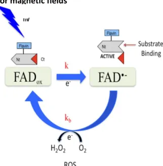
Figure 8: Diagram of the potential common mechanism leading to the production of ROS
triggered by blue light and magnetic field activation of Cryptochrome.
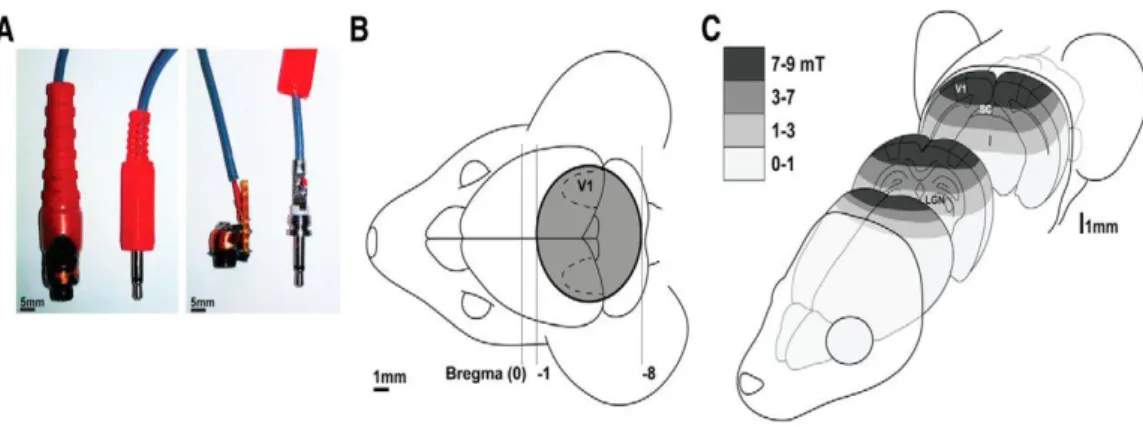
Figure 9: Methods for focal LI-rTMS delivery in mice.
Figure 10: Morphology and organisation of the adult mouse cerebellum. Figure 11: Cerebellar cytoarchitecture.
Figure 12 Topographical organization of the OCP into longitudinal zones and microzones. Figure 13: Morphological differentiation of Purkinje cells.
Figure 14: Postnatal development of CF-PC synapses.
aAPs Anterograde action potentials
AMPA α-amino-3- hydroxy-5-methyl-4 isoxazoleproprionic acid
AP Action potential
baAPs Backward propagating anterograde action potentials
BDNF Brain derived neurotrophic factor
CABP Calcium binding protein
cAMP Cycline adenosine monophosphate
CB Calbindin
CF Climbing fibers
CNS Central nervous system
CREB cAMP responsive element binding protein
CRY Cryptochrome
CS Complex spikes
CSF Cerebrospinal fluid
CSP Cortical silent period
CST Cortico spinal tract
cTBS Continuous theta burst stimulation
DCN Deep cerebellar nuclei
DLPFC Dorso lateral DFC
DTI Diffusion tensor imaging
EEG Electroencephalogram
ELF-MF Extremely low frequency magnetic fields
EMF Electromagnetic field
EPSP Excitatory post synaptic potential
FAD Flavin adenine dinucleotide
fMRI Functional magnetic resonance imaging
FSIs Fast spiking interneurous
GABA Gamma-aminobutyric acid
GAD Glutamic acid decarboxylase
GAP43 Growth associated protein
GC Granular cells
H2O2 Hydrogen peroxide
HF-rTMS High frequency repetitive transcranial magnetic stimulation
ICF Intracortical facilitation
IEGs Immediate early genes
ION Inferior olive neurons
iTBS Intermittent theta burst stimulation
KO Knock-out
LFMS Low field magnetic stimulation
LF-rTMS Low frequency repetitive transcranial magnetic stimulation
LI-rTMS Low intensity repetitive magnetic stimulation
LTD Long term depression
LTP Long term potentiation
L-VGCC L- voltage gated calcium thermal
M1 Primary motor cortex
MP Motor threshold
MRI Magnetic resonance imaging
NGF Nerve growth factor
NMDA N-methyl-D-ascorbic acid
NO Nitric oxide
NT3 Neurotrophin-3
O2°- Superoxide anion
OCP Olivo cerebellar pathway
PC Purkinje cells
PD Post-natal day
PEMF Pulsed electromagnetic fields
PET Positron emission tomography
PSD95 Post synaptic density
PV Paravalbumin
Px Pedunculotomy
RGCs Retinal ganglion cells
RMT Resting motor threshold
RORα RAR-related orphan receptor alpha
ROS Reactive oxygen species
rTMS Repetitive transcranial magnetic stimulation
SICI Short interval intracortical inhibition
SYP Synaptophysin
T Tesla
TBS Theta burst stimulation
TMS Transcranial magnetic stimulation
TrK B Tyrosine kinase receptor B
VDCC Voltage dependent calcium channel
VESCs Voltage gated sodium channels
1
2
I) Magnetic stimulation: a non-invasive approach to enhance neuroplasticity?
I.1) What is neuroplasticity?
Neuroplasticity is the ability of the brain to reorganize neural connections at the structural and functional level in response to extrinsic or intrinsic factors. This is a fundamental process during development of the central nervous system and its physiological functioning in the adult. Neuroplasticity allows the nervous system to adapt to environmental pressure, experience (psychological or somatic) and injury/lesion, beyond its genetically-determined structure (Pascual-Leone et al., 2005).
Neuroplasticity can be observed at the whole-brain level, when spatiotemporal patterns of activation occur in distinct brain regions; at the circuit level, as modulation of long-range or local contacts between different neuronal types; or at the synaptic level, where dynamic modifications adjust the strength of a preexisting connection and thus a neuron’s response to an input (Ganguly and Poo, 2013). These short-term processes may be followed by structural change, e.g. dendritic growth and arborization leading to the formation of new synapses. Plasticity is thought to be the neural basis for learning and memory formation (Hübener and Bonhoeffer, 2010; Johansen et al., 2011; Squire et al., 2004), through diverse mechanisms such as cellular long-term potentiation (LTP) in the hippocampus (Bliss and Lomo, 1973), long-term depression (LTD) in the cerebellum (Ito and Kano, 1982), and synaptic formation or dissolution.
Neuroplasticity also contributes to functional recovery after brain injury by modulation of neurogenesis, dendritic morphology, axon sprouting, and neural circuits in the peri-lesion area (Cramer, 2008; Taub et al., 2002). It is essential to understand which of the local and distant changes are optimal for recovery because these modified patterns of neural activation may themselves lead to maladaptive neuroplasticity and abnormal behavior. This adverse aspect of plasticity is illustrated in developmental, acquired and degenerative neuropathologies, involving either excessive (Naro et al., 2016; Saab, 2012; Sheehy and Marsden, 1982) or impaired plasticity (Cramer et al., 2011; Elbert et al., 1998; Parihar and Brewer, 2010). Impaired plasticity would not allow the brain to adjust to changing environmental situations, and an inappropriately-high level of plasticity would make the
3
structural connections unstable and therefore adversely affect functional systems required for cognition and behavior (Pascual-Leone et al., 2011).
To find ways of modulating cerebral plasticity to provide functional benefits, it is crucial to understand neuroplasticity mechanisms, and the relationships between these mechanisms, neuronal activity, and behavior. The aim is to suppress neural changes resulting in unwanted behavior and to increase those leading to improved behavior beneficial for a healthy subject or a patient.
I.2) Neuroplasticity over the lifespan
The brain can be seen as a structure constantly adapting to its environment and therefore it is essential to understand its plasticity over the lifespan. During development a highly plastic state has been characterized, termed the critical period. Plasticity during this critical period is essential for the physiological establishment of neural circuitry and function. The developmental critical period has been clearly demonstrated to interact with postnatal experience and activity-dependent plasticity in both the visual system (Anderson et al., 2011; Chapman and Stryker, 1993; Espinosa and Stryker, 2012) and in the descending corticospinal tract (CST) of the motor system (Anderson et al., 2011; Martin, 2005). Monocular deprivation on the one hand, or silencing of the CST on the other, during this critical period results in respectively a dramatic permanent loss of the response to the deprived eye (Hubel and Wiesel, 1963, 1998) and in an altered topography and axon terminal morphology of the CST leading to long-term motor disorders (Martin, 2005). This critical-period plasticity might partly explain the Kennard Principle which notes that lesions in childhood are characterized by better recovery than equivalent lesions in adults (Dennis, 2010). Conversely, this plasticity can also lead to devastating effects of some early brain injuries (Kolb et al., 2000). Investigation of the time window and factors that balance these opposite outcomes is required in order to find optimal interventions for functional recovery in the immature brain (Anderson et al., 2011).
In contrast, in the adult brain plasticity phenomena are different, occurring at different levels, in response to environmental factors (sensory input, motor act, learning, injury…). Functional short-term synaptic modifications (Abbott et al., 1997; Zucker and Regehr, 2002) allow immediate adaptation of signal-processing in neural circuits. Long-term synaptic
4
plasticity mechanism, such as LTP and LTD (Malenka and Nicoll, 1993), underlie more prolonged changes, e.g. learning and memory. These functional synaptic plasticity mechanisms are accompanied by structural modifications such as synapse turnover (Grutzendler et al., 2002; Trachtenberg et al., 2002) and formation of stable new dendritic spines (Hofer et al., 2009; Xu et al., 2005; Yang and Zhou, 2009). On a more cellular level, morphological plasticity of the neuronal dendritic tree was observed following sensorimotor learning (Gonzalez et al., 2005) or following peripheral activity (Churchill et al., 2004) such as exposure to an enriched environment (Jacobs et al., 1993; Volkmar and Greenough, 1972). At a larger scale, cortical map reorganization in response to sensory deprivation occurs in the mature brain (Buonomano and Merzenich, 1998; Feldman and Brecht, 2005). Adult brain neuroplasticity is nonetheless region dependent and is often described only in restricted areas such as the hippocampus, cerebellum or sensorimotor cortex. Although the mature brain can sometimes overcome injury through the creation of new connections between surviving neurons, its repair capacity is limited by a growth-inhibitory cellular environment, poor growth potential in mature neurons, and the restriction of stem cells necessary for neuronal replacement to specific brain areas (Fuchs and Flügge, 2014).
Although adult neurogenesis in mammalian brain is now well accepted in the subventricular zone (Doetsch et al., 1997; Saha et al., 2012, 2013), which migrate to the olfactory bulb (Lois and Alvarez-Buylla, 1994; Luskin, 1993) and subgranular zone of the hippocampus (Altman and Das, 1965; Gage et al., 1998; Saha et al., 2013), their capacity to repair cerebral lesions is unclear. Recently it was shown that motor cortical lesion in adult mice induces the proliferation of neural progenitors in the subventricular zone, which migrate to the lesion and differentiate mainly into glial cells and to a lesser degree into neurons (Saha et al., 2013). However, evidence of neurogenesis in areas such as neocortex, amygdala, striatum and substantia nigra lacks reproducibility and requires clarification (Gould, 2007; Saha et al., 2012). Also, the re-growth ability of axons in the mature central nervous system (CNS) sharply decreases after the critical period. The ability of the CNS to regenerate injured axons becomes restricted by the lack of intrinsic neuronal growth capacity and an extrinsic inhibitory molecular environment (Chen and Zheng, 2014). Axonal sprouting of uninjured axons is a natural repair mechanism after injury in the adult CNS. Sprouting from remaining axons, rather than regenerating the whole path, requires growth over shorter distances to make functional connections on appropriated targets (Geoffroy and Zheng, 2014). Therefore
5
modulation of axonal sprouting may represent a more realistic therapeutic goal to promote functional recovery, than trying to induce regeneration. Axonal sprouting can be promoted by extrinsic factors such as application of neurotrophins or genetic manipulation (Geoffroy and Zheng, 2014; Giger et al., 2010; Willson et al., 2008) but this is invasive and therefore not yet clinically applicable. There is an evident medical need to find non-invasive ways to promote neuroplasticity in the mature brain.
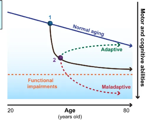
Later in life, age-related decline of synaptic plasticity correlated with neurocognitive alterations (Rosenzweig and Barnes, 2003). For example, hippocampal LTP declines faster after its induction in aged rats, and this is linked to altered memory (Barnes and McNaughton, 1980; Kelly et al., 2006). In humans, however, there is generally only indirect evidence of decreasing neuroplasticity as aging progresses (Fotenos et al., 2005; Scahill et al., 2003) through the assessment of cognitive and motor decline combined with imaging techniques that show structural (magnetic resonance imaging (MRI), diffusion tensor imaging (DTI) and functional functional MRI (fMRI), positron emission tomography (PET) deficits (Pascual-Leone et al., 2011). Neuroplasticity over the lifespan shows a downward trend in all individuals but it does so from variable “baseline” levels and with different slopes according to genetic factors and environmental influences (Figure 1)
In contrast, direct experimental evidence of decreasing corticomotor plasticity over human lifespan can be obtained through studies combining transcranial magnetic stimulation (TMS) together with electroencephalogram (EEG) and fMRI (Freitas et al., 2011a, 2011b). TMS can be used as a non-invasive exploration tool to understand and assess adult brain plasticity, or in its repetitive form (rTMS) as a non-invasive way to modulate cortical excitability and therefore to enhance plasticity. By using the same technique, these two aims are particularly complementary, since a better understanding of the underlying mechanism of the neuroplasticity is essential for the optimization of the parameters of the non-invasive way to induce it. In the scope of this thesis I will focus on the rTMS as a tool to induce neuroplasticity. It appears essential to consider the stage of brain development, the age of the subject, and if it is a healthy or injured brain, since the mechanisms and level of plasticity are not identical, in order to optimally induce neuroplasticity with rTMS.
In summary, brain plasticity has a central role from development to ageing that allows the brain to adapt to intrinsic or extrinsic factors and therefore optimizes function in healthy
6
subjects. The plasticity mechanisms are reduced in the mature and ageing brain so that there is an absence of appropriate compensatory changes to brain pathology. As plasticity is closely linked to neuronal activity, modulating activity, and thus plasticity, by non-invasive approach such as the rTMS represent an interesting strategy to reduce or even treat neurological dysfunction.
I.3) What is rTMS?
Using electric and magnetic field to stimulate the human brain and muscle has a long history. In 1791, Galvani was the first to show that electric focal stimulation can induce responses in isolated frog nerves and muscles (Galvani, 1791). This attracted broad interest Figure 1: Schematic representation of individual plasticity across the lifespan.
Neuroplasticity over the lifespan shows a downward trend in all individuals but it does so from variable “baseline” levels and with different slopes according to genetic factors and environmental influences. Alteration to local plasticity by brain injury, morbidity or unhealthy lifestyle will trigger secondary adaptive responses across neural networks, and they may be that adaptive or maladaptive for the individual. Non invasive interventions to promote adaptive network plasticity and suppress maladaptive plasticity mechanism are essential therapeutic goals. Modified from (Pascuale-Leone et al; 2011)
7
into the electrical excitability of biological tissues, and led to the discovery of muscle activation by electric stimulation of various motor cortex areas (Rothwell et al., 1991).
Since then, electrical stimulation has shown considerable efficiency in various clinical applications but has some technical limitations that require clear application guidelines (Antal et al., 2017). Stimulation can be painful due to activation of pain fibers in the scalp (Hallett, 2000; Merton and Morton, 1980), and deep structures are very difficult to stimulate non-invasively due to the high electrical resistance of the skull. An alternative approach is to apply time-varying magnetic fields.
The mechanism underlying electromagnetic induction was discovered by Michael Faraday in 1831. Faraday's law of induction is a law of electromagnetism predicting how a magnetic field will interact with an electric circuit to generate an electromotive force,a phenomenon called electromagnetic induction (Walsh 2003). The first magnetic activation of isolated frog muscle in vitro by Kollin and colleagues (Walsh 2003) paved the way to stimulation of the human brain and muscle via electromagnetic induction.
At that time the reliable generation of powerful and rapidly alternating electromagnetic fields was limited by the technology advancement. Therefore it was only in 1985 that Barker and colleagues were able to show for the first time hand muscle activation as a result of human motor cortex stimulation with alternating magnetic fields (Barker et al., 1985; Hallett, 2007). The technique has generated particular interest since, unlike electrical stimulation, rTMS does not produce pain and is not attenuated by hair, tissue, or skull (Hallett, 2007). Thus the intensity of the secondary electric field generated by the time-varying magnetic field in the underlying brain tissue is only affected by distance and time from the initial stimulation point (Pascual-Leone et al., 2000; Wagner et al., 2007). It allows the stimulation of discrete brain regions and increases the accuracy of the stimulation. Since its first application, the use of TMS in various domains such as neurology, neuroscience and psychiatry has grown widely, as an explorative tool in research applications. With technological advances, TMS devices were rapidly able to deliver multiple pulses within a short time period (10-20ms) (Pascual-Leone et al., 1991; Rossini and Caramia, 1992). This form of TMS, called repetitive TMS (rTMS), modifies a range of measures of brain function including cortical excitability for up to few weeks after the last stimulation. Thus it logically became an important research tool due to its therapeutic potential in the treatment of a variety of neurological and psychiatric disorders (Hallett, 2000; Rossini et al., 2015).
8
I.4) Basic principle of rTMS
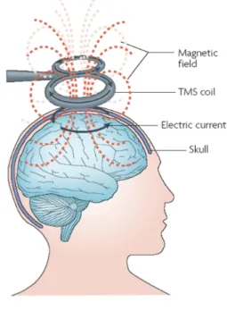
Transcranial magnetic stimulation (TMS) is based on Faraday’s principles of electromagnetic induction. In TMS studies, the stimulating wire coil is held over a subject’s head, a brief pulse of current flows through the coil, generates a magnetic field which passes through the subject’s scalp and skull with negligible attenuation (the field only decays by the square of the distance from the source) (Hallett, 2007). This time-varying magnetic field induces a secondary perpendicular electric field which can create an electric current of opposite direction in any adjacent conductor and thus stimulates the neural tissue (Pascual-Leone et al., 2000; Walsh and Rushworth, 1999) (Figure2). The field’s rate of change determines the size of the induced current (Maxwell-Faraday equation; Jackson 1962). This secondary electric current is thought to modify the membrane potential and therefore the excitability of the neurons underneath the coil. It requires a flow of electric charge through the excitable neuronal membrane and creates a transmembrane potential that propagate along the nerve. TMS can therefore stimulate both output and input connections of any cortical areas (Siebner et al., 2009). At the system level, the effects of the TMS are thus not restricted to the targeted region but produce altered activity in distant interconnected cortical, subcortical and spinal regions (Kobayashi and Pascual-Leone, 2003; Naro et al., 2016). The direction and strength of the electric field as well as the biological structure’s intrinsic conductivity influence the amount of charge going through the neuronal membrane. Change in neuronal membrane properties is the result of a gradient change of electric charge induced by the electric field at a specific location of the neuron (Di Lazzaro et al., 2003; Radman et al., 2009; Siebner et al., 2009). Some studies suggest that TMS stimulates axons more easily than the soma because axons are most efficiently activated by a brief electrical current, as in the TMS, whereas somata require longer pulses. Exactly which axons are activated is not clear and factors like the degree of myelinisation, neuronal type and geometry of the axons must be considered (Siebner et al., 2009). However it has also been suggested that excitation occurs in the cortical grey matter rather than in the subcortical white matter because of its location (just below the scalp surface) and lower electrical resistance (Edgley et al., 1997).
9
Various types of excitatory and inhibitory neurons from interconnected regions are activated by the TMS, thus it seems more rigorous to consider the TMS mechanism of action in terms of neural circuits rather than local excitability changes. Since neurons have different thresholds of electrical activation, lower stimulation intensities activates a much smaller number of neurons. Therefore if one wants to have focal stimulation it appears more logical to use low intensity stimulation to avoid unwanted activation of distant structures.
I.5) TMS and rTMS applications
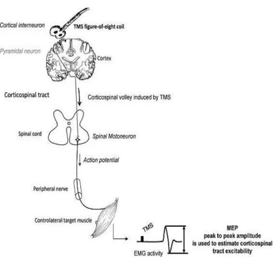
In clinical neurophysiology, TMS of the motor cortex is widely used as an exploratory tool to assess the excitability of descending cortico-nuclear and cortico-spinal circuits (Rossini et al., 2015). The motor cortex is a widely used region since modulation of motor cortex activation and excitability can be readily evaluated via recordings of motor evocated potentials (MEPs) (Figure 3). A wide variety of neurophysiological measures made with TMS are possible. Among the most commonly used are: corticomotor threshold (MT), MEP amplitude and latency, cortical silent period (CSP), cortical mapping of motor representations and, following paired TMS pulses, short interval intracortical inhibition (SICI) and intracortical
Figure 2: Overview of electromagnetic induction in TMS.
A brief pulsed (time varying) primary current flows through the coil and generates a changing (time varying) magnetic field. The magnetic field passes through the skull and induces a perpendicular electric field within the brain. This secondary electric field can create secondary electric currents, of opposite direction, in any conductive tissue, and thus stimulate the brain. The magnetic field decreases rapidly with distance from the coil, thus cortex and subcortical white matter are assumed to be the principal neural elements stimulated. Image adapted from Ridding & Rothwell (2007)
10
facilitation (ICF). They all provide evidence of pathology-related alteration of motor function in patients, or neuroplasticity in the motor cortex of healthy subjects (Rossini et al., 2015). Combination of neuroimaging techniques and EEG during TMS allows for non-invasive assessment of cortical excitability and good time resolution of connectivity in regions outside the motor cortex (Ilmoniemi and Kicić, 2010; Siebner et al., 2009; Ziemann, 2011).
This thesis will focus on the potential of rTMS to induce neuroplasticity and to treat neurological and psychiatric disorders. Its modulatory effect on cortical activity of various motor and non-motor areas that outlasts the stimulation period makes it a very interesting research tool. Studies of rTMS effects on cortical activity have been mostly done with stimulation of the primary motor cortex (M1), since the motor outputs can be easily measured. In studies of corticospinal motor output it is generally accepted that low frequency stimulation (≤1Hz) is mainly inhibitory, while high frequency stimulations (≥5Hz) are mainly facilitatory (Fitzgerald et al., 2006). More recent patterned protocols such as the popular “theta burst stimulation” (TBS) delivered as continuous (cTBS) or intermittent (iTBS) trains are described to be inhibitory and excitatory respectively. They have been suggested to have longer lasting effects on cortical excitability than classic low or high frequency rTMS protocols (Iezzi et al., 2011; Di Lazzaro et al., 2011). The stimulation frequency is thought to be the main parameter that influences the direction of the modulation. However the effect of a defined rTMS or TBS protocol do not always follow this rule (“high frequency and iTBS = excitatory”; “low frequency and cTBS=inhibitory”)(Fitzgerald et al., 2006; Gamboa et al., 2010; Houdayer et al., 2008). The influence of the stimulation frequency will be discussed in more detailed in the following section addressing stimulation parameters.
The long term-modulatory action of rTMS on cortical reactivity has driven interest in the use of rTMS to facilitate recovery from a broad range of neurological diseases (stroke, multiple sclerosis, epilepsy, Parkinson’s disease and tinnitus), psychiatric disorders (depression, anxiety, schizophrenia and obsessive-compulsive disorders) and pain syndromes (migraine and chronic pain). A recent evidence-based review demonstrated level “A” efficacy (definite efficiency) in the antidepressant effect of high-frequency rTMS (HF-rTMS) targeting the dorsolateral prefrontal cortex (DLPFC), and the analgesic effect of HF rTMS targeting M1, contralateral to the pain (Lefaucheur et al., 2014). Specific rTMS devices received FDA approval for the treatment of these two pathologies in 2008 and 2014 respectively (O’Reardon et al., 2007). Treatment of the negative symptoms of schizophrenia and chronic
11
motor stroke by HF-rTMS of the left DLPFC and by low-frequency rTMS (LF-rTMS) of contralesional M1 respectively, received level B recommendation (probable efficiency) (Lefaucheur et al., 2014). Some studies report improvements in neurological and psychological disorders after rTMS applications that last up to few months
(
Wassermann and Zimmermann, 2012).However a great discrepancy in outcomes, with studies failing to reproduce reported beneficial effects of rTMS, represent an obstacle for the acceptance of rTMS as a viable therapeutic tool (Héroux et al., 2015; Platz and Rothwell, 2010; Ridding and Rothwell, 2007; Figure 3: Simplified scheme of TMS mechanism of action on the motor cortex
Interneurons oriented in a plane parallel to the brain surface are preferentially activated by the TMS applied over the motor cortex. This leads to a transynaptic activation of pyramidal cells evoking descending volleys in the pyramidal axons projecting on spinal motoneurons (corticospinal tract). Motoneuron activation in response to corticospinal volleys induced by TMS leads to a contraction in the target muscle evoking a motor-evoked potential (MEP) on electromyography (EMG). Surface electrodes applied over the muscle belly are used to record the MEP. Its peak-to-peak amplitude is measured to estimate excitability of the corticospinal tract. MEP is the most common read-out of human cortical excitability.
12
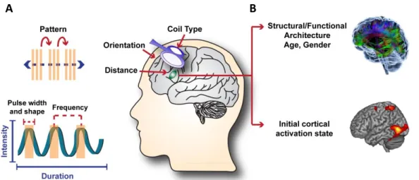
Wassermann and Zimmermann, 2012). This variability can be partially explained by several factors (Figure 4); among the most important:
1) Inter- and intra-individual variability of rTMS effects is one of the major sources of discrepancies between and within studies (Goldsworthy et al., 2014; Hannah et al., 2016; López-Alonso et al., 2014; Vallence et al., 2015). Each subject's individual baseline level of cortical excitability, prior to stimulation, strongly influences the direction of modulation produced (Daskalakis et al., 2006; Siebner and Rothwell, 2003). The susceptibility to induce neuroplasticity is generally influenced by previous neuronal activity, relying on mechanisms of homeostatic plasticity and metaplasticity (Abraham and Tate, 1997; Bienenstock et al., 1982; Turrigiano and Nelson, 2004). Other biological factors such as age, gender, genetics, physical fitness, hormone levels and brain anatomy also affect the rTMS effects and thus represent another cause of inter-individual variability of rTMS clinical outcomes (Hoogendam et al., 2010; Ridding and Ziemann, 2010).
2) rTMS parameters, including total duration of stimulation, pulse width, coil type, magnetic field orientation and, very importantly, the intensity and frequency pattern of the stimulation dramatically influence the neural and behavioral effects of the stimulation. (Chen et al., 1997; Di Lazzaro et al., 2011; Rothkegel et al., 2010; Rubens and Zanto, 2012) High variability in the stimulation parameters applied in different studies probably explains much of the lack of reproducibility between studies and even contradictory results. While consideration of the biological factors involved in inter-individual variability is essential, rTMS parameters are the most convenient and accessible factors of influence to carefully control.
I.6) rTMS parameters
A large number of stimulation parameters influence the neural and behavioral outcomes of rTMS. I will briefly describe the influence of different pulse shapes and coil types, with particular attention to the effects of different stimulation intensities and frequency patterns.
13
Direction and amplitude of the secondary electric field are determined by the rate of change of the magnetic field over time, typically occurring during the rise-time and fall-time of the
magnetic pulse as represented in the Maxwell-Faraday equation: ∇ x E = - 𝝏𝑩 𝝏𝒕
Where ∇ is the curl operator, E is the electric field, B is the magnetic field and t the time. Therefore the amplitude of the induced electric field is a function of the amplitude of the magnetic field and how fast it changes over time. Since axons have various strength-duration properties, it is possible to target specific sets of peripheral axons by changing the pulse width of the stimulation (Mogyoros et al., 1996). However all commercially-available stimulators deliver sinusoidal pulses that cannot be modified and therefore limit the possibility to target specific sets of neurons (Goetz et al., 2013; Hannah et al., 2016).
Stimulators can generate either a monophasic pulse or a biphasic/polyphasic pulse. Generally these two types of pulse have different neuromodulatory effects, with bi/polyphasic pulses suggested to be more efficient in single stimulus application and monophasic pulses thought to have greater neuromodulatory effects in rTMS application (Arai et al., 2005; Hamada et al., 2013; Sommer et al., 2006). Because of lower energy requirements, classical rTMS devices deliver high-frequency biphasic stimuli that activate different cortical circuits and induce a variety of effects that could partly explain inter-individual variability in outcomes (Hamada et al., 2013). However the implementation of a novel modifiable device, which can deliver a nearly-triangular monophasic pulse during high frequency rTMS (controllable TMS; (Peterchev et al., 2014), is thought to produce stronger and more reproducible effects on MEP than classical rTMS devices (Goetz et al., 2016). Various wire coil designs are available for TMS clinical applications, the two most popular being the classic round coil and the figure-of-eight-shaped coil (Cohen et al., 1990; Hallett, 2007) The shape of the magnetic field produced depends on the coil type, which will determine the area with the highest intensity stimulation (hotspot), the total area stimulated and the stimulation depth (Deng et al., 2013; Lang et al., 2006a). Commercially available TMS coils used in the clinic usually deliver a maximum magnetic field of 1.25-2 Tesla (T). Intensity of the magnetic field decreases with increasing the distance from the coil and the central focal hotspot, leaving surrounding cortical and sub-cortical regions to be stimulated albeit at lower intensities (Cohen et al., 1990; Deng et al., 2013).
14
I.6.2) Stimulation intensity
The rTMS as described to this point delivers focal high-intensity stimulation which depolarizes neurons of the targeted cortical regions sufficiently to induce action potential firing. Another area of research called low field magnetic stimulation (LFMS), pulsed electromagnetic field (PEMF) stimulation or extremely low frequency magnetic field (ELF-MF) stimulation use diffuse subthreshold low-intensity magnetic fields that induce a generalised electric current throughout the brain but do not directly depolarize neurons sufficiently to induce action potential firing. However these low intensity magnetic fields have wide effects on brain function that will be extensively described in chapter III.
I.6.2.1) High intensity rTMS
The intensity of an individual’s rTMS is usually determined by the cortical motor threshold (MT), defined as the minimal intensity at which TMS over the M1 induces a reliable electromyography (EMG) response around 100µV or a visible muscle twitch response in the target muscle (Rossini et al., 1994; Westin et al., 2014) Although twitch-based MT evaluation is easier to carry out, it is related to a high intra- and inter-rater variability and MTs assessed visually are about 10% higher than EMG-recorded MTs (Westin et al., 2014). Suprathreshold stimulations inducing action potential firing in stimulated neurons are required to induce such EMG or muscle twitch responses (Pell et al., 2011). MEP amplitude is commonly used to assess modulation of cortical excitability promoted by rTMS, and increases with increasing stimulation intensity (Rothwell et al., 1987). Intrinsic differences in cortical excitability induce inter and intra-subject variability in the MEP amplitude. While physiological fluctuations cannot be avoided, other physiological and technical parameters should be kept constant, including baseline activity of the target muscle, arousal and attention levels, environmental noise, and coil position/orientation (Cuypers et al., 2014; Rossini et al., 2015). The stimulation intensity needed to induce a response in a resting muscle is often expressed relative to the resting motor threshold (RMT) and represents the percentage of the stimulator output to reproducibly induce MEPs (Fitzgerald et al., 2006; Rothwell et al., 1987). Stimulation intensity in human rTMS is generally applied around 80-120% of the RMT and therefore is only characterized as a percentage of the stimulator output. This represents another factor of variability between studies since identical devices are not always used.
15
I.6.2.2) Low intensity magnetic stimulation
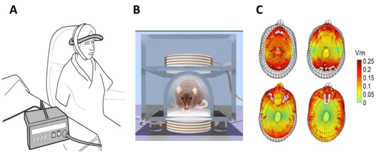
As for high intensity rTMS, low intensity magnetic fields are delivered by one or more coils in which time-variable current flows. Parameters of stimulation are usually in the microtesla to millitesla (µT-mT) range and generally 10-100 Hz (0-300Hz) frequency (Di Lazzaro et al., 2013). Waveforms also can be mono-phasic or biphasic. Extremly low frequency magnetic field (ELF-MF) studies use various waveforms that can be asymmetric, biphasic, quasi-rectangular, or quasi triangular (Bassett, 1989) although most ELF-MF sources of stimulation produce sinusoidal waveforms (Juutilainen and Lang, 1997). Pulsed electromagnetic fields (PEMF), a subset of ELF-MF, usually induces greater electrical current than sinusoidal waveforms due to their faster magnetic field rate of change (Tesla/seconds) (Di Lazzaro et al., 2013). Unlike rTMS which delivers central focal high intensity stimulation of a targeted brain region, ELF-MF generates a diffuse homogeneous magnetic field within the whole brain. Helmholtz coil-based exposure systems are the most commonly used and are made of two identical circular magnetic coils that deliver a nearly uniform magnetic field. ELF-MF experiments use a wide variety of stimulation devices and coils which induce a broad range of waveforms (sinusoidal or pulsed), intensities (µT-mT) and frequencies (0-300Hz) of stimulation, making comparisons between studies very difficult.
I.6.3) rTMS frequency
The temporal spacing between repeated magnetic stimuli (rTMS frequency) is the most widely studied and manipulated parameter of magnetic stimulation because of its crucial influence on the direction of cortical excitability modulation. As discussed above (Section I.5), the general consensus is that high frequencies ≥ 3 Hz and iTBS increase cortical excitability, while low frequencies ≤ 1 Hz and cTBS decrease cortical excitability (Fitzgerald et al., 2006; Huang et al., 2005; Rossini et al., 2015). (Huang et al., 2005), motivated by the limited efficiency of classical rTMS frequencies (1Hz and 10Hz) to modulate cortical excitability, took advantage of the high-efficiency of electrical theta burst stimulation to induce LTP in hippocampal slices (Hernandez et al., 2005), to design an alternative approach of rTMS in which a three-pulse 50-Hz burst is applied at 5 Hz (theta burst). In intermittent TBS (iTBS), trains of theta bursts are delivered intermittently, for 2s every 10s for about 190s (600pulses in total), while in continuous TBS (cTBS), theta bursts are delivered continuously
16
for 40s (600 pulses in total) (Huang et al., 2005). Both iTBS and cTBS were able to produce the most consistent and sustained effects on cortical excitability in human studies, while using lower intensity and shorter duration of stimulation than classical high- and low-frequency rTMS (Hoogendam et al., 2010; Huang et al., 2005). Thus these patterned stimulation frequencies represent promising tools for induction of neuroplasticity.
This strict distinction of excitatory and inhibitory rTMS may be a simplistic vision, and both types of rTMS might have more mixed inhibitory and excitatory effects (Houdayer et al., 2008; Matheson et al., 2016). It seems more appropriate to consider the effects of different “excitatory” or “inhibitory” rTMS protocols as the result of stimulation and modulation of various cortical circuits (Di Lazzaro et al., 2010, 2011). For example, the increased MEP following HF rTMS could be the consequence of decreased gamma-aminobutyric acid (GABA)-mediated intracortical inhibition rather than a direct increase of motor cortex excitability (Hamada et al., 2013; Di Lazzaro et al., 2001; Wu et al., 2000; Ziemann, 2004). Hamada and colleagues showed that the great variability between individuals in iTBS “excitatory” effects and cTBS “inhibitory” effects on MEP size is related to different interneuronal cortical networks recruited by the TMS (Hamada et al., 2013). Therefore comparison between experiments using different protocols described as “excitatory” or “inhibitory” should be made cautiously, especially with theta-burst patterns.
Similar distinctions between different types of modulation induced by different ELF-MF frequencies have not been described. A large number of studies have assessed the effects of sinusoidal 50-60Hz ELF-MF on the brain, since electromagnetic fields of this type are present in our environment.
Figure 4: Factors influencing the effects of transcranial magnetic stimulation (TMS). A) TMS mechanical
parameters that can be tailored for a desired outcome. B) Individual brain characteristics that also influence the outcomes of a given TMS protocol and potentially contribute to the discrepancy between results of rTMS studies. The different colors in the upper brain represent connections between different brain regions.
17
II) Potential mechanisms underlying high-intensity rTMS
As described above variability in the outcomes between studies on the effects of rTMS are partly due to different applied protocols (pulse shape, intensity, frequency pattern…) as well as intra and inter-individual variability due to different baseline level of cortical excitability and other individual biological factors. Altogether this will in turn specifically modify the intracellular pathways activated by the magnetic field and therefore the subsequent long-term changes. It is thus of major importance to search for neurophysiological mechanisms responsible for these long-term effects. An understanding of these mechanisms will help us to fine-tune our rTMS protocols in order to optimally activate those mechanisms, depending on the type of individual subject, the neural network targeted and the pathology implicated.
II.1) Synaptic plasticity: evidence from human studies
rTMS has the appealing potential to modulate cortical excitability beyond the simulation period (Pell et al., 2011) and in both directions, either excitatory or inhibitory. These after-effects are dependent on several parameters which were presented in the previous chapter. A main approach has been to explain those effects on the brain through LTP/LTD like mechanisms.
Long-term changes in synaptic strength like LTP (long-term potentiation) and LTD (long-term depression) are durable changes in synaptic efficacy (Malenka and Bear, 2004; Raymond, 2007). LTP results in potentiation of synaptic strength that may last for days, weeks or months. Brief high-frequency stimulations are used to induce this potentiation. LTD results in a long-lasting reduction in synaptic strength (Duffau, 2006). Synaptic plasticity obeys to key rules that are described in detail in different experimental models (Abraham, 2008; Cooke and Bliss, 2006; Malenka and Bear, 2004) Studies on rTMS and their effects on human brain have used several key concepts shared with classic synaptic plasticity which are summarized in figure 5. However it is essential to specify that LTP- and LTD-like mechanisms induced by rTMS differ from their classic form of synaptic plasticity, which use direct electric
18
stimulation of synapses in vivo and in vitro (Malenka and Bear, 2004; Pell et al., 2011). A main difference is related to the different conditions of stimulation: TMS activates a wide number of axons at presynaptic and postsynaptic terminals simultaneously (Funke and Benali, 2011) This could explain the significantly lower stimulation frequencies that induce LTP in rTMS studies compared to classic electrophysiology synaptic plasticity protocols (10Hz vs 100Hz) which induce facilitation (Vlachos et al., 2012).
Cortical excitability is modulated by magnetic stimulation in a frequency dependent manner. Brain activity is also affected by the stimulation as indicated by regional cerebral blood flow (Lee et al., 2003; Rounis et al., 2005), EEG responses (Esser et al., 2006; Huber et al., 2007; Litvak et al., 2007), and blood-oxygen level-dependent (BOLD) activation patterns (Hubl et al., 2008). High frequency rTMS for 10-20 minutes preceding a task produces prolonged increases in attentional control, consolidation of new motor skills, and tactile discrimination (Boyd and Linsdell, 2009; Hwang et al., 2010; Tegenthoff et al., 2005). All of these effects outlasted the stimulation period, which is an important component of synaptic and network plasticity.
The temporal pattern of stimulation is another key determinant of synaptic plasticity. In classic LTP/LTD studies the direction and strength of synaptic plasticity are generally frequency-dependent. High-frequency stimulation allows evocated post synaptic potential EPSP-summation, release of the N-methyl-D-aspartate receptor NMDA-receptor magnesium block, allowing calcium influx and triggering LTP. Low-frequency stimulation might allow for a lower calcium influx, leading to LTD (Collingridge, 2003; Malenka and Bear, 2004).
As discussed in section I.6.3, similarly to the synaptic plasticity mechanism elicited by electrical stimulations, rTMS stimulation frequency and pattern have a critical influence on the direction of the cortical excitability modulation induced by the rTMS (Cooke and Bliss, 2006; Ziemann, 2004)
Metaplasticity changes in synaptic plasticity over time is another key concept in synaptic plasticity which takes into account the previous history of activation of the synapse and can facilitate or inhibit synaptic plasticity, stabilize synapses or adjust cellular activity (Abraham, 2008). It allows homeostatic synaptic plasticity which allows adjustment of synaptic strengths to maintain stability in neural activity. Several rTMS studies have shown that the history of activity of the stimulated network influences the observed outcomes (Valero-Cabré et al., 2008). Studies on the after-effects of rTMS showed that prior stimulation would
19
"prime" subsequent stimulation effects and influence their direction (Karabanov et al., 2015; Todd et al., 2009). Physiological activity such as motor action or training is able to modify/reverse the rTMS effects on the motor cortex (Huang et al., 2008b). Thus rTMS effects are influenced by prior activation of a neural circuit.
The NMDA glutamate receptor has been extensively described as essential for LTP induction at most glutamatergic synapses (Collingridge et al., 1983; Morris et al., 1986). In support of this notion, studies in humans showed that the effects of 10 Hz rTMS were blocked by administration of the NMDA antagonists ketamine (de Andrade et al., 2014) or memantine (Huang et al., 2008b).
Brain-derived neurotrophic factor gene (BDNF) also modulates LTP (Aicardi et al., 2004; Figurov et al., 1996; Woo et al., 2005) and LTD (Woo et al., 2005) in the adult central nervous system. A common single nucleotide polymorphism (called "BDNF Val66Met") is present in 35% of the Caucasian population, and is associated with differences in hippocampal volume and episodic memory (Pezawas et al., 2004) and decreased experience-dependent plasticity in the motor cortex (Kleim et al., 2006). Effects of iTBS and cTBS on the excitability and plasticity of the motor cortex were reduced in subjects carrying this BDNF Val66Met allele (Cheeran et al., 2008); the authors suggest that this is due to the role of BDNF on the susceptibility of synapses to undergo LTP/LTD. These results reinforce the hypothesis that rTMS is acting in part through mechanisms similar to synaptic plasticity. Given that a specific BDNF gene allele can influence the response to rTMS it is possible that other genetic mutations could be an explanation for the great inter-individual variability in rTMS studies. LTP and LTD have been demonstrated in several animal experiments to be the neural substrate of learning (Rioult-Pedotti et al., 2000). Therefore to make a link between rTMS and synaptic plasticity it seems logical to test if the rTMS affects learning. Several studies showed that rTMS influence learning (Baraduc et al., 2004; Muellbacher et al., 2000) and suggests that rTMS has the potential to alter synaptic plasticity.
We have seen that there are several similarities between rTMS induced plasticity and classic synaptic plasticity (Figure 5), but no direct links can be demonstrated from only human studies. Animal studies allow more direct evidence ofthe underlying molecular and cellular mechanisms of synaptic plasticity induced by rTMS.A fundamental advantage of using appropriate animal or in vitro experiments to study mechanisms underlying rTMS is the fact that various forms of plasticity (synaptic plasticity of excitatory or inhibitory synapses,