Prof. Prof. Prof. Prof. Prof. Prof. Prof. Prof. Prof. Prof.
Academic and Research Staff Prof. T. F. Weisst, tt Prof. T. R. Willemain Dr. J. S. Barlowjf Dr. H. Duifhuis*** N. I. Durlach Dr. R. D. Hall Dr. A. J. M. Iloutsma Dr. N. Y. S. Kiangt Dr. L. U. E. Kbhlloffellttt Dr. E. C. Moxont Dr. M. J. Mulroy? . D. Braida . K. Burns [. S. Colburnt ,. S. Frishkopf . L. Goldsteint J. Guinan, Jr
L.
G. Mark** i. M. Oman V. T. PeakeT i. M. Siebert O. Bayer, Jr. E. Berliner L. Bonn R. Bourk II. Conrad M. Cronin IH. Domnitz E. Gorelick Ilasan L. flicks D. W. Altmannt D. J. Callahan A. HI. Cristt B. Gaiman P. W. Herman, Jr. W. F. Kelley Elizabeth M. Marrt J. M. Onorato, Jr.t L. H. Seifel J. B. Walters, Jr. M. Rabinowitz L. Rhyne A. Scudder T. Shepard Singer L. Smith M. Stern, Jr. L. Sulman Tom TungA. NORMOTHERMIC PERFUSION OF CANINE KIDNEYS - THE EFFECT OF PERFUSATES
National Institutes of Health (Grant 2 TO1 GM01555-06) C. H. Conrad, R. G. Mark
[With B. P. Kekis, C. D. Derry, and NI. Slapak, Sears Surgical Laboratory, Boston City Hospital.]
tAlso at the Eaton-Peabody Laboratory, Massachusetts Eye and Ear Infirmary, Cambridge, Massachusetts.
tOn leave - Department of Biophysics, University College, London.
Also Instructor in Medicine, Harvard Medical School, Boston, Massachusetts. ttAlso Instructor in Preventive and Social Medicine, Harvard Medical School, Boston, Massachusetts.
TResearch Affiliate in Communication Sciences from the Neurophysiological Labo-ratory of the Neurology Service of the Massachusetts General Hospital, Boston, Massa-chusetts.
Visiting Scientist from Institute for Perception Research, Eindhoven, The Netherlands.
Visiting Scientist from University of Stuttgart, Germany.
ITDepartment of Physiology, Harvard University, Cambridge, Massachusetts. Graduate Students P. Hochfeld D. II. Johnson M. S. Keshner M. C. LibermanjT Lynette L. Linden R. P. Lippmann N. D. Megill S. L. Moshier J. A. Myers V. Nedzelnitsky S. R. Purks
(X. COMMUNICATIONS BIOPHYSICS)
1. Introduction
Although hypothermic perfusion is now used as the basis of all long-term methods of organ preservation, a period of seven days is the longest for which preservation has been successfully achieved.I By using intermediate living hosts, perfusion at 37°C with whole blood has allowed kidney preservation for periods of more than three weeks.2 At present, however, limitations imposed by the materials used for artificial preservation
systems prohibit the perfusion of organs with whole blood for periods exceeding more than a few days, because of phenomena such as cell aggregation and hemolysis.
In a recent study3 it was shown that a 25°C 24-hour kidney perfusion could be suc-cessfully achieved with life supporting function after transplantation by the use of either platelet-poor or platelet-rich plasma. The advantages of plasma as a perfusate in a normothermic system are obvious. It contains many of the substrates required by the kidney and its proteins confer appropriate buffering and osmotic properties. The use
of plasma also eliminates problems of hemolysis and sludging. Although protein dena-turation might still be damaging, the use of a membrane oxygenator and the minimiza-tion of the air-plasma interface area might minimize this problem. Inherent in the use of plasma as a perfusate at 37CC, however, is the question of whether it is capable of carrying enough oxygen to support a normothermic kidney. Theoretical consideration of the oxygen consumption and blood flow of normal kidneys in vivo would suggest that it is. At the normal flow rate of 3-4 ml/gram of kidney tissue/minute, a very small arte-riovenous oxygen difference is found,4 of the order of 2 ml/100 ml as compared with 4 ml/100 ml for systemic venous blood. Since it has been shown5 that renal oxygen consumption during normal sodium filtration is of the order of 0. 08 ml/gm/min, the 2 ml/100 ml of oxygen dissolved in oxygen-saturated plasma might be enough to sup-port the kidney, provided that sufficiently high flows could be maintained. Thus, by using a 97% oxygen 3% CO2 mixture and flow rates of 3-4 ml/gm/min, an adequate
sup-ply of oxygen should theoretically be available.
The purpose of the present study, then, was to ascertain whether or not a normo-thermic kidney could be supported by using plasma as the perfusate. The methods and results have been described in detail elsewhere.7
2. Method
The experiment involved perfusion of kidneys from 19 apparently healthy 10-20 kg mongrel dogs. Three groups of 3-4 perfusions were done. Group A (9 kid-neys) was perfused with whole blood, Group B (4 kidkid-neys) with low hematocrit blood (4-7%), Group C (6 kidneys) with plasma. Anesthesia was induced with sodium thiamylal at an approximate dosage of 20 mg/kg, and additional thiam-ylal was given intravenously as needed. After intubation, a midline incision was
made, and the left kidney was dissected free after infiltration of the surrounding tissue with 5% Cyclaine (topical hexylcaine hydrochloride, 50 mg/ml). The artery, vein and ureter were freed and the ureter divided and cannulated with polyethylene tubing.
The dog was maintained in a diuretic state with a fluid load of 5% dextrose in normal saline or 25% mannitol in 5% dextrose in normal saline. The renal artery and vein were then clamped and divided and both cannulated with siliconized glass cannulae. The kid-ney was then weighed and transferred to the perfusion apparatus while the donor animal was maintained under light thiamylal anesthesia until reimplantation.
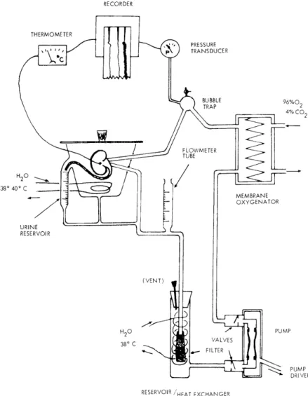
The perfusion circuit is shown schematically in Fig. X-1. It included a pulsatile
RECORDER THERMOMETER PRESSURE TRANSDUCER BUBBLE 96%O0 TRAP 2 4% CO2 H20 38' 40o C URINE RESERVOIR (VENT) H20 38' C MEMBRANE OXYGENATOR PUMP PUMP DRIVER
RESERVOIR /HEAT EXCHANGER
Fig. X-1. The perfusion system. The silastic pump is driven by a pneumatic oscillator which has been described previously.8
(X. COMMUNICATIONS BIOPHYSICS)
pump, membrane oxygenator (silicone rubber), bubble trap, organ chamber, and venous reservoir/heat exchanger with an incorporated filter. The pump was a silicone rubber ventricle with one-way valves, driven by a pneumatic oscillator with adjustable systolic and diastolic intervals and pressures similar to the one described by Slapak et al.8
The oxygenator was a Travenol 5M0321 membrane oxygenator ventilated by 97% oxygen with 3% CO 2 or 96% oxygen with 4% CO 2. There was a bubble trap in the arterial line,
and a graduated tube in the venous line which made possible direct measurement of flow by occlusion of the venous line between the graduated tube and the reservoir. The venous return was filtered by a blood administration set filter as it entered the reservoir. The organ rested in a glass organ chamber covered by a moist sponge and supported by a
silicone rubber coil through which water at 38°C was circulated. Urine was collected in a graduated reservoir and could be returned to the venous line if desired. The venous
reservoir contained a glass coil through which 38 0C water was circulated, to serve as a heat exchanger. The priming volume of the apparatus was approximately 200 ml.
The arterial perfusion pressure and organ surface temperature were recorded con-tinuously. Flow, arterial and venous pH, PCO2 and P0O2 were monitored periodically,
as were hematocrit, plasma glucose, and urine flow. Samples of perfusate and urine were also taken periodically for determination of osmolarity, sodium, potassium and creati-nine. After termination of perfusion, the kidney was flushed with cold lactated Ringer' s solution and reweighed. It was then reimplanted by end-to-end anastomosis of the renal artery to the external iliac artery and end-to-side anastomosis of the renal vein to the external iliac vein. Total ischemic times were from 25 to 35 minutes. The ureter was implanted directly in the bladder and contralateral nephrectomy was performed imme-diately following the implantation. Postoperatively, blood samples were drawn every few days for measurement of BUN, serum creatinine, sodium, potassium, and osmolarity. Since in all groups venous PO was above 150 mm Hg, a value at which hemoglobin remains fully saturated, oxygen consumption was calculated from the dissolved oxygen alone by using the formula
AO2
(ml/100 ml) 0 CONSUMPTION (ml/gm/min) = FLOW (ml/gm/min) X 100where FLOW is the perfusate flow, and AO2AV is the arteriovenous oxygen difference, given by
(PO
artPO 2ven)AO2AV (ml/100 ml)= 760 X a (ml/100 ml/atm) 6
where a is the solubility of oxygen in plasma as given by Sendroy et al. The solubility was corrected for temperature and for hematocrit, since acells is greater than aplasma.
Groups A,
planted or
B, and C that 6, 2, and 2 kidneys, respectively, were not reim-function was not evaluated because of unrelated technical failure.
Function after Reim -plantation
Oxygen
Consumption
(ml/gm/min)
Creatinine Clearance (ml/100 gm/min) I I .024 Sodium Reabsorption ( LEq/gm/min) 6711 - .020 6890 U .035 6924 U .038 2.7 3.7 6946 U .021 3. 6 4.7 6760 U .041 MEAN ± S. D. (n) .030 ±. 009(6) 3. 1 ±. 7(2) 4. 2 ±. 7(2) 6977 - .039 20. 2 29. 0 LOW 6206 . 049 44. 3 63. 5 HE MATOCRIT 6978 S .043 17. 1 24.4 (Group B) 6990 U .041 MIEAN S. D. (n) .041i.
008(4) 27. 2 ±(3) 39. 0 ±21.4(3) 6856 - .050 6860 - .042 6875 - .038 6872 - .039 WHOLE BLOOD 6903 - . 028 (Group A) 6925 - . 037 19. 0 28. 3 6885 S .025 5. 5 7. 9 6945 S .040 16. 8 25. 0 6966 S .034 12.3 17.3 MIEAN S. D. (n) .037 ±. 007(9) 13.4±6.
0(4) 19. 6±9.
1(4)Notes: - No reimplant, or function not evaluated. S Survival U Failure (uremic). Dog No. 6873 PLAS MA (Group C)
(X. COMMUNICATIONS BIOPHYSICS)
2. Results
Results are summarized in Table X-1.
a. Viability
None of the Futhermore, at
4 kidneys perfused by plasma were functional after reimplantation. reimplantation, these kidneys were dark and sometimes mottled. They
S6760 X 6946 --- x ---- x- PLASMA 6924---- LOW HEMATOCRIT x - -- WHOLE BLOOD 6945 990 6978 - 6966 10 20 30 DAYS POSTOPERATIVE Fig. X-2.
Postoperative renal function after reim-plantation for dog numbers indicated, measured by blood urea nitrogen values. Serum creatinine values not shown were similar in their behavior.
produced little, if any, urine. In contrast, all of the three kidneys perfused by whole blood and reimplanted regained normal color and were immediately life-sustaining; the animals were sacrificed some months later with normal renal function, as shown in Fig. X-2. One of the two kidneys perfused with low hematocrit blood was life-sustaining.
b. Oxygen Consumption and Other Results
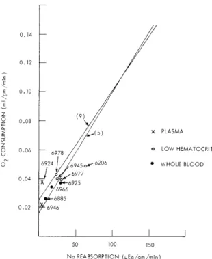
There were no statistically significant differences in the oxygen consumption of the three groups. Figure X-3 shows the relationship between Na reabsorption and 02
consumption in the three groups. There was, however, a statistically significant difference (P<. 05) in favor of the whole blood and low hematocrit groups for sodium reabsorption and creatinine clearance, although all groups showed impaired filtration. It was found in all three groups that renal venous PO 2 during perfusion was greater than
150 mm Hg, arterial PO 2 averaging approximately 560 mm Hg.
3. Discussion
The data indicate that inadequate oxygen supply or consumption does not seem to provide a convincing explanation of the striking difference in the viability between the plasma- and whole blood-perfused kidneys, although there is a possibility that failure of
0.14 0.12 -0.10 -Z (9) 0.08 -(5) x PLASMA z O 0.06 6978 o LOW HEMATOCRIT 0 - 6978 0( 6924 6945 o 6206 * WHOLE BLOOD ,6977 0.04 6925 x 6925 6966 6885 0.02 6946 50 100 150 Na REABSORPTION (pEq/gm/min)
Fig. X-3. Oxygen consumption and sodium reabsorption of perfused kidneys. Note that.although oxygen consumption is not significantly different in the three groups, Na reabsorption values in the plasma group (shown by X's) are much lower. The straight lines indicate the relations obtained by other researchers.5, 9
the plasma-perfused kidneys is related to oxygen supply or consumption. Renal cortical PO 2 is generally10 below renal venous PO 2 and localized areas of hypoxia might exist
even with a high venous PO 2 . The presence of intrarenal "shunt diffusion" has been
proposed to explain this effect, although limited oxygen diffusion at the capillary level would produce the same effect. These effects would allow oxygen to bypass respiring tissue, and make the organ incapable of extracting all seemingly available oxygen. A shunt diffusion effect would be most significant with high arterial PO 2 because of the
large gradient for diffusion that is thus created.
Thus, since the venous PO 2 was 150 mm Hg in all groups, the possibilities are
(i) there was more than adequate oxygen; (ii) the oxygen could not reach the renal paren-chyma because of a shunting effect; and (iii) oxygen diffusion at the capillary level was inadequate, or the cells were unable to utilize the available oxygen. Even if the plasma-perfused kidneys in these experiments did have adequate oxygen, the low creatinine clearances leave open the possibility that a normally filtering kidney perfused with plasma would become hypoxic as a result of increased sodium load and the consequent need for oxygen to provide energy for sodium reabsorption.
(X. COMMUNICATIONS BIOPHYSICS)
Work done by Gimbrone et al.12 suggests that platelets might play a role in the maintenance of vascular endothelium. In their studies, organs perfused at 370C showed diminished function and increased endothelial damage in the absence of platelets. In our experiment, platelets were present in Group A (whole blood) and Group B (low hematocrit), and the lack of platelets in Group C (plasma) might be responsible for the observed failure in this group. Although kidneys perfused with rich and platelet-poor plasma at 250C did not demonstrate advantages of platelet-rich plasma,3 results might differ at 37 0C. Further experiments including a platelet-rich plasma group would be necessary to test this possibility.
4. Conclusion
These data show that perfusion with plasma at 37 0C does not produce life-supporting function, whereas perfusion with whole or diluted blood can. The cause for the failure of the plasma-perfused group seems to be due to a fact or factors other than oxygen.
The absence of platelets is one of the possible factors.
References
1. J. E. Woods, "Successful Three-to-Seven Day Preservation of Canine Kidneys,"
Arch. Surg. (Chicago) 102, 614 (1971).
2. A. R. Lavender, M. Forland, J. J. Rams, J. S. Thompson, H. P. Russe, and
B. H. Spargo, "Extracorporeal Renal Transplantation in Man," J. Am. Med.
Assoc. 203, 265 (1968).
3. M. J. Wexler, A. D. Ginsburg, A. Latsena, R. A. Aster, and M. Slapak, "Twenty-four Hour Renal Preservation and Perfusion Utilizing Platelet-rich Plasma," Ann. Surg. 174, 811 (1971).
4. L. G. Wesson, Jr., Physiology of the Human Kidney (Grune and Stratton, New York, 1969).
5. N. A. Lassen, O. Munck, and J. H. Thaysen, "Oxygen Consumption and Sodium Reabsorption in the Kidney," Acta Physiol. Scand. 51, 371 (1961).
6. J. Sendroy, Jr. . R. T. Dillon, and D. D. van Slyke, "Studies of Gas and
Elec-trolyte Equilibria in Blood. XIX. Solubility and Physical State of Uncombined
Oxy-gen in Blood," J. Biol. Chem. 105, 597 (1934).
7. C. IH. Conrad, "A Study of the Oxygen Consumption of Isolated Perfused Normo-thermic Kidneys," S. M. Thesis, Department of Electrical Engineering, M. I. T., 1972.
8. M. Slapak, R. A. Wigmore, and R. Demers, et al., "Preservation Using a Simple Portable Apparatus Suitable for Controlled Hyperbarbic Conditions," in J. C. Norman (Ed.), Organ Perfusion and Preservation (Appleton-Century-Crofts, New York, 1968), Chap. 25, pp. 317-335.
9. P. Deetjen and K. Kramer, "Die Abhangigkeit des 02-Verbrauchs der Niere von der Na-Ruckresorption," Pflueger Arch. Ges. Physiol. 273, 636 (1961).
10. IH. P. Leichtweiss, D. W. Libbers, and C. Weiss, et al., "The Oxygen Supply
of the Rat Kidney. Measurements of Intrarenal PO2," Pflueger Arch. Ges. Physiol. 309, 328 (1969).
11. M. N. Levy and G. Sauceda, "Diffusion of Oxygen from Arterial to Venous
Seg-ments of Renal Capillaries," Amer. J. Physiol. 196, 1336 (1959).
12. MI. A. Gimbrone, Jr., R. H. Aster, and R. S. Cotran, et al., "Preservation of Vascular Integrity in Organs Perfused in vitro with a Platelet-rich Medium," Nature 222, 33 (1969).