HAL Id: inserm-00318934
https://www.hal.inserm.fr/inserm-00318934
Submitted on 5 Sep 2008HAL is a multi-disciplinary open access archive for the deposit and dissemination of sci-entific research documents, whether they are pub-lished or not. The documents may come from teaching and research institutions in France or abroad, or from public or private research centers.
L’archive ouverte pluridisciplinaire HAL, est destinée au dépôt et à la diffusion de documents scientifiques de niveau recherche, publiés ou non, émanant des établissements d’enseignement et de recherche français ou étrangers, des laboratoires publics ou privés.
on upon activation of a dimeric GPCR.
Veronika Hlavackova, Cyril Goudet, Julie Kniazeff, Alice Zikova, Damien
Maurel, Claire Vol, Johana Trojanova, Laurent Prézeau, Jean-Philippe Pin,
Jaroslav Blahos
To cite this version:
Veronika Hlavackova, Cyril Goudet, Julie Kniazeff, Alice Zikova, Damien Maurel, et al.. Evidence for a single heptahelical domain being turned on upon activation of a dimeric GPCR.. EMBO Journal, EMBO Press, 2005, 24 (3), pp.499-509. �10.1038/sj.emboj.7600557�. �inserm-00318934�
Evidence for a single heptahelical domain being turned
on upon activation of a dimeric GPCR
Veronika Hlavackova1,2,*, Cyril Goudet2,*, Julie Kniazeff2, Alice Zikova1, Damien Maurel2,3, Claire Vol2, Johana Trojanova1, Laurent Prézeau2, Jean-Philippe Pin2,4,# and Jaroslav Blahos1,4,#
1 Department of Molecular Pharmacology, Institute of Experimental Medicine, Czech Academy of Science, Videnska 1083, 142 20 Prague 4, Czech Republic
2 Department of Molecular Pharmacology, Laboratory of Functional Genomic, CNRS unité propre de Recherche 2580, 141 rue de la Cardonille, F-34094 Montpellier Cedex 5, France.
3 Cis Bio International, Bagnols-sur-Cèze, France. 4 co-last authors
* These authors contributed equaly to this work
#: corresponding authors:
[email protected] [email protected] phone: +33 467 14 2988 phone: +420 2 96 44 2725 fax: +33 467 54 2432 fax: +420 2 96 44 2109
Running title: Asymmetric functioning of dimeric mGlu heptahelical domains
Title: 91 characters Abstract: 176 words
Total number of characters including spaces: 54646
Abbreviations
GABA, γ-aminobutyric acid; mGlu, metabotropic glutamate; GPCR, G-protein coupled receptors; i3 loop, third intracellular loop; PLC, Phospholipase C; HEK 293 cells, human embryonic kidney cells; IP, inositol phosphates; HD: heptahelical domain; VFT: Venus Flytrap domain; mGlu: metabotropic glutamate; MPEP:
Abstract
GPCRs have been shown to form dimers, but the relevance of this phenomenon in G-protein activation is not known. Among the large GPCR family, metabotropic glutamate (mGlu) receptors are constitutive dimers. Here we examined whether both heptahelical domains (HDs) are turned on upon full receptor activation. To that aim, we measured G-protein coupling efficacy of dimeric mGlu receptors in which one subunit bears specific mutations. We show that a mutation in the i3 loop known to prevent G-protein activation in a single subunit, decreases coupling efficacy. However, when a single HD is blocked in its inactive state using an inverse agonist, MPEP, no decrease in receptor activity is observed. Interestingly, in a receptor dimer in which the subunit that binds MPEP is mutated in its i3 loop, MPEP enhances agonist-induced activity, reflecting a "better" activation of the adjacent HD. These data are consistent with a model in which a single HD is turned on upon activation of such homodimeric receptors and raise important issues in deciphering the functional role of GPCR dimer formation for G-protein activation.
Key words: metabotropic glutamate receptors – G-protein coupling – allostery – receptor
Introduction
G-protein coupled receptors (GPCRs) are major players in cell-cell communication (Bockaert and Pin, 1999). These receptors are encoded by more than one percent of the mammalian genes and are the target of about 50% of the drugs on the market. Although our knowledge of their activation mechanism, as well as of the various processes involved in their regulation, has expanded extensively within the last ten years, it is still unclear how these receptors stimulate the GDP-GTP exchange in heterotrimeric G-proteins. For many years it was assumed that GPCRs are monomers, one receptor molecule being activated by a single ligand and activating one heterotrimeric G-protein. However, recent studies revealed that these receptors can form dimers or higher ordered oligomers, but the functional significance of this phenomena remains unclear (Bouvier, 2001; Chabre et al., 2003; Fotiadis et al., 2003; Kühn, 1984; Salahpour et al., 2000). Some authors propose a dimer of GPCRs is required for G-protein activation (Baneres and Parello, 2003; Liang et al., 2003), but monomeric rhodopsins are capable of activating transducin (Jastrzebska et al., 2004; Kühn, 1984). This raises the question of whether both subunits in a dimeric receptor have to be turned on to activate a G-protein.
Several classes of GPCRs have been defined based on their sequence similarity (Bockaert and Pin, 1999; Fredriksson et al., 2003; Kolakowski, 1994). Whereas the rhodopsin-like receptors constitute the most abundant class (class A), the secretin-like and metabotropic glutamate-like receptors constitute smaller classes (B and C, respectively). Class C includes receptors for the two major neurotransmitters, glutamate and GABA, as well as the Ca2+-sensing and some taste and pheromone receptors (Pin et al., 2003). Most of these class C GPCRs are constitutive dimers, with the two subunits being covalently linked by a disulfide bridge (Pin and Acher, 2002; Romano et al., 1996; Tsuji et al., 2000). This has been firmly demonstrated for the mGlu and Ca2+-sensing receptors, and is likely the case for the taste and pheromone receptors, but not for the GABAB receptor (Pin et al., 2003). However, the latter is an obligatory heterodimer
composed of the GABAB1 and GABAB2 subunits stabilized by an intracellular coiled-coil
interaction (Calver et al., 2001). Such receptors therefore constitute an excellent model to examine the specific role of the two subunits in G-protein activation.
In addition to the heptahelical domain (HD), which is typical for all GPCRs, class C receptors possess a large extracellular domain consisting of a Venus Flytrap domain (VFT). Biochemical and structural studies further demonstrate direct interaction between the two VFTs in these dimeric receptors (Kunishima et al., 2000; Liu et al., 2004; Tsuji et al., 2000). Structural as well as functional analysis indicate that a important change in the relative orientation of the two VFTs resulting from their closure upon agonist binding is a necessary step for receptor activation (Bessis et al., 2002; Kniazeff et al., 2004b; Kunishima et al., 2000; Tsuchiya et al., 2002). As such, the dimeric nature of these receptors appears crucial for the intramolecular transduction – i.e. transfer of information from the VFT to the HD.
Despite these differences, HDs of class A and class C GPCRs likely function in a similar manner (Binet et al., 2004; Goudet et al., 2004). Of interest, non-competitive antagonists that bind in the HD have been identified for class C GPCRs (Carroll et al., 2001; Knoflach et al., 2001; Pagano et al., 2000). As observed for class A antagonists, most of these compounds stabilize the inactive state, as demonstrated by their inverse agonist activity both on full-length receptors (Carroll et al., 2001; Pagano et al., 2000) and receptors deleted of their VFT (Goudet et al., 2004).
In the present study, we examined whether a single or two HDs have to be turned on per mGlu1 receptor dimer for full activity. To that aim we used a system that allows the functional expression of mGlu1 dimeric receptors composed of two well defined subunits, each bearing specific mutations. Our data indicate that binding of a single inverse agonist per dimer does not affect receptor activity. This is in contrast to the decreased G-protein coupling efficacy observed when a mutation is introduced in the i3 loop of a single subunit. These, plus other data are consistent with a model in which a single HD is turned on upon activation of such a dimeric receptor.
RESULTS
Generation of "heterodimeric" mGlu1 receptors.
In order to analyze the role of each subunit in the activation process of homodimeric mGlu receptors, one need to have access to receptors in which engineered mutations are carried by a single subunit only. To that aim, we used the quality control system of the heterodimeric GABAB receptor. In this receptor dimer, the GB1 subunit
does not reach the cell surface alone due to the presence of an ER retention signal (RSRR) in its C-terminal intracellular tail. This signal is masked when associated with the C-terminal tail of GB2 (Couve et al., 1998; Margeta-Mitrovic et al., 2000; Pagano et al., 2001). Accordingly, two chimeric mGlu1 receptors called R1c1 and R1c2 were created by replacing the mGlu1 C-terminal tail by that of GB1 and GB2 subunits, respectively. As expected, R1c1 does not reach the cell surface alone (Fig. 1), whereas it does if the ER retention signal RSRR is mutated into ASAR (data not shown). As observed with the GABAB receptor, the C-terminal tail of GB2 in R1c2 did not prevent
this receptor from reaching the cell surface alone, but allowed R1c1 to be targeted to the surface (Fig. 1).
Heterodimers R1c1:R1c2 reach the cell surface
In order to firmly demonstrate R1c1:R1c2 heterodimers exist at the cell surface, TR-FRET experiments were performed with an anti-HA antibody labeled with the donor fluorophore EuCryptate, and an anti-myc antibody labeled with the acceptor fluorophore Alexa647. As shown in Fig. 2a, a large FRET signal was detected in cells expressing HA-R1c1 and myc-R1c2, as well as in cells expressing both HA-GB1 and myc-GB2. Such a signal was not observed after mixing cells expressing HA-R1c1 and cells expressing myc-R1c2, and only a small signal was obtained in cells co-expressing HA-R1c2 and the myc-tagged V2 vasopressin receptor (Fig. 2a) despite a similar expression level of each partner at the cell surface (Fig. 2b,c). Moreover, the FRET signal was directly proportional to the amount of HA-tagged subunit expressed at the cell surface (Fig. 2e).
These data do not exclude the possibility that myc-R1c2 allows targeting of preformed HA-R1c1 homodimers to the cell surface. This is unlikely the case since the
FRET signal detected between HA epitopes in cells expressing HA-R1c1 and myc-R1c2 remains low. It is indeed similar to that measured in cells expressing HA-GB1 and myc-GB2 (Fig. 2a and e) and thus, likely results from an over-expression of the receptors (Maurel et al., 2004). In contrast, a clear signal was detected between HA epitopes in cells expressing HA-R1c2 only, demonstrating that homodimers of HA-mGlu1 can be identified using this method (Fig. 2d).
The proportion of both populations of dimers (R1c1:R1c2 heterodimers and R1c2 homodimers) at the cell surface was further examined by quantifying the expression level of the subunits using ELISA on intact cells. Anti-HA antibody was used to detect either a single or both subunits. Our data revealed that the amount of R1c1 at the cell surface is more than one third of the total amount of subunits (Fig. 3). According to these data, we estimated that 72 ± 3 % (n=6) of the receptors corresponded to the R1c1:R1c2 combination when an equal amount of plasmid encoding each subunit was used for transfection. This proportion can be increased by augmenting the proportion of plasmid encoding R1c1 (data not shown).
Functional expression of R1c1 and R1c2 chimeras
In cells expressing R1c1, no quisqualate response could be measured due to the retention of this subunit in the ER (Fig. 4). However, the presence of the C-terminal tail of GB1 in R1c1 does not prevent coupling of this receptor to G-protein, as indicated by the normal functioning of the equivalent chimera in which the ER retention signal RSRR is mutated into ASAR (R1c1ASA, Fig. 4). Similarly, replacement of the C-terminal tail of
mGlu1 by that of GB2 did not prevent activation of PLC (Fig. 4). However, a clear decrease in the maximal effect of quisqualate was observed with either R1c1ASA or R1c2
even though care was taken to have a similar level of expression of each at the cell surface, as quantified using ELISA. This suggests that the C-terminal tail of either GB1 or GB2 decreases coupling efficacy of the mGlu1 receptor. Regardless, it is important to note that the C-terminal tails of GB1 and GB2 similarly affect the coupling efficacy of the chimeric receptors.
When both R1c1 and R1c2 were co-expressed in the same cells, a clear activation of PLC by quisqualate was observed (Fig. 4). In order to firmly demonstrate that the R1c1:R1c2 heterodimer was functional, a point mutation was introduced into the i3 loop of R1c2 (R1Xc2). This mutation (F781P) is known to suppress the ability of mGlu1 receptor to activate PLC and adenylyl cyclase (Francesconi and Duvoisin, 1998). Mutation of the equivalent residue in the Ca2+-sensing receptor to Ala also suppresses coupling (Chang et al., 2000). As shown in Fig. 4, when expressed alone R1Xc2 did not activate PLC as measured either by IP production or Ca2+ release though it was correctly targeted to the cell surface (data not shown). When R1c1 and R1Xc2 were co-expressed in the same cells, a clear response was observed. Since no response is expected from the R1c1 and R1Xc2 homodimers, this demonstrates a functional coupling of the R1c1:R1Xc2 heterodimer.
In cells expressing R1c1 and R1Xc2, the maximal effect was about one third of that measured in cells expressing R1c1 and R1c2 for a similar expression level of these constructs at the cell surface. The same is true with other similar combinations of subunits as long as the c2 version is mutated in the i3 loop (Fig. 5). Although the absence of functional R1c2 homodimer may explain part of this decrease, only a 30% decrease would be expected since 30% of the receptors are R1c2 homodimers, as described above. The larger decrease observed suggests that the R1c1:R1Xc2 heterodimer is not as efficient as the control combination in activating PLC. Accordingly, this suggests that both HDs must be able to activate G-proteins to obtain a full receptor activity.
A single non-competitive antagonist does not inhibit activation of dimeric mGlu receptors
In order to examine whether one or both HDs must reach its active state for dimeric receptor activation of G-proteins, we created a mutant mGlu1 receptor sensitive to the mGlu5 selective non-competitive inverse agonist MPEP, as described by others (Pagano et al., 2000). This mutant, named R1M, displays an agonist-induced activity similar to that of the control receptor (Fig. 5). However, in contrast to the wild-type receptor, R1M was fully antagonized by MPEP (IC50 of 3.7± 1.3 µM). Of interest, the
the mutated R1M receptor. The combination R1Mc1:R1Mc2 was also inhibited by MPEP with a similar IC50 (3.4 ± 1.4 µM) (Fig. 7 and 8).
Next, we examined the effect of MPEP on receptor combinations in which a single subunit was sensitive to MPEP (R1Mc1:R1c2, and R1c1:R1Mc2). As shown in Fig. 7, no inhibition by MPEP was observed in cells expressing both R1Mc1 and R1c2 although BAY 36-7620, which can bind both subunits, was able to fully block the response. When the MPEP site is included in the R1c2 subunit, MPEP inhibits 20% of the agonist-mediated response. This inhibition likely represents the component of the response mediated by the R1Mc2 homodimers, consistent with the heterodimer not being sensitive to MPEP.
To futher confirm that MPEP has no antagonist activity on receptor dimers possessing a single MPEP site, we performed additional experiments with dimer combinations made of R1c1 and a R1c2 subunit that does not form a functional receptor alone (R1Bc2) (Fig. 9). The latter possesses two mutations in the agonist binding site (Y236A and D318A). As show in Fig. 9, under such condition, only the R1c1:R1Bc2 heterodimer is functional, allowing the clear analysis of the effect of MPEP in such receptor dimer possessing a single site in either one subunit. As shown in Fig. 9, whether the MPEP site is introduced in R1Bc2 or in R1c1, the effect of quisqualate is not affected by MPEP. Only when both subunits possess the MPEP site can MPEP act as an antagonist. Although these data indicate that the presence of a single MPEP site per dimer is not sufficient to allow this inverse agonist to inhibit receptor activity, it is important to know whether or not MPEP binds in such a site and inhibits activation of this subunit.
MPEP fully antagonizes the R1Mc1:R1Xc2 combination
To verify that MPEP can still bind a receptor dimer possessing a single site, binding experiments could be performed. However, the only commercially available radioligand for the MPEP site is [3H]-MPEP. Unfortunately, the affinity of MPEP for the mutated R1M subunit (3 µM) is too low to expect any significant binding with this radioligand. As an alternative, we examined the effect of MPEP on a receptor dimer in which one subunit was unable to activate G-proteins, and the other contained an MPEP
site (R1Mc1:R1Xc2). Such a receptor combination is supposed to have the same ability as the R1Mc1:R1c2 combination to bind MPEP. Moreover, because the only functional HD is the one that possesses the MPEP site, we will be able to examine whether or not MPEP can stabilize it in an inactive state. As shown in Fig. 7, MPEP fully inhibited activation of this receptor combination with an IC50 (1.9±0.2 µM; n=4) and a hill
coefficient (1.24 ± 0.25; n=4) similar to those measured with the combination containing two MPEP sites per dimer (IC50 = 2.3±0.4 µM; nH = 1.16±0.21; n=4)(Fig. 7 and 8). This
demonstrates that MPEP can indeed bind a receptor dimer containing a single MPEP site, and can stabilize the occupied HD in its inactive state. These data also suggest there is no cooperativity for the MPEP binding in such dimeric receptors. Importantly, these data suggest that for receptor combinations in which one HD is maintained in its inactive state with MPEP, the associated subunit is able to generate the full response of the receptor.
MPEP enhances agonist activity at R1c1:R1MXc2 combination
The effect of intracellular loop mutations indicated that both HDs in a receptor dimer can potentially activate G-proteins. The above results suggest that stabilizing a single HD in an inactive state with an inverse agonist has no effect on the coupling efficacy of a receptor dimer. Taken together, these data suggest that stabilizing one HD in its inactive state favors coupling by the associated subunit.
To directly test this possibility, we examined the effect of MPEP on a receptor dimer in which one HD is wild-type, and the other possesses the MPEP site and is impaired in its ability to activate G-proteins. If the above proposal is correct, then MPEP binding in one subunit should favor receptor activity mediated exclusively by the second subunit. We therefore co-expressed R1c1 that contains a wild-type HD with R1MXc2 that has both an MPEP site and a mutation in the i3 loop. Note that when these subunits are co-expressed, only the R1c1:R1MXc2 combination is functional, since the other receptor combination reaching the cell surface, R1MXc2 homodimer is not functional. As expected according to the above proposal, MPEP was found to enhance the effect of quisqualate in a dose dependent manner (Fig. 10). This further suggests that preventing the HD of the R1MXc2 subunit to reach its active state, facilitates G-protein activation by the associated subunit.
Discussion
In the present study we examined whether one or both HDs in a dimeric mGlu1 receptor are turned on during receptor activation. To that aim we applied the quality control system of the GABAB receptor to control the formation of dimeric mGlu1
receptors composed of wild-type or differentially mutated subunits. Then we examined the effect of the non-competitive mGlu5 antagonist MPEP on mGlu1 receptor combinations in which a single subunit is made sensitive to MPEP.
Either HDs in homodimeric mGlu receptors can activate G-proteins
We show that mGlu1 receptor dimers in which one HD is impaired in its ability to activate G-proteins are still able to activate PLC. This is nicely illustrated when the i3 loop of R1c2 is mutated (R1Xc2). Indeed, in cells co-expressing this subunit and the R1c1, the R1Xc2 homodimers that are at the cell surface are not functional, such that the measured response can only be generated by the heterodimer in which a single HD is functional. This illustrates that a single HD able to couple to G-protein is sufficient to get a functional dimeric receptor. This is consistent with the finding that the GB2 HD is crucial for G-protein activation by the heterodimeric GABAB receptor. Indeed, a GABAB
receptor bearing a single mutation in the i2 or i3 loop of GB2 HD does not activate G-proteins, whereas the equivalent mutation in GB1 HD has a minor effect (Duthey et al., 2002; Robbins et al., 2001).
Although receptor dimers with one subunit mutated in the i3 loop is functional, a large decrease in the maximal response is observed, even when care was taken to control cell surface density of receptors. Indeed, this decrease is larger (about 60%) than that expected from the loss of function of R1Xc2 homodimers which represent less than 30% of the total number of dimers at the cell surface. This suggests that the R1c1:R1Xc2 heterodimer is less efficient in activating G-proteins than the control R1c1:R1c2 heterodimer. Accordingly, either HDs can activate a G-protein in mGlu dimers. This is reminiscent of our observation that in a GABAB receptor combination in which both
subunits possess a GB2 HD, either HD can activate G-proteins (Havlickova et al., 2002). This is also consistent with our recent data with the mGlu5 receptor (Kniazeff et al.,
2004a). In that case we used an R5c2 construct (mGlu5 with the C-terminal tail of GB2) made non functional by a mutation that prevents agonist activation of the receptor, and a R5c1 construct with a wild-type agonist binding site. As such, only the R5c1:R5c2 heterodimers are functional. In that case, whether the i3 loop mutation is introduced in either one of the subunits, a 2 fold decrease in the maximal response was observed. Although the interpretation of this may be that each HD is capable of activating a G-protein independently of the other, another explanation is also possible. Indeed, in an activated homodimeric GPCR, either one or the other (but not both) HD may be turned on at a time.
Blocking one HD in its inactive state with an inverse agonist does not impair receptor coupling
As reported previously, the simultaneous introduction of 3 point mutations (one in TM3, and 2 in TM7) into mGlu1 is sufficient to make it sensitive to the mGlu5 selective inverse agonist MPEP (Pagano et al., 2000). Such mutations did not impair the sensitivity of the receptor to the mGlu1 selective inverse agonist BAY36-7620. Of interest, if a single subunit within the dimer possesses such a site, no effect of MPEP was observed. However, the receptor was fully antagonized by BAY36-7620 that can bind in both subunits of the dimer. The absence of effect of MPEP is unlikely due to the inability of MPEP to act in a dimeric receptor possessing a single MPEP site. Indeed, MPEP fully blocks a receptor combination in which one subunit is sensitive to MPEP and the other is impaired in its ability to couple to G-protein by an i3 loop mutation (R1Mc1:R1Xc2 combination). Taken together, these data show that binding of an inverse agonist in one HD within a dimer does not impair G-protein coupling efficacy of the dimer.
Either one HD in a dimeric mGlu1 receptor is activated at a time.
How can one reconcile the two apparently opposite observations that i) impairment of G-protein coupling by i3 mutation decreases G-protein activation, whereas ii) binding of an inverse agonist in one HD does not?
If one accepts that the mutation in i3 loop impairs G-protein activation, but not the ability of the HD to reach an active conformation, then our data can be interpreted by a
single HD being turned on per dimer at a time (Fig. 11). According to this model, upon activation of the receptor, 50% of the receptors are active due to the active conformation of one HD (white HD on the left scheme, Fig. 11a), and 50% due to the active form of the other HD (black HD on the right scheme). If one HD is impaired in its ability to couple to G-proteins (the white one in Fig. 11b), then only half of the receptor dimers can couple to G-proteins, and as such the maximal response is decreased two fold. In contrast, if one HD (the black one, Fig. 11b) is blocked in an inactive state due to the presence of MPEP, then activation of the dimer of VFTs has no other possibility than to activate the other HD (the white one), leading to 100% of receptor dimers with the wild-type HD reaching the active state (compare Fig. 11c and 11a). Accordingly, MPEP is not expected to inhibit such a receptor dimer, as observed here.
In agreement with this proposal, in a receptor dimer comprised of one HD bearing the i3 loop mutation (indicated in white, Fig. 11d), and the other possessing an MPEP site (indicated in black, Fig. 11d), only the white HD can reach an active conformation in the presence of MPEP. Because this HD is not able to activate G-proteins, then MPEP is expected to fully block receptor activity (Fig. 11, compare panels d and b). This is what we observed with the R1Mc1:R1Xc2 combination.
This proposal also explains why MPEP enhances agonist effect in the R1c1:R1MXc2 combination. Indeed, in such a receptor dimer, both HDs may reach their active state in the absence of MPEP, but only one is capable of activating the G-protein. By adding MPEP, and thus preventing the R1MXc2 (black HD in Fig. 11e) from reaching the active state, we would expect to observe a higher proportion of R1c1 subunits (white HD in Fig. 11e) reach the active state, thereby leading to an increase in the maximal response to agonists (Fig. 11, compare panels e and b).
Why a receptor dimer for G-protein activation?
Within the last five years, accumulating data indicate that class A rhodopsin-like GPCRs can form dimers, including heterodimers (Angers et al., 2002; Bouvier, 2001; Javitch, 2004; Milligan, 2004; Terrillon and Bouvier, 2004). In some cases, functional interactions between the associated receptors have been observed. For example, activation of a single receptor type has been shown to induce or prevent the
desensitization of the associated receptor (Jordan et al., 2001; Lavoie et al., 2002). There are also some examples where agonist binding in one subunit decreases the signaling of the other (Jordan et al., 2003), whereas antagonist of one subunit increases agonist affinity or signaling of the associated subunit (Gomes et al., 2004). Although these observations may well be explained by our proposal that a single HD is activated in a receptor dimer, positive synergistic effect resulting from the activation of both receptor subunits have also been observed (Jordan and Devi, 1999). However, such effects can result from a cross-talk between the signaling pathways activated by each individual receptor without a prerequisite for heterodimer formation. In addition, the relative proportion of heterodimers versus monomers and homodimers is generally not known, thus making it difficult to draw a clear conclusion as to the functioning of such heterodimers.
Other observations suggest that our proposal that a single HD reaches the active state in a dimeric mGlu receptor may well be relevant to other GPCRs. Indeed, data reported almost 20 years ago (Kühn, 1984), as well as recently (Jastrzebska et al., 2004), demonstrated that monomeric rhodopsin can activate transducin, suggesting that in a receptor dimer there is no need for both subunits to be turned on to activate G-proteins. Moreover, it is generally accepted that a single photon can be detected by the retina, such that a single activated rhodopsin can signal. If rhodopsin exists in a dimeric form in disk membranes, as revealed recently (Fotiadis et al., 2003; Fotiadis et al., 2004; Liang et al., 2003), then a single photon will activate a single subunit only, leaving the associated subunit covalently linked with the inverse agonist cis-retinal. Accordingly, a rhodopsin dimer with a single subunit in an active state, and the other stabilized in its inactive state by an inverse agonist is expected to activate transducin. Also of interest, in GPCRs known to function exclusively in a heterodimeric form, such as the GABAB receptor and
the sweet and umami taste receptors, only one HD appears to play a pivotal role in G-protein activation (Duthey et al., 2002; Galvez et al., 2001; Havlickova et al., 2002; Xu et al., 2004).
Taken together, these data raise the question of the role of GPCR dimerization in G-protein activation. In the case of the class C GPCRs, structural as well as mutational studies indicate that dimerization in required for intramolecular transduction – i.e.
transfer of the signal from the VFTs to the HDs. Indeed, it is proposed that a change in the relative orientation of the two VFTs in the dimer (Kniazeff et al., 2004a; Kunishima et al., 2000; Tsuchiya et al., 2002) leads to a different relative position of the HDs (Tateyama et al., 2004) and allows for their activation. As such, a dimeric structure appears intimately linked to function in class C GPCRs. In the case of class A GPCRs, as shown for rhodopsin, although a monomer can activate G-proteins, recent data suggest that the dimeric form couples more efficiently (Jastrzebska et al., 2004). This is supported by recent data obtained with the yeast α-factor receptor illustrating that each receptor subunit in this dimeric GPCR can be activated independently, but function in concert to activate G-proteins (Chinault et al., 2004).
Why is the activated dimer of HDs not symmetric?
The most surprising observation of the present study is that the HD dimer in mGluRs does not function in a symmetrical way. This is particularly surprising when one considers that the dimer of VFTs apparently remains symmetric during the activation process, with full activation being observed only when both VFTs are occupied by an agonist and when both are in a closed state (Kniazeff et al., 2004a). Although other possibilities exist, the simplest way to explain such a rupture of symmetry in a receptor dimer composed of two identical proteins is that there is an external constraint that prevents both HDs from behaving similarly. Recent data suggest that a single heterotrimeric G-protein interacts with a dimer of GPCRs (Baneres and Parello, 2003). Based on the known surface area of the G-protein that contacts the receptor, it has been proposed that both subunits of the receptor dimer contact the G-protein, one HD interacting with the α subunit, and the other with βγ (and the N-terminal alpha helix of the α subunit) (Filipek et al., 2004; Liang et al., 2003). This proposal is supported by biophysical analysis of the dimer of BLT1 receptors associated with one G-protein heterotrimer (Baneres and Parello, 2003), as well as by modeling studies (Filipek et al., 2004; Liang et al., 2003). Accordingly, it is tempting to speculate that the G-protein heterotrimer acts as an external constraint to allow only one of the two HDs to reach the active state. This proposal is in agreement with recent modeling data suggesting that
transducin can associate well with a rhodopsin dimer in which only one subunit is in the active state (Filipek et al., 2004; Liang et al., 2003).
Materials and methods
Chemicals including L-quisqualic acid (quisqualate) and MPEP (2-methyl-6-(phenylethynyl)pyridine) were obtained from Tocris Cookson Ltd. (Bristol, U.K.), BAY 36-7620 has been synthesized by Bayer. [3H]Myo-inositol (23.4 Ci/mol) (1 Ci = 37 GBq) was obtained from Amersham Pharmacia (Perkin-Elmer Life Science (NEN), Paris, France). Glutamate-pyruvate transaminase was purchased from Roche (Roche Bioscience, Meylan, France) and used in 1 U/ml concentration.
Mutagenesis and construction of chimeric mGlu1a receptor – Unless noted otherwise, the mutants were generated using the QuickChange site-directed mutagenesis kit from Stratagene (Chemos, Czech Republic). The entire coding region of all point mutants were sequenced from leading strand using the Big Dye Terminator v. 3,1 Cycle Sequencing Kit (Applied Biosystems, Foster City, CA).
Chimeric mGlu1 receptors bearing the C-terminal tail of either GB1 (R1c1) or GB2 (R1c2), starting at position Met873 and Gln761, respectively, were constructed by taking advantage of the restriction site Sph-I (GB1 or GB2 tails inserted after His859) in the mGlu1 sequence.
In most constructs an HA (hemagglutinin) or a myc tag was introduced in the N-terminal end after the signal peptide. To that aim, the coding sequence of the mature mGlu1 receptor was introduced after the unique Mlu-I restriction site located after the epitope tag of pRKGB1-HA or pRKGB1-myc (Galvez et al., 2001). The resulting constructs consist of the signal peptide of mGlu5, then either the HA or myc epitope, followed by the mGlu1 coding sequence starting at Ser34. As previously reported for other mGlu (Ango et al., 2000; Havlickova et al., 2003) or GABAB receptors (Galvez et
al., 2001; Pagano et al., 2000), the presence of these tags did not modify the functional expression and pharmacological properties of the mGlu1 receptor.
Cell culture and transfection - HEK293 and COS-7 cells were cultured in Dulbecco's modified Eagle's medium (BRL-Life Technologies, Inc., Cergy Pontoise, France), without sodium pyruvate, supplemented with 10% fetal calf serum, penicillin and
streptomycin (100 U/ml final). Electroporation was performed as described elsewhere (Maurel et al., 2004). For the functional assays we also added the high-affinity glutamate transporter EAAC1 to prevent the influence of glutamate in the medium.
Quantification of cell surface receptors using ELISA - Cell surface expression level of the N-terminal HA-tagged receptors was determined using ELISA as previously described (Balasubramanian et al., 2004; Goudet et al., 2004; Zerangue et al., 1999). Transfected cells were fixed with 4% paraformaldehyde, then permeabilized or not using 0.05% Triton X100 (5 min) and incubated for 1 hour with rat monoclonal anti-HA antibody coupled to horseradish peroxydase (clone 3F10 (Roche) at 0.5 µg/ml). Antibodies were quantified by chemiluminescence using SuperSignal® ELISA femto maximum sensitivity substrate (Pierce) and a Wallac Victor2 luminescence counter (Molecular Devices).
Time-resolved fluorescence resonance energy transfer (TR-FRET) analysis – These experiments were conducted as previously described (Maurel et al., 2004). Briefly, COS-7 cells expressing the indicated tagged receptor subunits were labeled with monoclonal anti-HA (12CA5) and/or anti-myc (9E10; American Type Culture Collection no. CRL-1729) carrying either Eu3+-Cryptate PBP or AlexaFluor 647 (provided by Cis Bio International Research). The bound Alexa647 antibodies were quantified after excitation at 640 nm and emission monitored at 682 nm using an AnalystTM reader (Molecular Devices). Eu3+ cryptate fluorescence and TR-FRET signal were measured 50 µsec after excitation at 337 nm at 620 and 665 nm, respectively, using a RubyStar fluorimeter (BMG Labtechnologies, Champigny-sur-Marne, France). The FRET signal was measured either as Delta665 (Delta665=R665pos – R665neg where R665pos is the fluorescence
intensity measured at 665 nm in the presence of both fluorophores, and R665neg is that measured in the absence of the acceptor molecule), or Delta F (%) (Delta F(%) =
[(R665/620)pos - (R665/620)neg] x 100/(R665/620)neg,where (R665/620)pos is the ratio of
the 665 signal over that at 620 measured in the presence of both antibodies, and
Intracellular Ca2+ mobilization assay – Measurement of intracellular Ca2+ mobilisation in transfected cells was performed in 96 well plates using the Ca2+-sensitive fluorescent
dye Fluo-4AM (Molecular Probes) as already described (Berridge and Irvine, 1984; Goudet et al., 2004). The fluorescence signal (excitation 485 nm, emission 525 nm) at 1.5 s intervals for a period of 60 s using the microplate reader FlexStation (Molecular Devices). The effect of added compounds was examined after 20 s of recording.
Determination of inositol phosphates (IP) accumulation - The determination of IP accumulation in transfected cells was adapted for a 96 well plate format as previously described (Berridge and Irvine, 1984; Goudet et al., 2004). Briefly, after an overnight labeling with [3H]myo-inositol, HEK293 cells were stimulated in the presence of 10 mM LiCl and indicated compounds for 30 min. The reaction was stopped with 0.1 M formic acid. IP produced were purified in 96-well plates by ion-exchange chromatography. Radioactivity was measured using a Wallac 1450 MicroBeta microplate liquid scintillation counter (Molecular Devices). Results are expressed as the ratio between IP and the total radioactivity present in the membranes.
Acknowledgements:
The authors wish to thank Drs B. Mouillac, P. Rondard, T. Durroux, C. Barberis, C. Brock, A Brady (Montpellier, France) and M. Chabre (Sophia Antipolis, France) for constructive discussion and critical reading of the manuscript. The authors also thanks Dr C. Brock for sharing constructs, F. Malhaire for technical help, and Dr. A. Brady for correction of the english.
This work was supported by the Grant Agency of Czech Republic (GACR 301/03/1183 and 309/03/H095), Grant Agency of Academy of Science of Czech Republic (GAAV B5039402), 5thFW EC (QLG3-CT-2001-00929 "Epileptosome") and 5thFW EC (ICA1-CT-2000-70028 “MEDIPRA”) and CEZ (AVOZ 2039906 and AVOZ5008914), to JB, and by grants from the CNRS, the Action Concertée Incitative "Molécules et Cibles Thérapeutiques" of the french ministry of Research and Technology, the Comité Parkinson of the Fondation de France, the fondation Paul Hamel, Addex Pharmaceuticals and the European Community (grant LSHB-CT-200-503337) to JPP. CG was supported by a fellowship from the Fondation pour la Recherche Médicale, JK by a bourse de Docteur Ingénieur from the CNRS, and DM was supported by both Cis Bio International and the french government (CIFRE fellowship).
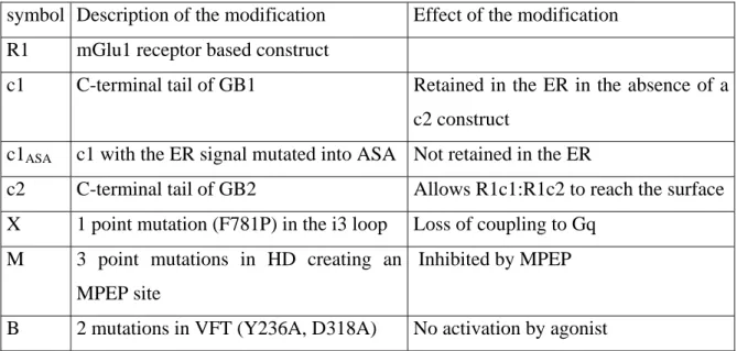
Table 1: Nomenclature used for the constructs described in this study:
symbol Description of the modification Effect of the modification R1 mGlu1 receptor based construct
c1 C-terminal tail of GB1 Retained in the ER in the absence of a c2 construct
c1ASA c1 with the ER signal mutated into ASA Not retained in the ER
c2 C-terminal tail of GB2 Allows R1c1:R1c2 to reach the surface X 1 point mutation (F781P) in the i3 loop Loss of coupling to Gq
M 3 point mutations in HD creating an MPEP site
Inhibited by MPEP
Legends to figures
Figure 1. Determination of cell surface expression of GABAB and wt or chimeric
mGlu1 receptors.
ELISA assay was conducted on intact cells (control, black columns), or on cells permeabilized with Triton X100 (white columns) using an HA-antibody. HA-tagged GB1, mGlu1 (R1), R1c1 or R1c2 were transfected alone or together with myc-tagged GB2 or R1c2, as illustrated in the figure. Mock represents the signal obtained with pRK6-transfected cells. Values are means ± S.E.M. of triplicate determinations from one representative experiment out of three independent experiments.
Figure 2. Heterodimerization of chimeric mG1 receptor subunits
(a) The TR-FRET signal was measured using anti-HA-EuCryptate and anti-myc-Alexa647 antibodies using intact cells expressing the indicated subunits. (b) The specific fluorescent signal (signal measured in the indicated cells minus the signal measured in mock transfected cells) was measured with the anti-HA-EuCryptate antibody, and is used here to estimate the level of surface expression of the HA-tagged subunits. (c) same as in (b) with the anti-Alexa647 antibody to estimate the surface expression of the myc-tagged subunits. (d) same as in (a) but using an equimolar amount of anti-HA-EuCryptate and anti-HA-Alexa647 to visualize any possible interaction between the HA-tagged subunits. (e) The TR-FRET signal was measured in cells transfected with 1µg HA-R1c1 and various amounts of myc-R1c2 (from 0 to 1µg). Anti-HA-EuCryptate and anti-myc-Alexa647 antibodies were used to estimate HA-R1c1:myc-R1c2 heterodimers (open
circles), while anti-HA-EuCryptate and anti-HA-Alexa647 antibodies were used to
estimate the amount of HA-R1c1:HA-R1c1 homodimers (closed circles). The plot shows the TR-FRET signal as a function of the surface expression of the HA-tagged subunits as determined by the specifically bound anti-HA-EuCryptate antibody. All values are means ± S.E.M. of triplicate determinations from a representative experiment out of three. Data shown in a-d are from a single experiment.
Figure 3. Quantification of the relative expression of R1c1:R1c2 heterodimers and R1c2:R1c2 homodimers by ELISA on intact cells.
(a) Schematic representation of the expected surface expression of the chimeric receptors. Mb – plasma membrane; ER – endoplasmic reticulum; HA indicates the subunit is HA-tagged; (HA) means this subunit is either or not HA-tagged, as indicated in (b). (b) Determination of the luminescence signal obtained by ELISA using HA antibody measured in cells expressing the indicated subunits, of which one or both are HA-tagged. Values are means ± S.E.M. of triplicate determinations from a typical experiment out of three and correspond to the raw values obtained with the luminometer.
Figure 4. Effect of increasing doses of quisqualate on mutated mGlu1 receptors
Increase in Ca2+ (a) or IP formation (b) in cells expressing the indicated receptor subunits is plotted as a function of quisqualate concentration. Values are normalized to the quisqualate-evoked maximal response obtained with wt mGlu1 receptor (100%) and are means ± S.E.M. of at least 3 independent experiments performed in triplicate.
Figure 5. Effect of F781P point mutation within the i3 loop on quisqualate-evoked Ca2+ signal in cells expressing various types of receptor dimer combinations
(a) Response measured in cells expressing wt, chimeric or M-mutated receptors alone. (b) Response measured in cells co-expressing R1c1 and the indicated R1c2 constructs. (c) Response measured in cells co-expressing R1Mc1 and the indicated R1c2 constructs. In each case, basal (open columns) and quisqualate-induced (grey columns) responses were determined. For each individual experiments, both the Ca2+ signal and surface expression of the HA-tagged subunit were measured. Values are means ± S.E.M. of the Ca2+ over the ELISA signals of 3-4 independent experiments performed in triplicate.
Figure 6. Effect of MPEP and BAY 36-7620 on quisqualate-evoked Ca2+ signal on wt, R1c2, R1M and R1Mc2 homodimers.
Effect of quisqualate (1µM) alone (open columns) or together with MPEP (100 µM) (black columns) or BAY 36-7620 (10 µM) (grey columns) on Ca2+
-signals was measured in cells expressing the indicated subunits. Values are expressed as percentage of the
maximal quisqualate effect and are mean ± S.E.M. of 3 independent experiments performed in triplicate.
Figure 7. Two MPEP sites per dimer appear necessary for MPEP inhibition of receptor activity.
Effect of quisqualate (1µM) alone (open columns) or together with MPEP (100 µM) (black columns) or BAY 36-7620 (10 µM) (grey columns) on Ca2+
-signals was measured in cells expressing the indicated subunits. Values are expressed as a percentage of the maximal quisqualate effect and are means ± S.E.M. of 3 independent experiments performed in triplicates.
Figure 8. Dose-dependent effect of MPEP on the quisqualate-stimulated IP production.
HEK293 expressing R1Mc2 (circles) or co-expressing R1Mc1 with R1Mc2 (triangles) and R1Mc1:R1Xc2 (squares) were monitored for changes in IP formation upon stimulation with quisqualate (1µM) in the presence of various concentrations of MPEP. Results are expressed as IP production over the total radioactivity remaining in the membrane fraction of the cells. Values are means ± S.E.M of triplicate determinations from a representative experiment out of three independent experiments.
Figure 9. MPEP does not inhibit activity of receptor dimers containing a single MPEP site.
a) Scheme illustrating that in cells expressing R1c1 and R1Bc2, the R1c1 homodimer is retained in the ER, the R1Bc2 homodimer is not functional due to the agonist site mutations, such that only the R1c1:R1Bc2 combination is functional. b) IP production measured under basal condition (grey columns), in the presence of quisqualate (white columns) or in the presence of both quisqualate and MPEP (black columns) in cells expressing the indicated combinations of subunits. Data are means ± s.e.m. of triplicate determinations from a representative experiments out of five.
Figure 10. MPEP enhances quisqualate-induced responses in cells expressing R1c1 and R1MXc2.
a) Schematic representation of the expected receptor dimers at the surface of cells expressing both R1c1 and R1MXc2 subunits. Note that only the heteromer can generate a signal upon agonist activation. b) The Ca2+-signal (left panel) and IP production (right
panel) induced by quisqualate alone (open columns) or together with MPEP (black columns) or BAY 36-7620 (grey columns) were measured in cells expressing the
indicated subunits. c) Effect of increasing concentrations of MPEP on IP production induced by quisqualate in cells expressing R1c1 and R1MXc2. Values are expressed as a percentage of the maximal quisqualate effect and are means ± S.E.M. of 3-7 independent experiments performed in triplicate.
Figure 11. Proposed model : one HD being turned on at a time.
The receptor is represented as a dimer of HDs, one in white, the other in black. The inactive conformation of the HD is represented by a rectangle, whereas the active form is represented by a trapezoid. The presence of the i3 loop mutation that prevents G-protein activation is indicated by a star. The presence of an MPEP site is indicated by an M. The expected effect of MPEP, according to our model proposing that only one HD can reach the active state at a time, is indicated on the right. a) control condition, with either one HD being turned on. b) One HD is mutated in its i3 loop such that only 50% of the dimers activate the G-protein. c) When MPEP prevents the black HD from reaching the active state, then only the white HD is turned on in every receptor dimers. d) MPEP fully blocks receptor dimer activity when bound to the only active subunit. e) By preventing the black HD from reaching its active state, MPEP increases the probability of the white HD to be turned on, thus leading to a enhancement of the agonist effect. n.e. – no effect.
References
Angers S, Salahpour A and Bouvier M (2002) Dimerization: an emerging concept for G protein-coupled receptor ontogeny and function. Annu Rev Pharmacol Toxicol,
42: 409-435.
Ango F, Pin J-P, Tu JC, Xiao B, Worley PF, Bockaert J and Fagni L (2000) Dendritic and axonal targeting of type 5 metabotropic glutamate receptor (mGluR5) is regulated by homer1 proteins and neuronal excitation. J Neurosci, 20: 8710-9716.
Balasubramanian S, Teissere JA, Raju DV and Hall RA (2004) Hetero-oligomerization between GABAA and GABAB receptors regulates GABAB receptor trafficking. J
Biol Chem, 279: 18840-18850.
Baneres J-L and Parello J (2003) Structure-based analysis of GPCR function. Evidence for a novel pentameric assembly between the dimeric leukotriene B4 Receptor
BLT1 and the G-protein. J Mol Biol, 329: 815-829.
Bessis A-S, Rondard P, Gaven F, Brabet I, Triballeau N, Prézeau L, Acher F and Pin J-P (2002) Closure of the Venus Flytrap module of mGlu8 receptor and the activation process: insights from mutations converting antagonists into agonists. Proc Natl
Acad Sci (USA), 99: 11097-11102.
Binet V, Brajon C, Le Corre L, Acher F, Pin JP and Prézeau L (2004) The heptahelical domain of GABAB2 is directly activated by CGP7930, a positive allosteric
modulator of the GABAB receptor. J Biol Chem, 279: 29085-29091.
Bockaert J and Pin J-P (1999) Molecular tinkering of G-protein coupled receptors: an evolutionary success. EMBO J, 18: 1723-1729.
Bouvier M (2001) Oligomerization of G-protein-coupled transmitter receptors. Nat Rev
Neurosci, 2: 274-286.
Calver AR, Robbins MJ, Cosio C, Rice SQ, Babbs AJ, Hirst WD, Boyfield I, Wood MD, Russell RB, Price GW, Couve A, Moss SJ and Pangalos MN (2001) The C-terminal domains of the GABAB Receptor subunits mediate intracellular
trafficking but are not required for receptor signaling. J Neurosci, 21: 1203-1210. Carroll FY, Stolle A, Beart PM, Voerste A, Brabet I, Mauler F, Joly C, Antonicek H,
Bockaert J, Müller T, Pin JP and Prézeau L (2001) BAY36-7620: a potent non-competitive mGlu1 receptor antagonist with inverse agonist activity. Mol
Pharmacol, 59: 965-973.
Chabre M, Cone R and Saibil H (2003) Biophysics: is rhodopsin dimeric in native retinal rods? Nature, 426: 30-31.
Chang W, Chen TH, Pratt S and Shoback D (2000) Amino acids in the second and third intracellular loops of the parathyroid Ca2+-sensing receptor mediate efficient coupling to phospholipase C. J Biol Chem, 275: 19955-19963.
Chinault SL, Overton MC and Blumer KJ (2004) Subunits of a yeast oligomeric G protein-coupled receptor are activated independently by agonist but function in concert to activate G protein heterotrimers. J Biol Chem, 279: 16091-16100. Couve A, Filippov AK, Connolly CN, Bettler B, Brown DA and Moss SJ (1998)
Intracellular retention of recombinant GABAB receptors. J Biol Chem, 273:
Duthey B, Caudron S, Perroy J, Bettler B, Fagni L, Pin J-P and Prézeau L (2002) A single subunit (GB2) is required for G-protein activation by the heterodimeric GABAB receptor. J Biol Chem, 277: 3236-3241.
Filipek S, Krzysko K, Fotiadis AD, Liang Y, Saperstein DA, Engel A and Palczewski K (2004) A concept for G protein activation by G protein coupled receptor dimers: the transducin/ rhodopsin interface. Photochem Photobiol Sci, 3: 628-638. Fotiadis D, Liang Y, Filipek S, Saperstein DA, Engel A and Palczewski K (2003)
Atomic-force microscopy: Rhodopsin dimers in native disc membranes. Nature,
421: 127-128.
Fotiadis D, Liang Y, Filipek S, Saperstein DA, Engel A and Palczewski K (2004) The G protein-coupled receptor rhodopsin in the native membrane. FEBS Lett, 564: 281-288.
Francesconi A and Duvoisin RM (1998) Role of the second and third intracellular loops of metabotropic glutamate receptors in mediating dual signal transduction activation. J Biol Chem, 273: 5615-5624.
Fredriksson R, Lagerstrom MC, Lundin LG and Schioth HB (2003) The g-protein-coupled receptors in the human genome form five main families. Phylogenetic analysis, paralogon groups, and fingerprints. Mol Pharmacol, 63: 1256-1272. Galvez T, Duthey B, Kniazeff J, Blahos J, Rovelli G, Bettler B, Prézeau L and Pin J-P
(2001) Allosteric interactions between GB1 and GB2 subunits are required for optimal GABAB receptor function. EMBO J, 20: 2152-2159.
Gomes I, Gupta A, Filipovska J, Szeto HH, Pintar JE and Devi LA (2004) A role for heterodimerization of mu and delta opiate receptors in enhancing morphine analgesia. Proc Natl Acad Sci U S A, 101: 5135-5139.
Goudet C, Gaven F, Kniazeff J, Vol C, Liu J, Cohen-Gonsaud M, Acher F, Prezeau L and Pin JP (2004) Heptahelical domain of metabotropic glutamate receptor 5 behaves like rhodopsin-like receptors. Proc Natl Acad Sci U S A, 101: 378-383.
Havlickova M, Blahos J, Brabet I, Liu J, Hruskova B, Prezeau L and Pin JP (2003) The second intracellular loop of metabotropic glutamate receptors recognizes C termini of G-protein α-subunits. J Biol Chem, 278: 35063-35070.
Havlickova M, Prezeau L, Duthey B, Bettler B, Pin J-P and Blahos J (2002) The intracellular loops of the GB2 subunit are crucial for G-protein coupling of the heteromeric gamma-aminobutyrate B receptor. Mol Pharmacol, 62: 343-350. Jastrzebska B, Maeda T, Zhu L, Fotiadis D, Filipek S, Engel A, Stenkamp RE and
Palczewski K (2004) Functional characterization of rhodopsin monomers and dimers in detergents. J Biol Chem, in press.
Javitch JA (2004) The ants go marching two by two: oligomeric structure of G-protein-coupled receptors. Mol Pharmacol, 66: 1077-1082.
Jordan BA and Devi LA (1999) G-protein-coupled receptor heterodimerization modulates receptor function. Nature, 399: 697-700.
Jordan BA, Gomes I, Rios C, Filipovska J and Devi LA (2003) Functional interactions between mu opioid and alpha 2A-adrenergic receptors. Mol Pharmacol, 64: 1317-1324.
Jordan BA, Trapaidze N, Gomes I, Nivarthi R and Devi LA (2001) Oligomerization of opioid receptors with beta 2-adrenergic receptors: a role in trafficking and
mitogen-activated protein kinase activation. Proc Natl Acad Sci U S A, 98: 343-348.
Kniazeff J, Bessis A-S, Maurel D, Ansanay H, Prezeau L and Pin J-P (2004a) Closed state of both binding domains of homodimeric mGlu receptors is required for full activity. Nat Struct Mol Biol, 11: 706-713.
Kniazeff J, Saintot P-P, Goudet C, Liu J, Charnet A, Guillon G and Pin J-P (2004b) Locking the dimeric GABAB G-protein coupled receptor in its active state. J
Neurosci, 24: 370-377.
Knoflach F, Mutel V, Jolidon S, Kew JN, Malherbe P, Vieira E, Wichmann J and Kemp JA (2001) Positive allosteric modulators of metabotropic glutamate 1 receptor: Characterization, mechanism of action, and binding site. Proc Natl Acad Sci U S
A, 98: 13402-13407.
Kolakowski LF (1994) GCRDb: a G-protein-coupled receptor database. Receptors
Channels, 2: 1-7.
Kühn H (1984) Interactions between photoexcited rhodopsin and light-activated enzymes in rods. In Osborne, N. and Chader, J. (eds.), Progress in Retinal Research. Pergamon press, p. 123–156.
Kunishima N, Shimada Y, Tsuji Y, Sato T, Yamamoto M, Kumasaka T, Nakanishi S, Jingami H and Morikawa K (2000) Structural basis of glutamate recognition by a dimeric metabotropic glutamate receptor. Nature, 407: 971-977.
Lavoie C, Mercier JF, Salahpour A, Umapathy D, Breit A, Villeneuve LR, Zhu WZ, Xiao RP, Lakatta EG, Bouvier M and Hebert TE (2002) Beta 1/beta 2-adrenergic receptor heterodimerization regulates beta 2-adrenergic receptor internalization and ERK signaling efficacy. J Biol Chem, 277: 35402-35410.
Liang Y, Fotiadis D, Filipek S, Saperstein DA, Palczewski K and Engel A (2003)
Organization of the G Protein-coupled Receptors Rhodopsin and Opsin in Native Membranes. J Biol Chem, 278: 21655-21662.
Liu JF, Maurel D, Etzol S, Brabet I, Ansanay H, Pin JP and Rondard P (2004) Molecular determinants of the allosteric control of agonist affinity in GABAB receptor by the
GABAB2 subunit. J Biol Chem, 279: 15824-15830.
Margeta-Mitrovic M, Jan YN and Jan LY (2000) A trafficking checkpoint controls GABAB receptor heterodimerization. Neuron, 27: 97-106.
Maurel D, Kniazeff J, Mathis G, Trinquet E, Pin J-P and Ansanay H (2004) Cell surface detection of membrane protein interaction with homogeneous time-resolved fluorescence resonance energy transfer technology. Anal Biochem, 329: 253-262. Milligan G (2004) G protein-coupled receptor dimerization: function and ligand
pharmacology. Mol Pharmacol, 66: 1-7.
Pagano A, Rovelli G, Mosbacher J, Lohmann T, Duthey B, Stauffer D, Ristig D, Schuler V, Meigel I, Lampert C, Stein T, Prézeau L, Blahos J, Pin J-P, Froestl W, Kuhn R, Heid J, Kaupmann K and Bettler B (2001) C-terminal interaction is essential for surface trafficking but not for heteromeric assembly of GABAB receptors. J
Neurosci, 21: 1189–1202.
Pagano A, Rüegg D, Litschig S, Stoehr N, Stierlin C, Heinrich M, Floersheim P, Prézeau L, Carroll F, Pin J-P, Cambria A, Vranesic I, Flor PJ, Gasparini F and Kuhn R (2000) The non-competitive antagonists 2-Methyl-6-(phenylethynyl)pyridine and 7-hydroxyiminocyclopropan[b]chromen-1a-carboxylic acid ethyl ester interact
with overlapping binding pockets in the transmembrane region of group I metabotropic glutamate receptors. J Biol Chem, 275: 33750–33758.
Pin J-P and Acher F (2002) The metabotropic glutamate receptors: structure, activation mechanism and pharmacology. Cur Drug Targets - CNS & Neur Dis, 1: 297-317. Pin J-P, Galvez T and Prezeau L (2003) Evolution, structure and activation mechanism of
family 3/C G-protein coupled receptors. Pharmacol Ther, 98: 325-354. Robbins MJ, Calver AR, Filippov AK, Hirst WD, Russell RB, Wood MD, Nasir S,
Couve A, Brown DA, Moss SJ and Pangalos MN (2001) GABAB2 is essential for
G-protein coupling of the GABAB receptor heterodimer. J Neurosci, 21:
8043-8052.
Romano C, Yang W-L and O'Malley KL (1996) Metabotropic glutamate receptor 5 is a disulfide-linked dimer. J Biol Chem, 271: 28612-28616.
Salahpour A, Angers S and Bouvier M (2000) Functional significance of oligomerization of G-protein-coupled receptors. Trends Endocrinol Metab, 11: 163-168.
Tateyama M, Abe H, Nakata H, Saito O and Kubo Y (2004) Ligand-induced
rearrangement of the dimeric metabotropic glutamate receptor 1alpha. Nat Struct
Mol Biol, 11: 637-642.
Terrillon S and Bouvier M (2004) Roles of G-protein-coupled receptor dimerization.
EMBO Rep, 5: 30-34.
Tsuchiya D, Kunishima N, Kamiya N, Jingami H and Morikawa K (2002) Structural views of the ligand-binding cores of a metabotropic glutamate receptor
complexed with an antagonist and both glutamate and Gd3+. Proc Natl Acad Sci U
S A, 99: 2660-2665.
Tsuji Y, Shimada Y, Takeshita T, Kajimura N, Nomura S, Sekiyama N, Otomo J,
Usukura J, Nakanishi S and Jingami H (2000) Cryptic dimer interface and domain organization of the extracellular region of metabotropic glutamate receptor
subtype 1. J Biol Chem, 275: 28144-28151.
Xu H, Staszewski L, Tang H, Adler E, Zoller M and Li X (2004) Different functional roles of T1R subunits in the heteromeric taste receptors. Proc Natl Acad Sci U S
A, 101: 14258-14263.
Zerangue N, Schwappach B, Jan YN and Jan LY (1999) A new ER trafficking signal regulates the subunit stoichiometry of plasma membrane KATP channels. Neuron,
Figure 1
0 2 4 6 8Luminescence signal (Cps x10
5)
HA-R1c1
+R1c2
HA-R1c2
HA-R1c1
HA-R1
HA-GB1
+GB2
HA-GB1
mock
Permeabilized
0 1 3 5 Europium fl uorescence C p s x 10 4 0 1 3 5 Alexa 647 fl uorescence C p s x 10 5
delt
a
F ( % ) anti-myc-Alexa 647 0 50 150 250 R1c2 GB2 R1c2 ≠ cell V2 myc R1c1 GB1 R1c1 R1c2 HAb)
c)
delt
a
F ( % ) anti-HA-EuCryptate anti-HA-Alexa 647 0 50 150 250 350d)
0 500 1000 1500 0 500 1000 1500 HA-myc HA-HAdelt
a665
HA surface expression
e)
0 0.2 0.4