HAL Id: hal-00817464
https://hal.archives-ouvertes.fr/hal-00817464
Submitted on 23 Nov 2020
HAL is a multi-disciplinary open access
archive for the deposit and dissemination of
sci-entific research documents, whether they are
pub-lished or not. The documents may come from
teaching and research institutions in France or
abroad, or from public or private research centers.
L’archive ouverte pluridisciplinaire HAL, est
destinée au dépôt et à la diffusion de documents
scientifiques de niveau recherche, publiés ou non,
émanant des établissements d’enseignement et de
recherche français ou étrangers, des laboratoires
publics ou privés.
Distributed under a Creative Commons Attribution| 4.0 International License
Stable tumor vessel normalization with pO2 increase
and endothelial PTEN activation by inositol
trispyrophosphate brings novel tumor treatment.
Claudine Kieda, Bouchra El Hafny-Rahbi, Guillaume Collet, Nathalie
Lamerant-Fayel, Catherine Grillon, Alan Guichard, Jozef Dulak, Alicja
Jozkowicz, Jerzy Kotlinowski, Konstantina C Fylaktakidou, et al.
To cite this version:
Claudine Kieda, Bouchra El Hafny-Rahbi, Guillaume Collet, Nathalie Lamerant-Fayel, Catherine
Grillon, et al.. Stable tumor vessel normalization with pO2 increase and endothelial PTEN activation
by inositol trispyrophosphate brings novel tumor treatment.. Journal of Molecular Medicine, Springer
Verlag, 2013, epub ahead of print. �10.1007/s00109-013-0992-6�. �hal-00817464�
1 23
Journal of Molecular Medicine
ISSN 0946-2716
J Mol Med
DOI 10.1007/s00109-013-0992-6
Stable tumor vessel normalization with pO
2
increase and endothelial PTEN activation
by inositol trispyrophosphate brings novel
tumor treatment
Claudine Kieda, Bouchra El
Hafny-Rahbi, Guillaume Collet, Nathalie
Lamerant-Fayel, Catherine Grillon, Alan
Guichard, et al.
1 23
Commons Attribution Non-Commercial
license which allows users to read, copy,
distribute and make derivative works for
noncommercial purposes from the material,
as long as the author of the original work is
cited. All commercial rights are exclusively
held by Springer Science + Business Media.
You may self-archive this article on your own
website, an institutional repository or funder’s
repository and make it publicly available
immediately.
ORIGINAL ARTICLE
Stable tumor vessel normalization with pO
2
increase
and endothelial PTEN activation by inositol
trispyrophosphate brings novel tumor treatment
Claudine Kieda&Bouchra El Hafny-Rahbi&
Guillaume Collet&Nathalie Lamerant-Fayel&
Catherine Grillon&Alan Guichard&Jozef Dulak&
Alicja Jozkowicz&Jerzy Kotlinowski&
Konstantina C. Fylaktakidou&Aurélien Vidal&
Philippe Auzeloux&Elisabeth Miot-Noirault&
Jean-Claude Beloeil&Jean-Marie Lehn&Claude Nicolau
Received: 20 September 2012 / Revised: 24 December 2012 / Accepted: 2 January 2013 # The Author(s) 2013. This article is published with open access at Springerlink.com
Abstract Tumor hypoxia is a characteristic of cancer cell growth and invasion, promoting angiogenesis, which facil-itates metastasis. Oxygen delivery remains impaired be-cause tumor vessels are anarchic and leaky, contributing to tumor cell dissemination. Counteracting hypoxia by normal-izing tumor vessels in order to improve drug and radio therapy efficacy and avoid cancer stem-like cell selection is a highly challenging issue. We show here that inositol trispyrophosphate (ITPP) treatment stably increases oxygen tension and blood flow in melanoma and breast cancer
syngeneic models. It suppresses hypoxia-inducible factors (HIFs) and proangiogenic/glycolysis genes and proteins cascade. It selectively activates the tumor suppressor phos-phatase and tensin homolog (PTEN) in vitro and in vivo at the endothelial cell (EC) level thus inhibiting PI3K and reducing tumor AKT phosphorylation. These mechanisms normalize tumor vessels by EC reorganization, maturation, pericytes attraction, and lowering progenitor cells recruit-ment in the tumor. It strongly reduces vascular leakage, tumor growth, drug resistance, and metastasis. ITPP
C. Kieda and B. El Hafny-Rahbi contributed equally to this study. Electronic supplementary material The online version of this article (doi:10.1007/s00109-013-0992-6) contains supplementary material, which is available to authorized users.
C. Kieda (*)
:
B. El Hafny-Rahbi:
G. Collet:
N. Lamerant-Fayel
:
C. Grillon:
A. Guichard:
J.-C. Beloeil Centre de Biophysique Moléculaire, UPR CNRS 4301, rue Charles Sadron,45071 Orléans Cedex 2, France e-mail: claudine.kieda@cnrs-orleans.fr J. Dulak
:
A. Jozkowicz:
J. KotlinowskiDepartment of Medical Biotechnology, Faculty of Biochemistry, Biophysics and Biotechnology, Jagiellonian University, 30-387 Kraków, Poland
K. C. Fylaktakidou
:
J.-M. Lehn:
C. NicolauISIS, Université de Strasbourg, 8 Allée Gaspard Monge, 67083 Strasbourg, France
J.-M. Lehn
e-mail: lehn@isis.u-strasbg.fr C. Nicolau
e-mail: claude.nicolau@normoxys.com
A. Vidal
:
P. Auzeloux:
E. Miot-NoiraultUMR 990 INSERM/Université d’Auvergne, “Imagerie moléculaire et thérapie vectorisée”, 58, rue Montalembert, 63005 Clermont-Ferrand, France
C. Nicolau
NormOxys Inc., 300 Market Street, Boston, MA 02135, USA
C. Nicolau
Friedman School of Nutrition Science and Policy, Tufts University, Boston, MA 02115, USA
Present Address: K. C. Fylaktakidou
Molecular Biology and Genetics Department, Democritus University of Thrace, Dimitras 19,
68100 Alexandropoulis, Greece DOI 10.1007/s00109-013-0992-6
treatment avoids cancer stem-like cell selection, multidrug resistance (MDR) activation and efficiently enhances chemotherapeutic drugs activity. These data show that counteracting tumor hypoxia by stably restoring healthy vas-culature is achieved by ITPP treatment, which opens new therapeutic options overcoming hypoxia-related limitations of antiangiogenesis-restricted therapies. By achieving long-term vessels normalization, ITPP should provide the adjuvant treatment required in order to overcome the subtle definition of therapeutic windows for in vivo treatments aimed by the cur-rent strategies against angiogenesis-dependent tumors. Keywords Angiogenesis . Normalization . Oxygen . PTEN . Tumor hypoxia
Introduction
Tumor hypoxia, decisive for cancer progression, upregulates
the hypoxia-inducible factors (HIFs), O2sensors in animal
cells. Hypoxic tumor cells become resistant to radiotherapy and chemotherapy, getting to be highly aggressive and
metastatic [1]. HIF-1α is associated with increased vessel
numbers, tumor grade severity, poor prognostic, and
treat-ment failure. Hypoxia-induced tumor angiogenesis [2] to
build new vessels for oxygen and nutrients supply to tumor cells is in fact inefficient. It leads to incomplete vessels that are permeable and allow metastatic spreading of tumor cells
escaping through nonsealed endothelial cells (ECs) [3].
Antiangiogenic strategies aiming at inhibition of tumor neo-vascularization have not provided lasting benefits be-cause, by increasing tumor hypoxia, they result in selection
of drug-resistant, aggressive cancer stem-like cells [4].
Tu-mor vessel normalization [5], rather than destruction, is a
promising approach to cancer therapy since vessel
abnorm-alization is now recognized as a hallmark of cancer [6]. The
challenge is to counteract the vicious circle of hypoxia-induced abnormal vessels and tumor hypoxia maintained
because of vessel defects [7].
New strategies aim at regulating intratumor vessels by reducing the activity of hypoxia sensors like PHD1-3 enzymes (prolyl hydroxylases), which target HIFs for
deg-radation [8]. Vessel normalization beneficial effects were
confirmed by the double antiangiogenic protein [9] targeting
both vascular endothelial growth factor (VEGF) A [10] and
angiopoietins, which restored tumor vessels efficacy. Vessel
normalization prevents tumor cell dissemination [11],
allows efficient delivery of cytotoxic drugs, and increases
efficacy of radiotherapy [5] through control of HIFs activity
[12].
Such approaches have allowed treatment protocols at time lapses defined as therapeutic windows during which
vessels are normalized [13]. The technical difficulties to set
adequate therapeutic windows prompted the search for long term normalization as an alternative goal for cancer
antian-giogenesis therapy [14].
Hypoxia-induced angiogenesis is inhibited when human microvascular endothelial cells are cultured under hypoxic and flow conditions in the presence of RBCs loaded in vitro
with ITPP [15] that overcame low oxygen tension (pO2). In
vitro, ITPP was shown to act as an allosteric effector of
haemoglobin, and it was observed to reduce HIF-1α [16].
Given the central role that hypoxia plays in initiation and progression of neoplasms, these findings suggested a high
potential for“oxygen tension compensation” in cancer
ther-apy [15,17] [18].
We tested here this hypothesis and the potential utility of ITPP to reach this challenge by treatment of melanoma and
mammary cancer-bearing mice [19]. We show that ITPP
treatment reduces tumor growth and eradicates lung metas-tasis. Biochemical changes in tumor and its microenviron-ment, upon ITPP treatmicroenviron-ment, appear to result predominantly, from the selective reversal of tumor hypoxia through vessel normalization. As an inositol phosphate derivative, ITPP molecule was tested for potential activation of endothelial phosphatase and tensin homolog (PTEN) and thus for its ability to bring a new tool to regulate angiogenesis indepen-dently of the cancer cell type. Indeed, drugs affecting the
PTEN-regulated PI3K/AKT/mTOR pathway [20] that
indu-ces HIF-1α in a hypoxia-independent mechanism [21] may
act on ECs to normalize tumor vessels. As PTEN is one of the most frequently mutated tumor suppressor, its inactiva-tion leads to permanent AKT phosphorylainactiva-tion that main-tains tumor growth. Consequently, this work was undertaken to check the hypothesis that ITPP treatment efficiently contributes to long-term vessel normalization through both dependent and oxygenation-independent control of HIF. We could show that angiogen-esis regulation by ITPP treatment occurred through the downregulation of HIF-dependent proangiogenic genes. It regulated PTEN/AKT pathway by activating the endothelial
PTEN that controls angiogenesis [22]. Resulting functional
vessels were shown to facilitate access of chemotherapeutic agents to tumor cells. VEGF-induced leakiness was re-duced, and invasive metastatic cell escape was abolished. Compensating intratumor hypoxia, treatment reduced the number of hypoxia- and multidrug-resistant as well as stemness-marker-positive tumor cells, reduced anaerobic glycolysis, and stopped the recruitment of HIF-mediated
bone marrow derived CXCR4+ precursor cells [23]. This
work deciphers some aspects of the potent multifactorial therapeutic effects of ITPP treatment on neoplastic angio-genesis, inhibition of tumor growth, and prevention of me-tastasis through maturation of tumor vasculature and opens to new angiogenesis-based therapies of hypoxia-inducing diseases. It shows that ITPP provides means to reach the
goal of persistent angiogenesis normalization as an
alterna-tive to antiangiogenic therapies [14].
Materials and methods Cells
Endothelial cells are FVB mouse lung microvascular
endo-thelial cells (MLMEC FVB) [24, 25]. 4T1 murine breast
cancer cells [19] were kindly provided by Professor Danuta
Dus (IITD, PAN, Wroclaw, Poland). B16F10LucGFP are B16F10 murine melanoma cells, transduced with retroviral vectors containing a firefly luciferase complementary DNA (cDNA) driven by 5'LTR promoter followed by IRES se-quence and enhanced green fluorescent protein cDNA (see
Supplemental data). B16F10LucGFP cells were compared to B16F10 cells to validate their use in terms of similarity of growth and metastatic potential in vivo. Luciferase activity was shown not to be impaired by hypoxia, which could potentially affect the detection sensitivity (supplementary
Fig.S1A).
Cell culture and oxygen regulation
The B16F10, B16F10LucGFP cell lines (seeSupplemental
data) and the 4T1 breast cancer cell line were cultured,
respectively, in Dulbecco’s modified Eagle medium and
RPMI (Gibco) with 10 % fetal bovine serum (FBS) (PAA). The MLMEC-FVB endothelial cells were cultured in OptiMEM/2 % FBS. Cells were routinely cultured in a humidified incubator in 19.5 % oxygen and were
oxygen-deprived in a Biotronix incubator allowing p02 regulation
(1 %) and time setting.
Mouse subcutaneous melanoma and breast cancer models C57BL6 mice and BALB/c mice were from Janvier Labo-ratory (France). Animal care and experimental procedures were approved by the CNREEA 03 Ethics Committee.
B16F10 or B16F10LucGFP cells were implanted in C57BL6 mice leg as subcutaneous tumors by injection of
a plug constituted by 2×10⁵cells in 100μl Matrigel™ (BD
Biosciences) to help angiogenesis. 4T1 murine mammary
carcinoma (104cells in Matrigel) cells were injected in the
mammary fat pad of BALB/c mice.
For the experimental metastases, see “Supplementary
methods.”
ITPP treatment and chemotherapeutic protocols
ITPP, prepared as described [16], was injected
intraperito-neally (1.5 g/kg: in saline). Protocol consisting of serial
treatments, over 4 weeks, was selected. It was started on day7 and repeated on day8 posttumor inoculation (day0). The following serial treatments were applied on days15 and 16, 21 and 22, and 28 and 29.
When combined with ITPP treatment, chemotherapeutic drugs, paclitaxel (2 mg/kg, in 50 % ethanol, 50 % chremo-phor EL; Calbiochem, per os) and cisplatine (cis-dichlorodi-amine platinum) (10 mg/kg in saline; Sigma-Aldrich, intraperitoneally) were administered on days 9, 17, and 23, and the analyses were conducted on day 25.
Luciferase activity
Luciferase Assay System (Promega) was used as described by the manufacturer and luminescence quantified as relative light units per number of cells or per milligram of tissue with manual luminometer (Lumat LB9507).
Bioluminescence imaging was performed at the Center for Small Animal Imaging (TAAM, CNRS Orleans) Cell staining for flow cytometry
Tumor cells were analyzed after tissue dissociation by col-lagenase/dispase (Gibco). For membrane antigen detection, single cell suspensions were incubated at 4 °C with respec-tive primary antibodies for 1 h then with secondary anti-bodies for 30 min. For intracellular staining, cells were permeabilized by Cytofix/Cytoperm solution (BD Bioscien-ces) as indicated, for 20 min before incubation with anti-bodies. Labeled primary antibodies used were the following: antimouse-PE (rat Ig-G2a, eBioscience) or CD31-PerCP (rat IgG2a, R&D), -VEGF R1-PE (Rat IgG2b, R&D), -VEGF R2-PE (Rat IgG2a, R&D), -CXCR4-PE (Rat IgG2b, R&D), -CD45-PerCP (rat IgG2b, R&D) or CD45-PE-Cy7 (rat IgG2b, R&D), -CD34-A700 (rat IgG2a, R&D). Unconjugated primary antibodies were the follow-ing: Firefly Luciferase (Rabbit IgG, Abcam), anti-mouse -CD202 (Rat IgG1, eBiosciences), -HIF-2α (rabbit IgG, Abcam), -Lox (rabbit Ig-G, Novus Biologicals), -iNos (rabbit G, Transduction Laboratories), -Glut-1 (rabbit Ig-G, Santa-Cruz), -HO-1 (mouse Ig-G1, Abcam), -LDH (rabbit IgG, SantaCruz), CAIX (rab(rabbit IgG, SantaCruz), -CD133 (Rat IgG1, eBioscience), -Oct-3-4 ( Rat IgG2a, R&D), or -ABCG2 (Rat IgG, Abcam). Binding was revealed by fluorescently-labeled anti-isotype secondary antibodies.
In specified experiments, tumors were depleted from
CD45+ and/ or CD31+cells by magnetic separation (Easy
Sep magnet, StemCell Technologies Inc). Cytofluorimetry analyses were conducted on a FACS Sort (Becton Dick-inson, Sunnyvale, CA, USA). Data were acquired on 5×
104 to 105 cells and analyzed using CellQuest software
Immunohisto/cytological staining
Tumor tissues were embedded in tissue freezing medium (Tissue-Tek; Sakura) and snap frozen in liquid nitrogen. Tumor cryosections or cells were fixed and stained with mouse anti-CD31 (rat monoclonal IgG2a) (eBiosciences), anti-SMA (Rabbit IgG, Abcam), anti-P-glycoprotein (mouse IgG2a, Calbiochem), anti-CD133 (Rat IgG1, eBio-sciences), anti-Phospho-AKT (Ser473) (Rabbit IgG), or anti-PTEN (rabbit IgG) (Cell Signalling) before tetramethyl rhodamine isothiocyanate or fluorescein isothiocyanate sec-ondary antibodies were added. Nuclei were stained with bis-benzimide H 33258 (Sigma-Aldrich).
To assess hypoxic areas, pimonidazole (75 mg/kg, Hypo-xyprobe™) was injected intravenously, 1 h before mice killing and tissue collection. Tumor cryosections were incu-bated with primary antipimonidazole antibodies (Mouse IgG1, Hypoxyprobe) before PE-labeled anti-mouse IgG antibodies were added.
PTEN localization and activation was studied on murine endothelial cells from lung (MLNEC FVB) that have been treated by hypoxia (22 h) and/or reoxygenation (25 h) in the presence or absence of ITPP (25 mM).
Fluorescent microscopy detection was performed on a Zeiss 200M inverted fluorescence microscope (Le Pecq, France), video microscopy station with controlled tempera-ture, hygrometry, and gaz composition. Analysis was done with the Axiovision, software. Tumor necrosis was analyzed after hematoxylin–eosin staining of tumor sections. Quantitative real-time PCR Total RNA was extracted from whole tumor using RNeasy Plus RNA extraction kit (Quiagen). cDNA was made with Transcriptor First strand cDNA Synthesis kit (Roche), and quantitative real-time PCR was performed with the QuantiTect SYBR Green RT-PCR kit (Qiagen), using LightCycler 480 (Roche). The data were analyzed with LightCycler 480 Software. Primers were supplied by Qiagen.
Magnetic resonance imaging
MR experiments on mice were performed on 9.4T horizon-tal magnet dedicated to small animal (94/21 USR Bruker Biospec, Wissembourg, France), equipped with a 950mT/m
gradient set. Detailed method is given in“Supplementary
methods.” The animals were put under gaseous anesthesia
during MRI exams (50 % N2O, 0.7 l/min–50 % O2,
0.7 l/min–Isoflurane, 1.5 %). Breathing rate was monitored by an air pillow placed on the mouse chest to adjust the anesthetic output. Magnetic resonance angiography-time of
flight (MRA-TOF) experiment [26] allowed visualizing the
3D structure of the vascular tree of the tumor on the same
animal. The total duration of the MRA experiment was 50 min. MRA sensitivity was increased by working at high field (9.4 T). Angiograms were produced using maximum intensity projections (MIPs) using Paravision 4.0 (Bruker). MRA-TOF is used for angiography, but this pulse sequence
could also be seen as a“saturation recovery” pulse sequence
(T1 sensitive). We have used it for detection of necrosis
(Fig. 1b). Measurement of the tumor size was performed
with a classical morphological spin-echo pulse sequence.
PO2and blood flow measurements
Mice were anesthetized by ketamine (Imalgène® 500, Rhone Mérieux, France) and xylazine (Rompun® 2 %, Bayer, France) (4:1 ratio) intraperitoneally injected
(40 μl/20 g mouse) before the oxylite probe tips (Oxford
optronics) were installed inside the tumor and oxygen
pres-sure or blood flow recorded by the oxylite 2000E pO2or
OxyFlow 2000 systems (Oxford Optronics) [27]. pO2was
computed by determining the O2-dependent fluorescence
lifetime of ruthenium chloride on the tip of an optical fiber probe. The fluorescence lifetime is inversely proportional to
the pO2in the tissue. The OxyFlow, a microvascular
perfu-sion monitor, uses laser Doppler flowmetry established method for the measurement of blood perfusion with probe
coupled to the O2probe tip.
[18F]-FMISO PET imaging
For each animal anesthetized by ketamine/xylazine, [18
F]-FMISO PET imaging was performed using a device dedi-cated to small animal (eXplore VISTA®, GE Healthcare, USA). The spatial resolution of this system is given as 1.4 mm full width at half maximum at the center of the field
of view [28].
Whole body images were acquired 1.30 h after i.v.
ad-ministration of [18F]-FMISO (8.9±1.3 MBq/mouse) for a
total acquisition time of 20 min, an energy window set at 250–700 keV, two bed positions, and 6 ns coincidence-timing window. Image reconstruction used an ordered-subset expectation maximization (FORE/2D OSEM) meth-od including corrections for radioactive decay, scanner dead time, and scattered radiation. No correction was applied for partial volume or attenuation.
Quantitative analysis of scans were performed using eX-plore VISTA® workspace. Volume of interest (VOI) was delineated over tumor, and whole body, by summing multi-ple two-dimensional regions of interest from consecutive tomographic planes encompassing the entire uptake volume. The VOI of reference tissue was drawn on paw muscle and considered as background.
For each VOI, total activity was obtained as counts per minute (cpm), as well as mean activity in cpm per pixel and
3.00E+09 4.00E+09 5.00E+09 6.00E+09 hotons/sec) D18 D24
a
Control mice ITPP treated mice
b
0.00E+00 1.00E+09 2.00E+09 3.00E 09
Untreated ITPP Treated
Total flux (p
h
Untreated tumors D23 ITP-treated tumors D23
(D7,8 14,15 21,22)
Mean tumor size D23
c
1000 1500 2000 2500 cm 1 cm 1d
Untreated Tumor 0 500control ITPP treated
T
u
mor siz
e mm3
**
0 cm 1 0 cm 1 0 0 Tumor + ITPPe
0 0Tumor size Necrotic areas
*
SSC
M2
*
Anti Luciferase Cy5
Anti Luciferase Cy5
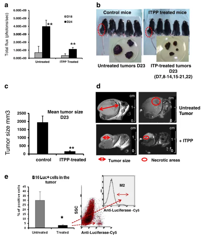
Fig. 1 ITPP reduces melanoma tumor growth and improves mice survival. a Effect of ITPP treatment on the kinetics of tumor growth measured by bioluminescence in treated and nontreated animals at days18 and 24. Endpoint was fixed at 2 cm3(n=6 animals per group, one representative experiment out of N>10, **p<0.001). b Compari-son of tumor size, 23 days after B16F10LucGFP cells injection, show-ing reduced tumor growth in treated mice. Representative groups of five animals among groups of n=10 animals. One experiment out of N≥5 separate experiments. Insets illustrate the extreme size ranges (minimal and maximal) that tumor reached in nontreated compared to treated mice. c Mean size of the tumors in treated and non treated animals at day23 (n=10 in each group; number of experiments N>20, **p < 0.001). d Magnetic resonance imaging of B16 F10 induced
tumor. Morphological pulse sequence (left). Strong volume variation of the tumor (untreated/ITPP=1163 mm3/121 mm3) was observed by image analysis after volume reconstruction. One typical example out of n=10/experimental group. MRA-TOF/saturation recovery pulse se-quence (right): Necrotic areas appear darker. After ITPP treatment, their size decreased. One typical example out of n=10/experimental group. e Analysis by flow cytometry of B16LucGFP cells in tumors. Luciferase was detected intracellularly by specific antibodies and la-beled by PerCP-Cy7 antirabbit IgG confirming: the reduced growth of tumor cells in ITPP-treated mice (%) and counts by direct cytometry analysis. Cells were numbered on the basis of intracellular Luciferase detection (n = 8; *p< 0.05) from dot plots or inset from histogram analysis for quantification of B16F10LucGFP in the tumor
converted to cpm per milliliter using a calibration constant (obtained by imaging a mouse-size cylindrical calibration
phantom containing a known activity of18F). By assuming a
tissue density of 1 g/ml, VOI activity in cpm per milliliter was converted to cpm per gram. Tumor uptake was calcu-lated by dividing total tumor activity (in cpm) by total whole body activity (in cpm).
Tumor capillary leakiness
It was assessed by Evans blue dye extravasion to the tumor
interstitium. The dye was extracted by formamide [29]. The
concentration measured spectrophotometrically was corre-lated to tumor weight.
Elisa detection of circulating VEGF
VEGF was assessed in serum (100μl) by a typical sandwich
ELISA kit for Mouse VEGF (Duo Set from R&D Systems). Assays were conducted according to the manufacturer’s instructions.
Statistical analysis
Data represent mean ±SD of 5 or 10 (when specified) rep-resentative experiments on 5≤n≤10 animals in each group. Statistical significance was calculated by Student’s t test (p= 0.05; p=0.001; n=6 animals per group, one representative experiment out of N>10, p<0.001).
Results
ITPP treatment counteracts melanoma tumor growth and lung metastases and improves mice survival
ITPP treatments produced a strong reduction of tumor
growth assessed in terms of visible tumor size (Fig. 1b).
The mean tumor weight at day23 was 2.5±0.5 g and re-duced to 0.5±0.2 g in treated animals (n=10 per group and per experiment out of N≥5 separate experiments). This was precisely quantified by measurement of bioluminescence emission from B16F10Luc tumors measured at days 18 and 24, which increased 5.5-fold as compared to 2.8-fold
in the treated animals, shown in Fig.1a (n=6 animals per
group, one representative experiment out of N > 10, p < 0.001). Tumor growth reduction was quite visible in
Fig. 1b showing the maximal and minimal sizes reached
by tumors and the treatment effect, which is expressed as
mean values of the tumor volumes at day 23 on Fig.1c. MRI
calculation of the tumor volume is precised on Fig.1d. This
was confirmed by the numbers of tumor cells detected in the tumor mass ranging from 30±7 % in nontreated animals,
down to 3±1 % after ITPP treatment (Fig. 1e). Moreover,
the metastatic invasion was drastically diminished. Indeed, in the subcutaneous model, no visible invasion but a faint Luciferase activity was detectable in lungs of treated
ani-mals (Fig. 2a). This difference was confirmed by artificial
metastases assessment, after i.v. injection of B16F10LucGFP cells, which weakly developed in ITPP-treated animals
com-pared to controls (Fig.2b) (n=20 in each group). These results
corroborate the observed survival rate (Fig.2d). Further on, no
control animal was ethically allowed to survive longer than 30 days, as opposed to 100 % survival of ITPP-treated ani-mals, which were killed on day 60 for further analyses.
End-points were then fixed when tumors reached 2 cm3. Indeed,
Fig.2cindicates that the total body weight of control animals
clearly decreased compared to ITPP-treated tumor-bearing animals, despite the tumor growth. ITPP itself did not change the weight of the mice compared to saline injected mice, daily
(Fig.2c, inset).
ITPP treatment selectively counteracts hypoxia in the tumor microenvironment and normalizes tumor vessels
pO2 was measured directly inside tumors before, during,
and after ITPP treatment by intratumor assessment of the
O2-dependent fluorescence quenching of ruthenium.
Non-treated tumors were strongly hypoxic (pO2<2 mmHg). pO2
measurement in real time inside the tumor showed that a
first injection of ITPP caused a rapid pO2increase detectable
after 30 min, reaching 40 mmHg within next 5 min (Fig.3a).
After serial injections of ITPP, as indicated in“Materials and
methods,” we show that the pO2 level was stabilized at a
high level for, at least, 72 h (Fig.3b). Moreover, the pO2
increase specifically concerned the hypoxic tumor site as, in the muscle of the contra lateral healthy leg of the same
animal, no pO2change was detected by concomitant
meas-urements (Fig.3a). The validation of such hypoxia
compen-sation by ITPP treatment was assessed and confirmed by performing the same experiments on murine breast cancer
4T1 model. Figure3cgives typical data registered from one
mouse out of 10 treated animals and showing a similar behaviour. A moderate increase was obtained after the first
injection, and pO2 strongly and stably increased after the
second injection of ITPP. The reversal of intratumor hypoxia upon ITPP treatment was functionally paralleled by in-creased intratumor blood flow, measured concomitantly to
pO2, by laser Doppler and reported for day 22 tumors
(Fig.3d). Blood flow change may contribute to the rapid
pO2 increase and indicate a normalization of the tumor
vessel function. Chemical confirmation of this process was assessed by histochemical detection of hypoxic sites in tumor. Hypoxic areas were evidenced by pimo-nidazole adducts formation with reduced proteins. Exist-ing blood vessels, detected by double labelExist-ing for CD31
did not insure proper tissue oxygenation (Fig. 2e), con-firming the poor efficacy of tumor angiogenesis. ITPP treatment prevented the formation of hypoxic areas, influencing deeply blood vessels structure, size, and
density (Fig. 4a). These data were confirmed in living
mice injected with [18F]-FMISO to quantify hypoxia in
the tumor (Fig. 4b). ITPP-treated mice clearly displayed
restricted tumor growth and lower intratumor hypoxia. The tumor incorporation was expressed as Tact% = (total tumor activity/total whole body activity ) × 100. Upon ITPP treatment, Tact decreased from 14.58 ±0.52 % to 7.6 ± 0.6 % (n = 8 per group N= 4).
ITPP treatment of tumor-bearing animals normalizes structure and function of vessels in the tumor
Vessel normalization was validated in live tumor-bearing animals upon ITPP treatment. Magnetic resonance im-aging of tumor vasculature indicated strong structural changes. Typical chaotic tumor vessel architecture was observed by magnetic resonance angiography (MRA), while in ITPP-treated mice, vasculature appeared less
dense but organized (Fig. 5a). Intratumor examination
after treatment revealed CD31 labeling typical for endo-thelial cell, which delineates vessel-like structures after
ITPP treatment as opposed to CD31+ aggregates in
controls (Fig. 5a). Furthermore, in treated tumors,
vessel-like structures appear at the tumor periphery, surrounded by pericytes, positive for smooth muscle
antigen (SMA+) (Fig. 5a). Comparison with the
dis-persed SMA+ labeled cells in the nontreated tumor mass
suggests vessel normalization upon ITPP treatment. Confirmation of ITPP-induced vessel normalization is
brought by pericytes recruited and lining the CD31+
endothelial cells of the treated tumor vessels (Fig. 3a,
d) as opposed to the random distribution of SMA+ cells
in nontreated tumor, shown by confocal microscopy
(Fig. 5a). “Normalization” accompanied a strong
reduc-tion of tumor size (Figs. 1 and 5a).
Normalization was confirmed in terms of vessel function, first by reduction of tumor vessels permeability. Evans blue leakage was significantly diminished after two sequential
treatments by ITPP (Fig. 5b) correlating with the reduced
concentration of circulating VEGF, the main vessel
Fig. 2 Reduction of colonization and increased survival in cancer bearing mice treated by ITPP. a Reduced luciferase activity after ITPP treatment in lungs of mice bearing subcutaneously implanted melanoma, indicating reduced metastasis (n=8 ; p<0,001, one experiment out of 10). b Reduced lung colonization, after treatment, in artificial metastasis model where melanoma cells were injected intravenously. Representative samples out of 20 mice in each group. c Evolution of the animal body weights. Nontreated animals are losing weight compared to ITPP-treated animals (n=10 in each group, the number of experi-ments is N>10), inset shows the effect of daily ITPP injection compared to saline. d Survival curve showing the rescue of melanoma bearing mice treated by ITPP (n=10 in each group, one typical experiment out 5)
permeant growth factor (Fig.5c). ITPP-induced vessel
mat-uration was shown by reduction of the invasive index [30].
CD105+/CD31+ ratio measures endoglin (CD105) versus
PECAM-1 (CD31) expressing endothelial cells and reflects
tumor neo-angiogenic activity. CD105+/CD31+ cells ratio,
calculated among tumor CD45− cells, was lowered upon
ITPP action (Fig.5d), mainly because CD31+ cell number
increased (Fig. 5a, d). The strong enhancement of VEGF
receptors 1 and 2 on nonleukocyte CD31+CD45−cells upon
treatment (Fig. 5e) confirms maturation of blood vessels
reflecting direct interactions of endothelial cells with mural
pericytes (Fig.5a).
CD31+ cell numbers increased among the whole tumor
population (Fig.5f) and was accompanied by enhancement
of the hypoxia-dependent, endothelial tyrosine kinase Tie-2
receptor for angiopoietins 1 and 2 (Fig.5fand
Supplemen-tary Fig.2d), a marker of matured vessels [6,10].
ITPP treatment induces tumor vessels maturation by regulating hypoxia-sensitive molecules
Hypoxia-sensitive genes turning on tumor angiogenesis dis-played drastic changes upon ITPP treatment of tumor-bearing animals. Since the ITPP effect was associated with
pO2 changes in the tumor, the levels of HIF-1 and HIF-2,
crucial for cell response to oxygen, were analyzed by
quan-titative PCR. Figure6ashows the strong downregulation of
messenger RNA (mRNAs) for HIF1−and HIF2,
corroborat-ing the reduction of HIF1 and HIF2 protein expression
(Supplementary Fig.S2and Fig.6a, respectively) and
indi-cating that regulation occurred at the transcriptional level.
HIFs mRNA and O2-dependent molecules like VHL and the
tumor protective and proangiogenic enzyme HO-1 mRNAs
were considerably reduced by ITPP (Fig6a). Main oxygen
sensors in angiogenesis, PHD-1, PHD-2, and PHD-3, are
Fig. 3 Improved tumor oxygenation in cancer bearing mice treated by ITPP. a ITPP treatment increases oxygen pressure specifically inside a melanoma tumor within 30 min. No pO2modification was observed in
the healthy muscle. The probe was maintained in the same place, and pO2was recorded in real time. Figure reports a representative example
out of five animals treated the same day; identical data were acquired from n>50 mice treated by ITPP. b Tumor oxygen pressure increase after double injection of ITPP is stable up to 72 h. Tumor oxygen pressure is enhanced 30 min after the first injection of ITPP. A second injection of ITPP applied after 24 h increases and stabilizes the pO2
increase for at least 72 h. Figure reports typical measurements
randomly performed in treated animals (n=10 per experiment). c ITPP treatment increases oxygen pressure inside a 4T1 breast tumor within 10 min. The increased pO2(12 mmHg at day14) is stable over 1 week
(20 mmHg at day22) and enhanced once more after a second ITPP injection. Picture shows data typically reporting several measurements randomly performed in treated animals (n=8 per experiment). d Laser Doppler signals showing tumor blood flow improvement in ITPP-treated mice. Measurements were done randomly in each tumor ITPP-treated as described in“Materials and methods,” day22 (n=8 out of 10 per
considerably underexpressed in treated animals, indicating a
direct regulation by pO2, as described for VHL mRNA
induction by hypoxia.
Increase in CD31, VEGFR1, and VEGFR2 mRNAs
(Fig. 6a) confirmed the enhancement of corresponding
protein-expressing cell numbers and the maturation effect
observed among CD45− cells (Fig. 5d) with the general
increase in Tie2+ cell numbers (Fig.5e). mRNA for
osteo-pontin, a key molecule of the tumor stroma, decisive for
tumor invasion and known to be regulated by the PTE-N/AKT pathway in melanoma, was strikingly reduced by
ITPP treatment (Fig.6a).
In accordance with the finding that ITPP treatment did
not influence endothelial cells growth in vitro [15] but
controlled the angiogenic process, its in vivo effect might implicate activation of PTEN that controls both hypoxia-dependent and hypoxia-inhypoxia-dependent mechanisms of tumor angiogenesis. We observed a clear reduction of the SDF1/CXCR4-dependent recruitment of endothelial precur-sor cells from the bone marrow. They cooperate to tumor angiogenesis by integrating neovessels in a
PTEN/PI3-K/AKT/eNOS dependent process. Figure6bshows the
dras-tic reduction of the proportion of CXCR4+, CD34+, and
CD45− endothelial precursors cells, recruited inside the
tumor upon ITPP treatment.
ITPP-induced tumor vessel normalization regulates energetic metabolism molecules linked to oxidative stress The number of cells expressing stress- and metastasis-related markers not only HIF2 but also lysyl oxydase (LOX) was assessed. Hypoxia-regulated LOX, involved in
invasiveness [31] and responsible for H2O2production that
inactivates PTEN [32], was drastically reduced by ITPP in
the whole tumor (Fig.6c).
Moreover, upon ITPP treatment, LOX and HIFs were both efficiently downregulated in their mRNA expression
(Supplementary Fig. S2). In parallel, the
endothelium-restricted enzyme, inducible NO synthase, produces NO that induces vessel dilatation and VEGF production responsible for permeabilization. Here, the number of cells expressing
INOS [33] was significantly reduced (Fig.6c), similarly to
HO-1. The artificial metastasis model confirmed the
bene-ficial effects of ITPP treatment (Supplementary Fig.S2).
Tumor cells resist to poor oxygen supply using the anaerobic glycolysis as source of energy. This rescue pathway starts with enhanced Glut-1 receptor expression ending with lactate release, activating glycolytic path-way enzymes, such as lactate dehydrogenase (LDH)
[34] and carbonic anhydrase IX (CAIX), a key enzyme
allowing tumor cell survival in hypoxia and acidic pH
[35]. Numbers of cells expressing Glut-1 receptors,
LDH and CAIX, were drastically reduced upon ITPP
treatment (Fig. 6d), indicating a significant reversal of
hypoxia-induced resistance.
ITPP-treatment-induced tumor vessel maturation involves activation of endothelial PTEN
As the above data point to PTEN-mediated controls of angiogenesis upon ITPP treatment, PTEN activation was first studied in situ. In tumors, endothelial cells displayed a
Fig. 4 Reduction of hypoxia in cancer bearing mice treated by ITPP. a Images showing CD31+ blood vessels (green) and hypoxic sites detected by pimonidazole staining (red), in tumor sections. Nuclei are in blue. Pimonidazole was i.p. injected, 1 h before killing then staining by anti pimonidazole antibodies. After ITPP treatment, no staining for hypoxia was visible in tumors and weakly detected by image analysis estimating the fluorescence intensities and distribution in the green and red channels. One representative picture out N>10 experiments (n = 10 animals per group). b Quantification by PET imaging of hypoxia by [18F]-FMISO fixation in melanoma bearing mice 14 days after subcutaneous implantation of the tumor cells (left, control) and upon serial ITPP treatments (right). The tumor radioac-tivity incorporation was quantified and expressed as: % of tumor activity =(total tumor activity/total whole body activity)×100. After ITPP treatment, the tumor activity decreased from 14.58± 0.52 % (controls) to 7.6±0.6 % (n=8 animals in each group) showing the reversal of tumor hypoxia (N =4). Representative PET imaging of control and treated animals, normalized with the same color scaling (0–21 on both images)
clear redistribution of PTEN getting distinct from CD31
labeling (Fig. 7a) while they colocalized before treatment
(Fig.7a). Concomitantly, a strong general decrease in AKT
phosphorylation (Fig.7b) in endothelial cells and the tumor
tissue confirmed PTEN control of angiogenesis [22] and the
efficient reversal of tumor hypoxia (Fig.4a).
As PTEN activity requires its relocation from the
cyto-plasm towards the membrane [36], we attempted to decipher
Fig. 5 Morphological and functional tumor vessel normalization in-duced by ITPP treatment. a ITPP-inin-duced vessel normalization in ITPP-treated mice compared to untreated tumors imaged by: magnetic resonance angiography, 20 days after tumor induction showing chaotic vessel architecture in nontreated tumor (left panel, yellow arrows), vessel reorganization (normalization, yellow arrows, right panel) in ITPP-treated melanoma-bearing mice accompanied by tumor size re-duction (see scale). Representative images from a typical example among 10 separate experiments. CD31 immunostaining of vascular endothelial cells in tumor sections showing disorganized aggregates in non treated tumors (n=10) and vessel-like structures (green arrow) in treated mice (n=10). Nuclei are stained with DAPI (blue); immuno-histochemical staining for smooth muscle actin (SMA+) pericytes (white arrows) in frozen sections showing vessel organization in tumors from ITPP-treated mice (right panel, n=10) compared to non-treated animals (left panel, n=10). Tumor vessel architecture observed
by confocal microscopy (Zeiss, LSM) imaging showing the close proximity of smooth muscle actin (SMA)-stained pericytes (green) with CD31+ endothelial cells (red) in the organized and normalized
vessels, after ITPP treatment (n=10) compared to nontreated tumor-bearing mice (n=10). Scale bars represent 50 μm. b Reduction of tumor vessel leakiness measured by permeability to Evans Blue diffu-sion in tumor (n=8; *p=0,001). c Assessment of circulating VEGF by ELISA (n = 8; *, p = 0,001). d Angiogenesis-associated Endoglin (CD105) related to CD31 endothelial cell marker, quantified by flow cytometry among CD45-depleted tumor population, predicting endo-thelial cells lower activity and motility after treatment (n=8; **p= 0.001). e Increased CD31+ and VEGF-Rs+ cells, in CD45 depleted tumor population after ITPP treatment (n=10/group; *p=0.05). f Flow cytometry analysis showing endothelial cells maturation markers: en-hanced CD31 and Tie-2 (CD202) expressing cell numbers (n=10/ group; *p=0.05)
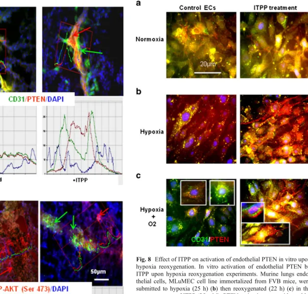
in vitro the direct effect of ITPP on PTEN activation in endothelial cells, by hypoxia/reoxygenation experiments conducted in the presence and/or absence of ITPP. Organo-specific murine lung endothelial cells showed a
reorganisation of PTEN in the presence of ITPP (Fig. 8).
PTEN first detected in the whole cytoplasm, colocalizing
mainly with CD31 (Fig.8a) migrated upon treatment with
ITPP, towards the plasma membrane more efficiently in
hypoxia (Figs.5 and 7b) than in normoxia (Fig.8a). This
effect of ITPP was clearly enhanced in experiments
involving hypoxia/reoxygenation (Fig. 8c) performed to
mimic the in vivo sequence of events that occur during angiogenesis as shown by the preferential relocation in
elongated endothelial cells (Fig.8cinsets).
ITPP-induced tumor vessels normalization prevents resistant cancer stem-like cells formation
In the ITPP-treated animals, reduction of p-glycoprotein
expression among cells in the tumor (Fig.9a) suggests that
Fig. 6 Phenotypic effect of ITPP treatment on tumor metabolism. a RT-PCR analysis revealing: downregulation of hypoxia/oxygen sensing genes and prometastasis genes; upre-gulation of genes implicated in endothelial cells maturation. Results are percent of non trea-ted samples level (n=8 animals, five separate experiments; **p =0.001; ***p=0.0001). b Re-duction of the number of CXCR4+CD34+CD45−
pre-cursor cells among tumor cells upon ITPP treatment. Quantifi-cation by flow cytometry from separate tumor samples (n=10/ group p<0.001). c Flow cytometry quantitative analysis showing a dramatic decrease in the number of cells expressing hypoxia-, stress-, and metastasis- related markers in primary tumors (day22) after ITPP treatments as described in “Materials and methods” (n=8/
group; 5 separate experiments, **p=0.001; ***p=0.0001). d Flow cytometry quantification analysis showing a dramatic decrease in the number of cells expressing markers of high en-ergetic metabolism in primary tumors (day22) after ITPP treatments as described in “Materials and methods” (n=8/
group; five separate experi-ments; ***p=0.0001)
hypoxia-induced loss of sensitivity to drugs, due to multi-drug efflux pumps (MDRs), could be reversed by tumor reoxygenation. This is confirmed by the reduction upon ITPP treatment of the number of cells positive for
ABCG-2 [35], which is a drug exclusion pump typical for stem
cells, as well as other stemness markers, i.e., CD133 and Oct 3–4 that were detected in highly positive tumor cell
sub-populations before treatment (Fig.9b).
ITPP-induced tumor vessels normalization favors chemotherapy
As ITPP treatment improves O2delivery to hypoxic tissues
and normalizes vessels, we studied its effect on melanoma treatment by drugs such as paclitaxel and cisplatin. Com-bined ITPP and drug treatments acted positively and led to
Fig. 7 Effect of ITPP treatment on activation of endothelial PTEN and loss of tumor AKT phosphorylation. a PTEN, P-AKT (Ser473) and CD31 immunostainings. PTEN was expressed (red arrows) and colo-calized with CD31+ endothelial cells (green arrows and green/red channels analysis of the label distribution, by image analysis) in non-treated tumor-bearing animals (left panel, n=10/group). Markers sep-arately localized after ITPP treatment (right panel, n=10/group). The red/green channels display separate distribution by image analysis. b P-AKT distribution over the tumor (red arrows and red curve of the image analysis) observed in tumor stroma and endothelial cells, colo-calized with CD31 staining (green arrows and green curve) in non-treated tumors. Expression of P-AKT was strongly reduced to punctual sites upon ITPP treatment. Image analysis point the separate localiza-tion with CD31 (right four panels, n=10/group)
Fig. 8 Effect of ITPP on activation of endothelial PTEN in vitro upon hypoxia reoxygenation. In vitro activation of endothelial PTEN by ITPP upon hypoxia reoxygenation experiments. Murine lungs endo-thelial cells, MLuMEC cell line immortalized from FVB mice, were submitted to hypoxia (25 h) (b) then reoxygenated (22 h) (c) in the presence or not of ITPP (25 mM). PTEN activation was evidenced by its relocalization from the cytoplasm in hypoxia towards the inner face of the plasma membrane upon ITPP treatment. ITPP induced localiza-tion effect of PTEN was as enhanced by reoxygenalocaliza-tion
eradication of metastatic tumor cells from lungs as shown
for day22 in Fig.9c. The CD31+microvessels density was
reduced when animals were treated by ITPP/drugs as com-pared to numerous and poorly structured microvessels,
CD31+ endothelial cells in controls (Fig. 8d). pO2 and
vessel normalization preceding drug treatment favored drugs c ytox icity, a s indica te d b y n ec ro tic ar ea s corresponding to diffuse CD31 positivity and delineated
by H&E staining (Fig.9d). These data stress the potential
of ITPP in combined therapies.
Discussion
When pO2in tumor microenvironment is brought to normal
levels, tumor cells do not invade surrounding tissues and do not metastasize. This work shows that this effect is due to normalization of tumor angiogenesis into matured vessels
resulting from selective compensation of hypoxia and con-trol of PTEN/AKT pathway through endothelial cell mem-brane PTEN activation by ITPP treatment.
Intratumor neo-vessel strengthening may explain the re-duction of metastatic cells escape from primary tumors. ITPP treatment indeed resulted in vessels normalization, through endothelial cells acquisition of a matured phenotype and reorganization of the vessel tumor microenvironment. Here, vessels strengthening, shown by pericyte alignment and maturation was confirmed by the induction of VEGF
receptors in response to the pericyte/EC cross-talk [37].
Moreover, Tie-2, a specific endothelial tyrosine kinase re-ceptor reduced in hypoxia and essential for normal blood vessel maturation by attracting pericytes upon binding of angiopoietin-1, was increased, corroborating the ITPP effect on vessel maturation. This was confirmed by the reduced invasive index reporting that the tumor angiogenic activity
[30] through CD31-positive endothelial cell increases, while
Fig. 9 Effect of ITPP treatment on tumor hypoxia-induced resistance, stem cell selection, and enhancement of chemotherapeutic efficacy. a The P-glycoprotein immunostaining showing a reduced number of multidrug resistance positive tumor cells after ITPP treatment. Frozen sections of primary tumors from experiments described in Fig.6were histochemically labeled (day 22, ITPP treatments as described in “Materials and methods” (n=8/group; five separate experiments).
Scale bars=50μm. b Quantification by flow cytometry showing the reduction of cells positive for precursor and stem cell-associated markers (CD133, Oct3-4, ABCG-2) after ITPP treatment. CD133+ immunostaining corroborated the reduction visible on frozen section staining of primary tumors as in a. Scale bars = 50 μm. c Lung
metastasis is suppressed by chemotherapeutic drugs (Paclitaxel and Cisplatin), when treatment is preceded by ITPP injection. Tumor cells are detected by their Lucifease activity in the lungs of animals from control, ITPP, CisPt plus Paclitaxel and combined treatments ITPP+
drugs as described in“Materials and methods.” Data are reported for day22 (n=10/group; 5 experiments; ***; p=0.001). d CD31 staining of endothelial cells (green) and eosin/hematoxylin staining obtained in primary tumor frozen sections from experiment described in c. Effi-cient tissue necrosis was obtained when chemotherapeutic treatment is preceded by ITPP injection as described in“Materials and methods.” Scale bars=50μm
CD31, ensuring endothelial cell junction and vessel efficacy,
is reduced by hypoxia [38]. ITPP-treatment effect on the
number and function of CD31+ cells confirms vessel
nor-malization and differs deeply from CD31+ cell reduction,
which results from antiangiogenic treatments.
The O2-dependent HIFs pathway is regulated by ITPP
treatment. PHD/HIF regulatory axis is described as a prom-ising therapeutic target to disable tumor capacity to adjust to hypoxia and control cell survival. Inhibition of residual PHDs shown here would avoid feedback protection of HIF
and reduce tumor resistance to hypoxia [39].
Moreover, similarly to HIF-1α mRNA [40], other O2
-dependent mRNAs like VHL mRNA, regulated by the O2
-sensitive angiomiR 92-1 [41] and the tumor protective and
proangiogenic enzyme HO-1 [42], were considerably
re-duced by ITPP treatment. Our data on PHDs and VHL mRNA reduction in long-term-treated tumors might reflect the whole tumor stroma response to stable reoxygenation and to reduced level of HIFs mRNAs, downregulating their
regulatory proteins mRNA as described [43].
Vessel strengthening control of tumor cell escape is accompanied by a remarkable reduction of mRNA for osteopontin. Disappearance of such key molecule of
tumor stroma helps explain the reduction of tumor cell dissemination. Osteopontin is indeed decisive for tumor
invasion [44] and known to be regulated by the
PTE-N/AKT pathway in melanoma [45]. This strong
modifi-cation of the tumor stroma reaction upon ITPP treatment was linked to the PTEN activation at the endothelial cell level, thus independent of the PTEN status of the tumor cells.
Contributing to control cell dissemination, activation of PTEN is favored here by downregulation of LOX
expres-sion. Indeed, local production of H2O2 by LOX would
inactivate the tumor suppressor—PTEN—explaining the positive regulation loop between LOX and HIF in cancer
development [32]. As PTEN activation was shown at the
endothelial cell level, it implies that ITPP treatment has for main target the vascular cell biology. It restores the PTEN-mediated control on tumor angiogenesis due to activated
AKT through the PDK/PI3K/AKT/mTOR pathway [22].
Effect on endothelial cells was indeed associated with a strong general decrease in AKT phosphorylation in the tumor mass confirming PTEN control of normal vs patho-logical angiogenesis and the efficient reversal of tumor
hypoxia [46].
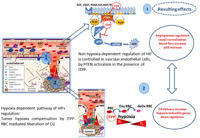
Fig. 10 Schematic outline of the proposed action of ITPP on HIFs regulation. 1 ITPP regulates angiogenesis by activating PTEN that inhibits PI3K action, AkT phosphorylation and mTOR actitivy towards HIF at the endothelial cell level. This regulates the vessels and increases the blood flow, while 2 hypoxia mediated O2 delivery by
ITPP, allosteric effector of hemoglobin [15–18], increases the intra tumor pO2, which also acts to destabilize HIF1α and down regulates
VEGFs and VEGFs-related gene cascade. This directly reduces the endothelial cell mobilization, activation and growth, thus regulating the tumor VEGF-mediated pathological angiogenesis
ITPP treatment is clearly targeting the vasculature. It is thus applicable to angiogenesis-dependent pathologies inde-pendently of the PTEN tumor suppressor mutations that occur in the majority of tumors.
The biological significance of the above described effect on PTEN activation is further illustrated by its effect on the recruitment of endothelial precursors, the second main
mechanism by which tumors build angiogenesis [47].
Among the bone marrow derived cells that are recruited by the tumor to help its progression, endothelial precursors are mobilized to integrate tumor forming neovessels mainly by the SDF1/CXCR4 axis. This work shows the drastic
reduc-tion of the proporreduc-tion of CXCR4+, CD34+, CD45−cells, and
endothelial precursors recruited in response to tumor SDF-1
chemoattraction, which is PTEN/AKT dependent [48].
Consequently, ITPP treatment contributes to restore ves-sel wall integrity and efficient blood supply by counteract-ing both hypoxia-dependent and hypoxia-independent HIF
induction as summarized on Fig.10. ITPP action appears to
contribute in control of both mechanisms. The part played
by PTEN vs O2delivery could not be directly shown using
endothelial cell-specific mutation of Pten (Tie2CrePten) in
mice. Tie2CrePtenflox/+ mice only being viable [22]. Such
normalization is known to result in improved chemothera-peutics delivery by efficient and, as shown here, nonperme-able mature vessels. It also reverted stem-like resistant invasive phenotype of tumor cells, prevented activation of glycolysis pathway shown by the numbers of cells express-ing Glut-1 receptors, LDH and CAIX, which were drasti-cally reduced upon ITPP treatment. Indicating an efficient reversal of hypoxia-induced resistance towards drug and cancer stem-like cells selection ITPP treatment contributed
to the efficiency of chemotherapy [33].
Indeed, ITPP-induced vessel normalization was accom-panied by the reduction of drug efflux pumps thus
counter-acting chemo-resistance built by MDRs [34]. It also reduced
drastically the number of cancer stem cells as opposed to their selection operated by the strong intratumor hypoxia, which results from antiangiogenic therapies using monoclo-nal antibodies—bevacuzimab (Avastin) against VEGF-A or
VEGFR2 inhibitors (Sunitinib) as documented [4,49]. Our
data help to explain why antiangiogenic cancer therapies provide poor results and why drug-induced improvement of vascular health correlates with better cancer prognosis. This work shows the strength of such an approach allowing stable vessel normalization. This important effect of ITPP should overcome the problem of adequate therapeutic win-dows for future therapies.
Our data stress the potential of ITPP in combined thera-pies. ITPP should provide the adjuvant needed to
chemo-and radiotherapy efficacy providing enhanced O2 supply
and vessel normalization [5,12], an alternative to
antiangio-genic strategies [14].
Collectively, these findings highlight the multifactor and potent therapeutic use of ITPP and demonstrate its funda-mental interest for advancing therapy of hypoxia and angiogenesis-dependent pathologies.
Acknowledgments Thanks are due to Dr. Michael Morin (OncoVax, Boston, MA, USA) and to Dr Agata Matejuk (Le_Studium, senior research fellow) for reading the manuscript. The authors thank David Gosset, Michèle Mitterrand, and Frédéric Szeremeta, for their skillful technical help in flow cytometry, biological and MRI experiments, respectively; Emilie Cardamone is thanked for technical help for in vivo imaging acquisition.
Grant support/disclosure This work was partly supported by the grant no. 347/N-INCA/2008/0 from the Polish Ministry of Science and Higher Education and CNRS and by the French National Research Agency (ANR 3Sens). AJ is a recipient of the Wellcome Trust Senior Research Fellowship in Basic Biomedical Science. This work was financed by Normoxys Inc. under contract, within the research agree-ment between the Company and the University of Strasbourg. Claude Nicolau is shareholder in NormOxys Inc.
Open Access This article is distributed under the terms of the Creative Commons Attribution License which permits any use, distribution, and reproduction in any medium, provided the original author(s) and the source are credited.
References
1. Semenza GL (2004) Intratumoral hypoxia, radiation resistance, and HIF-1. Cancer Cell 5:405–406
2. Folkman J (2007) Angiogenesis: an organizing principle for drug discovery? Nat Rev Drug Discov 6:273–286
3. Carmeliet P (2003) Angiogenesis in health and disease. Nat Med 9:653–660
4. Paez-Ribes M, Allen E, Hudock J, Takeda T, Okuyama H, Vinals F, Inoue M, Bergers G, Hanahan D, Casanovas O (2009) Anti-angiogenic therapy elicits malignant progression of tumors to increased local invasion and distant metastasis. Cancer Cell 15:220–231
5. Jain RK (2005) Normalization of tumor vasculature: an emerging concept in antiangiogenic therapy. Science 307:58–62
6. Goel S, Duda DG, Xu L, Munn LL, Boucher Y, Fukumura D, Jain RK (2011) Normalization of the vasculature for treatment of can-cer and other diseases. Physiol Rev 91:1071–1121
7. Carmeliet P, Jain RK (2011) Principles and mechanisms of vessel normalization for cancer and other angiogenic diseases. Nat Rev Drug Discov 10:417–427
8. Kaelin WG Jr, Ratcliffe PJ (2008) Oxygen sensing by meta-zoans: the central role of the HIF hydroxylase pathway. Mol Cell 30:393–402
9. Koh YJ, Kim HZ, Hwang SI, Lee JE, Oh N, Jung K, Kim M, Kim KE, Kim H, Lim NK et al (2010) Double antiangiogenic protein, DAAP, targeting VEGF-A and angiopoietins in tumor angiogene-sis, metastaangiogene-sis, and vascular leakage. Cancer Cell 18:171–184 10. Yancopoulos GD, Davis S, Gale NW, Rudge JS, Wiegand SJ,
Holash J (2000) Vascular-specific growth factors and blood vessel formation. Nature 407:242–248
11. Mazzone M, Dettori D, Leite de Oliveira R, Loges S, Schmidt T, Jonckx B, Tian YM, Lanahan AA, Pollard P, Ruiz de Almodovar C et al (2009) Heterozygous deficiency of PHD2 restores tumor
oxygenation and inhibits metastasis via endothelial normalization. Cell 136:839–851
12. Semenza GL (2012) Hypoxia-inducible factors: mediators of can-cer progression and targets for cancan-cer therapy. Trends Pharmacol Sci 33:207–214
13. Carmeliet P, Jain RK (2011) Molecular mechanisms and clinical applications of angiogenesis. Nature 473:298–307
14. Sato Y (2011) Persistent vascular normalization as an alternative goal of anti-angiogenic cancer therapy. Cancer Sci 102:1253–1256 15. Kieda C, Greferath R, Crola da Silva C, Fylaktakidou KC, Lehn JM, Nicolau C (2006) Suppression of hypoxia-induced HIF-1alpha and of angiogenesis in endothelial cells by myo-inositol trispyrophosphate-treated erythrocytes. Proc Natl Acad Sci U S A 103:15576–15581
16. Fylaktakidou KC, Lehn JM, Greferath R, Nicolau C (2005) Inosi-tol tripyrophosphate: a new membrane permeant allosteric effector of haemoglobin. Bioorg Med Chem Lett 15:1605–1608
17. Sihn G, Walter T, Klein JC, Queguiner I, Iwao H, Nicolau C, Lehn JM, Corvol P, Gasc JM (2007) Anti-angiogenic properties of myo-inositol trispyrophosphate in ovo and growth reduction of implanted glioma. FEBS Lett 581:962–966
18. Aprahamian M, Bour G, Akladios CY, Fylaktakidou K, Greferath R, Soler L, Marescaux J, Egly JM, Lehn JM, Nicolau C (2011) Myo-inositoltrispyrophosphate treatment leads to HIF-1alpha sup-pression and eradication of early hepatoma tumors in rats. Chem-BioChem 12:777–783
19. Chen L, Huang TG, Meseck M, Mandeli J, Fallon J, Woo SL (2007) Rejection of metastatic 4 T1 breast cancer by attenuation of Treg cells in combination with immune stimulation. Mol Ther 15:2194–2202
20. Qayum N, Muschel RJ, Im JH, Balathasan L, Koch CJ, Patel S, McKenna WG, Bernhard EJ (2009) Tumor vascular changes mediated by inhibition of oncogenic signaling. Can-cer Res 69:6347–6354
21. Harada H, Itasaka S, Kizaka-Kondoh S, Shibuya K, Morinibu A, Shinomiya K, Hiraoka M (2009) The Akt/mTOR pathway assures the synthesis of HIF-1alpha protein in a glucose- and reoxygenation-dependent manner in irradiated tumors. J Biol Chem 284:5332–5342
22. Hamada K, Sasaki T, Koni PA, Natsui M, Kishimoto H, Sasaki J, Yajima N, Horie Y, Hasegawa G, Naito M et al (2005) The PTEN/ PI3K pathway governs normal vascular development and tumor angiogenesis. Genes Dev 19:2054–2065
23. Du R, Lu KV, Petritsch C, Liu P, Ganss R, Passegue E, Song H, Vandenberg S, Johnson RS, Werb Z et al (2008) HIF1alpha induces the recruitment of bone marrow-derived vascular modulatory cells to regulate tumor angiogenesis and invasion. Cancer Cell 13:206–220 24. Bizouarne N, Denis V, Legrand A, Monsigny M, Kieda C (1993) A
SV-40 immortalized murine endothelial cell line from peripheral lymph node high endothelium expresses a new alpha-L-fucose binding protein. Biol Cell 79:209–218
25. Denis V, Dupuis P, Bizouarne N, de O Sampaio S, Hong L, Lebret M, Monsigny M, Nakache M, Kieda C (1996) Selective induction of peripheral and mucosal endothelial cell addressins with periph-eral lymph nodes and Peyer’s patch cell-conditioned media. J Leukoc Biol 60:744–752
26. Fauconnier M, Bourigault ML, Meme S, Szeremeta F, Palomo J, Danneels A, Charron S, Fick L, Jacobs M, Beloeil JC et al (2011) Protein kinase C-theta is required for development of experimental cerebral malaria. Am J Pathol 178:212–221
27. Elas M, Ahn KH, Parasca A, Barth ED, Lee D, Haney C, Halpern HJ (2006) Electron paramagnetic resonance oxygen images corre-late spatially and quantitatively with oxylite oxygen measure-ments. Clin Cancer Res 12:4209–4217
28. Wang Y, Seidel J, Tsui BM, Vaquero JJ, Pomper MG (2006) Performance evaluation of the GE healthcare eXplore VISTA
dual-ring small-animal PET scanner. J Nucl Med 47:1891– 1900
29. Dolinay T, Wu W, Kaminski N, Ifedigbo E, Kaynar AM, Szilasi M, Watkins SC, Ryter SW, Hoetzel A, Choi AM (2008) Mitogen-activated protein kinases regulate susceptibility to ventilator-induced lung injury. PLoS One 3:e1601
30. Tachezy M, Reichelt U, Melenberg T, Gebauer F, Izbicki JR, Kaifi JT (2010) Angiogenesis index CD105 (Endoglin)/CD31 (PECAM-1) as a predictive factor for invasion and proliferation in intraductal papillary mucinous neoplasm (IPMN) of the pancre-as. Histol Histopathol 25:1239–1246
31. Erler JT, Bennewith KL, Nicolau M, Dornhofer N, Kong C, Le QT, Chi JT, Jeffrey SS, Giaccia AJ (2006) Lysyl oxidase is essential for hypoxia-induced metastasis. Nature 440:1222–1226
32. Sonveaux P, Vegran F, Schroeder T, Wergin MC, Verrax J, Rabbani ZN, De Saedeleer CJ, Kennedy KM, Diepart C, Jordan BF et al (2008) Targeting lactate-fueled respiration selec-tively kills hypoxic tumor cells in mice. J Clin Invest 118:3930– 3942
33. Airley RE, Loncaster J, Raleigh JA, Harris AL, Davidson SE, Hunter RD, West CM, Stratford IJ (2003) GLUT-1 and CAIX as intrinsic markers of hypoxia in carcinoma of the cervix: relation-ship to pimonidazole binding. Int J Cancer 104:85–91
34. Song X, Liu X, Chi W, Liu Y, Wei L, Wang X, Yu J (2006) Hypoxia-induced resistance to cisplatin and doxorubicin in non-small cell lung cancer is inhibited by silencing of HIF-1alpha gene. Cancer Chemother Pharmacol 58:776–784
35. Monzani E, Facchetti F, Galmozzi E, Corsini E, Benetti A, Cavazzin C, Gritti A, Piccinini A, Porro D, Santinami M et al (2007) Melanoma contains CD133 and ABCG2 positive cells with enhanced tumourigenic potential. Eur J Cancer 43:935–946
36. Vazquez F, Matsuoka S, Sellers WR, Yanagida T, Ueda M, Devreotes PN (2006) Tumor suppressor PTEN acts through dy-namic interaction with the plasma membrane. Proc Natl Acad Sci U S A 103:3633–3638
37. Lorquet S, Berndt S, Blacher S, Gengoux E, Peulen O, Maquoi E, Noel A, Foidart JM, Munaut C, Pequeux C (2010) Soluble forms of VEGF receptor-1 and−2 promote vascular maturation via mural cell recruitment. FASEB J 24:3782–3795
38. Carreau A, Kieda C, Grillon C (2011) Nitric oxide modulates the expression of endothelial cell adhesion molecules involved in angiogenesis and leukocyte recruitment. Exp Cell Res 317:29–41
39. Henze AT, Riedel J, Diem T, Wenner J, Flamme I, Pouyseggur J, Plate KH, Acker T (2010) Prolyl hydroxylases 2 and 3 act in gliomas as protective negative feedback regulators of hypoxia-inducible factors. Cancer Res 70:357–366
40. Galban S, Gorospe M (2009) Factors interacting with HIF-1alpha mRNA: novel therapeutic targets. Curr Pharm Des 15:3853–3860
41. Ghosh AK, Shanafelt TD, Cimmino A, Taccioli C, Volinia S, Liu CG, Calin GA, Croce CM, Chan DA, Giaccia AJ et al (2009) Aberrant regulation of pVHL levels by microRNA promotes the HIF/VEGF axis in CLL B cells. Blood 113:5568–5574
42. Was H, Cichon T, Smolarczyk R, Rudnicka D, Stopa M, Chevalier C, Leger JJ, Lackowska B, Grochot A, Bojkowska K et al (2006) Overexpression of heme oxygenase-1 in murine melanoma: in-creased proliferation and viability of tumor cells, dein-creased sur-vival of mice. Am J Pathol 169:2181–2198
43. Turcotte S, Desrosiers RR, Beliveau R (2004) Hypoxia upregulates von Hippel-Lindau tumor-suppressor protein through RhoA-dependent activity in renal cell carcinoma. Am J Physiol Renal Physiol 286:F338–F348
44. Shevde LA, Das S, Clark DW, Samant RS (2010) Osteopontin: an effector and an effect of tumor metastasis. Curr Mol Med 10:71–81
45. Packer L, Pavey S, Parker A, Stark M, Johansson P, Clarke B, Pollock P, Ringner M, Hayward N (2006) Osteopontin is a down-stream effector of the PI3-kinase pathway in melanomas that is inversely correlated with functional PTEN. Carcinogenesis 27:1778–1786
46. Obara D, Utsugisawa K, Nagane Y, Tohgi H (2002) Hypoxic condi-tion interferes with phosphorylacondi-tion of Akt at Thr(308) in cultured rat pheochromocytoma-12 cells. Neurosci Lett 332:167–170
47. Chouaib S, Kieda C, Benlalam H, Noman MZ, Mami-Chouaib F, Ruegg C (2010) Endothelial cells as key determinants of the tumor
microenvironment: interaction with tumor cells, extracellular ma-trix and immune killer cells. Crit Rev Immunol 30:529–545 48. Zheng H, Fu G, Dai T, Huang H (2007) Migration of endothelial
progenitor cells mediated by stromal cell-derived factor-1alpha/ CXCR4 via PI3K/Akt/eNOS signal transduction pathway. J Car-diovasc Pharmacol 50:274–280
49. Ebos JM, Lee CR, Cruz-Munoz W, Bjarnason GA, Christensen JG, Kerbel RS (2009) Accelerated metastasis after short-term treatment with a potent inhibitor of tumor angiogenesis. Cancer Cell 15:232–239