HAL Id: inserm-01701177
https://www.hal.inserm.fr/inserm-01701177
Submitted on 5 Feb 2018
HAL is a multi-disciplinary open access
archive for the deposit and dissemination of
sci-entific research documents, whether they are
pub-lished or not. The documents may come from
teaching and research institutions in France or
abroad, or from public or private research centers.
L’archive ouverte pluridisciplinaire HAL, est
destinée au dépôt et à la diffusion de documents
scientifiques de niveau recherche, publiés ou non,
émanant des établissements d’enseignement et de
recherche français ou étrangers, des laboratoires
publics ou privés.
Progression
Audrey Lamora, Julie Talbot, Mathilde Mullard, Benedicte Brounais-Le
Royer, Françoise Redini, Franck Verrecchia
To cite this version:
Audrey Lamora, Julie Talbot, Mathilde Mullard, Benedicte Brounais-Le Royer, Françoise Redini, et
al.. TGF-β Signaling in Bone Remodeling and Osteosarcoma Progression. Journal of Clinical Medicine
Research, Elmer Press, 2016, 5 (11), �10.3390/jcm5110096�. �inserm-01701177�
Clinical Medicine
Review
TGF-β Signaling in Bone Remodeling and
Osteosarcoma Progression
Audrey Lamora1,2,3, Julie Talbot1,2, Mathilde Mullard1,2, Benedicte Brounais-Le Royer1,2, Françoise Redini1,2and Franck Verrecchia1,2,*
1 INSERM, UMR 957, Equipe Labellisée Ligue contre le Cancer 2012, Faculté de Médecine, 1 rue Gaston Veil, 44035 Nantes cedex, France; [email protected] (A.L.); [email protected] (J.T.);
[email protected] (M.M.); [email protected] (B.B.-L.R.); [email protected] (F.R.)
2 Laboratoire de Physiopathologie de la Résorption Osseuse et Thérapie des Tumeurs Osseuses Primitives, Université de Nantes, 44000 Nantes, France
3 INSERM Liliane Bettencourt School, 75014 Paris, France
* Correspondence: [email protected]; Tel.: +33-240-412-842; Fax: +33-240-412-860 Academic Editor: Andrei Turtoi
Received: 12 October 2016; Accepted: 28 October 2016; Published: 3 November 2016
Abstract: Osteosarcomas are the most prevalent malignant primary bone tumors in children. Despite intensive efforts to improve both chemotherapeutics and surgical management, 40% of all osteosarcoma patients succumb to the disease. Specifically, the clinical outcome for metastatic osteosarcoma remains poor; less than 30% of patients who present metastases will survive five years after initial diagnosis. Treating metastatic osteosarcoma thus remains a challenge. One of the main characteristics of osteosarcomas is their ability to deregulate bone remodelling. The invasion of bone tissue by tumor cells indeed affects the balance between bone resorption and bone formation. This deregulation induces the release of cytokines or growth factors initially trapped in the bone matrix, such as transforming growth factor-β (TGF-β), which in turn promote tumor progression. Over the past years, there has been considerable interest in the TGF-β pathway within the cancer research community. This review discusses the involvement of the TGF-β signalling pathway in osteosarcoma development and in their metastatic progression.
Keywords:bone remodeling; osteosarcoma; TGF-β; primary tumor growth; metastasis
1. Introduction
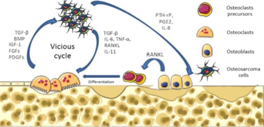
Osteosarcoma are the most common malignant primary bone tumors affecting children and young adults, with 2–3 cases per million per year [1–3]. Osteosarcomas arise from mesenchymal bone-forming cells, and mainly occur in long bone extremities, such as the distal femur, the proximal tibia, or the humerus [4]. Molecular mechanisms underlying osteosarcoma formation are characterized by complex karyotype and multiple genomic alterations [5,6]. Osteosarcomas are pathologies that affect bone remodeling, involving alterations in both osteoblast and osteoclast functions. They are characterized by the direct formation of osteoid matrix by tumor cells, associated with severe osteolytic lesions. To explain these dysregulations of bone cell functions, a vicious cycle between tumor and bone cells has been described during osteosarcoma development (Figure1). In brief, cancer cells produce soluble factors, such as cytokines (IL-6, IL-11, TNF-α, RANKL, etc.) that activate osteoclastogenesis, leading to bone degradation. Following bone resorption, growth factors trapped in the bone matrix, such as IGF-1 or transforming growth factor-β (TGF-β), are released in the bone microenvironment and stimulate tumor growth [7].
The current treatments include the combination of surgical tumor resection with limb salving and systemic multidrug neoadjuvant and adjuvant chemotherapy [8,9]. Before the introduction of chemotherapy in the early 1980s, amputation was the only therapeutic approach, and survival rates were around 20% at five years. Since then, overall survival had evolved with a five-year survival of about of 70%–75% for localized forms, but still very poor for patients with metastasis at diagnosis [10] or resistant to chemotherapy (approximately 20% at 5 years). New molecular approaches attempt to better understand this disease in order to identify new markers and new therapeutic targets. Among developing treatments, various strategies have been developed, such as targeting of the tumor microenvironment, induction of apoptosis, or inhibition of different signaling pathways [11]. Despite advances in diagnosis and treatments of osteosarcoma, no substantial improvement in survival rate has been achieved over the past few decades, and the mortality rate remains high for high-risk patients [12]. In this context, developing a better understanding of osteosarcoma biology with the aim of identifying new therapeutic targets is a major challenge in order to improve the outcome in osteosarcoma patients with poor prognosis.
J. Clin. Med. 2016, 5, 96 2 of 11 The current treatments include the combination of surgical tumor resection with limb salving and systemic multidrug neoadjuvant and adjuvant chemotherapy [8,9]. Before the introduction of chemotherapy in the early 1980s, amputation was the only therapeutic approach, and survival rates were around 20% at five years. Since then, overall survival had evolved with a five‐year survival of about of 70%–75% for localized forms, but still very poor for patients with metastasis at diagnosis [10] or resistant to chemotherapy (approximately 20% at 5 years). New molecular approaches attempt to better understand this disease in order to identify new markers and new therapeutic targets. Among developing treatments, various strategies have been developed, such as targeting of the tumor microenvironment, induction of apoptosis, or inhibition of different signaling pathways [11]. Despite advances in diagnosis and treatments of osteosarcoma, no substantial improvement in survival rate has been achieved over the past few decades, and the mortality rate remains high for high‐risk patients [12]. In this context, developing a better understanding of osteosarcoma biology with the aim of identifying new therapeutic targets is a major challenge in order to improve the outcome in osteosarcoma patients with poor prognosis.
Figure 1. Vicious cycle between primary tumor cell and bone cells. Cancer cells produce soluble
factors that activate the osteoclast differentiation and maturation directly or indirectly via osteoblasts. In turn, during bone degradation, osteoclasts allow the release of growth factors stored in the mineralized bone matrix that are able to stimulate tumor growth. TGF‐β: transforming growth factor‐β.
2. TGF‐β Signaling Pathways
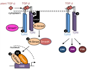
The transforming growth factor‐β (TGF‐β) family of secreted cytokines comprises at least 30 members in humans [13]. Three isoforms—TGF‐β1, ‐β2 and ‐β3—have been identified in mammals. TGF‐βs are secreted as latent precursor molecules requiring activation into a mature form for receptor binding [14]. Once activated, TGF‐βs signal from the membrane to the nucleus by binding to two heteromeric cell surface receptors, named type I (TβRI) and type II (TβRII) receptors. Ligand binding induces the assembly of TβRI and TβRII into complexes, within which TβRII phosphorylates and activates TβRI. This phosphorylation event is associated with the activation of TβRI kinase and subsequent downstream signaling [15–19].
TGF‐βs thus activate the Smads cascade, known as the canonical TGF‐β signaling pathway. Briefly, receptor‐regulated Smads (R‐Smads), including Smad1, ‐2, ‐3, ‐5, and ‐8, are phosphorylated and activated by TβRI. Then, R‐Smads recruit the common‐mediator Smad (co‐Smad), Smad4. This protein complex is translocated into the nucleus and regulates target gene expression (Figure 2). At the regulatory DNA binding sequence of genes, the R‐Smad/co‐Smad complex activates transcription through physical interaction and functional cooperation of DNA‐binding Smads with sequence‐specific transcription factors [19,20]. The minimal Smad‐binding element (SBE) contains four base pairs (5′‐AGAC‐3′), but binding to other G/C‐rich sequences has also been reported [21,22]. TGF‐β signalling may be controlled by several inhibitory mechanisms. Among them,
Figure 1.Vicious cycle between primary tumor cell and bone cells. Cancer cells produce soluble factors that activate the osteoclast differentiation and maturation directly or indirectly via osteoblasts. In turn, during bone degradation, osteoclasts allow the release of growth factors stored in the mineralized bone matrix that are able to stimulate tumor growth. TGF-β: transforming growth factor-β.
2. TGF-β Signaling Pathways
The transforming growth factor-β (TGF-β) family of secreted cytokines comprises at least 30 members in humans [13]. Three isoforms—TGF-β1, -β2 and -β3—have been identified in mammals. TGF-βs are secreted as latent precursor molecules requiring activation into a mature form for receptor binding [14]. Once activated, TGF-βs signal from the membrane to the nucleus by binding to two heteromeric cell surface receptors, named type I (TβRI) and type II (TβRII) receptors. Ligand binding induces the assembly of TβRI and TβRII into complexes, within which TβRII phosphorylates and activates TβRI. This phosphorylation event is associated with the activation of TβRI kinase and subsequent downstream signaling [15–19].
TGF-βs thus activate the Smads cascade, known as the canonical TGF-β signaling pathway. Briefly, receptor-regulated Smads (R-Smads), including Smad1, -2, -3, -5, and -8, are phosphorylated and activated by TβRI. Then, R-Smads recruit the common-mediator Smad (co-Smad), Smad4. This protein complex is translocated into the nucleus and regulates target gene expression (Figure2). At the regulatory DNA binding sequence of genes, the R-Smad/co-Smad complex activates transcription through physical interaction and functional cooperation of DNA-binding Smads with sequence-specific transcription factors [19,20]. The minimal Smad-binding element (SBE) contains four base pairs (50-AGAC-30), but binding to other G/C-rich sequences has also been reported [21,22]. TGF-β signalling may be controlled by several inhibitory mechanisms. Among them,
Smad7—induced by TGF-β—competes with R-Smads for binding to activated TβRI, and thus inhibits R-Smads phosphorylation and/or recruits E3-ubiquitin ligases to activated TβRI, resulting in receptor degradation [17,23]. Additionally, Smad7 may recruit protein phosphatases to the receptor complex, resulting in its dephosphorylation [24], and thus in its inactivation.
In addition to this canonical pathway, TGF-βs are also able to activate Smad-independent or non-canonical pathways such as PI3K/AKT, ERK1/2, JNK, and p38 cascades (Figure2) [25,26].
J. Clin. Med. 2016, 5, 96 3 of 11
Smad7—induced by TGF‐β—competes with R‐Smads for binding to activated TβRI, and thus inhibits R‐Smads phosphorylation and/or recruits E3‐ubiquitin ligases to activated TβRI, resulting in receptor degradation [17,23]. Additionally, Smad7 may recruit protein phosphatases to the receptor complex, resulting in its dephosphorylation [24], and thus in its inactivation.
In addition to this canonical pathway, TGF‐βs are also able to activate Smad‐independent or non‐canonical pathways such as PI3K/AKT, ERK1/2, JNK, and p38 cascades (Figure 2) [25,26].
Figure 2. TGF‐β signaling pathways. Schematic representation of the canonical and non‐canonical
TGF‐β signaling pathways. R‐Smad: receptor‐regulated Smad; SBE: Smad‐binding element; TF: transcription factor.
3. TGF‐β and Bone Remodeling
Bone remodeling mainly depends on the differentiation and activity of two cell lineages: the mesenchymal osteoblastic lineage, and the hematopoietic osteoclastic lineage. At the molecular level, the differentiation and activation processes of these cell lineages are tightly regulated by various cytokines and growth factors, including TGF‐βs.
Although TGF‐β1 is the most abundant in bone [27,28], the three mammalian isoforms (TGF‐β1, ‐β2, and ‐β3) are found in bone, particularly expressed by the perichondrium, the periosteum, and the epiphyseal growth plate [29]. Latent precursor molecules of TGF‐β1 are in part synthetized by osteoblasts, deposited in the bone matrix, and activated by acids and matrix metalloproteinases secreted from osteoclasts [30].
The role of TGF‐β1 in skeleton development, and specifically during bone remodeling is complex. Regarding the mesenchymal osteoblastic lineage, TGF‐β1 favors bone formation by stimulating the proliferation and migration of mesenchymal stem cells during the early stages of osteoblastogenesis [30,31]. In contrast, during the late stages of osteoblastogenesis, TGF‐β1 inhibits the differentiation of mesenchymal stem cells into osteoblasts and the mineralization of mature osteoblasts in culture [32,33]. Regarding the hematopoietic osteoclastic lineage, TGF‐β1 affects bone resorption in a dose‐dependent manner [29]. Low concentrations of TGF‐β1 stimulate the migration of osteoclast precursors to the bone resorption site, and their differentiation into mature osteoclasts. In contrast, at high doses, TGF‐β1 inhibits the migration of osteoclast precursors and their differentiation through the modulation of RANKL and OPG expression by osteoblasts [34,35]. In vivo experiments indicate that TGF‐βs favor bone resorption and destruction [36–38].
4. TGF‐β and Cancer
TGF‐βs are able to regulate tumor initiation, progression, and metastatic development. It is widely accepted that TGF‐βs act as both tumor suppressors and tumor promoters, depending on the cancer type and tumor development timing [39–44].
Figure 2.TGF-β signaling pathways. Schematic representation of the canonical and non-canonical TGF-β signaling pathways. R-Smad: receptor-regulated Smad; SBE: Smad-binding element; TF: transcription factor.
3. TGF-β and Bone Remodeling
Bone remodeling mainly depends on the differentiation and activity of two cell lineages: the mesenchymal osteoblastic lineage, and the hematopoietic osteoclastic lineage. At the molecular level, the differentiation and activation processes of these cell lineages are tightly regulated by various cytokines and growth factors, including TGF-βs.
Although TGF-β1 is the most abundant in bone [27,28], the three mammalian isoforms (TGF-β1, -β2, and -β3) are found in bone, particularly expressed by the perichondrium, the periosteum, and the epiphyseal growth plate [29]. Latent precursor molecules of TGF-β1 are in part synthetized by osteoblasts, deposited in the bone matrix, and activated by acids and matrix metalloproteinases secreted from osteoclasts [30].
The role of TGF-β1 in skeleton development, and specifically during bone remodeling is complex. Regarding the mesenchymal osteoblastic lineage, TGF-β1 favors bone formation by stimulating the proliferation and migration of mesenchymal stem cells during the early stages of osteoblastogenesis [30,31]. In contrast, during the late stages of osteoblastogenesis, TGF-β1 inhibits the differentiation of mesenchymal stem cells into osteoblasts and the mineralization of mature osteoblasts in culture [32,33]. Regarding the hematopoietic osteoclastic lineage, TGF-β1 affects bone resorption in a dose-dependent manner [29]. Low concentrations of TGF-β1 stimulate the migration of osteoclast precursors to the bone resorption site, and their differentiation into mature osteoclasts. In contrast, at high doses, TGF-β1 inhibits the migration of osteoclast precursors and their differentiation through the modulation of RANKL and OPG expression by osteoblasts [34,35]. In vivo experiments indicate that TGF-βs favor bone resorption and destruction [36–38].
4. TGF-β and Cancer
TGF-βs are able to regulate tumor initiation, progression, and metastatic development. It is widely accepted that TGF-βs act as both tumor suppressors and tumor promoters, depending on the cancer type and tumor development timing [39–44].
During the early stage of tumor development, TGF-β1 acts as a tumor suppressor mainly by its ability to inhibit the proliferation of epithelial cells. TGF-β1 can cause G1 cell cycle arrest by inducing the expression of CDK inhibitors such as p21Cip1and p15lnk4b, and/or reducing the expression of proliferative drivers such as c-Myc and ID [45–47]. Other tumor-suppressing properties have been correlated to the ability of TGF-βs to induce the apoptosis [48] or senescence of cancer cells [49]. In this context, alterations of the TGF-β cascade have been associated with many cancers [40,50,51]. For example, Smad4 gene mutations have been identified in most pancreatic [52] and colorectal [53] cancers, and in a lesser proportion in other cancers, such as hepatocellular, ovarian, intestinal, and lung carcinomas [40]. Mutations of TβRI and TβRII have also been identified in many cancers; TBRII has been associated with colon, gastric, pancreatic, lung, and brain tumors, for example [50,54–56], and mutations of the TβRI gene have been identified in ovarian tumors [51].
During the last decades, studies of TGF-β expression in epithelial cancers have correlated the levels of TGF-β with the metastatic potential of many tumors, such as breast, colon, and prostate [57–59], suggesting a role of TGF-β in tumor progression. It is now well accepted that TGF-βs act as tumor promoters during the late stages of carcinogenesis, by their ability to induce epithelial–mesenchymal transition (EMT), to stimulate angiogenesis, and to favor immune evasion.
EMT—characterized by the loss of E-cadherin, the expression of mesenchymal cytoskeleton proteins such as vimentin and fibronectin, and the expression of transcription factors such as Snail, Slug, Twist, and FoxC3—is induced by TGF-β in many cancer cells [41,60–63]. The loss of E-cadherin has been associated with Smad-dependent and Smad-independent signaling pathways [64–66]. Interestingly, several other signaling cascades, such as the Wnt, Hippo, and Sonic Hedgehog (SHH) cascades cooperate with the Smad cascade to regulate EMT in cancer cells [63]. During the EMT process, the epithelial cells trans-differentiate into mesenchymal cells able to migrate through the extracellular matrix and form metastases at distant secondary sites [67,68]. In this context, TGF-β1 is able to stimulate the expression and activity of MMP-2 and MMP-9, two matrix metalloproteinases implicated in the ability of cancer cells to invade surrounding tissue [69,70]. EMT seems to be a transient and reversible process during carcinogenesis, allowing the promotion of cancer cells’ intravasation into the blood or lymph systems; however, the phenotype of the tumor cells at the metastatic site seems to be mainly determined by the stromal site itself rather than the innate properties of the cancer cells [68].
Tumor-associated angiogenesis also plays a crucial role during tumor progression [71]. This process favors the formation of new blood vessels, allowing the supply of nutrients and providing an entry point for the metastatic cells [72]. As an example, high levels of TGF-β1 mRNA in breast cancers are associated with an increase in the density of blood vessels [73]. Other studies suggest that the level of TGF-β1 in the circulating plasma is associated with the induction of tumor angiogenesis [59,74–76]. It seems that TGF-β stimulates angiogenesis in part by stimulating the expression of vascular endothelial growth factor (VEGF) and connective tissue growth factor (CTGF) [40].
A third crucial step in cancer progression is the selective suppression of the immune system. TGF-βs produced by several immune cells, such as macrophages, dendritic cells, NK cells, B cells, and T cells play a crucial role in the suppression of the immune system, as demonstrated by the autoimmunity developed in TGF-β1 null mice [43,77].
5. TGF-β and Osteosarcoma
In contrast with the dual effects of TGF-βs on carcinoma progression, TGF-βs seem to mainly have a pro-tumoral effect on sarcoma specifically in osteosarcoma.
The expression of TGF-βs is increased in the sera of patients with osteosarcoma compared to the sera of healthy donors [38]. Interestingly, this increase of TGF-βs production is associated with the presence of metastases in lung or in other sites [78,79], and is correlated with high-grade osteosarcoma and a lack of osteosarcoma response to chemotherapy [80,81].
In vitro experiments have demonstrated the pro-migratory effect of TGF-β1 on several osteosarcoma cell lines [38,82–84], this effect being associated with the ability of TGF-βs to promote
an EMT-like phenomena [85]. TGF-β1 also exerts pro-angiogenic properties in osteosarcoma [86,87]. In addition, the anti-tumor effects of an anti-TGF-β antibody combined with dendritic cells has been associated with the restoration of the immune response in osteosarcoma [88].
More recently, using molecular (over-expression of the inhibitor Smad, Smad7) and pharmacological (SD-208 and/or halofuginone) approaches, we clearly demonstrated that TGF-βs affect osteosarcoma tumor growth and lung metastatic development [38,89]. Of note, SD-208 is a chemical inhibitor of TβRI, and halofuginone is an alkaloid known for its inhibitory properties on the TGF-β signaling pathway [90]. Using a xenograft murine model of osteosarcoma, we specifically demonstrated that Smad7 overexpression slows primary tumor growth. Interestingly, this effect seems to involve the bone tumor microenvironment rather than the tumor cells directly. Using micro-computed tomography analysis, we indeed demonstrated that Smad7 inhibits tumor-associated bone destruction by both promoting ectopic bone formation and preventing trabecular bone osteolysis. Our hypothesis is that blocking the TGF-β cascade in tumor cells inhibits the expression of TGF-β target genes involved in the establishment of the vicious cycle between tumor cells and bone cells. In this context, we demonstrated that Smad7 overexpression in osteosarcoma cells blocks their ability to produce RANKL, a cytokine that plays a central role in osteoclast differentiation and activation [91]. In addition, we demonstrated that blocking the TGF-β cascade in tumor cells inhibits the expression of TGF-β target genes, such as IL-11, CXCR4, and osteopontin, known to enhance bone metastasis formation from breast cancer cells or melanoma [92–95]. Of note, in contrast to Smad7, the effects of halofuginone appear to be mainly due to its pro-apoptotic properties in osteosarcoma, regardless of its ability to inhibit the TGF-β signaling pathway [89]. The role of the non-canonical TGF-β signaling pathway in osteosarcoma progression is poorly documented. Although we have not observed an effect of Smad7 on the ability of TGF-β to stimulate the MAPK pathway in osteosarcoma cells (suggesting a crucial role of the TGF-β/Smad cascade in osteosarcoma progression), the role of TGF-β/MAPK pathways cannot be ruled out.
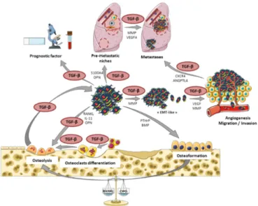
Finally, we showed that Smad7, SD-208, and halofuginone strongly affect the ability of the primary bone tumor to develop lung metastases, mainly by their ability to block the capacity of TGF-β1 to stimulate osteosarcoma migration and invasion, as previously described in the context of melanoma bone metastasis [93–95]. The roles of TGF-β in the progression of osteosarcoma and the development of lung metastases are summarized in Figure3.
J. Clin. Med. 2016, 5, 96 5 of 11
an EMT‐like phenomena [85]. TGF‐β1 also exerts pro‐angiogenic properties in osteosarcoma [86,87]. In addition, the anti‐tumor effects of an anti‐TGF‐β antibody combined with dendritic cells has been associated with the restoration of the immune response in osteosarcoma [88].
More recently, using molecular (over‐expression of the inhibitor Smad, Smad7) and pharmacological (SD‐208 and/or halofuginone) approaches, we clearly demonstrated that TGF‐βs affect osteosarcoma tumor growth and lung metastatic development [38,89]. Of note, SD‐208 is a chemical inhibitor of TβRI, and halofuginone is an alkaloid known for its inhibitory properties on the TGF‐β signaling pathway [90]. Using a xenograft murine model of osteosarcoma, we specifically demonstrated that Smad7 overexpression slows primary tumor growth. Interestingly, this effect seems to involve the bone tumor microenvironment rather than the tumor cells directly. Using micro‐computed tomography analysis, we indeed demonstrated that Smad7 inhibits tumor‐associated bone destruction by both promoting ectopic bone formation and preventing trabecular bone osteolysis. Our hypothesis is that blocking the TGF‐β cascade in tumor cells inhibits the expression of TGF‐β target genes involved in the establishment of the vicious cycle between tumor cells and bone cells. In this context, we demonstrated that Smad7 overexpression in osteosarcoma cells blocks their ability to produce RANKL, a cytokine that plays a central role in osteoclast differentiation and activation [91]. In addition, we demonstrated that blocking the TGF‐β cascade in tumor cells inhibits the expression of TGF‐β target genes, such as IL‐11, CXCR4, and osteopontin, known to enhance bone metastasis formation from breast cancer cells or melanoma [92–95]. Of note, in contrast to Smad7, the effects of halofuginone appear to be mainly due to its pro‐apoptotic properties in osteosarcoma, regardless of its ability to inhibit the TGF‐β signaling pathway [89]. The role of the non‐canonical TGF‐β signaling pathway in osteosarcoma progression is poorly documented. Although we have not observed an effect of Smad7 on the ability of TGF‐β to stimulate the MAPK pathway in osteosarcoma cells (suggesting a crucial role of the TGF‐β/Smad cascade in osteosarcoma progression), the role of TGF‐β/MAPK pathways cannot be ruled out.
Finally, we showed that Smad7, SD‐208, and halofuginone strongly affect the ability of the primary bone tumor to develop lung metastases, mainly by their ability to block the capacity of TGF‐β1 to stimulate osteosarcoma migration and invasion, as previously described in the context of melanoma bone metastasis [93–95]. The roles of TGF‐β in the progression of osteosarcoma and the development of lung metastases are summarized in Figure 3.
Figure 3. The central role of TGF‐β in osteosarcoma tumor and metastases development. Roles of
TGF‐β as a main player in the vicious cycle between osteosarcoma cells and the bone tumor microenvironment, thus contributing to tumor development and lung metastases dissemination. Figure 3. The central role of TGF-β in osteosarcoma tumor and metastases development. Roles of TGF-β as a main player in the vicious cycle between osteosarcoma cells and the bone tumor microenvironment, thus contributing to tumor development and lung metastases dissemination. EMT: epithelial–mesenchymal transition; MMP: matrix metalloproteinase; VEGF: vascular endothelial growth factor.
6. TGF-β Cascade Blockers in Cancer Clinical Trials
During the last decade, numerous strategies against TGF-β signaling have been used in preclinical or clinical applications, especially in end-stage cancer, including anti-ligand antisense oligonucleotides, antibodies that target ligands or receptors, and drugs against TGF-β receptor kinases (reviewed in [40,96]). Anti-TGF-β2 antisense strategies have thus been developed. For example, Trabedersen (AP12009)—a synthetic 18-mer phosphorothioate antisense oligonucleotide able to bind human TGF-β2 mRNA—has been successfully used in clinical trials for oncological applications [40,96], such as glioblastoma [97,98]. Strategies using monoclonal antibodies have also been developed. For example, GC-1008 (Fresolimumab, a humanized mAB against TGF-β) was developed and tested in Phase I/II clinical trials on patients with advanced malignant melanoma or renal carcinoma [99]. Many chemical compounds able to block the transduction of TGF-β signal, such as inhibitors of TβRI or TβRII (SB-431542, LY2109761, or LY2157299, etc.) have been developed in preclinical models. Among them, only one (LY2157299) was recently tested in clinical trial on patients with Grade IV glioma [100].
7. Conclusions and Perspective
In conclusion, blocking TGF-β signaling may represent a promising therapeutic approach to treat osteosarcoma patients. However, despite the existence of many tools allowing us to block TGF-β signalling pathways, such as neutralizing antibodies, soluble TGF-β receptors, or receptor kinase inhibitors, the lack of spectacular success in clinical trials reinforces the need to continue research on this TGF-β signaling pathway, specifically on the crosstalk between this pathway and others implicated in osteosarcoma tumor progression, such as Wnt, Hippo, or SHH cascades.
Acknowledgments: Research at INSERM U957 is supported by grants from INSERM, Ligue contre le Cancer (Equipe labellisée 2012), Fédération Enfants et Santé, Etoile de Martin and Société Française des Cancers de l’Enfant (SFCE).
Conflicts of Interest:The authors declare no conflict of interest. The founding sponsors had no role in the writing of the manuscript.
References
1. Ottaviani, G.; Jaffe, N. The epidemiology of osteosarcoma. Cancer Treat. Res. 2009, 152, 3–13. [PubMed] 2. Gatta, G.; Van der Zwan, J.M.; Casali, P.G.; Siesling, S.; Dei Tos, A.P.; Kunkler, I.; Otter, R.; Licitra, L.;
Mallone, S.; Tavilla, A.; et al. Rare cancers are not so rare: The rare cancer burden in Europe. Eur. J. Cancer
2011, 47, 2493–2511. [CrossRef] [PubMed]
3. Schwab, J.H.; Springfield, D.S.; Raskin, K.A.; Mankin, H.J.; Hornicek, F.J. What’s new in primary bone tumors. J. Bone Jt. Surg. 2012, 94, 1913–1919. [CrossRef] [PubMed]
4. Bielack, S.S.; Kempf-Bielack, B.; Delling, G.; Exner, G.U.; Flege, S.; Helmke, K.; Kotz, R.; Salzer-Kuntschik, M.; Werner, M.; Winkelmann, W.; et al. Prognostic factors in high-grade osteosarcoma of the extremities or trunk: An analysis of 1702 patients treated on neoadjuvant cooperative osteosarcoma study group protocols. J. Clin. Oncol. 2002, 20, 776–790. [CrossRef] [PubMed]
5. Chen, X.; Bahrami, A.; Pappo, A.; Easton, J.; Dalton, J.; Hedlund, E.; Ellison, D.; Shurtleff, S.; Wu, G.; Wei, L.; et al. Recurrent somatic structural variations contribute to tumorigenesis in pediatric osteosarcoma. Cell Rep. 2014, 7, 104–112. [CrossRef] [PubMed]
6. Shaikh, A.B.; Li, F.; Li, M.; He, B.; He, X.; Chen, G.; Guo, B.; Li, D.; Jiang, F.; Dang, L.; et al. Present Advances and Future Perspectives of Molecular Targeted Therapy for Osteosarcoma. Int. J. Mol. Sci. 2016, 17, 506. [CrossRef] [PubMed]
7. Wittrant, Y.; Théoleyre, S.; Chipoy, C.; Padrines, M.; Blanchard, F.; Heymann, D.; Rédini, F. RANKL/RANK/OPG: New therapeutic targets in bone tumours and associated osteolysis. Biochim. Biophys. Acta 2004, 1704, 49–57. [CrossRef] [PubMed]
8. European Society for Medical Oncology (ESMO)/European Sarcoma Network Working Group. Bone sarcomas: ESMO Clinical Practice Guidelines for diagnosis, treatment and follow-up. Ann. Oncol. 2014, 3, 113–123.
9. Ferrari, S.; Serra, M. An update on chemotherapy for osteosarcoma. Expert Opin. Pharmacother. 2015, 16, 2727–2736. [CrossRef] [PubMed]
10. Wu, P.K.; Chen, W.M.; Chen, C.F.; Lee, O.K.; Haung, C.K.; Chen, T.H. Primary osteogenic sarcoma with pulmonary metastasis: Clinical results and prognostic factors in 91 patients. Jpn. J. Clin. Oncol. 2009, 39, 514–522. [CrossRef] [PubMed]
11. Kansara, M.; Teng, M.W.; Smyth, M.J.; Thomas, D.M. Translational biology of osteosarcoma. Nat. Rev. Cancer
2014, 14, 722–735. [CrossRef] [PubMed]
12. Whelan, J.; McTiernan, A.; Cooper, N.; Wong, Y.K.; Francis, M.; Vernon, S.; Strauss, S.J. Incidence and survival of malignant bone sarcomas in England 1979–2007. Int. J. Cancer 2012, 131, 508–517. [CrossRef] [PubMed] 13. Feng, X.H.; Derynck, R. Specificity and versatility in TGF-β signaling through Smads. Annu. Rev. Cell
Dev. Biol. 2005, 21, 659–693. [CrossRef] [PubMed]
14. Harrison, C.A.; Al-Musawi, S.L.; Walton, K.L. Prodomains regulate the synthesis, extracellular localisation and activity of TGF-β superfamily ligands. Growth Factors 2011, 29, 174–186. [CrossRef] [PubMed]
15. Cárcamo, J.; Zentella, A.; Massagué, J. Disruption of transforming growth factor beta signaling by a mutation that prevents transphosphorylation within the receptor complex. Mol. Cell. Biol. 1995, 15, 1573–1581. [CrossRef] [PubMed]
16. Moustakas, A.; Heldin, C.H. The regulation of TGFbeta signal transduction. Development 2009, 136, 3699–3714. [CrossRef] [PubMed]
17. Massagué, J. TGFβ signalling in context. Nat. Rev. Mol. Cell Biol. 2012, 13, 616–630. [CrossRef] [PubMed] 18. Hata, A.; Chen, Y.G. TGF-β Signaling from Receptors to Smads. Cold Spring Harb. Perspect. Biol. 2016,
8, a022061. [CrossRef] [PubMed]
19. Heldin, C.H.; Moustakas, A. Signaling Receptors for TGF-β Family Members. Cold Spring Harb. Perspect. Biol.
2016, 8, a022053. [CrossRef] [PubMed]
20. Chang, C. Agonists and Antagonists of TGF-β Family Ligands. Cold Spring Harb. Perspect. Biol. 2016, 8, a021923. [CrossRef] [PubMed]
21. Dennler, S.; Itoh, S.; Vivien, D.; ten Dijke, P.; Huet, S.; Gauthier, J.M. Direct binding of Smad3 and Smad4 to critical TGF beta-inducible elements in the promoter of human plasminogen activator inhibitor-type 1 gene. EMBO J. 1998, 17, 3091–3100. [CrossRef] [PubMed]
22. Weiss, A.; Attisano, L. The TGFbeta superfamily signaling pathway. Wiley Interdiscip. Rev. Dev. Biol. 2013, 2, 47–63. [CrossRef] [PubMed]
23. Shi, Y.; Massagué, J. Mechanisms of TGF-beta signaling from cell membrane to the nucleus. Cell 2003, 113, 685–700. [CrossRef]
24. Shi, W.; Sun, C.; He, B.; Xiong, W.; Shi, X.; Yao, D.; Cao, X. GADD34-PP1c recruited by Smad7 dephosphorylates TGFbeta type I receptor. J. Cell Biol. 2004, 164, 291–300. [CrossRef] [PubMed]
25. Lutz, M.; Knaus, P. Integration of the TGF-beta pathway into the cellular signalling network. Cell Signal.
2002, 14, 977–988. [CrossRef]
26. Javelaud, D.; Mauviel, A. Crosstalk mechanisms between the mitogen-activated protein kinase pathways and Smad signaling downstream of TGF-β: Implications for carcinogenesis. Oncogene 2005, 24, 5742–5750. [CrossRef] [PubMed]
27. Seyedin, S.M.; Thomas, T.C.; Thompson, A.Y.; Rosen, D.M.; Piez, K.A. Purification and characterization of two cartilage-inducing factors from bovine demineralized bone. Proc. Natl. Acad. Sci. USA 1985, 82, 2267–2271. [CrossRef] [PubMed]
28. Matsumoto, T.; Abe, M. TGF-β-related mechanisms of bone destruction in multiple myeloma. Bone 2011, 48, 129–134. [CrossRef] [PubMed]
29. Wu, M.; Chen, G.; Li, Y.P. TGF-β and BMP signaling in osteoblast, skeletal development, and bone formation, homeostasis and disease. Bone Res. 2016, 4, 16009. [CrossRef] [PubMed]
30. Janssens, K.; ten Dijke, P.; Janssens, S.; Van Hul, W. Transforming growth factor-β1 to the bone. Endocr. Rev.
2005, 26, 743–774. [CrossRef] [PubMed]
31. Juárez, P.; Guise, T.A. TGF-β in cancer and bone: Implications for treatment of bone metastases. Bone 2011, 48, 23–29. [CrossRef] [PubMed]
32. Alliston, T.; Choy, L.; Ducy, P.; Karsenty, G.; Derynck, R. TGF-β-induced repression of CBFA1 by Smad3 decreases cbfa1 and osteocalcin expression and inhibits osteoblast differentiation. EMBO J. 2001, 20, 2254–2272. [CrossRef] [PubMed]
33. Maeda, S.; Hayashi, M.; Komiya, S.; Imamura, T.; Miyazono, K. Endogenous TGF-beta signaling suppresses maturation of osteoblastic mesenchymal cells. EMBO J. 2004, 23, 552–563. [CrossRef] [PubMed]
34. Karst, M.; Gorny, G.; Galvin, R.J.; Oursler, M.J. Roles of stromal cell RANKL, OPG, and M-CSF expression in biphasic TGF-beta regulation of osteoclast differentiation. J. Cell Physiol. 2004, 200, 99–106. [CrossRef] [PubMed]
35. Crane, J.L.; Xian, L.; Cao, X. Role of TGF-β Signaling in Coupling Bone Remodeling. Methods Mol. Biol. 2016, 1344, 287–300. [PubMed]
36. Balooch, G.; Balooch, M.; Nalla, R.K.; Schilling, S.; Filvaroff, E.H.; Marshall, G.W.; Marshall, S.J.; Ritchie, R.O.; Derynck, R.; Alliston, T. TGF-β regulates the mechanical properties and composition of bone matrix. Proc. Natl. Acad. Sci. USA 2005, 102, 18813–18818. [CrossRef] [PubMed]
37. Mohammad, K.S.; Chen, C.G.; Balooch, G.; Stebbins, E.; McKenna, C.R.; Davis, H.; Niewolna, M.; Peng, X.H.; Nguyen, D.H.; Ionova-Martin, S.S.; et al. Pharmacologic inhibition of the TGF-beta type I receptor kinase has anabolic and anti-catabolic effects on bone. PLoS ONE 2009, 4, e5275. [CrossRef] [PubMed]
38. Lamora, A.; Talbot, J.; Bougras, G.; Amiaud, J.; Leduc, M.; Chesneau, J.; Taurelle, J.; Stresing, V.; Le Deley, M.C.; Heymann, M.F.; et al. Overexpression of smad7 blocks primary tumor growth and lung metastasis development in osteosarcoma. Clin. Cancer Res. 2014, 20, 5097–5112. [CrossRef] [PubMed]
39. Roberts, A.B.; Wakefield, L.M. The two faces of transforming growth factor beta in carcinogenesis. Proc. Natl. Acad. Sci. USA 2003, 100, 8621–8623. [CrossRef] [PubMed]
40. Katz, L.H.; Li, Y.; Chen, J.S.; Muñoz, N.M.; Majumdar, A.; Chen, J.; Mishra, L. Targeting TGF-β signaling in cancer. Expert Opin. Ther. Targets 2013, 17, 743–760. [CrossRef] [PubMed]
41. Principe, D.R.; Doll, J.A.; Bauer, J.; Jung, B.; Munshi, H.G.; Bartholin, L.; Pasche, B.; Lee, C.; Grippo, P.J. TGF-β: Duality of function between tumor prevention and carcinogenesis. J. Natl. Cancer Inst. 2014, 106. [CrossRef] [PubMed]
42. Han, J.; Alvarez-Breckenridge, C.A.; Wang, Q.E.; Yu, J. TGF-β signaling and its targeting for glioma treatment. Am. J. Cancer Res. 2015, 5, 945–955. [PubMed]
43. Sheng, J.; Chen, W.; Zhu, H.J. The immune suppressive function of transforming growth factor-β (TGF-β) in human diseases. Growth Factors 2015, 33, 92–101. [CrossRef] [PubMed]
44. Wang, J.; Shao, N.; Ding, X.; Tan, B.; Song, Q.; Wang, N.; Jia, Y.; Ling, H.; Cheng, Y. Crosstalk between transforming growth factor-β signaling pathway and long non-coding RNAs in cancer. Cancer Lett. 2016, 370, 296–301. [CrossRef] [PubMed]
45. Pardali, K.; Kurisaki, A.; Morén, A.; ten Dijke, P.; Kardassis, D.; Moustakas, A. Role of Smad proteins and transcription factor Sp1 in p21(Waf1/Cip1) regulation by transforming growth factor-beta. J. Biol. Chem.
2000, 275, 29244–29256. [CrossRef] [PubMed]
46. Staller, P.; Peukert, K.; Kiermaier, A.; Seoane, J.; Lukas, J.; Karsunky, H.; Möröy, T.; Bartek, J.; Massagué, J.; Hänel, F.; et al. Repression of p15INK4b expression by Myc through association with Miz-1. Nat. Cell Biol.
2001, 3, 392–399. [CrossRef] [PubMed]
47. Seoane, J.; Le, H.V.; Shen, L.; Anderson, S.A.; Massagué, J. Integration of Smad and forkhead pathways in the control of neuroepithelial and glioblastoma cell proliferation. Cell 2004, 117, 211–223. [CrossRef] 48. Siegel, P.M.; Massagué, J. Cytostatic and apoptotic actions of TGF-beta in homeostasis and cancer.
Nat. Rev. Cancer 2003, 3, 807–821. [CrossRef] [PubMed]
49. Senturk, S.; Mumcuoglu, M.; Gursoy-Yuzugullu, O.; Cingoz, B.; Akcali, K.C.; Ozturk, M. Transforming growth factor-beta induces senescence in hepatocellular carcinoma cells and inhibits tumor growth. Hepatology 2010, 52, 966–974. [CrossRef] [PubMed]
50. Levy, L.; Hill, C.S. Alterations in components of the TGF-beta superfamily signaling pathways in human cancer. Cytokine Growth Factor Rev. 2006, 17, 41–58. [CrossRef] [PubMed]
51. Drabsch, Y.; Ten Dijke, P. TGF-β signalling and its role in cancer progression and metastasis. Cancer Metastasis Rev.
2012, 31, 553–568. [CrossRef] [PubMed]
52. Hahn, S.A.; Schutte, M.; Hoque, A.T.; Moskaluk, C.A.; da Costa, L.T.; Rozenblum, E.; Weinstein, C.L.; Fischer, A.; Yeo, C.J.; Hruban, R.H.; et al. DPC4, a candidate tumor suppressor gene at human chromosome 18q21.1. Science 1996, 271, 350–353. [CrossRef] [PubMed]
53. Miyaki, M.; Iijima, T.; Konishi, M.; Sakai, K.; Ishii, A.; Yasuno, M.; Hishima, T.; Koike, M.; Shitara, N.; Iwama, T.; et al. Higher frequency of Smad4 gene mutation in human colorectal cancer with distant metastasis. Oncogene 1999, 18, 3098–3103. [CrossRef] [PubMed]
54. Izumoto, S.; Arita, N.; Ohnishi, T.; Hiraga, S.; Taki, T.; Tomita, N.; Ohue, M.; Hayakawa, T. Microsatellite instability and mutated type II transforming growth factor-β receptor gene in gliomas. Cancer Lett. 1997, 112, 251–256. [CrossRef]
55. Grady, W.M.; Myeroff, L.L.; Swinler, S.E.; Rajput, A.; Thiagalingam, S.; Lutterbaugh, J.D.; Neumann, A.; Brattain, M.G.; Chang, J.; Kim, S.J.; et al. Mutational inactivation of transforming growth factor-β receptor type II in microsatellite stable colon cancers. Cancer Res. 1999, 59, 320–324. [PubMed]
56. Bierie, B.; Moses, H.L. Tumour microenvironment: TGFbeta: The molecular Jekyll and Hyde of cancer. Nat. Rev. Cancer 2006, 6, 506–520. [CrossRef] [PubMed]
57. Walker, R.A.; Dearing, S.J. Transforming growth factor beta 1 in ductal carcinoma in situ and invasive carcinomas of the breast. Eur. J. Cancer 1992, 28, 641–644. [CrossRef]
58. Friedman, E.; Gold, L.I.; Klimstra, D.; Zeng, Z.S.; Winawer, S.; Cohen, A. High levels of transforming growth factor-β1 correlate with disease progression in human colon cancer. Cancer Epidemiol. Biomark. Prev. 1995, 4, 549–554.
59. Wikström, P.; Stattin, P.; Franck-Lissbrant, I.; Damber, J.E.; Bergh, A. Transforming growth factor beta1 is associated with angiogenesis, metastasis, and poor clinical outcome in prostate cancer. Prostate 1998, 37, 19–29. [CrossRef]
60. Zavadil, J.; Cermak, L.; Soto-Nieves, N.; Böttinger, E.P. Integration of TGF-beta/Smad and Jagged1/Notch signalling in epithelial-to-mesenchymal transition. EMBO J. 2004, 23, 1155–1165. [CrossRef] [PubMed] 61. Han, G.; Lu, S.L.; Li, A.G.; He, W.; Corless, C.L.; Kulesz-Martin, M.; Wang, X.J. Distinct mechanisms
of TGF-β1-mediated epithelial-to-mesenchymal transition and metastasis during skin carcinogenesis. J. Clin. Investig. 2005, 115, 1714–1723. [CrossRef] [PubMed]
62. Katsuno, Y.; Lamouille, S.; Derynck, R. TGF-β signaling and epithelial-mesenchymal transition in cancer progression. Curr. Opin. Oncol. 2013, 25, 76–84. [CrossRef] [PubMed]
63. Derynck, R.; Muthusamy, B.P.; Saeteurn, K.Y. Signaling pathway cooperation in TGF-β-induced epithelial-mesenchymal transition. Curr. Opin. Cell Biol. 2014, 31, 56–66. [CrossRef] [PubMed]
64. Peinado, H.; Quintanilla, M.; Cano, A. Transforming growth factor beta-1 induces snail transcription factor in epithelial cell lines: Mechanisms for epithelial mesenchymal transitions. J. Biol. Chem. 2003, 278, 21113–21123. [CrossRef] [PubMed]
65. Peinado, H.; Ballestar, E.; Esteller, M.; Cano, A. Snail mediates E-cadherin repression by the recruitment of the Sin3A/histone deacetylase 1 (HDAC1)/HDAC2 complex. Mol. Cell. Biol. 2004, 24, 306–319. [CrossRef] [PubMed]
66. Vogelmann, R.; Nguyen-Tat, M.D.; Giehl, K.; Adler, G.; Wedlich, D.; Menke, A. TGFbeta-induced downregulation of E-cadherin-based cell-cell adhesion depends on PI3-kinase and PTEN. J. Cell Sci. 2005, 118, 4901–4912. [CrossRef] [PubMed]
67. Polyak, K.; Weinberg, R.A. Transitions between epithelial and mesenchymal states: Acquisition of malignant and stem cell traits. Nat. Rev. Cancer 2009, 9, 265–273. [CrossRef] [PubMed]
68. Connolly, E.C.; Freimuth, J.; Akhurst, R.J. Complexities of TGF-β targeted cancer therapy. Int. J. Biol. Sci.
2012, 8, 964–978. [CrossRef] [PubMed]
69. Hagedorn, H.G.; Bachmeier, B.E.; Nerlich, A.G. Synthesis and degradation of basement membranes and extracellular matrix and their regulation by TGF-β in invasive carcinomas. Int. J. Oncol. 2001, 18, 669–681. [CrossRef] [PubMed]
70. Padua, D.; Massagué, J. Roles of TGFbeta in metastasis. Cell Res. 2009, 19, 89–102. [CrossRef] [PubMed] 71. Hanahan, D.; Weinberg, R.A. Hallmarks of cancer: The next generation. Cell 2011, 144, 646–674. [CrossRef]
[PubMed]
72. Carmeliet, P.; Jain, R.K. Principles and mechanisms of vessel normalization for cancer and other angiogenic diseases. Nat. Rev. Drug Discov. 2011, 10, 417–427. [CrossRef] [PubMed]
73. De Jong, J.S.; van Diest, P.J.; van der Valk, P.; Baak, J.P. Expression of growth factors, growth-inhibiting factors, and their receptors in invasive breast cancer. II: Correlations with proliferation and angiogenesis. J. Pathol. 1998, 184, 53–57. [CrossRef]
74. Ito, N.; Kawata, S.; Tamura, S.; Shirai, Y.; Kiso, S.; Tsushima, H.; Matsuzawa, Y. Positive correlation of plasma transforming growth factor-beta 1 levels with tumor vascularity in hepatocellular carcinoma. Cancer Lett.
75. Ivanovic, V.; Melman, A.; Davis-Joseph, B.; Valcic, M.; Geliebter, J. Elevated plasma levels of TGF-β-1 in patients with invasive prostate cancer. Nat. Med. 1995, 1, 282–284. [CrossRef] [PubMed]
76. Junker, U.; Knoefel, B.; Nuske, K.; Rebstock, K.; Steiner, T.; Wunderlich, H.; Junker, K.; Reinhold, D. Transforming growth factor beta 1 is significantly elevated in plasma of patients suffering from renal cell carcinoma. Cytokine 1996, 8, 794–798. [CrossRef] [PubMed]
77. Shull, M.M.; Ormsby, I.; Kier, A.B.; Pawlowski, S.; Diebold, R.J.; Yin, M.; Allen, R.; Sidman, C.; Proetzel, G.; Calvin, D.; et al. Targeted disruption of the mouse transforming growth factor-beta 1 gene results in multifocal inflammatory disease. Nature 1992, 359, 693–699. [CrossRef] [PubMed]
78. Yang, R.S.; Wu, C.T.; Lin, K.H.; Hong, R.L.; Liu, T.K.; Lin, K.S. Relation between histological intensity of transforming growth factor-beta isoforms in human osteosarcoma and the rate of lung metastasis. Tohoku J. Exp. Med. 1998, 184, 133–142. [CrossRef] [PubMed]
79. Xu, S.; Yang, S.; Sun, G.; Huang, W.; Zhang, Y. Transforming growth factor-beta polymorphisms and serum level in the development of osteosarcoma. DNA Cell Biol. 2014, 33, 802–806. [CrossRef] [PubMed]
80. Franchi, A.; Arganini, L.; Baroni, G.; Calzolari, A.; Capanna, R.; Campanacci, D.; Caldora, P.; Masi, L.; Brandi, M.L.; Zampi, G. Expression of transforming growth factor beta isoforms in osteosarcoma variants: Association of TGF-β1 with high-grade osteosarcomas. J. Pathol. 1998, 185, 284–289. [CrossRef]
81. Mintz, M.B.; Sowers, R.; Brown, K.M.; Hilmer, S.C.; Mazza, B.; Huvos, A.G.; Meyers, P.A.; Lafleur, B.; McDonough, W.S.; Henry, M.M.; et al. An expression signature classifies chemotherapy-resistant pediatric osteosarcoma. Cancer Res. 2005, 65, 1748–1754. [CrossRef] [PubMed]
82. Huang, Y.; Yang, Y.; Gao, R.; Yang, X.; Yan, X.; Wang, C.; Jiang, S.; Yu, L. RLIM interacts with Smurf2 and promotes TGF-β induced U2OS cell migration. Biochem. Biophys. Res. Commun. 2011, 414, 181–185. [CrossRef] [PubMed]
83. Kunita, A.; Kashima, T.G.; Ohazama, A.; Grigoriadis, A.E.; Fukayama, M. Podoplanin is regulated by AP-1 and promotes platelet aggregation and cell migration in osteosarcoma. Am. J. Pathol. 2011, 179, 1041–1049. [CrossRef] [PubMed]
84. Chen, J.; Song, Y.; Yang, J.; Gong, L.; Zhao, P.; Zhang, Y.; Su, H. The up-regulation of cysteine-rich protein 61 induced by transforming growth factor beta enhances osteosarcoma cell migration. Mol. Cell. Biochem. 2013, 384, 269–277. [CrossRef] [PubMed]
85. Sung, J.Y.; Park, S.Y.; Kim, J.H.; Kang, H.G.; Yoon, J.H.; Na, Y.S.; Kim, Y.N.; Park, B.K. Interferon consensus sequence-binding protein (ICSBP) promotes epithelial-to-mesenchymal transition (EMT)-like phenomena, cell-motility, and invasion via TGF-β signaling in U2OS cells. Cell Death Dis. 2014, 5, e1224. [CrossRef] [PubMed]
86. Quan, G.M.; Choong, P.F. Anti-angiogenic therapy for osteosarcoma. Cancer Metastasis Rev. 2006, 25, 707–713. [CrossRef] [PubMed]
87. Tsubaki, M.; Yamazoe, Y.; Yanae, M.; Satou, T.; Itoh, T.; Kaneko, J.; Kidera, Y.; Moriyama, K.; Nishida, S. Blockade of the Ras/MEK/ERK and Ras/PI3K/Akt pathways by statins reduces the expression of bFGF, HGF, and TGF-β as angiogenic factors in mouse osteosarcoma. Cytokine 2011, 54, 100–107. [CrossRef] [PubMed]
88. Kawano, M.; Itonaga, I.; Iwasaki, T.; Tsuchiya, H.; Tsumura, H. Anti-TGF-β antibody combined with dendritic cells produce antitumor effects in osteosarcoma. Clin. Orthop. Relat. Res. 2012, 470, 2288–2294. [CrossRef] [PubMed]
89. Lamora, A.; Mullard, M.; Amiaud, J.; Brion, R.; Heymann, D.; Redini, F.; Verrecchia, F. Anticancer activity of halofuginone in a preclinical model of osteosarcoma: Inhibition of tumor growth and lung metastases. Oncotarget 2015, 6, 14413–14427. [CrossRef] [PubMed]
90. Xavier, S.; Piek, E.; Fujii, M.; Javelaud, D.; Mauviel, A.; Flanders, K.C.; Samuni, A.M.; Felici, A.; Reiss, M.; Yarkoni, S.; et al. Amelioration of radiation-induced fibrosis: Inhibition of transforming growth factor-beta signaling by halofuginone. J. Biol. Chem. 2004, 279, 15167–15176. [CrossRef] [PubMed]
91. Boyle, W.J.; Simonet, W.S.; Lacey, D.L. Osteoclast differentiation and activation. Nature 2003, 1423, 337–342. [CrossRef] [PubMed]
92. Yin, J.J.; Selander, K.; Chirgwin, J.M.; Dallas, M.; Grubbs, B.G.; Wieser, R.; Massagué, J.; Mundy, G.R.; Guise, T.A. TGF-beta signaling blockade inhibits PTHrP secretion by breast cancer cells and bone metastases development. J. Clin. Investig. 1999, 103, 197–206. [CrossRef] [PubMed]
93. Javelaud, D.; Delmas, V.; Möller, M.; Sextius, P.; André, J.; Menashi, S.; Larue, L.; Mauviel, A. Stable overexpression of Smad7 in human melanoma cells inhibits their tumorigenicity in vitro and in vivo. Oncogene 2005, 24, 7624–7629. [CrossRef] [PubMed]
94. Javelaud, D.; Mohammad, K.S.; McKenna, C.R.; Fournier, P.; Luciani, F.; Niewolna, M.; André, J.; Delmas, V.; Larue, L.; Guise, T.A.; et al. Stable overexpression of Smad7 in human melanoma cells impairs bone metastasis. Cancer Res. 2007, 67, 2317–2324. [CrossRef] [PubMed]
95. Mohammad, K.S.; Javelaud, D.; Fournier, P.G.; Niewolna, M.; McKenna, C.R.; Peng, X.H.; Duong, V.; Dunn, L.K.; Mauviel, A.; Guise, T.A. TGF-beta-RI kinase inhibitor SD-208 reduces the development and progression of melanoma bone metastases. Cancer Res. 2011, 71, 175–184. [CrossRef] [PubMed]
96. Akhurst, R.J.; Hata, A. Targeting the TGFβ signalling pathway in disease. Nat. Rev. Drug Discov. 2012, 11, 790–811. [CrossRef] [PubMed]
97. Hau, P.; Jachimczak, P.; Schlingensiepen, R.; Schulmeyer, F.; Jauch, T.; Steinbrecher, A.; Brawanski, A.; Proescholdt, M.; Schlaier, J.; Buchroithner, J.; et al. Inhibition of TGF-beta2 with AP 12009 in recurrent malignant gliomas: From preclinical to phase I/II studies. Oligonucleotides 2007, 17, 201–212. [CrossRef] [PubMed]
98. Bogdahn, U.; Hau, P.; Stockhammer, G.; Venkataramana, N.K.; Mahapatra, A.K.; Suri, A.; Balasubramaniam, A.; Nair, S.; Oliushine, V.; Parfenov, V.; et al. Targeted therapy for high-grade glioma with the TGF-β2 inhibitor trabedersen: Results of a randomized and controlled phase IIb study. Neuro-Oncology
2011, 13, 132–142. [CrossRef] [PubMed]
99. Morris, J.C.; Tan, A.R.; Olencki, T.E.; Shapiro, G.I.; Dezube, B.J.; Reiss, M.; Hsu, F.J.; Berzofsky, J.A.; Lawrence, D.P. Phase I study of GC1008 (fresolimumab): A human anti-transforming growth factor-beta (TGFβ) monoclonal antibody in patients with advanced malignant melanoma or renal cell carcinoma. PLoS ONE 2014, 11, e90353. [CrossRef] [PubMed]
100. Rodon Ahnert, J.; Baselga, J.; Calvo, E.; Seoane, J.; Brana, I.; Sicart, E.; Gueorguieva, I.; Cleverly, A.; Lahn, M.M.; Pillay, S.; et al. First human dose (FHD) study of the oral transforming growth factor-beta receptor I kinase inhibitor LY2157299 in patients with treatment-refractory malignant glioma. J. Clin. Oncol.
2011, 29, 3011.
© 2016 by the authors; licensee MDPI, Basel, Switzerland. This article is an open access article distributed under the terms and conditions of the Creative Commons Attribution (CC-BY) license (http://creativecommons.org/licenses/by/4.0/).