Dazl regulates mouse embryonic germ cell
development
by
Mark E. Gill
B.S. Biochemistry and Molecular Biology and Mathematics
University at Albany, State University of New York
SUBMITTED TO THE DEPARTMENT OF BIOLOGY IN PARTIAL FULFILLMENT
OF THE REQUIREMENTS FOR THE DEGREE OF
DOCTOR OF PHILOSOPHY IN BIOLOGY
AT THE
MASSACHUSETTS INSTITUTE OF TECHNOLOGY
FEBRUARY 2010
@ 2010 Massachusetts Institute of Technology. All rights reserved.
ARCHIVES
. ISignature of Author:
Department of Biology
November 2, 2009
Certified by:
'X
David C. Page
Professor of Biology
Jhesis Supervisor
Accepted by:
Tania Baker
Professor of Biology
Chair, Committee for Graduate Students
MASSACHUS TS INT E
OF TECHNOLOGY
FEB 0
4 2010
Dazl regulates mouse embryonic germ cell development
by
Mark E. Gill
Submitted to the Department of Biology
on November 2, 2009 in Partial Fulfillment of the
Requirements for the Degree of Doctor of Philosophy in
Biology
ABSTRACT
In the mouse, germ cells can undergo differentiation to become either oocytes or
spermatozoa in response to sex of their gonadal environment. The nature of the germ
cell-intrinsic aspects of this signaling have not been well studied. The earliest known
sex-specific difference in germ cells is the initiation of meiosis in female, but not male,
embryonic germ cells. Experiments were performed showing that germ cells of both
sexes transit through a state, the meiosis competent germ cell, that is required for
initiation of meiosis. Acquisition of this state requires the function of the germ
cell-specific RNA binding protein DAZL.
The sufficiency for the absence of meiosis to drive male germ cell differentiation was
then tested by examining non-meiotic XX germ cells in the Dazl-deficient ovary. These
cells did not exhibit male differentiation indicating that the absence of meiosis is not
sufficient for male differentiation. XX Dazl-deficient germ cells also failed to exhibit
normal female differentiation. In addition, XY Dazl-deficient germ cells do not display
characteristics of either male or female germ cells. Taken together, these results indicate
that germ cells must first undergo a sex non-specific differentiation step prior to
acquiring sexual fate.
Thesis Supervisor: David C. Page
Title: Professor of Biology
Acknowledgements
The work described here would not have been possible without the aid of some amazing collaborators. Yanfeng Lin established the system that enabled this work to be possible. Her initial work on this project, as well as her mentorship of me at the beginning of my thesis research were essential for the completion of this project. Jana Koubova has been both an excellent colleague and a great friend, and without her insights we would have never connected this story to the retinoic acid signaling pathway. Yueh-Chiang Hu has also been a very good friend over the last several years and was also the first person to suggest a link between Dazl and DNA methylation many years ago.
All of the members of the Page lab during the last seven years have provided me with a great deal of help and needed criticism, and it is fair to say that this work would not have been possible without their help.
My time in graduate school has been a great deal of fun. This was largely because of the support of my friends and family, in particular my parents. I would also like to thank the members of my graduate class, Biograd2002, and in particular my former roommates: Mauro Calabrese, Lucas Dennis, Rami Rahal and Adam Saffer.
I have received a great deal of helpful advice from the members of my thesis committee: Richard Hynes, Hazel Sive and Rudolf Jaenisch. I would also like to thank Joel Richter and Terry Orr-Weaver for agreeing to serve on my thesis defense committee.
Finally, I would like to thank David for being an amazing advisor throughout the time that I have been in his lab. I have learned a great deal from David both about how to conduct research and how the scientific community operates.
Table of Contents
Title Page 1
Abstract 2
Acknowledgements 4
Table of Contents 5
Chapter 1. Introduction: Germ cell development in the mouse 7
1. Introduction 8
2. Fertilization 9
3. Early embryogenesis 12
4. Germ cell specification 14
5. Germ cell migration 20
6. Gonadal homecoming 25
7. Germ cell sex determination 32
8. Spermatogenesis 35
9. Oogenesis 44
10. Conclusions 53
References 54
Chapter 2. Germ cell-intrinsic and -extrinsic factors govern meiotic initiation in mouse
embryonic germ cells 86
Summary 87
Methods 94
References
Chapter 3. Licensing gametogenesis: a novel transition in mouse embryonic germ cell
development
100
Abstract
101
Methods
110
References
112
Chapter 4. Conclusions and future directions
115
References
121
Appendix 1: Additional meiotic and PGC expression defects in Dazl-deficient embryonic
Chapter 1:
1. Introduction
Germ cells represent the physical link between generations of multi-cellular organisms. As such the development of these cells has been extensively studied. Germ cell development can be seen as a cycle composed of discrete stages leading from one generation to the next. In order to fully understand these individual stages they must be viewed within the context of the complete cycle (Fig. 1).
Morula Blastocyst
Zygote
Epiblast0 Oocyte
Primordial germ cell Spermatozoan
Migration
Gonadal homecoming
Fig
I.
Cycle of the mammalian germ lineage. In this figure cells that have the potential to contribute to the germ cell lineage are labeled in green. Processes listed over arrows will be covered in greater detail in subsequent sections of this introduction.The purpose of this introduction is to provide an overview of the developmental processes in the mouse leading from zygote to sexually mature gametes and back again.
The research described in Chapters 2 and 3 of this thesis is focused upon the development of the post-migratory embryonic germline, and as such this introduction will focus extensively on this area of development. For a more extensive treatment of lineage restriction during early mammalian embryogenesis I recommend (Rossant and Tam, 2009). Morelli and Cohen (2005) provide an excellent review of the details of
mammalian meiosis in male and female germ cells. Russell et al. (1990) and Edson et al. (2009) provide a much more comprehensive review of postnatal spermatogenesis and oogenesis respectively.
2. Fertilization
While the development of the germline is cyclical and thus has neither a
beginning nor an end, a logical place to begin considering the development of these cells is with fertilization. Fertilization is triggered by the binding of mature spermatozoa (sperm) to the zona pellucida (ZP), a glycoprotein coat surrounding the mature oocyte Fig. 2). The components of the ZP display significant sequence similarity with components of the vitelline envelope found in fish, birds and amphibians (Kubo et al.,
1997; Tian et al., 1999). However, the exact structure of the ZP varies widely even
between different species of mammals and is thought to be a major barrier against inter-specific fertilization events (Gwatkin, 1977; Yanagimachi, 1994). In mice, three genes encode the major components of the ZP, Zpl, Zp2 and Zp3. Zp3 encodes a glycoprotein capable of binding to the sperm head plasma membranes through its extensive
oligosaccharide modifications (Bleil and Wassarman, 1980; Florman and Wassarman,
sperm, the identity of the complementary component within the sperm head has been
elusive, with more than two-dozen different components being proposed to act as egg
binding proteins (EBPs). The wide variety of species in which EBPs have been identified
might have confounded these studies, as the means of sperm to ZP binding may not be
conserved (Wassarman et al., 2001). Alternatively, while only a single sperm-binding
component exists within the ZP, this function might be subdivided among many
components in the sperm head.
A Acrosome
NucleuS
Centrioles
B Meiotic Spindle
Zona pellucida
Sperm binding to ZP3 Acrosome Reaction Egg Activation Corticle Reaction
2nd polar body
I-.
Chromosomes assemble Paternal mitochondna Sperm nuclear Zona Reaction Progression into degraded de-condensation Oocyte completion of cleavage divisions Pronuclear envelopes form 2nd meiotic division
Pronuclear migration
& DNA replication
Figure 2. Fertilization in the mouse. (A) Structure of the mature spermatozoan. (13) Structure of a
metaphase II oocyte. (C) Progression through fertilization from sperm binding to preparation for lirst
cleavage division Oocyte plasma membrane Perivitelline space .. ... ...
Binding of the sperm to ZP3 results in the outer membrane of the acrosome, a secretory vesicle located on the sperm head, fusing with the sperm plasma membrane, an event known as the acrosome reaction (Florman et al., 2008). This results in release of the acrosomal contents, which include several proteases. These proteases then digest a portion of the ZP allowing the sperm entry to the peri-vitelline space, the area between the ZP and the oocyte plasma membrane. Once within the perivitelline space the sperm
and egg plasma membranes fuse.
The fusion of the sperm and oocyte plasma membranes results in a process known as egg activation. This process results in an increase in intracellular levels of calcium, which in turn activate several signaling pathways (Swann and Yu, 2008). Additionally, egg activation results in the completion of the second meiotic division and extrusion of the second polar body. The fusion of the sperm and egg plasma membranes also triggers the cortical reaction, which helps to prevent polyspermy, the fertilization of one oocyte by more than one sperm (Gardner et al., 2007).
Cortical granules (CGs) exist just beneath the plasma membrane of the mature oocyte. CGs are secretory vesicles that, upon fusion with the sperm plasma membrane, are exocytosed into the perivitelline area. Exocytosis of the CGs activates a process known as the zona reaction (Sun, 2003). This reaction alters the ZP by eliminating the sperm-binding properties of ZP3 and cross-linking the glycoproteins.
Following fusion with the oocyte, the sperm head, midpiece and a portion of the sperm tail enter the oocyte cytoplasm. The midpiece of the sperm provides the zygote with paternal centrioles and mitochondria. These mitochondria, however, are actively degraded by factors within the cytoplasm and are eliminated before the development of
the eight cell stage embryo (Cummins, 2000). Following introduction of the sperm head
into the oocyte cytoplasm, the nuclear lamina is disassembled and the sperm chromatin
then undergoes extensive de-condensation (McLay and Clarke, 2003). Nuclear
membranes then reform around the chromosomes and both maternal and paternal
pronuclei move towards the center of the oocyte, undergoing DNA replication as they
migrate. Once at the center of the oocyte, the nuclear membranes surrounding the
pronuclei break down. The chromosomes then assemble on the spindle and the first
mitotic division can commence.
3. Early embryogenesis
Once the chromosomes of the sperm and the oocyte have aligned, the zygote can
undergo its first mitosis to form a two-cell embryo. Transcription, which has been
inactive in the oocyte since its arrest at meiotic metaphase II, is reactivated in multiple
waves starting in the two cell embryo and increasing dramatically in four- and eight-cell
embryos (Hamatani et al., 2006). For the first three divisions of the embryo (up through
the eight-cell stage) all cells of the embryo are considered equivalent. Following the
eight-cell stage, embryos undergo a process known as compaction, developing into
morulae with a polarized epithelium. At this stage the cells of the embryo undergo their
first lineage specification. This decision separates the trophectoderm (TE), which will
eventually develop into the placenta, from the primitive endoderm and epiblast. The
earliest factor known to be required for TE development is the transcription factor Tead4
(TEA domain family member 4) (Nishioka et al., 2008; Yagi et al., 2007). Tead4 is
required for expression of the transcription factor Cdx2 (caudal type homeobox 2) (Yagi
et al., 2007). Cells located on the outer layer of the morula express on average higher
levels of Cdx2 than of the transcription factor Oct4 (octamer binding 4, Mouse Genome
Informatics: Pou5fl, POU domain, class 5, transcription factor 1) (Johnson and
McConnell, 2004). Despite these differences in the eight- and sixteen-cell stages, all
cells of the 16-cell embryo retain the capacity to produce all developmental lineages (i.e.
they are totipotent) (Johnson and McConnell, 2004; Suwinska et al., 2008). Despite this
capacity, lineage biases among blastomeres are seen based on the cleavage plane of the
four-cell stage embryo (Piotrowska-Nitsche et al., 2005), suggesting that while an early
lineage specification decision is made, cell fates are not firmly restricted until later in
development.
Three and a half days after fertilization (E3.5) the embryo has developed into a
ball of cells, called a blastocyst, containing a fluid filled cavity (the blastocoel). Cells
that expressed high levels of Cdx2 have developed into TE. Cdx2 is not required for
these cells to acquire trophoblast identity, but it is required down-stream of this decision
in the TE lineage (Ralston and Rossant, 2008). These cells have deactivated expression
of Oct4 and continue to express high levels of Cdx2, whose expression is reinforced
through the activity of the transcription factor ElfS (E74-like factor 5) (Ng et al., 2008).
While the trophectoderm forms the surface of the blastocyst, the cells of the inner
cell mass are composed to two different cell lineages: the primitive endoderm (PE also
known as the hypoblast) and the epiblast. The PE will form the yolk-sac of the embryo,
while the epiblast will form the embryo proper (including the germ cells) as well as the
amnion and the allantois. Prospective epiblast and PE cells can be distinguished by their
expression of the transcription factors Nanog (Nanog homeobox) and Gata6 (GATA
binding protein 6) respectively (Chazaud et al., 2006). Specification of PE appears to
require active Grb2 MAP kinase activity, as in the absence of Grb2, PE formation fails
and only epiblast develops within the inner cell mass (Chazaud et al., 2006). During
morula and early blastocyst development Nanog is expressed at varying levels in all
blastomeres (Dietrich and Hiiragi, 2007). How its expression becomes restricted solely
to the epiblast by late blastocyst stage remains unclear.
Five days after fertilization, the zona pellucida, which has continued to surround
the developing embryo, is degraded by enzymes produced by cells of the TE
(Wassarman, 1984). This process, known as hatching, is required for implantation of the
embryo into the uterus (Enders and Schlafke, 1969). As the embryo is undergoing
implantation the process of gastrulation begins.
4. Germ cell specification
The earliest studies attempting to identify the origins of embryonic germ cells
utilized the enzymatic activity of alkaline phosphatase, which appears in a small number
of embryonic cells by E7.25, to localize the primordial germ cells (PGCs) of the mouse
(Chiquoine, 1954; Ginsburg et al., 1990). While alkaline phosphatase was and remains
one of the most useful markers for identifying primordial germ cells, it is not required for
their specification, survival or migration (MacGregor et al., 1995). Lineage tracing
experiments using retroviral integration indicate that all primordial germ cells arise from
at least three progenitor cells (Soriano and Jaenisch, 1986). These results were more
recently verified using a multiple fluorescent imaging approach (Ueno et al., 2009).
In the most studied model systems the germ cell lineage can be traced from first embryonic cell division via the cytoplasmic segregation of a specialized cytoplasm known as germplasm (Cinalli et al., 2008). In mammals, despite extensive investigation, no germplasm appears to be present and germ cell identity is thought to be induced by extracellular signals. The first such signal identified in the mouse was BMP4 (bone morphogenetic protein 4), which is secreted from extraembryonic ectoderm (Lawson et al., 1999). Active BMP signaling can be detected in the epiblast cells most proximal to the extraembryonic ectoderm, however, transplantation experiments have shown that
epiblast cells more distal to the extraembryonic ectoderm can also become PGCs and are only prevented from doing so in vivo by distance from the signaling source (Tam and Zhou, 1996).
Further studies have identified additional members of the BMP signaling family as having crucial functions in the specification of germ cells. BMP8b, also from the extraembryonic ectoderm (Ying et al., 2000), and BMP2 (Ying and Zhao, 2001), from the visceral endoderm, play critical roles in PGC specification. These signals are transduced via the down-stream signaling activities of the SMADs (MAD homologs (Drosophila)),
SMAD1, SMAD4, and SMAD5, but not SMAD8 (Arnold et al., 2006; Chang and
Matzuk, 2001; Chu et al., 2004; Hayashi et al., 2002; Tremblay et al., 2001). More recent studies have shown that the BMP4 signaling required for germ cell specification actually functions indirectly via activation of visceral endodermal signaling which in turn acts on epiblast cells to induce germ cell identity (de Sousa Lopes et al., 2004).
Following the discovery of extracellular regulation of germ cell specification, much effort was placed into identifying germ cell-intrinsic markers expressed
differentially from the very beginning of the specification process. Germ cells were thought to retain expression of genes associated with pluripotency such as Oct4, Nanog and Sox2 for much longer in embryogenesis than somatic cells (Scholer et al., 1989; Western et al., 2005; Yamaguchi et al., 2005). More recently it has been suggested that expression of these genes is actually transiently lost in the PGC population and their expression is re-activated following crucial events in specification (Yabuta et al., 2006). However, these markers are difficult to study in the context of germ cell specification, as they are initially expressed quite broadly in the epiblast. One of the first markers
identified that is expressed specifically in PGCs shortly after germ cell specification is
Dppa3 (developmental pluripotency associated gene 3; also known as stella or Pgc7)
(Bortvin et al., 2003; Saitou et al., 2002; Sato et al., 2002). Dppa3 was identified
independently in three separate expression screens utilizing an in silico approach (Bortvin et al., 2003), a serial analysis of gene expression (SAGE) approach (Sato et al., 2002) and a single cell cDNA sequencing approach (Saitou et al., 2002). Dppa3 has proven very useful as a marker of PGCs from E7.5 onwards, however, lineage-tracing experiments suggest that germ cells are specified significantly earlier than this (Soriano and Jaenisch,
1986), suggesting that other factors function upstream of Dppa3 in germ cell
specification. Additionally, Dppa3 function is not required for germ cell specification (Bortvin et al., 2004; Payer et al., 2003), so while it is a useful marker, it has not provided significant insight into the mechanism of germ cell specification.
One of the studies that identified Dppa3 as a PGC marker also identified an additional gene expressed more highly in PGCs than the surrounding epiblast known as
fragilis is expressed throughout the proximal epiblast in more cells than are specified as
germ cells. It has been proposed BMP signaling activates expression offragilis which in turn makes proximal epiblast cell competent to respond to further specification signals. Unfortunately as with Dppa3, fragilis function is not required for germ cell specification
(Lange et al., 2008).
Identification of a germ cell-intrinsic factor involved in germ cell specification finally occurred in 2005, when Ohinata et al. reported the discovery of role for Blimpl (B-lymphocyte induced maturation protein 1; MGI: Prdml, PR domain containing 1, with ZNF domain) in germ cells (Ohinata et al., 2005). Blimpl is expressed in six
fragilis-positive cells at E6.25. These cells then continue to proliferate and by E7.5 all Blimpl expressing cells also express alkaline phosphatase and Dppa3. Mice
heterozygous for a Blimpl null allele exhibit decreased numbers of germ cells, while homozygous Blimpl null animals possess very few PGC-like cells. These PGC-like cells do not proliferate or migrate like normal PGCs and express Hoxbl, a somatic
mesodermal marker normally not expressed in specified germ cells (Ohinata et al., 2005). These results suggest that one aspect of germ cell specification is prevention of somatic differentiation.
The protein encoded by Blimpl is a member of the PRDM family, members of which are characterized by the presence of a PR (PRDI-BF 1 and RIZ) domain associated with a variable number of zinc fingers. PRDM family members function as
transcriptional repressors via interactions with chromatin modifying enzymes. In PGCs
induces repression of somatic mesoderm target genes via dimethylation of arginine 3 of
histones H2A and H4 (Ancelin et al., 2006).
Given the phenotype of Blimpl-deficient mice, Yabuta et al. sought to determine
if additional PRDM genes might be involved in germ cell specification (Yabuta et al.,
2006). They examined the mRNA levels of the 16 PRDM genes in cDNA derived from
single PGCs and somatic mesoderm cells from E6.75 through E8.5 embryos using
quantitative RT-PCR. They found that Prdml4 (PR domain containing 14) is expressed
in PGCs but not somatic cells for all of the time points examined. In 2008, Yamaji et al.
used transgenic reporter mice to show that Prdml4 is expressed even more specifically in
germ cells than Blimpl (which is also expressed in visceral endoderm) (Yamaji et al.,
2008). They also showed that Prdml4 is a critical player in early germ cell specification
by generating Prdml4-deficient mice. Interestingly, Prdm14-deficient mice show a
failure in the re-activation of expression of the pluripotency factor Sox2 but unlike
Blimpl-deficient mice do not ectopically express the somatic marker Hoxbl. Based on
temporal expression and genetic data (Yabuta et al., 2006; Yamaji et al., 2008), a model
for germ cell specification has emerged in which initially Blimp] serves to specify a
small population of germ cells that in turn express Prdml4 (Fig. 3A). These cells then
require the activity of both Prdml4 and Blimpl to activate expression of Sox2 and
Somatic Mesoderm Expression
Re-activation of Pluripotency Expression
PGC Expression
Figure 3: Regulation of primordial germ cell specification. (A) Model foIbr the genetic regulation of PGC specification. Early in specification. Blimpl is expressed in nascent PGCs. These cells then induce expression of Prdml4. Blimpl alone represses somatic mesoderm fate, while Blimpl and
Prdml4 in conjunction induce expression of pluripotency genes (such as Sox2 and Oct4) and induce
expression of PGC markers (such as Dppa3 and alkaline phosphatase). (B) Model for the extrinsic signaling involved in PGC specification. Extraembryonic ectodermal cells (red) secrete BMP4 to proximal epiblast cells (orange and green) and posterior visceral endodermal cells (PVE, blue). PVE cells in turn secrete BMP8b to proximal epiblast establishing highest BMP levels in posterior proximal epiblast. In addition anterior visceral endoderm (AVE) cells, determined by expression of Smad2 and Foxhl. secrete anteriorization signals, which inhibit germ cell fate. All cells of the epiblast intrinsically express WNT3 between E6.0 and E6.25, making these cells responsive to BMP signaling. PGC identity (green) is specified by areas of highest BMP signaling and lowest anteriorization signals.
The model for germ cell specification was further refined in 2009 by the work of Ohinata et al. (Ohinata et al., 2009) (Fig. 3B). This group noted that only epiblast cells proximal to the extraembryonic ectoderm and in the posterior end of the embryo are
specified to express Blimpl and Prdml4. BMP signaling provides a clear signal differentiating proximal from distal epiblast, but it provides no information as to the anterior or posterior identity of epiblast cells. Ohinata et al. therefore hypothesized the
existence of an additional signal operating on the anterior to posterior axis that would combine with BMP signaling to select a small number of cells for germ cell specification. Ohinata et al. examined embryos mutant for Smad2 (MAD homolog 2 (Drosophila)) or
Foxhl (forkhead box H1) in which anterior development is prevented and the entire
epiblast acquires posterior fate and showed that in these mutants all proximal epiblast cells are specified as germ cells. The function of Smad2 and Foxhl in establishing anterior identity occurs via their role in the establishment of anterior visceral endoderm (AVE). The results of Ohinata et al. suggest that AVE acts as an inhibitor of germ cell specification. Ohinata et al. also established that Wnt3 (wingless-related MMTV
integration site 3) is required for epiblast cells to acquire competence to respond to BMP signaling and thus be specified as germ cells. Given Wnt3's limited timing of expression, it is thought that Wnt3 establishes a temporal signaling axis that interacts with the two spatial signaling axes (BMP's and AVE) to specify a specific number of epiblast cells as PGCs only at the correct developmental time. These results have greatly enhanced understanding of which the cells of the epiblast will become PGCs, however, a detailed mechanism of how this process occurs remains to be seen.
5. Germ cell migration
PGCs are specified at a site in the embryo distant from the location where the somatic gonad that will house them develops. Early studies utilizing alkaline
phosphatase activity suggested the migratory path of the PGCs from E8.0 until their arrival in the somatic gonad between E10.5 and E11.0. More recently, live-imaging studies using a GFP transgene driven by a modified Oct4 promoter (Boiani et al., 2004)
have enabled a more detailed picture of the process and timing of PGC migration to emerge (Anderson et al., 2000; Molyneaux et al., 2001).
These live imaging studies have suggested that germ cell migration in the mouse can be divided into six discrete stages (Anderson et al., 2000; Molyneaux and Wylie, 2004; Molyneaux et al., 2001). Firstly germ cells migrate from their site of specification through the primitive streak and into the definitive endoderm, parietal endoderm and
allantois (Anderson et al., 2000). Via an unknown mechanism, those germ cells that are present in the definitive endoderm are integrated into the hindgut pocket. Whether this is
an active process or a passive by-product of embryonic morphogenesis remains unknown. Within the hindgut at E9.0 PGCs undergo a burst of motility that appears random with respect to direction (Molyneaux et al., 2001). About 12 hours later PGCs exit from the dorsal side of the hindgut and begin to migrate towards the urogenital ridges (the somatic gonadal anlagen). The PGCs then split into two streams of cells and migrate into the left and right genital ridges.
Studies of germ cell migration have been greatly aided and hindered by the need to perform experiments in modified PGC culture systems. Early studies of migration revealed that PGCs in culture are driven to migrate towards the developing E10.5 urogenital ridge (Godin et al., 1990). Continuation of these studies suggested that TGFB1 (transforming growth factor, beta 1) was produced by the urogenital ridge and possessed PGC chemo-attractant properties (Godin and Wylie, 1991). However, when
embryos deficient for the TGFB1 receptor Tgfbrl (transforming growth factor, beta receptor I, also known as Aik5) were examined, it was found that PGC migration and
number was unaffected, suggesting that in vivo TGFB1 is not a crucial migratory cue for PGCs (Chuva de Sousa Lopes et al., 2005).
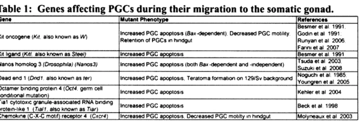
Identification of factors involved in germ cell migration is made particularly difficult by the fact that mis-migrating germ cells undergo apoptosis fairly shortly after mis-migration (Runyan et al., 2006). Thus most mutations that effect PGCs during their migration have been shown to affect germ cell survival and have not been shown to directly regulate migration. A list of genes affecting germ cells during their migration and their mutant phenotypes can be found in Table 1.
Table 1: Genes affecting PGCs during their migration to the somatic gonad.
Gene Mutant Phnotype References
Besmer et at. 1991
Increased PGC apoptosis (Bax-dependent) Decreased PGC motility Godin et al 1991 Retention of PGCs in hindgut Runyan et al 2006
Fanni et al 2007
Kit Igand (Kiti also known as Steel) Increased PGC apoptosis Besmer et al 1991
Tsuda et al 2003 Nanos homolog 3 (Drosophda) (Nanos3) Increased PGC apoptosis (both Bax -dependent and -independent) Suzuki et al 2008
Dead end 1 (Dndi. also known as ter) Increased PGC apoptosis, Teratoma formation on 1291Sv background Noguchi et al 1985
Youngren 01 al 2005 Octamer binding protein 4 (Oct4 germ cell Increased PGC apoptosis Kehler et al 2004 conditional mutation)
Tial cytotoxic granule-associated RNA binding Increased PGC apoptosis Beck et al 1998 protein-like 1 (TwiT also known as Tiar)
Chemokine (C-X-C motif) receptor 4 (Cxcr4) Increased PGC apoptosis. Decreased PGC motilty in hindgut . Molyneaux et at 2003
Of all factors required in PGCs during their migration, Kit, has the longest and
perhaps most complex history. Mutations of the dominant white spotting locus (W, which was later found to encode the Kit receptor (Chabot et al., 1988)) were easily discernable because of their effect on coat color (Mintz and Russell, 1957). To date over
120 alleles of Kit have been isolated (MGI, 2009). The difficulty in analyzing the role of
Kit in the migration of germ cells lies in the fact that Kit is also required for germ cell
survival and proliferation during their migratory phase (Besmer et al., 1993; Godin et al., 1991). However a role for Kit as a direct migratory cue has been suggested by several studies. First, in mice homozygous for the We allele of Kit, PGCs are retained in the hindgut at E 10.5, suggesting a role for this factor in migration of PGCs from the hindgut
into the urogenital ridge (Buehr et al., 1993). Consistent with this finding Runyan et al. showed that when Kitl-deficient PGCs are prevented from undergoing apoptosis by elimination of the pro-apoptotic Bax gene, they fail to migrate efficiently into the urogenital ridge (Runyan et al., 2006). Finally, Farini et al. using a novel feeder-free PGC culture system (Farini et al., 2005), established that PGCs are attracted to both urogenital ridges from E11.5 embryos and to purified recombinant KITL (Farini et al.,
2007). These results suggest that in addition to providing pro-survival and proliferation cues to PGCs, activation of the KIT receptor by KITL provides directional cues to migrating PGCs. The exact nature and organization of the KITL signal in the PGC migratory path has not been determined with enough precision to fully understand how this process proceeds in greater detail.
While PGCs are migrating, they proliferate and undergo significant epigenetic modification. The early changes in PGC chromatin are thought to re-establish an epigenetic landscape more similar to that of an early embryo than the partially
differentiated epiblast from which PGCs are specified (Hajkova et al., 2008; Seki et al., 2005; Seki et al., 2007; Surani et al., 2008). While there appears to be an orderly transition from one set of chromatin marks to another, no function for these changes has been shown.
One interesting epigenetic change in migrating PGCs involves the X
chromosome. It has been known for more than 50 years that both X chromosomes are active in the female germline (Epstein, 1969). Early studies suggested that the
reactivation of a previously inactive X chromosome occurred shortly before meiosis in ovarian germ cells (Monk and McLaren, 1981; Tam et al., 1994). Monk and McLaren
examined the enzymatic activity of X-linked HPRT (hypoxanthine phospho-ribosyl transferase) relative to autosomally encoded APRT (adenine phosphoribosyl transferase) and found a increase in the relative levels of HPRT at between E12 and E 13 in XX but not XY or XO germ cells, suggesting that X reactivation occurs in germ cells starting around E13 (Monk and McLaren, 1981). Tam et al. utilized an X-linked beta-galactosidase transgene to examine X chromosome activity and found that expression was induced in all female germ cells by E 13.5. Later results using XXY mice indicated that X chromosome re-activation occurs independently of the sex of the gonad in which PGCs find themselves (Mroz et al., 1999). Most recently, Chuva de Sousa Lopes et al., using an X-linked GFP transgene showed signals from XX but not XY urogenital ridges were required for X chromosome re-activation and that this reactivation occurred between E11.5 and E13.5 (Chuva de Sousa Lopes et al., 2008). These results were all limited by analysis of single X-linked loci as proxies for the behavior of the entire X chromosome.
To further study the regulation of X re-activation in germ cells, both Sugimoto and Abe and de Napoles et al. examined the expression of canonical markers of X inactivation, such as Xist (inactive X specific transcripts (Brockdorff et al., 1992)) and H3K27me3 (trimethylation of histone 3 on lysine 27 (Silva et al., 2003)) (de Napoles et
al., 2007; Sugimoto and Abe, 2007). These groups found that as early as E7.75 these markers of X inactivation were missing from some PGCs. The number of PGCs lacking these markers increased with time such that by E9.5 70-90% of PGCs did not possess Xist
RNA within their nuclei (de Napoles et al., 2007; Sugimoto and Abe, 2007). To further examine this process in detail Sugimoto and Abe examined loci across the entire X
chromosome and found that the timing of X re-activation was much more complex than
previously thought (Sugimoto and Abe, 2007). Some X-linked loci were biallelically
expressed even at E7.75 and many more became biallelically expressed by E10.5.
Interestingly, even at E14.5 (the last time point examined) not all loci exhibited biallelic
expression, suggesting that X re-activation is not complete even after germ cells initiate
meiosis. These results highlight the difficulties in studying X chromosome re-activation
and suggest the need to examine large number of loci across the entire chromosome in
order to truly understand this process.
6. Gonadal homecoming
Around the time of their arrival in the developing somatic gonad, PGCs undergo
several major changes: their nuclear diameter increases, they become connected into
cysts via intracellular bridges, they display a significant loss of DNA methylation and
they activate a program of gene expression specific to the post-migratory germline. The
last two of these changes are linked, with DNA de-methylation likely a pre-requisite for
the expression of post-migratory germ cell-specific genes.
The earliest difference between migratory PGCs and post-migratory germ cells to
be discovered was an increase in the diameter of the nucleus of post-migratory germ cells
relative to that of migratory PGCs. This difference is readily detectable by routine
histological staining. Both the cause and consequence of this change is unknown.
Following their arrival in the embryonic gonad, germ cells are localized in
clusters, referred to as cysts, throughout the gonad. These cysts are composed of
germ cell cysts are connected by intracellular bridges containing the germ cell specific
protein TEX14 (testis expressed 14) (Greenbaum et al., 2009). What if any role cyst
formation plays in embryonic germ cell development has not been determined. However,
germ cells deficient for TEX14 do not form intracellular bridges yet are capable of
undergoing normal embryonic differentiation, suggesting that cyst formation is not
essential for mouse germ cell development (Greenbaum et al., 2009).
While the specific timing of germline DNA de-methylation has not been
determined, it is likely to occur around the time of germ cell arrival in the gonad. Studies
assaying DNA de-methylation have yielded varying results, likely due to different
experimental methodologies. While these studies are somewhat contradictory, certain
aspects of the regulation of germline DNA methylation are generally agreed upon. Early
in their development, primordial germ cells possess a DNA methylation pattern similar to
that of somatic cells (Hajkova et al., 2002; Lee et al., 2002). Around the time of their
arrival in the gonad, between E10.5 and E 11.5, embryonic germ cell DNA methylation
begins to decrease (Hajkova et al., 2002; Lee et al., 2002; Szabo and Mann, 1995; Walsh
et al., 1998). Loss of DNA methylation occurs independently of the sex of the embryo
(Hajkova et al., 2002; Lee et al., 2002; Szabo and Mann, 1995). Regulation of the timing
of DNA de-methylation varies with particular classes of sequences and even particular
loci of the same sequence class (Hajkova et al., 2002; Lee et al., 2002; Walsh et al.,
1998). Finally, some level of variability exists within the population of germ cells with
respect to the rate their of DNA de-methylation (Lee et al., 2002). Studies of DNA
methylation in embryonic germ cells require the tissue microenvironment within which
the germ cells reside to be disrupted. This disruption means that it is not currently
feasible to determine whether DNA de-methylation status correlates with the location of germ cells within the embryo.
In addition to the outstanding questions about the timing of germline genome de-methylation, very little is also known about the mechanism by which this occurs.
Whether germline DNA de-methylation occurs passively by prevention of the activity of
Dnmtl (DNA methyltransferase 1), the enzyme required for maintenance of DNA
methylation during normal cell cycle progression (Hirasawa et al., 2008; Li et al., 1992), or is an active process is not known. However, the relative rapidity with which this process occurs suggests that an active mechanism may be involved (Hajkova et al.,
2008). The absence of an enzyme encoded within the mammalian genome known to possess the capacity to catalyze the energetically unfavorable conversion of
5-methylcytosine to cytosine makes the study of this process particularly challenging. Recent evidence in plants shows that DNA de-methylation can be accomplished by utilizing DNA repair enzymes (Choi et al., 2002; Gong et al., 2002; Zhu et al., 2007). It remains to be seen whether DNA de-methylation in mammalian cells could utilize a
similar mechanism.
Post-migratory germ cells are known to specifically express four genes. These include two germ cell specific RNA binding proteins: Mvh (mouse vasa homolog; MGI:
Ddx4) (Fujiwara et al., 1994; Toyooka et al., 2000) and Dazl (deleted in
azoospermia-like) (Seligman and Page, 1998), a component of the axial element of the synaptonemal complex: Sycp3 (synaptonemal complex protein 3) (Di Carlo et al., 2000), and an antigen
of unknown identity: GCNA1 (germ cell nuclear antigen 1) (Enders and May, 1994). It has been suggested that the continuing differentiation program of post-migratory germ
cells is coordinately regulated by the removal of DNA methylation (Maatouk and
Resnick, 2003). For the three of these markers whose genomic loci are known, a region
5' to their transcription start site containing DNA methylation has been identified
(Maatouk et al., 2006). These regions display high levels of DNA methylation in
migrating PGCs and somatic tissues, but significantly lower levels of DNA methylation
in post-migratory germ cells. Functional evidence for DNA methylation playing a role in
the control of these genes has been obtained by examining embryos with decreased
Dnmtl function (Maatouk et al., 2006). These embryos display aberrant somatic
expression of the normally germ cell specific GCNA1. In addition, migratory PGCs in
these mutant embryos express Mvh, Dazl, and Sycp3. The long-term consequences of
this aberrant gene expression have yet to be studied, as Dnmtl depleted embryos die
shortly after germ cells arrive in the gonad (Li et al., 1992). Taken together these data
suggest that the gene expression landscape of post-migratory germ cells is regulated by
the loss of DNA methylation. In order to further characterize the transition of migratory
PGCs to migratory germ cells, studies of the specific factors expressed in
post-migratory germ cells have been carried out.
Mouse vasa homolog (Mvh)
The DEAD-box RNA helicase Mvh is the mouse homolog of the highly conserved
germ cell gene vasa (Fujiwara et al., 1994). In a wide variety of metazoans vasa family
members are required for germ cell development (Komiya et al., 1994; Noce et al., 2001;
Schupbach and Wieschaus, 1986; Tsunekawa et al., 2000; Yoon et al., 1997). It is thus
somewhat surprising that the mouse homolog of this family is only expressed in germ
cells following their arrival in the gonad (Fujiwara et al., 1994). Interestingly, expression of Mvh is not observed in cultured germ cells, unless those cells are cultured in the presence of gonadal somatic cells (Toyooka et al., 2000). However, presence or absence of Mvh expression in vivo does not appear to strictly correlate with distance from the urogenital ridge (Noce et al., 2001). Thus it is not clear whether Mvh expression is
driven by germ cell-intrinsic or extrinsic environmental factors. It has not been
determined whether the induction of Mvh in the culture system developed by Toyooka et al. is accompanied by de-methylation of the region 5' of the Mvh transcriptional start site.
To understand the functional significance of Mvh expression in the mouse
germline, Mvh-deficient mice were created (Tanaka et al., 2000). In these mice, the only defects detected thus far occur in male germ cells; female mice deficient for Mvh display normal fertility and fecundity (Tanaka et al., 2000). Mvh-deficient male germ cells exhibit significantly reduced proliferation after their arrival in the gonad at E11.5 and later undergo apoptosis in the zygotene stage of meiotic prophase I (Tanaka et al., 2000). The molecular mechanism behind both of these phenotypes remains unknown.
Deleted in azoospermia-like (Dazl)
Dazl encodes an RNA binding protein with an RRM type RNA binding domain
and a DAZ domain, a domain of unknown function, which is characteristic of the DAZ family of proteins to which Dazl belongs. DAZ family proteins are germ cell specific RNA binding proteins that have been identified in all examined metazoans. In mice two members of this family are known, Dazl and Boll (boule-like; named for the Drosophila DAZ family member boule (Eberhart et al., 1996)). While Dazl is expressed in germ
cells beginning at the time of their arrival in the gonad (Seligman and Page, 1998), Boll is
expressed only in postnatal male meiotic germ cells (Xu et al., 2001). The regulation of
Dazl's transcription and translation in the embryonic germ lineage has not been well
studied. It is currently unknown whether the induction of Dazl expression is intrinsically
timed or induced by the somatic environment.
All studies of Dazl function to date have utilized a single mutant allele (Ruggiu et
al., 1997). All reports examining these Dazl-deficient mice have revealed an infertility
phenotype (Haston et al., 2009; Lin and Page, 2005; McNeilly et al., 2000; Ruggiu et al.,
1997; Saunders et al., 2003; Schrans-Stassen et al., 2001). However, mice deficient for
Dazl exhibit a wide range of germ cell defects. Defects range from late embryonic germ
cell apoptosis (Lin and Page, 2005; Ruggiu et al., 1997), to defects in postnatal male
germ cell differentiation (Schrans-Stassen et al., 2001) and meiotic prophase of both
sexes (Saunders et al., 2003). A major source of the variability observed between these
phenotypes is thought to be due to genetic modifiers introduced by varying non-inbred
strain backgrounds. Dazl-deficiency combined with an inbred genetic background
appears to give a clear and completely penetrant embryonic germ cell defect (Lin and
Page, 2005).
Germ cell nuclear antigen 1 (GCNA1)
An antibody against Germ Cell Nuclear Antigen 1 (GCNA1) was created by
injecting mouse pachytene spermatocytes into rats and purifying antibodies displaying
reactivity with mouse germ cells (Enders and May, 1994). GCNA1 is expressed in germ
cells of both sexes beginning at E 11.5 (Enders and May, 1994). Because the genomic
locus that encodes GCNA1 is still unknown, studies of its regulation and function are extremely limited. In one of the few studies to examine GCNA1 regulation, expression of GCNA1 was examined in mice deficient for the orphan nuclear receptor Sf-i (steroidogenic factor 1, MGI: Nr5al)(Wang et al., 1997), whose expression is required for production of the somatic gonad (Luo et al., 1994). Wang et al. found that germ cells in Sf-l-deficient embryos express GCNA1 at the appropriate time despite the failure of somatic gonad development in these embryos (Wang et al., 1997). In addition, primary cultures of migratory PGCs exhibit GCNA1 expression despite lacking contact with gonadal somatic cells (Wang et al., 1997). These results suggest that the gonadal soma may not be required for the expression profile of post-migratory germ cells.
Synaptonemal complex protein 3 (Sycp3)
Sycp3 encodes a protein that is a component of the axial element of the
synaptonemal complex (Dobson et al., 1994; Lammers et al., 1994). Sycp3 is necessary for normal homologous chromosome synapsis in both male and female meiosis (Yuan et al., 2002; Yuan et al., 2000). However, Sycp3 transcript and protein encoded can be detected in post-migratory embryonic germ cells of both sexes (Di Carlo et al., 2000). In embryonic male germ cells, this protein then disappears until the postnatal period (Di Carlo et al., 2000). Studies of gene expression in pre-meiotic male germ cells have identified expression of Sycp3 in spermatogonia as well (Wang et al., 2001). The expression of Sycp3 in embryonic male germ cells is particularly striking, as male germ cells do not initiate meiotic prophase until a week after birth. Why male embryonic germ cells produce this protein remains unclear. However, it has been suggested that Sycp3
may have a function beyond its role in the synaptonemal complex. This suggestion comes from observations that elimination of Sycp3 in an E2fl-deficient background causes a severe pre-meiotic germ cell defect, which is greater than that observed in the testes of either single mutant (Hoja et al., 2004). Whether a similar pre-meiotic role for
Sycp3 exists in embryonic germ cells is unknown.
In all, the process of gonadal homecoming has not been studied in as great a depth as other processes in mammalian germ cell development. Several major questions remain surrounding this developmental transition. Firstly, what is the precise timing of genome-wide DNA de-methylation and is this correlated with germ cell age (i.e. number of divisions following specification) or location in the embryo? Secondly, how is genome-wide DNA de-methylation accomplished? Thirdly, does proximity to specific signals induce expression of post-migratory germ cell genes or does this expression program activate independently of cellular environment? Finally, is this developmental transition an important event in the cycle of the germline, and what are the consequences of failing in this transition?
7. Germ cell sex determination
Prior to their arrival in the developing somatic gonad germ cells do not exhibit sexually dimorphic characteristics. Even following their arrival in the gonad the early changes observed in germ cells (specifically DNA de-methylation and gene expression changes) also appear similar in both sexes. The first morphological difference observed between male and female germ cells is the presence at E 13 of meiotic figures in female but not male germ cells (McLaren, 1983).
Meiotic figures are easily detectable by routine hematoxylin staining. The ease of this detection has led the field to use meiotic figures as the defining feature of female embryonic germ cells. Operating under a simple binary model, the field adopted the convention of defining embryonic germ cells without meiotic figures as male. This has
led the field to essentially define the question of germ cell sex determination as the question of whether embryonic germ cells initiate meiosis or not.
In order to address the timing at which germ cells commit to a meiotic or non-meiotic fate, Adams and McLaren transplanted XX and XY germ cells into E12.5 developing gonads (Adams and McLaren, 2002). Germ cells from XX embryos E12.5 or younger do not produce meiotic figures if placed in a developing testis. On the other hand germ cells from XY embryos E 11.5 or younger produce meiotic figures if placed within a
developing ovary. These results suggest that between E11.5 and E12.5 the commitment to either a meiotic or non-meiotic fate is determined. More recently, a more
comprehensive study was performed using germ cells isolated from E13.5 embryos (Iwahashi et al., 2007). This study cultured E13.5 germ cells with or without gonadal somatic cells for six days and showed that XX germ cells progressed into meiotic prophase, while XY germ cells did not even in the absence of further somatic gonadal cues. In addition this study also examined other characteristics of male and female germ cells (specifically establishment of imprinting and changes in the cell cycle) and found that germ cells do not require interaction with somatic cells after E13.5 to maintain their
sexual identity (Iwahashi et al., 2007).
Studies of meiotic initiation in the mammalian germline have focused on two predominant models (Kocer et al., 2009). One of these models proposes that germ cells
autonomously initiate meiosis unless prevented from doing so by a substance present in
the embryonic testis, but not the embryonic ovary, known as the meiosis preventing
substance (MPS). The other predominant model suggests that germ cells must be
induced to enter meiosis by the presence of a meiosis inducing substance (MIS) that is
present in the embryonic ovary and absent from the embryonic testis.
Over a period of three decades data were presented suggesting that either one or the
other of these models was correct. The earliest study of this process was performed by
Byskov and showed that signals from the rete ovary (also known as the mesonephros)
were capable of causing germ cells in embryonic testes to initiate meiosis (Byskov,
1974). McLaren and Southee suggested that if a MIS existed it must not be limited in its
location to the embryonic ovary, but must instead be present in several different
embryonic locations including the embryonic lung and adrenal (McLaren and Southee,
1997). They also showed that disruption of the structure of the embryonic testes was
sufficient to cause male germ cells to initiate meiosis.
In 2006 work from Koubova et al., Bowles et al., and Baltus et al. revealed molecular
identities to the previously theoretical substances proposed over the prior thirty years
(Baltus et al., 2006; Bowles et al., 2006; Koubova et al., 2006).
Stra8
(stimulated by
retinoic acid gene 8), was discovered to be required for early steps in embryonic female
meiosis (Baltus et al., 2006). This gene was in turn discovered to require active retinoic
acid signaling in order to be expressed in embryonic germ cells (Bowles et al., 2006;
Koubova et al., 2006). Thus retinoic acid was characterized as an MIS. This discovery
was consistent with the previous results of McLaren and Southee, which stated that the
MIS must be present broadly in the embryo, which retinoic acid is (McLaren and Southee, 1997).
In addition to identifying the MIS, the studies of Koubova et al. and Bowles et al. revealed how the sex specificity of gonadal retinoic acid signaling is established (Bowles et al., 2006; Koubova et al., 2006). Male but not female gonadal somatic cells express
Cyp26bl, a member of the cytochrome p450 class of enzymes, whose molecular function
is to alter retinoic acid and mitigate its signaling functions. Thus, Cyp26bl functions as the hypothesized MPS. These results suggested that both an MIS and an MPS existed to regulate the timing of meiotic initiation in embryonic germ cells.
It remains unclear whether meiotic initiation can be used as a proxy for the sex of embryonic germ cells. While the timing of meiotic initiation is different between male and female germ cells, the process of meiosis itself is not sex-specific. Many processes in germ cells that are sex-specific exist and the connection of these processes to meiosis has not been examined.
8. Spermatogenesis
Male germ cell sex determination is thought to occur by E12.5 (Adams and McLaren, 2002). However, the earliest male specific germ cell mutant phenotype (that of
Mvh-deficient mice) occurs even earlier, at E11.5. Male Mvh-deficient mice fail to
undergo normal proliferation after their arrival in the gonad (Tanaka et al., 2000).
Whether the sex specificity of this phenotype is caused by somatic gonadal cues or by the sex chromosome constitution of the germ cells has not been determined.
Gene expression screens for male specific expression in embryonic gonads have discovered significant numbers of sex-specific somatic cell markers, but surprisingly few early germ cell markers (Bowles et al., 2000; Menke and Page, 2002). Among the earliest known genes expressed in male but not female germ cells is Nanos2 (nanos homolog 2 (Drosophila)), which is detectable in male germ cells by E13.5 (Tsuda et al., 2003). Nanos2 function is required in male germ cells to prevent meiosis after E15.5 (Suzuki and Saga, 2008). It is thought that this function in prevention of meiosis occurs as a back-up to the earlier role of Cyp26b in preventing the activity of RA in activate
Stra8 expression (Saga, 2008).
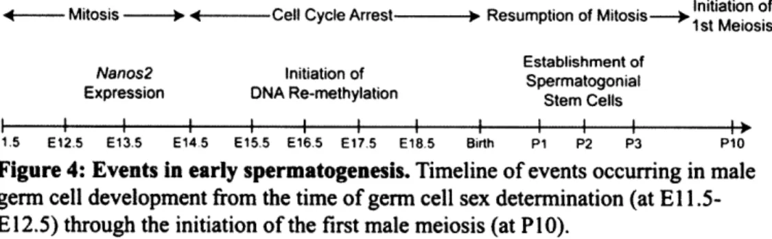
Male embryonic germ cells, sometimes referred to as pro-spermatogonia, are characterized by entry into a period of cell cycle arrest. This process begins in some germ cells as early as E12.5 and has occurred in most germ cells by E14.5 (Western et al., 2008). This arrest continues until a few days after birth when male germ cells, now referred to as spermatogonia, resume mitotic divisions (de Rooij, 1998).
Initiation of
-- Mitosis --- - Cell Cycle Arrest - Resumption of MitosisI---+ t Meiosis
Establishment of
Nanos2 Initiation of Spermatogonial
Expression DNA Re-methylation Stem Cells
I I I I I I I I I I I I
E11.5 E12.5 E13.5 E14 5 E15,5 E16.5 E17.5 E18 5 Birth P1 P2 P3 P10
Figure 4: Events in early spermatogenesis. Timeline of events occurring in male
germ cell development from the time of germ cell sex determination (at El
1.5-E12.5) through the initiation of the first male meiosis (at P10).
Pro-spermatogonia also initiate the process of global DNA re-methylation. As such, pro-spermatogonia express enzymes of the DNMT3 (DNA methyltransferase 3) family, which possess de novo DNA methylation functions (Lees-Murdock et al., 2005; Okano et al., 1999). Interspersed repeat DNA, such as LINEs and IAPS, are partially
de-methylated in the germline of both sexes between El 1.5 and E12.5, and are fully re-methylated in pro-spermatogonia beginning at E15.5 (Hajkova et al., 2002; Lees-Murdock et al., 2003). In addition paternally imprinted loci such as H19, Rasgrfl and
Gtl21/Dlk are biallelically methylated beginning by E14.5 (Davis et al., 2000; Li et al.,
2004; Ueda et al., 2000). It has been proposed that the pro-spermatogonial cell cycle arrest is required for the initiation of male specific DNA re-methylation (Schaefer et al., 2007), however, this hypothesis has not been experimentally tested.
The population of spermatogonia existing after birth is composed of at least two different sub-populations. One population of spermatogonia will progress immediately into differentiation, while the other will form a pool of long-term spermatogonial stem cells (SSCs). Confirmation of the existence of these sub-populations was obtained by tracing the spermatogonial lineage expressing Ngn3 (neurogenin 3) (Yoshida et al., 2006). It was found that sperm produced in the first round of spermatogenesis following birth do not pass through an Ngn3-expressing stage, while sperm produced in all other rounds of spermatogenesis do (Yoshida et al., 2006).
While it remains unclear how the SSC population is initially established, several studies have identified factors that are required for SSC self-renewal and differentiation. Within SSCs themselves two factors, Plzf(promyelocytic leukemia zinc finger protein, also known as luxoid; Zfpl45, zinc finger protein 145; MGI: Zbtbl6, zinc finger and BTB domain containing 16) (Buaas et al., 2004; Costoya et al., 2004) and Taf4b (TATA box-binding protein (TBP)-associated factor 4b) (Falender et al., 2005), have been shown to be required for SSC self-renewal.
In addition to SSC-intrinsic factors, two factors produced in testicular somatic
cells have also been shown to be required for SSC self-renewal. While mice
homozygous for mutations in Gdnf(glial cell line derived neurotrophic factor) die within
a day after birth, heterozygotes display an eventual loss of spermatogenesis because of a
loss of SSCs (Meng et al., 2000). In addition to showing that loss of Gdnffunction
caused eventual loss of SSCs, Meng et al. also showed that when Gdnfis over-expressed
in the testis, SSCs accumulate. Gdnffunctions by binding to the SSC expressed receptor
Gfral (glial cell line derived neurotrophic factor family receptor alpha 1) and activating
the Ret signaling pathway (Naughton et al., 2006). The relevant down-stream targets of
this pathway in SSCs remain unkown. The extrinsic factor that supports SSC
self-renewal is Erm (Ets related molecule, MGI: Etv5 (ets varient gene 5), which is produced
by the supporting cells of the testis, the Sertoli cells (Chen et al., 2005). Erm is a
transcription factor and the mechanism by which it regulates SSC differentiation or
self-renewal remains unknown.
Once an SSC population has been established, spermatogenesis proceeds in an
orderly and strictly timed manner. Spermatogenic cells form chains connected via
intercellular bridges, containing the previously mentioned TEX14 protein (Greenbaum et
al., 2006). Stem cell function (as assayed by transplantation assays) can be found within
chains of cells that are composed of less than 32 cells (de Rooij and Russell, 2000).
These cells, known collectively as the undifferentiated spermatogonia can be further
sub-divided, based on chain length, into Asingle (As, single unconnected cells), Apaired (Apr, two
connected cells) and
Aaligned (Aal,four to thirty-two connected cells). It has proven
difficult to separate these cells from each other to determine which sub-population(s)
possesses SSC activity. It has been hypothesized that As represent functional stem cells
in vivo and that Apr and Aai possess stem cell activity only when placed into an artificially
emptied niche, as is required for SSC transplantation assays (Yoshida et al., 2007). Without clear molecular markers to separate these cell types and lineage tracing experiments to track their in vivo development, it will not be possible to resolve this question.
In the adult mouse, SSCs initiate differentiation every 8.6 days (Oakberg, 1956). This commitment to differentiation begins with the transition of Aal cells into the first of six "differentiated" spermatogonial types, the A1 cells. The Aa to A1 transition represents
a major regulatory point in spermatogenesis. Unlike many other transitions in
spermatogenesis this transition is not accompanied by a mitotic cell division (de Rooij and Russell, 2000). This transition is sensitive to dietary vitamin A levels (van Pelt et al., 1995), temperature (de Rooij et al., 1999; Nishimune and Haneji, 1981) and perturbation of several genetic factors, including Dazl (Schrans-Stassen et al., 2001), jsd (de Rooij et al., 1999) and Kitl (de Rooij et al., 1999).
Dazi
jsd
Kitl
Vitamin A
Temperature Sohlh2 Stra8
Asingle --+Apaired-- Aaligned - Al - A2 - A3 - A4 -- Intermediate-* B --, Pre-leptotene- Leptotene
Undifferentiated Differentiated
Spermatogonia Spermatogonia
Figure 5: Pre-meiotic differentiation in adult spermatogenesis. Spermatogenic cell
types of the adult testis from SSCs through entry into meiotic prophase. Factors listed above arrows regulate these transitions. Names listed below cell types refer to common groupings of types of spermatogonia.
Following the transition from Aai to A1, A1 cells undergo a series of five mitoses