HAL Id: hal-02487079
https://hal-amu.archives-ouvertes.fr/hal-02487079
Submitted on 25 May 2020
HAL is a multi-disciplinary open access
archive for the deposit and dissemination of sci-entific research documents, whether they are pub-lished or not. The documents may come from teaching and research institutions in France or abroad, or from public or private research centers.
L’archive ouverte pluridisciplinaire HAL, est destinée au dépôt et à la diffusion de documents scientifiques de niveau recherche, publiés ou non, émanant des établissements d’enseignement et de recherche français ou étrangers, des laboratoires publics ou privés.
Whey protein isolate modulates beta-carotene
bioaccessibility depending on gastro-intestinal digestion
conditions
Mohammed Iddir, Celal Degerli, Giulia Dingeo, Charles Desmarchelier,
Thomas Schleeh, Patrick Borel, Yvan Larondelle, Torsten Bohn
To cite this version:
Mohammed Iddir, Celal Degerli, Giulia Dingeo, Charles Desmarchelier, Thomas Schleeh, et al.. Whey protein isolate modulates beta-carotene bioaccessibility depending on gastro-intestinal digestion con-ditions. Food Chemistry, Elsevier, 2019, 291, pp.157-166. �10.1016/j.foodchem.2019.04.003�. �hal-02487079�
Version preprint
Accepted Manuscript
Whey protein isolate modulates beta-carotene bioaccessibility depending on gastro-intestinal digestion conditions
Mohammed Iddir, Celal Degerli, Giulia Dingeo, Charles Desmarchelier, Thomas Schleeh, Patrick Borel, Yvan Larondelle, Torsten Bohn
PII: S0308-8146(19)30651-X
DOI: https://doi.org/10.1016/j.foodchem.2019.04.003
Reference: FOCH 24602
To appear in: Food Chemistry Received Date: 25 May 2018
Revised Date: 25 March 2019
Accepted Date: 1 April 2019
Please cite this article as: Iddir, M., Degerli, C., Dingeo, G., Desmarchelier, C., Schleeh, T., Borel, P., Larondelle, Y., Bohn, T., Whey protein isolate modulates beta-carotene bioaccessibility depending on gastro-intestinal digestion conditions, Food Chemistry (2019), doi: https://doi.org/10.1016/j.foodchem.2019.04.003
This is a PDF file of an unedited manuscript that has been accepted for publication. As a service to our customers we are providing this early version of the manuscript. The manuscript will undergo copyediting, typesetting, and review of the resulting proof before it is published in its final form. Please note that during the production process errors may be discovered which could affect the content, and all legal disclaimers that apply to the journal pertain.
Version preprint
Whey protein isolate modulates beta-carotene bioaccessibility depending on gastro-intestinal
digestion conditions
Mohammed Iddir1, Celal Degerli1,2, Giulia Dingeo1, Charles Desmarchelier3, Thomas Schleeh4,
Patrick Borel3, Yvan Larondelle5, Torsten Bohn1*
1
Luxembourg Institute of Health, Department of Population Health, Strassen, Luxembourg
2
Ege University, Engineering Faculty, Food Engineering Department, Izmir, Turkey
3
C2VN, INRA, INSERM, Aix-Marseille Univ, Marseille, France
4
Luxembourg Institute of Science and Technology, Belvaux, Luxembourg
5
University Louvain-la-Neuve, Institute of Life Sciences, Louvain-la-Neuve, Belgium
*To whom correspondence should be addressed:
Torsten Bohn
Luxembourg Institute of Health
1 A-B, rue Thomas Edison
L-1445 Strassen, Luxembourg
E-mail: torsten.bohn@gmx.ch
Phone: +621-216-637
Version preprint
Abstract
Carotenoids are lipophilic phytochemicals; their intake has been associated with reduced chronic
diseases. However, their absorption depends on emulsification during digestion and incorporation
into mixed micelles, requiring digestive enzymes, gastric peristalsis, bile, and dietary lipids. In this
study, we investigated whether whey-protein-isolate (WPI), a commonly consumed protein source,
can modulate β-carotene bioaccessibility in vitro, especially under incomplete digestive conditions,
i.e. under low digestive enzyme conditions. Thus, pepsin and pancreatin, kinetic energy, gastric
digestion time, and amount bile and co-digested lipids were modified, and WPI at concentrations
equivalent to 0/25/50% of the protein recommended dietary allowance (approx. 60 g/d) were added
to β-carotene dissolved in oil. WPI enhanced bioaccessibility by up to 20% (p<0.001), especially
under higher simulated peristalsis or reduced dietary lipids. Conversely, they impaired
bioaccessibility to one third (p<0.001) under incomplete digestive conditions. WPI modulated
β-carotene bioaccessibility depending on digestive conditions.
Version preprint
Introduction
Carotenoids and their metabolites are recognized as playing a role in the prevention of human
diseases. Based on epidemiological studies, a positive association has been suggested between
dietary intake and tissue concentrations of carotenoids and lowered risk of several chronic diseases
(Elliott, 2005), such as cardiovascular diseases and type 2 diabetes (Sluijs, Cadier, Beulens, van der
A, Spijkerman, & van der Schouw, 2015), cancer (Sharoni, Linnewiel-Hermoni, Khanin, Salman,
Veprik, Danilenko, et al., 2011), as well as reduced risk of age-related macular degeneration
(Arunkumar, Calvo, Conrady, & Bernstein, 2018). These correlations may be attributed to their
antioxidant and anti-inflammatory activity (Krinsky, 2001), as well as their influence on the
immune system (Rao & Rao, 2007), both possibly related to a number of transcription factors and
nuclear receptors such as NF-κB (Kaulmann & Bohn, 2014) and RAR/RXR (Caris-Veyrat, Garcia,
Reynaud, Lucas, Aydemir, & Rühl, 2017). In addition to these health relevant aspects, certain
carotenoids, including β-carotene, constitute vitamin A active compounds, following their cleavage by β-carotene oxygenase 1 (BCO-1) in vivo and metabolism into retinal.
Before being absorbed, carotenoids must be released from the food matrix and dissolved in lipid
droplets. Then, under the influence of digestive enzymes (especially lipase), pH, gastric peristalsis,
and bile, carotenoids are incorporated into mixed micelles, allowing their uptake by the mucosa of
the small intestine (Bohn, Desmarchelier, Dragsted, Nielsen, Stahl, Rühl, et al., 2017). Due to their
lipophilic nature, the bioavailability of carotenoids, including β-carotene, is relatively low, typically
between 10 and 20% (Bohn, 2008; Desmarchelier & Borel, 2017). Consequently, several studies
have been investigating potential dietary as well as host-related factors influencing carotenoid
bioavailability aspects, including their bioaccessibility and absorptive processes.
Regarding matrix-related factors, several studies have concluded that dietary lipids can
significantly enhance carotenoid absorption, fostering micellization (Bohn, 2008; Huo, Ferruzzi,
Version preprint
micellization of carotenoids, possibly by limiting enzymatic access and activity (O'Connell, Ryan,
O'Sullivan, Aherne-Bruce, & O'Brien, 2008). Recently, divalent minerals at higher concentrations
were suggested to compromise carotenoid bioaccessibility, likely by precipitating fatty acids and
bile salts (Biehler, Hoffmann, Krause, & Bohn, 2011; Borel, Desmarchelier, Dumont, Halimi,
Lairon, Page, et al., 2016). In addition to these dietary factors, it was shown that host-related factors
can modulate carotenoid bioavailability (reviewed in Bohn, et al., 2017), including conditions that
alter the gastro-intestinal (GI) surface area or digestive enzyme secretion. For example, bariatric
surgery has been associated with several physiological modifications, among them, gastric
peristalsis movement, at least in part explaining the decrease of plasma carotenoid concentrations
(Granado-Lorencio, Simal-Anton, Blanco-Navarro, Gonzalez-Dominguez, & Perez-Sacristan,
2011). In another study, it was reported that Crohn’s disease resulted in carotenoid malabsorption
(Edes, Walk, Thornton, & Fritsche, 1991). Other anomalies such as cholecystectomy (reducing the
available amount of bile), and pancreatitis (leading to secretory insufficiency of digestive enzymes)
have been proposed to compromise carotenoid bioavailability (Desmarchelier & Borel, 2017).
One factor that may impinge on carotenoid absorption, but has not yet received much attention, is
the presence of proteins. For example, caseins have been successfully employed for micro- and
nano-encapsulation of β-carotene, significantly improving its bioaccessibility (Soukoulis & Bohn,
2018; Yi, Li, Zhong, & Yokoyama, 2014). It was proclaimed that proteins may aid in the
bioaccessibility of liposoluble dietary constituents in several ways. Following their adsorption to
lipid droplet surfaces, proteins may stabilize oil-in-water (o/w) emulsions in the GI tract, attributed
to the fact that proteins can be highly surface-active molecules (Mun, Kim, McClements, Kim, &
Choi, 2017; Soukoulis & Bohn, 2018), and the formed particles tend to be highly negatively
charged, preventing the aggregation of lipid droplets (Qiu, Zhao, Decker, & McClements, 2015).
However, the same study also showed that gliadin decreased the enzymatic degradation of lipids,
Version preprint
directly, also implying potential negative influences of proteins on the micellization process. Whey
protein isolate (WPI) in contrast was effective at inhibiting lipid oxidation of o/w emulsions (Qiu,
Zhao, Decker, & McClements, 2015), which may contribute to limit oxidative degradation of
sensitive molecules, as were caseino-phosphopeptides, which were proposed to chelate iron which
may trigger oxidative degradation (Kim, Jang, & Kim, 2007). Furthermore, WPI also facilitated the
formation of smaller lipid droplets; Salvia-Trujillo and co-workers showed that reducing the
diameter of these particles was related to increased β-carotene bioaccessibility (Salvia-Trujillo,
Qian, Martin-Belloso, & McClements, 2013).
It is assumed that the emulsifying properties are greatly influenced by the composition and
structure of proteins. For instance, both hydrophobic and hydrophilic groups are abundant in WPI,
highlighting its amphiphilic behavior (Sharif, Williams, Sharif, Abbas, Majeed, Masamba, et al.,
2017). The structure of WPI is comprised of two main fractions, i.e. α-lactalbumin (123 amino acids) and β-lactoglobulin (162 amino acids), together accounting for approx. 75% of the whey
proteins, and their conformation (predominantly α-helix and β-pleated sheet, respectively) affect
their properties at the oil/water interface (Chatterton, Smithers, Roupas, & Brodkorb, 2006).
In the current study, we aimed to investigate the effect of a commonly consumed protein source,
WPI, on the bioaccessibility of pure β-carotene, under a range of digestive conditions, as proteins
were hypothesized to rather enhance bioaccessibility under marginal, i.e. somewhat insufficient
digestive conditions. For this purpose, we employed a previously established in vitro GI consensus
model (Minekus, Alminger, Alvito, Ballance, Bohn, Bourlieu, et al., 2014), and WPI was added at
different concentrations, reflecting various fractions of the recommended dietary allowance (RDA)
of proteins, i.e. 0, 25 and 50%. Micellization of co-digested β-carotene thus measured also under
insufficient digestion parameters (i.e. shortened gastric digestion duration, limited concentrations of
digestive enzymes, gastric peristalsis, bile, and a surplus of dietary lipids), such as possible in
Version preprint
Materials and Methods
2.1 Enzymes and chemicals
Pepsin from porcine gastric mucosa (powder, ≥250 U/mg, Art. No. P7000), pancreatin from
porcine pancreas (activity equivalent to 4x USP specifications. Art. No. P1750), porcine bile extract
(Art. No. B8631), as well as β-carotene standard (≥97% UV, >95% all-trans form according to
supplier and own HPLC analysis, Art. No. 22040) were purchased from Sigma-Aldrich (Overijse,
Belgium). Whey protein isolate (WPI) was obtained from Pure Nutrition USA (95% purity,
California, USA) while the peanut oil, typically free of native carotenoids (according to the USDA
database and own blank examinations), was purchased from a local supermarket (Delhaize,
Strassen) and used without further purification.
Unless otherwise specified, all chemicals were of analytical grade or superior. Potassium chloride (≥99%, Art. No. P9541), potassium phosphate (monobasic ≥99, Art. No. P5655), sodium
bicarbonate (≥99, Art. No. S5761), sodium chloride (≥99.5%, Art. No. 71376), magnesium chloride
hexahydrate (Art. No. M2393), ammonium carbonate (Art. No. 68392), sodium hydroxide solution
(1 M, Art. No.1.09137), calcium chloride dihydrate (≥99, Art. No. C3306), nile red (Art. No.
19123), fluorescein isothiocyanate isomer I (FITC) (Art. No. F7250), and phenolphthalein (Art. No.
105945) were acquired from Sigma-Aldrich. Hexane (≥95%, Art. No. 24574), acetone (≥99%, Art.
No. 20063), and hydrochloric acid (1 M, Art. No. 30024.290) were obtained from VWR (Leuven,
Belgium).
2.2 β-Carotene, WPI and enzyme solutions
β-Carotene standard solution was first prepared by dissolving 1 mg of β-carotene standard in 2
mL of hexane. The solution was sonicated for 5 min. and warmed up at 30 °C for 5 min. (Ultrasonic
Version preprint
dissolution of β-carotene by sonication and warming at 30 °C for 5 min. The hexane was removed
by evaporation under a stream of nitrogen for ± 20 min. at 30 °C (TurboVap LV from Biotage®
,
Uppsala, Sweden). The standard β-carotene solution was made by the addition of 4 mL of peanut
oil to the remaining 1 mL to reach a concentration of 0.2 mg/mL, and two other concentrations (0.1
and 0.4 mg/mL) were also prepared for further investigation by adding 9 and 1.5 mL (instead of 4
mL) of peanut oil, respectively. The concentration of the standard solutions was later verified
spectrophotometrically as explained below (section 2.5). Aliquots of the standard solutions were
pipetted into 15 mL falcon tubes, and stored at -80 °C until usage.
Assuming a total volume of 2 L of intestinal fluids during GI digestion, the amount of β-carotene added to each digesta was 30 μg, by pipetting 75, 150 or 300 μL from the standard solutions (0.4,
0.2 or 0.1 mg/mL, respectively), representing 1.2 mg/L of β-carotene and 3, 6 or 12 g/L of oil
(Table 1).
In the same way, the amounts of WPI tested were 0, 7.5 and 15 g/L (Table 1), corresponding to 0,
25 and 50% of the recommended dietary allowance (RDA) (60 g/d for human adults) (Hautvast,
Baya, Amorim Cruz, de Backer, Ducimetiere, Durnin, et al., 1989). Practically, 0, 187.5 or 375 mg
of WPI were dissolved in 6.25 mL of pure water, and together with the added β-carotene solution in
oil, the final ratio of matrix (WPI+β-carotene solution) to simulated gastric fluid of 50:50 (v/v) was
reached. A brief mixing of the WPI solution with the beta-carotene in peanut oil and the simulated
gastric fluid (SGF) was achieved by vortexing for 15 sec.
SGF and the simulated intestinal fluid (SIF) were made as recommended by the European
consensus digestion model (Minekus, et al., 2014). Pepsin solution was prepared in SGF to reach a
final concentration of 2000 U/mL of the final gastric mixture. In addition to the standardized
Version preprint
Regarding pancreatin and bile extract, pancreatin was weighed based on a trypsin activity of 100,
200 or 400 U/mL, while the bile extract was weighed in order to reach a concentration of 3.4, 6.8 or
13.6 mg/mL of the final digestion volume (Table 1); both were prepared in the same SIF solution.
It is noteworthy that the European consensus suggested a standardized amount of pancreatin
based on trypsin activity at 100 U/mL, but in the present study, pancreatin amount was doubled
following pre-experiments (200 U/mL of trypsin), in order to enhance the digestibility of both WPI
and oil (by increasing at the same time the lipase activity). As explained above, two other
concentrations of pancreatin (100 and 400 U/mL) as well as bile extract (3.4 and 13.6 mg/mL) were
tested. Conditions with low pancreatin (100 U/mL) and/or low bile (3.4 mg/mL) are denoted in the
following as marginal digestive conditions in this investigation.
2.3 Simulation of gastro-intestinal (GI) digestion
In vitro simulated GI digestion was carried out according to the harmonized INFOGEST
protocol, with some modifications as explained above (section 2.2). The model was used to test the
influence of proteins at three different concentrations on β-carotene bioaccessibility, including also
rather marginal, i.e. insufficient digestion parameters. The oral phase was omitted as the matrix
employed did not include significant amounts of carbohydrates and was liquid.
For the gastric phase, 6.25 mL of protein solution at the desired concentration (equivalent to 0, 25
or 50% of the RDA) was added to each sample, followed by the addition of 75, 150 or 300 μL of
β-carotene solution in oil (corresponding to a total of 30 μg of β-β-carotene per digest, with amounts
similar to former in vitro experiments (Biehler et al. 2011)). Before the addition of 4 mL of SGF
(1.25 x concentrate), 1 mL of pepsin solution was added to the mixture in order to reach enzymatic
activities of 1000, 2000, 4000 U/mL of the final gastric volume. Then, 31 μL of calcium dichloride
Version preprint
adjustment to 3 by hydrochloric acid (1 M) was carried out, bringing the volume of each sample to
12.5 mL with pure water.
Finally, the samples were incubated in a shaking water bath (GFL 1083 from VEL®, Leuven,
Belgium) for 1 or 2 h at 37 °C, with a shaking speed of 75 or 100 rounds per minute (equal to a
doubling of the kinetic energy, which is proportional to the square of the speed), depending on the
desired condition. In the standardized INFOGEST digestion model (Minekus, et al., 2014), no
specific shaking speed was suggested.
At the end of the gastric incubation, 7.5 mL of SIF, and 2.5 mL of pancreatin and bile extract
solution at various concentrations (100, 200 or 400 U/mL based on trypsin activity for pancreatin,
and 3.4, 6.8 or 13.6 mg/mL for the bile extract) were added to the chyme. Then, 250 μL of calcium
dichloride (0.03 M) was added to reach a concentration of 0.3 mM in the final mixture. Before
bringing the volume to 25 mL, the pH was adjusted to 7 by the addition of sodium hydroxide
solution (1 M), then the samples were incubated for 2 h at 37 °C, with a shaking speed of 75 or 100
rounds per minute (rpm).
2.4 β-Carotene extraction
At the end of the intestinal incubation, 12 mL were removed from the digesta and transferred into
15 mL falcon tubes. The samples were centrifuged for 1h at 3200 x g (4 °C), then 5 mL were
collected from the middle aqueous phase and filtered through 0.2 μm nylon membrane syringe
filter. In a new 15 mL falcon tube, 4 mL of hexane:acetone (2:1, v:v) were added to 2 mL of the
filtered aqueous phase, vortexed for 30 sec. and centrifuged for 2 min. at 3200 x g (4 °C). The
supernatant, which represents the hexane phase, was transferred into a new falcon tube, then the
extraction process was repeated two times with 4 mL of hexane. All extracts were combined in the
Version preprint
2.5 Spectrophotometric analysis
Spectrophotometric analysis of β-carotene was done as described previously (Corte-Real,
Richling, Hoffmann, & Bohn, 2014). The dried residue was reconstituted in 1 mL of cold (4oC)
hexane and transferred to a 1 mL quartz cuvette (absorption cuvette, Hellma®, New York, USA).
The absorbance was measured between 300 and 600 nm (GENESYSTM 10S UV-Vis
Spectrophotometer, Thermo Fisher Scientific, Massachusetts, USA). The concentration was
calculated by applying the Beer-Lambert law, taking into account the molar absorption coefficient
(Britton, Liaaen-Jensen, & Pfander, 2004), and different dilution steps:
where C = concentration of the measured solution (mg/mL), absmax = maximum measured absorbance of β-carotene at approx. 450 nm, Fd = dilution factor adjusting for extraction or
reconstitution process, ελ = molar absorption coefficient (Lmol-1cm-1) of β-carotene in hexane
(138824), d = length of light pathway through cuvette (cm) and M = β-carotene molar mass
(536.89 g/mol).
Finally, the percentage of β-carotene micellization was used as a measure of bioaccessibility, and
was expressed as the percentage of the solubilized amount of β-carotene present in the aqueous
phase of the filtered digesta after in vitro GI digestion, compared to the initial amount added to the
sample.
2.6 Further physico-chemical characterization of digesta
To study further physico-chemical properties of the digesta, macroviscosity and surface tension,
micelle size and zeta potential, fatty acid titration, and the emulsion structures were investigated, as
these parameters were also believed to possibly impinge on β-carotene bioaccessibility.
Version preprint
Flow curves were determined as a function of the shear rate between 0.1 s-1 and 130 s-1 on an
Anton-Paar rheometer (MCR 302, WESP, Graz, Austria), equipped with a double gap cell at 5°C.
Each measurement cycle did comprise a temperature equilibration phase, a pre-shear phase at 5 s-1
of 60 s, a flow curve with increasing shear rates, an intermediate equilibration phase at 131 s-1 of 60
s and a flow curve with decreasing shear rates. At the end of each cycle, a regression flow curves
was calculated for the shear rate range between 0 s-1 and 100 s-1 for the shear stress respectively
between 1 s-1 and 100 s-1 for the viscosity. Each measurement cycle was performed three times,
because of the fluidity of the samples. The regression curves were averaged and plotted as viscosity
and shear stress as a function of the shear rate.
Surface tension of digesta samples, pre-conditioned at 25 ± 0.1 °C, were determined by the
weight-drop method as previously described by (Permprasert & Devahastin, 2005). The air-water
interfacial properties of digesta were calculated as follows:
H2O
where σH2O= 71.99 dyn cm−1 is the surface tension of pure water.
2.6.2 Micelle size and zeta potential analysis
Aliquots of the aqueous micellar fraction obtained after GI digestion under standard conditions
(Table 1) were filtered through a 0.2 μm nylon membrane syringe filter for the analysis of the
micelle size and zeta potential. The intensity-weighted mean hydrodynamic radius and zeta
potential were determined by dynamic light scattering and laser doppler micro-electrophoresis,
respectively. Measurements were done at room temperature with at least four replicates, by using a
Zetasizer Nano Zs instrument (Malvern Instruments, Malvern, UK).
Version preprint
The amount of free fatty acids in the digesta after GI digestion was estimated as described
previously (Corte-Real, Desmarchelier, Borel, Richling, Hoffmann, & Bohn, 2018), based on the
titration against sodium hydroxide (NaOH 0.1 M), using phenolphthalein as an indicator.
Sodium dodecyl sulfate-polyacrylamide gel-electrophoresis (SDS-PAGE) was carried out to study
completeness of protein digestion. Samples were denatured at 95oC for 5 min. in Lämmli buffer.
About 22 µg of protein per pocket was loaded to a 15% acrylamide and 0.4% bisacrylamide gel
(gastro-intestinal digestion) and 45 g of protein per pocket was loaded to a 10% acrylamide/0.3% bisacrylamide gel (gastric digestion). Marker was Invitrogen novex see blue plus 2 (Invitrogen,
Carlsbad, CA). Gel was run at 80 V (30 min.) plus 100 V (90 min.). Running buffer contained SDS
(0.1%), glycine (190 mM), tris-HCl (25 mM). Fixation/staining was done with methanol/acetic
acid/coomassie brilliant blue R (50%, 10%, 0.1%) (30 minutes), destaining in methanol/glacial
acetic acid/water (30%, 10%, 60%, 3 h). Pictures were taken in a Kodak Gel Logic 2200 Imaging
System (Kodak, Rochester, NY).
2.6.4 Confocal laser scanning microscopy (CLSM)
Confocal imaging of emulsion structures before and after GI digestion under standard conditions
(Table 1) was carried out at room temperature with a confocal laser scanning microscope (Zeiss
LSM 880, Airyscam SR, Jena, Germany), using a 63x objective. Nile Red dissolved in ethanol and
FITC dissolved in acetone were mixed into the aliquots of digesta for fat and protein staining with
final concentrations in the samples of 1 µg/mL. A small-sample aliquot was placed on a slide for
visualization. The fluorescent dyes were excited by an Argon 488 nm laser and the emitted light
was collected at 522 nm for proteins and 635 nm for the fat phase. The diameter of lipid droplets
(n=20/group) at various concentration of WPI was measured by using a modular image-processing
and analysis software for digital microscop (Zen 2.3 blue edition, Carl Zeiss Microscopy GmbH,
Version preprint
2.7 Statistical analyses and data treatment
In order to minimize day-to-day variations between experiments, bioaccessibility of β-carotene
was normalized to a daily control which was assessed for each digestion. Unless otherwise stated,
all values are expressed as the mean ± standard deviation. Replicates were obtained from at least 2
individual sets of analyses obtained at different days (N≥2), and at least 4 replicates were obtained
for each digestive condition during 1 set of analyses.
Statistical analysis was performed using SPSS 22 software (SPSS Inc., Chicago, IL). Normal
distribution of data was verified by Q-Q-plots and equality of variance by box-plots and Levene’s
test. For statistical evaluation, β-carotene bioaccessibility values were log-transformed to achieve a
normal distribution. Linear mixed models were developed with the effect of WPI concentration,
amount of lipid source, bile extract concentration, kinetic energy and duration of gastric phase,
pepsin and pancreatin concentration as fixed factors and log-β-carotene bioaccessibility as the
observed dependent factor. As a model including all factors was not fully factorial, but several
significant interactions were obtained, additional linear mixed models were run to keep constant
certain parameters and to better allow further group-wise comparisons. P-values < 0.05 were
considered statistically significant different (2-sided). Where needed, Fisher F-tests were followed
by post hoc tests (Bonferroni, for comparing >3 groups) or LSD tests (for comparison of ≤ 3
groups).
3. Results
3.1 Interactions of WPI with matrix related effects affecting β-carotene bioaccessibility
3.1.1 Bioaccessibility under standard conditions and with escalating concentrations of WPI
Under control conditions (no addition of WPI), bioaccessibility of pure β-carotene following the
Version preprint
the aqueous micellar fraction at the end of the in vitro digestion, compared to the initial amount
added at the beginning of digestion (Fig. 1A). Although the addition of an equivalent of 50%
protein RDA of WPI slightly enhanced bioaccessibility (Fig. 1A) compared to the control (no
protein added), this effect was not significant under standard conditions (Minekus, et al., 2014).
3.1.2 Influence of WPI at different amounts of co-digested oil on β-carotene bioaccessibility
Following linear mixed models, the effect of oil (p<0.001) was statistically significant, while the
influence of WPI had no significant effect. In addition, a significant interaction between the amount
of added oil and WPI concentration was encountered (p<0.001), thus the effects were further
studied at individual WPI levels.
When no WPI was added, increasing the volume of oil (up to 300 μL) negatively affected the
bioaccessibility (p<0.001) compared to the standard conditions (150 μL of oil). No significant effect on β-carotene bioaccessibility was observed with a lower amount of oil (75 μL of oil, Fig. 1B,
Table 2).The addition of 25% RDA of WPI to samples with a reduced amount of oil further
enhanced the bioaccessibility (by up to 20%) compared to the control, while the addition of WPI at
50% RDA had no significant effect (Fig. 1B). However, adding WPI to the simulated GI digestion
with a high volume of oil (300 L) caused an additional decrease in bioaccessibility, in a dose dependent manner (p<0.001). In this scenario, adding the equivalent of 25 and 50% RDA of WPI significantly decreased β-carotene bioaccessibility by 20% (p=0.017) and up to 35% (p<0.001),
respectively, compared to the control (no WPI).
3.2 Influence of WPI on β-carotene bioaccessibility at various gastric conditions
3.2.1 Impact of WPI at altered simulated peristalsis on β-carotene bioaccessibility
Following linear mixed models, the effect of peristalsis conditions on β-carotene bioaccessibility
Version preprint
the interaction between the added amount of WPI and peristalsis movements had a significant
(p=0.019) effect on the bioaccessibility of β-carotene, thus effects were further studied at individual
shaking speed.
The higher shaking water speed of 100 rpm induced an up to two times higher β-carotene
micellization (p<0.001) compared to the lower speed of 75 rpm (Fig. 2, Table 2), across all 3 levels
of proteins present (0, 25 or 50% of RDA), at 2 h of gastric digestion time.
Following all-group-wise comparisons (post hoc-tests), the addition of 25% RDA of WPI
significantly increased (p<0.001) β-carotene bioaccessibility when a higher kinetic energy was
applied, relative to the control (Fig. 2A). However, no significant influence was observed with the
addition of 50% RDA of WPI, i.e. WPI did not influence β-carotene bioaccessibility in a linear
dose-dependent manner.
3.2.2 Influence of WPI on β-carotene bioaccessibility at altered gastric phase duration
Following linear mixed models, the effect of gastric phase duration (p<0.001) was statistically
significant, while the influence of WPI had no overall significant effect. However, the impact of
gastric digestion time (2 vs. 1 h) depended significantly on the shaking speed (p=0.0016, Fig. 2A),
with larger differences observed at lower simulated peristalsis.
Reducing gastric transition to 1 h decreased bioaccessibility by 25% at 75 rpm (in general for all
levels of WPI addition), while the negative effect was less pronounced, yet significant, with a
higher shaking speed, compared to the standardized 2 h of gastric phase (p<0.001, Table 2).
At 75 rpm, no further influence was noticed on β-carotene bioaccessibility following the addition
of WPI, with a reduced gastric digestion time (Fig. 2A), while a significant (p<0.001) interaction
was found at 100 rpm between protein concentration and gastric phase duration (Fig. 2A). Here, the
addition of WPI significantly (p<0.001), though only slightly, increased the bioaccessibility of
Version preprint
3.2.3 Effect of varying concentrations of pepsin
Following linear mixed models, the effects of both WPI (p<0.001) and of pepsin concentration
(p<0.001) were statistically significant. A statistical significant interaction of pepsin x protein
(p=0.008) was also found regarding β-carotene bioaccessibility.
Under control conditions (no WPI), no influence of varying the amount of pepsin on β-carotene
micellization (Fig. 3A) was found. Similarly to the standard conditions (2000 U/mL of pepsin),
addition of WPI to the simulated GI digestion with 4000 U/mL of pepsin did not influence
β-carotene bioaccessibility compared to the daily control (Table 2). However, the addition of WPI to
the samples with a lower concentration of pepsin decreased β-carotene bioaccessibility by up to
40% (p<0.001), compared to the control (no WPI).
3.3 Influence of WPI at altered intestinal conditions
3.3.1 Effect of varying concentrations of bile extract
Following linear mixed models, the effects of both WPI (p=0.002) and of bile concentration
(p<0.001) were statistically significant. In addition, a significant interaction between added amounts
of bile extract and protein concentration was found (p<0.001).
A maximum of β-carotene micellization (around 25%) was obtained with the higher amounts of
bile extract (6.8 and 13.6 mg/mL), while 3.4 mg/mL of bile extract significantly (p<0.001)
decreased β-carotene bioaccessibility, compared to the standard conditions (6.8 mg/mL, Fig. 3B).
There was no significant effect of adding WPI at high amounts of bile (6.8 and 13.6 mg/mL per
digesta), except for a slight decrease of β-carotene bioaccessibility when 50% RDA of WPI was
added to the samples with highest amounts of bile extract (p=0.0011). However, the addition of
WPI to the digesta with reduced amount of bile extract (3.4 mg/mL) negatively affected the
Version preprint
RDA of WPI decreased the bioaccessibility to half and 1/3, respectively, compared to the control
(p<0.001, Table 2).
3.3.2 Effect of varying concentrations of pancreatin
Following linear mixed models, the effects of both WPI amount (p=0.009) and of pancreatic
enzyme concentrations (p<0.001) were statistically significant. Under control conditions (no WPI),
increasing the pancreatin concentrations positively affected the bioaccessibility (p<0.001) in a
concentration dependent manner (Fig. 2B), i.e. increasing β-carotene micellization from 25% up to
34% (75 rpm), and from 45% up to 68% (100 rpm).
Furthermore, a statistical interaction (pancreatin x peristalsis x protein, p=0.004) regarding
β-carotene bioaccessibility was also found. At 75 rpm, the addition of WPI to the simulated GI
digestion with half the typical amount of pancreatin negatively affected (p<0.001) β-carotene
bioaccessibility, by up to 50%, compared to the control (Fig. 2B, Table 2), in a dose-dependent
fashion. When doubling the amount of pancreatin to 400 U/mL, a significant (p<0.001) negative effect of WPI on β-carotene micellization was found only with the addition of 25% RDA, compared
to the control.
A similar effect was observed for conditions resembling a stronger peristalsis effects (100 rpm).
Using the double amount of pancreatin (400 U/mL) slightly decreased β-carotene micellization
following the addition of 50% RDA of WPI (p=0.006, Fig. 4). However, employing half the amount
of pancreatin compared to standard digestive conditions resulted in ambivalent effects, with 25%
RDA of WPI resulting in enhanced β-carotene bioaccessibility (p=0.004), while the addition of
50% RDA of WPI slightly decreased β-carotene bioaccessibility (p=0.015), compared to the
control.
Version preprint
In general, the shear stress and viscosity of digested samples at various concentrations of WPI
was found very close to that of aqueous systems (Supplementary Fig. 1A, left panel), and samples
had a very small yield point (0.01 Pa or even lower), with lower yield points after digestion.
Non-digested WPI solutions at 0% RDA and all Non-digested solutions showed almost Newtonian behaviour
above shear rates of 5 s-1 (Supplementary Fig. 1A, right panel).
Surface tension for control and supplemented digesta with 25% of WPI showed no significant
difference (Supplementary Fig. 2). The addition of 50% WPI slightly reduced surface tension
compared to the control digesta (p<0.001), being in line with the higher macroviscosity data.
Regarding mixed micelle size and zeta potential, both 25% and 50% RDA of WPI additions
resulted in significantly reduced micelle size of 3.3 ± 0.1 nm compared to WPI free control digesta
(supplementary Fig. 3A, p<0.001). However, the average absolute zeta potential of the samples
digested with 25% and 50% RDA of WPI was somewhat reduced (from −46.8 ± 1 mV to -25.3 ±
6.2 and -28.8 ± 1.7 mV, respectively (Supplementary Fig. 3B, p<0.001).
Lipid digestion results obtained by fatty acid titration were not fully informative and rather
reflected the buffering capacity of the added proteins, and/or additional protein hydrolysis, thus it
was not feasible to separate their contribution to lipolysis (Supplementary Fig. 4). The size of lipid
droplets and the co-location of lipids and proteins at 25% and 50% RDA samples were measured by
confocal microscope. Prior to digestion, the lipid droplets were distributed as large aggregates of
lipid droplets in the mixture. With increasing concentration of WPI, the lipid droplets formed
smaller aggregates in the digesta (Fig. 4A). The higher concentration of WPI resulted in a smaller
average lipid droplet size compared to the control digesta, decreasing from 13.42 ± 9.28 to 4.84 ±
1.52 and to 3.35 ± 1.76 µM for 0, 25 and 50% WPI, respectively (Supplementary table I), with the
first group being significantly different from the last 2 groups (p<0.001).
Regarding protein digestion, there was no detectable difference between the various digestive
Version preprint
gastro-intestinal digestion (Supplementary fig. 5, upper panel). Gastro-intestinal digestion
suggested the complete digestion of WPI into smaller fragments during all digestive conditions, as
no major bands were visible following digestion. Only bands originating from enzyme additions
were detectable after gastro-intestinal digestion. Following only gastric digestion however
(Supplementary fig. 5, lower panel), fragments around 18 kDa were clearly visible after WPI
digestion, for all digestive conditions, likely representing either α-lactalbumin (14.2 kDa) and or
β-lactoglobulin (18.4 kDa).
Discussion
Micellization of β-carotene constitutes one of the major steps determining its absorption and
bioavailability, and previous studies have shown that they are well correlated (Biehler & Bohn,
2010; Reboul, Richelle, Perrot, Desmoulins-Malezet, Pirisi, & Borel, 2006). Several reports have
highlighted matrix-related as well as host-related factors influencing carotenoid bioaccessibility and
absorptive process, as reviewed earlier (Bohn, 2008; Bohn, et al., 2017). In this regard, proteins
have been proclaimed to aid in emulsifying liposoluble dietary constituents (Soukoulis & Bohn,
2018), but their presence has also shown a negative influence on lipid degradation, which could
have an impact on the micellization process (Qiu, Zhao, Decker, & McClements, 2015). In the
present study, we investigated the effect of WPI, a commonly consumed protein, on β-carotene
bioaccessibility under selective simulated digestion conditions to study its influence at more
physiological vs. somewhat sub-optimal conditions regarding lipid and protein digestion.
The most important finding was that the influence of WPI on β-carotene depended on digestive
conditions. More specifically, WPI reduced β-carotene bioaccessibility by approx. 70% when lipid
digestion was rather compromised by lower concentrations of bile, enzymes, or when reducing
shaking speed. However, under certain conditions, enhanced bioaccessibility, compared to standard
Version preprint
encountered, but appeared less consistent. Under standard digestion conditions (Table 1, Fig. 1A),
adding WPI reflecting 0, 25 and 50 % of the RDA for a 70 kg adult (assuming a digestion volume
of 2 L) during simulated GI digestion, had no significant effect on the micellization of β-carotene,
in spite of resulting in lower lipid droplet size after GI digestion. To our knowledge, this is the first
report systematically investigating carotenoid bioaccessibility in the presence of a co-digested
protein, under varying simulated digestion conditions.
Whey protein consists primarily of β-lactoglobulin (50-55%) and α-lactalbumin (20-25%).
Regarding its emulsion stabilizing capability, it was emphasized that the high percentage of
β-pleated sheet (~50%) conformation and the relatively low content of the α-helix form (~15%)
allows β-lactoglobulin (due to its higher hydrophobicity) to adsorb and produce better
conformational changes at the o/w interface, compared to α-lactalbumin (β-sheet ~ 9%, α-helix ~
45%) (Carbonaro, Maselli, & Nucara, 2012; Schröder, Berton-Carabin, Venema, & Cornacchia,
2017). In addition, during the gastric phase, a complete hydrolysis of α-lactalbumin was found,
while a portion of β-lactoglobulin remained resistant after 1 h (Malaki Nik, Wright, & Corredig,
2011). Thus, in whey protein-stabilized emulsions, β-lactoglobulin may play a greater role
regarding emulsion stability, compared to α-lactalbumin.
The strongest positive significant effects of WPI addition were observed when halving the
amount of oil (Fig. 1B) and doubling the peristalsis energy (Fig. 2A), reflecting more efficient
digestive conditions. The effects were an approx. 20% increase, compared to their respective
controls (no WPI addition), but only at protein concentrations equivalent to 25% RDA, not 50%
RDA. It was reported earlier that β-lactoglobulin can interact with β-carotene within a pocket of
hydrophobic residues, with strong non-covalent interactions (Mensi, Choiset, Rabesona, Haertle,
Borel, & Chobert, 2013), suggesting that WPI can act as a vehicle for hydrophobic compounds
during the GI digestion. This may explain the positive effect observed when reducing the amount of
carotenoid-Version preprint
protein complexes with glutathione-S-transferase and crustacyanin are examples of well
characterized carotenoid-binding proteins (Pilbrow, Garama, & Carne, 2012). In addition, native
whey proteins are highly surface-active molecules (Mun, Kim, McClements, Kim, & Choi, 2017). It
was demonstrated that the proteins and peptides liberated during GI digestion can affect the stability
of o/w emulsions (van der Ven, Gruppen, de Bont, & Voragen, 2001), as well as retarding
triglyceride oxidation (Qiu, Zhao, Decker, & McClements, 2015). It was also reported that after
gastric digestion, some of the protein fragments remained adsorbed to the lipid droplet surfaces.
However, these appeared to be removed into the surrounding aqueous phase following further
proteolysis by pepsin, the low pH, and peristalsis movements in the stomach, as well as further
action of the bile salts in the small intestine (Mun, Kim, McClements, Kim, & Choi, 2017),
allowing access to lipolytic enzymes. This is in line with results from the present study, as no more
adsorption of proteins to lipid droplets in fully digested samples was seen by confocal microscopy.
These considerations are in line with the present outcomes (Fig. 2A) and the positive influence of
peristalsis movements on the bioaccessibility.
Contrarily, the higher dose of WPI (reflecting 50% RDA) resulted in reducing bioaccessibility to
the level of the standard conditions (Fig. 2B). We can only speculate for the reasons. It is possible
that a higher amount of protein, perhaps in conjunction with a less complete digestion, resulted in
liberating large peptides at the o/w interface, which may obstruct the displacement of WPI from
lipid droplet surfaces and/or hindering digestive enzymes to adsorb to the droplet surfaces as
reported previously (Qiu, Zhao, Decker, & McClements, 2015). In general, the negative effects of
WPI were more pronounced (reducing bioaccessibility by up to 70%) and consistent compared to
the positive effects, and were observed especially at conditions reflecting incomplete lipid digestion
(low amount of bile and/or pancreatin, or high content of oil).
It is proposed, as supported by the observed smaller lipid droplet size with higher protein addition
Version preprint
droplet size, and by the formation of a viscoelastic film at the oil droplet interface, formed by
peptides liberated during the GI digestion of whey proteins (Schröder, Berton-Carabin, Venema, &
Cornacchia, 2017). In general, it is accepted that smaller lipid droplet size enhances lipid digestion
and thus carotenoid micellization (Salvia-Trujillo, Qian, Martin-Belloso, & McClements, 2013),
though in this study, we were unable to measure the effect of proteins on lipid digestion due to the
over-layering effect of protein buffering capacity and/or hydrolysis.
Also, it was also shown earlier that the generated peptides strongly affected the stability of the
o/w emulsions during the gastric phase, dictating the resulting size of lipid droplets (Malaki Nik,
Wright, & Corredig, 2011). Thereupon, WPI-coated droplets, at least during earlier stages of
digestion, may hinder the access of pancreatic lipase (and pancreatic co-lipase) to the droplet
surface, especially at low bile salt concentration, due to the large and/or compact peptide film
adsorbed to the lipid droplet surfaces (Qiu, Zhao, Decker, & McClements, 2015). In our study, this
effect was corroborated by lower amounts of pepsin (Fig. 3A), and perhaps a less complete protein
digestion, though this could not be differentiated in the present SDS-page (supplementary fig. 5).
However, protein digestion following gastric phase was found to be incomplete, with remaining bands of α-lactalbumin and/or β-lactoglobulin, which may have restricted the subsequent adsorption
of lipase required for lipid digestion. This could have resulted in the observed decrease in
β-carotene bioaccessibility (by 40%), which was not observed with regular or elevated amounts of
pepsin (2000 or 4000 U/mL, respectively).
Likewise in line, reducing the amount of pancreatin (Fig. 2B) or doubling the amount of oil (Fig.
1B) resulted in negative effects on β-carotene bioaccessibility, by up to 50%. Pancreatin contains
lipase, allowing for fat hydrolysis, and either doubling the amount of oil, or halving the amount of
pancreatin could cause a relative lack of lipolytic enzymes, thereby promoting incomplete droplet
Version preprint
of WPI further impeded the bioaccessibility in a dose-dependent manner, decreasing β-carotene
bioaccessibility by half with a higher dose of WPI, compared to the control conditions.
Finally, halving the amount of bile (Fig. 3B) resulted in one of the most drastic negative effects on β-carotene bioaccessibility, with the addition of WPI reducing bioaccessibility of β-carotene to
one third (Fig. 3B). The results obtained by Nik et al. showed that WPI undergoes further
displacement from lipid droplet surfaces in the presence of other surface-active compounds, such as
bile salts during the intestinal phase, offering finally access to lipolytic enzymes (Malaki Nik,
Wright, & Corredig, 2011). In their study, this induced a higher degree of lipolysis and led to the
formation of a smaller lipid droplets. This suggests that, in addition to proteolytic enzymes and
peristalsis movements, the presence of bile at the interfacial composition of the original emulsion
plays a major role in determining the extent of lipolysis.
The additional physico-chemical properties investigated following GI digestion under standard
conditions yielded additional insights regarding potential effects of WPI on digestive processes and
carotenoid bioaccessibility. Firstly, the addition of proteins did not have a marked effect on the
macro-viscosity or surface tension of the digesta, precluding that changes in these parameters
effected bioaccessibility. This is further corroborated by the small changes in sheer-forces, and in
line with a rather complete hydrolysis of added proteins following GI digestion to smaller, non
SDS-PAGE detectable fragments, under all digestive conditions (suppl. Fig. 5). In the future, it
would be interesting to follow the fate of smaller protein fragments, e.g. by LC-MS-MS, which was
not carried out in the present study. However and secondly, protein addition, via their emulsifying
properties, reduced lipid droplet size after digestion, and also resulted in lower mixed micelle size,
which may have contributed to the positive effect on carotenoid bioaccessibility. It is unclear why
the absolute zeta-potential was somewhat decreased with higher protein addition, suggesting a less
Version preprint
In summary, the addition of WPI during simulated GI digestion influenced the bioaccessibility of β-carotene positively as well as negatively, depending on the digestion conditions. The results
obtained in this study are in line with previous findings regarding the ability of WPI to adsorb to
lipid droplet surfaces and playing a major role in stabilizing o/w emulsions, dictating the fate of
lipophilic compounds. In this study, the ability of WPI to improve β-carotene bioaccessibility was
somewhat limited (around 20%), and found when applying a higher peristalsis energy (rather more
suggestive of a healthy situation), or when reducing dietary lipids. The negative effects resulted
following halving the amount of enzymes (pancreatin and pepsin) or doubling the amount of oil,
and halving the amount of bile, decreasing the bioaccessibility of β-carotene up to one third after
the addition of WPI. These results suggest that WPI and possibly other proteins may modulate
carotenoid bioaccessibility, likely via their interaction during the processing of lipid droplets into
mixed micelles during digestion as suggested by the altered droplet sizes found. When hypothetically transferring results to humans, a negative influence of WPI on β-carotene may be
expected when digestion is less complete as characterized by lower enzyme secretion, while under
rather normal and healthy conditions no negative effects may occur. Finally, a potential influence of
WPI on other carotenoids (such as xanthophylls), and/or the influence of other proteins on
β-carotene remains to be shown, as well as the possible influence of the food-matrix on
protein-carotenoid interactions, and importantly, a confirmation of such in vitro results by in vivo studies.
Acknowledgements
The authors are grateful for the support of the Fonds National de la Recherche Luxembourg
(Grant No. C16/BM/11320230). The insights obtained from the EU-COST actions
EUROCAROTEN (CA15136) and POSITIVE (FA 1403) are much appreciated. The authors thank
Version preprint
The help of Bernadette Leners during digestions, fatty acid titrations and SDS-page is much
appreciated.
The authors declare that there are no conflicts of interest.
M.I. conducted the majority of the analyses and wrote the first version of the manuscript. T.B.
designed the trials and aided in statistical evaluation and manuscript preparation. C.D. conducted a
large number of digestion experiments. Y.L. participated in the design of the study and the final
manuscript version. T.S. conducted viscosity measurements, G.D. performed surface tension and
Version preprint
References
Arunkumar, R., Calvo, C. M., Conrady, C. D., & Bernstein, P. S. (2018). What do we know about the macular pigment in AMD: the past, the present, and the future. Eye (Lond), 32(5), 992-1004.
Biehler, E., & Bohn, T. (2010). Methods for assessing aspects of carotenoid bioavailability. Current Nutrition & Food Science, 6(1), 44-69.
Biehler, E., Hoffmann, L., Krause, E., & Bohn, T. (2011). Divalent minerals decrease micellarization and uptake of carotenoids and digestion products into Caco-2 cells. Journal of Nutrition, 141(10), 1769-1776.
Bohn, T. (2008). Bioavailabilty of non-provitamin A carotenoids. Current Nutrition and Food Science, 4(4), 240-258.
Bohn, T., Desmarchelier, C., Dragsted, L. O., Nielsen, C. S., Stahl, W., Rühl, R., Keijer, J., & Borel, P. (2017). Host-related factors explaining interindividual variability of carotenoid bioavailability and tissue concentrations in humans. Molecular Nutrition and Food Research, 61(6), in press.
Borel, P., Desmarchelier, C., Dumont, U., Halimi, C., Lairon, D., Page, D., Sebedio, J. L., Buisson, C., Buffiere, C., & Remond, D. (2016). Dietary calcium impairs tomato lycopene bioavailability in healthy humans. British Journal of Nutrition, 116(12), 2091-2096.
Britton, G., Liaaen-Jensen, S., & Pfander, H. (2004). Carotenoids Handbook (Eds), Compiled by A. Z. Mercadante and E. S. Egeland, Birkhauser Verlag, Basle, Switzerland, 2004. Free Radical Research, 38(8), 885-885.
Carbonaro, M., Maselli, P., & Nucara, A. (2012). Relationship between digestibility and secondary structure of raw and thermally treated legume proteins: a Fourier transform infrared (FT-IR) spectroscopic study. Amino Acids, 43(2), 911-921.
Caris-Veyrat, C., Garcia, A. L., Reynaud, E., Lucas, R., Aydemir, G., & Rühl, R. (2016). Lycopene-induced nuclear hormone receptor signalling in inflammation and lipid metabolism via still unknown endogenous apo-10´-lycopenoids. International Journal for Vitamimn and Nutrition Research, 86, 62-70.
Chatterton, D. E. W., Smithers, G., Roupas, P., & Brodkorb, A. (2006). Bioactivity of β-lactoglobulin and α-lactalbumin—Technological implications for processing. International Dairy Journal, 16(11), 1229-1240.
Corte-Real, J., Desmarchelier, C., Borel, P., Richling, E., Hoffmann, L., & Bohn, T. (2018). Magnesium affects spinach carotenoid bioaccessibility in vitro depending on intestinal bile and pancreatic enzyme concentrations. Food Chemistry, 239, 751-759.
Corte-Real, J., Richling, E., Hoffmann, L., & Bohn, T. (2014). Selective factors governing in vitro beta-carotene bioaccessibility: negative influence of low filtration cutoffs and alterations by emulsifiers and food matrices. Nutrition Research, 34(12), 1101-1110.
Desmarchelier, C., & Borel, P. (2017). Overview of carotenoid bioavailability determinants: From dietary factors to host genetic variations. Trends in Food Science & Technology, 69, 270-280.
Edes, T. E., Walk, B. E., Thornton, W. H., Jr., & Fritsche, K. L. (1991). Essential fatty acid sufficiency does not preclude fat-soluble-vitamin deficiency in short-bowel syndrome. American Journal of Clinical Nutrition, 53(2), 499-502.
Elliott, R. (2005). Mechanisms of genomic and non-genomic actions of carotenoids. Biochimica et Biophysica Acta (BBA) - Molecular Basis of Disease, 1740(2), 147-154.
Version preprint
Granado-Lorencio, F., Simal-Anton, A., Blanco-Navarro, I., Gonzalez-Dominguez, T., & Perez-Sacristan, B. (2011). Depletion of serum carotenoid and other fat-soluble vitamin concentrations following obesity surgery. Obesity Surgery, 21(10), 1605-1611.
Hautvast, J. G., Baya, C., Amorim Cruz, J. A., de Backer, G. G., Ducimetiere, P., Durnin, J. V., Faivre, J., Ghione, S., Gibney, M. J., Gustafsson, J. A., & et al. (1989). Recommended dietary allowances for Europe. Lancet, 2(8673), 1220.
Huo, T., Ferruzzi, M. G., Schwartz, S. J., & Failla, M. L. (2007). Impact of fatty acyl composition and quantity of triglycerides on bioaccessibility of dietary carotenoids. Journal of the Agricultural and Food Chemistry, 55(22), 8950-8957.
Kaulmann, A., & Bohn, T. (2014). Carotenoids, inflammation, and oxidative stress--implications of cellular signaling pathways and relation to chronic disease prevention. Nutrition Research, 34(11), 907-929. Kim, G.-N., Jang, H.-D., & Kim, C.-I. (2007). Antioxidant capacity of caseinophosphopeptides prepared from
sodium caseinate using Alcalase. Food Chemistry, 104(4), 1359-1365. Krinsky, N. I. (2001). Carotenoids as antioxidants. Nutrition, 17(10), 815-817.
Malaki Nik, A., Wright, A. J., & Corredig, M. (2011). Impact of interfacial composition on emulsion digestion and rate of lipid hydrolysis using different in vitro digestion models. Colloids Surf B Biointerfaces, 83(2), 321-330.
Mensi, A., Choiset, Y., Rabesona, H., Haertle, T., Borel, P., & Chobert, J. M. (2013). Interactions of beta-lactoglobulin variants A and B with Vitamin A. Competitive binding of retinoids and carotenoids. Journal of the Agricultural and Food Chemistry, 61(17), 4114-4119.
Minekus, M., Alminger, M., Alvito, P., Ballance, S., Bohn, T., Bourlieu, C., Carriere, F., Boutrou, R., Corredig, M., Dupont, D., Dufour, C., Egger, L., Golding, M., Karakaya, S., Kirkhus, B., Le Feunteun, S., Lesmes, U., Macierzanka, A., Mackie, A., Marze, S., McClements, D. J., Menard, O., Recio, I., Santos, C. N., Singh, R. P., Vegarud, G. E., Wickham, M. S., Weitschies, W., & Brodkorb, A. (2014). A standardised static in vitro digestion method suitable for food - an international consensus. Food and Function, 5(6), 1113-1124.
Mun, S., Kim, J., McClements, D. J., Kim, Y. R., & Choi, Y. (2017). Fluorescence imaging of spatial location of lipids and proteins during digestion of protein-stabilized oil-in-water emulsions: A simulated gastrointestinal tract study. Food Chemistry, 219, 297-303.
O'Connell, O., Ryan, L., O'Sullivan, L., Aherne-Bruce, S. A., & O'Brien, N. M. (2008). Carotenoid micellarization varies greatly between individual and mixed vegetables with or without the addition of fat or fiber. International Journal for Vitamin and Nutrition Research, 78(4-5), 238-246.
Permprasert, J., & Devahastin, S. (2005). Evaluation of the effects of some additives and pH on surface tension of aqueous solutions using a drop-weight method. Journal of Food Engineering, 70(2), 219-226.
Pilbrow, J., Garama, D., & Carne, A. (2012). Carotenoid-binding proteins; accessories to carotenoid function. Acta Biochimica Polonica, 59(1), 163-165.
Qiu, C., Zhao, M., Decker, E. A., & McClements, D. J. (2015). Influence of protein type on oxidation and digestibility of fish oil-in-water emulsions: gliadin, caseinate, and whey protein. Food Chemistry, 175, 249-257.
Rao, A. V., & Rao, L. G. (2007). Carotenoids and human health. Pharmacological Research, 55(3), 207-216. Reboul, E., Richelle, M., Perrot, E., Desmoulins-Malezet, C., Pirisi, V., & Borel, P. (2006). Bioaccessibility of
carotenoids and vitamin E from their main dietary sources. Journal of the Agricultural and Food Chemistry, 54(23), 8749-8755.
Version preprint
Salvia-Trujillo, L., Qian, C., Martin-Belloso, O., & McClements, D. J. (2013). Influence of particle size on lipid digestion and beta-carotene bioaccessibility in emulsions and nanoemulsions. Food Chemistry, 141(2), 1472-1480.
Schröder, A., Berton-Carabin, C., Venema, P., & Cornacchia, L. (2017). Interfacial properties of whey protein and whey protein hydrolysates and their influence on O/W emulsion stability. Food Hydrocolloids, 73, 129-140.
Sharif, H. R., Williams, P. A., Sharif, M. K., Abbas, S., Majeed, H., Masamba, K. G., Safdar, W., & Zhong, F. (2017). Current progress in the utilization of native and modified legume proteins as emulsifiers and encapsulants – A review. Food Hydrocolloids, 76, 2-16.
Sharoni, Y., Linnewiel-Hermoni, K., Khanin, M., Salman, H., Veprik, A., Danilenko, M., & Levy, J. (2011). Carotenoids and apocarotenoids in cellular signaling related to cancer: A review. Molecular Nutrition and Food Research, 56, 259-269.
Sluijs, I., Cadier, E., Beulens, J. W. J., van der A, D. L., Spijkerman, A. M. W., & van der Schouw, Y. T. (2015). Dietary intake of carotenoids and risk of type 2 diabetes. Nutrition, Metabolism and Cardiovascular Diseases, 25(4), 376-381.
Soukoulis, C., & Bohn, T. (2018). A comprehensive overview on the micro- and nano-technological encapsulation advances for enhancing the chemical stability and bioavailability of carotenoids. Critical Reviews in Food Science and Nutrition, 58, 1-36.
van der Ven, C., Gruppen, H., de Bont, D. B. A., & Voragen, A. G. J. (2001). Emulsion Properties of Casein and Whey Protein Hydrolysates and the Relation with Other Hydrolysate Characteristics. Journal of Agricultural and Food Chemistry, 49(10), 5005-5012.
Yi, J., Li, Y., Zhong, F., & Yokoyama, W. (2014). The physicochemical stability and in vitro bioaccessibility of beta-carotene in oil-in-water sodium caseinate emulsions. Food Hydrocolloids, 35, 19-27.
Version preprint
Tables
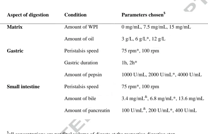
Table 1. Overview of digestion conditions studied in dependence of various concentrations of
whey protein isolate (WPI).
Aspect of digestion Condition Parameters chosen$
Matrix Amount of WPI
Amount of oil
0 mg/mL, 7.5 mg/mL, 15 mg/mL
3 g/L, 6 g/L*, 12 g/L
Gastric Peristalsis speed
Gastric duration
Amount of pepsin
75 rpm*, 100 rpm
1h, 2h*
1000 U/mL, 2000 U/mL*, 4000 U/mL
Small intestine Peristalsis speed
Amount of bile
Amount of pancreatin
75 rpm*, 100 rpm
3.4 mg/mL&, 6.8 mg/mL*, 13.6 mg/mL
100 U/mL&, 200 U/mL*, 400 U/mL
$
all concentrations are per final volume of digesta at the respective digestive step
*Termed standard conditions , according to the European consensus digestion model (Minekus,
et al., 2014), except RPM which was not defined.
&
Version preprint
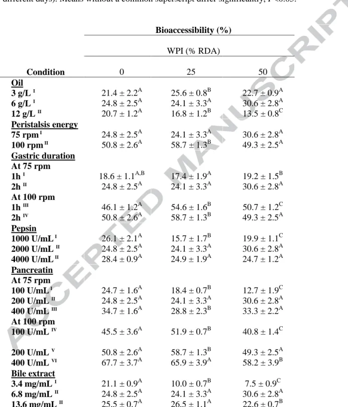
Table 2. Average bioaccessibility (%)1 of pure β-carotene digested with whey protein isolate (WPI) at varying concentrations2. Values represent mean ± SD of n = 4 and N≥2 (sets repeated at different days). Means without a common superscript differ significantly, P<0.05.
Bioaccessibility (%) WPI (% RDA) Condition 0 25 50 Oil 3 g/L I 6 g/L I 12 g/L II 21.4 ± 2.2A 24.8 ± 2.5A 20.7 ± 1.2A 25.6 ± 0.8B 24.1 ± 3.3A 16.8 ± 1.2B 22.7 ± 0.9A 30.6 ± 2.8A 13.5 ± 0.8C Peristalsis energy 75 rpm I 100 rpm II 24.8 ± 2.5A 50.8 ± 2.6A 24.1 ± 3.3A 58.7 ± 1.3B 30.6 ± 2.8A 49.3 ± 2.5A Gastric duration At 75 rpm 1h I 2h II At 100 rpm 1h III 2h IV 18.6 ± 1.1A,B 24.8 ± 2.5A 46.1 ± 1.2A 50.8 ± 2.6A 17.4 ± 1.9A 24.1 ± 3.3A 54.6 ± 1.6B 58.7 ± 1.3B 19.2 ± 1.5B 30.6 ± 2.8A 50.7 ± 1.2C 49.3 ± 2.5A Pepsin 1000 U/mL I 2000 U/mL II 4000 U/mL II 26.1 ± 2.1A 24.8 ± 2.5A 28.4 ± 0.9A 15.7 ± 1.7B 24.1 ± 3.3A 24.9 ± 1.9A 19.9 ± 1.1C 30.6 ± 2.8A 24.7 ± 1.2A Pancreatin At 75 rpm 100 U/mL I 200 U/mL II 400 U/mL III At 100 rpm 100 U/mL IV 200 U/mL V 400 U/mL VI 24.7 ± 1.6A 24.8 ± 2.5A 34.7 ± 1.6A 45.5 ± 3.6A 50.8 ± 2.6A 67.7 ± 3.7A 18.4 ± 0.7B 24.1 ± 3.3A 28.8 ± 2.3B 51.9 ± 0.7B 58.7 ± 1.3B 65.9 ± 3.9A 12.7 ± 1.9C 30.6 ± 2.8A 33.3 ± 2.2A 40.8 ± 1.4C 49.3 ± 2.5A 58.2 ± 3.9B Bile extract 3.4 mg/mL I 6.8 mg/mL II 13.6 mg/mL II 21.1 ± 0.9A 24.8 ± 2.5A 25.5 ± 0.7A 10.0 ± 0.7B 24.1 ± 3.3A 26.5 ± 1.1A 7.5 ± 0.9C 30.6 ± 2.8A 22.6 ± 0.7B
RDA: recommended dietary allowance. 1Bioaccessibility is expressed as the percentage of β-carotene recovered at the end of the in vitro GI digestion, compared to the amount added at the beginning of digestion. 2Concentrations given in final digestion stage (small intestine) are 0, 25 and 50% based-RDA.
Version preprint
Figure Headings
Fig. 1. Influence of whey protein isolate (WPI) (A), and oil, co-digested with WPI (B) on
β-carotene bioaccessibility at 75 rpm water bath shaking speed. β-Carotene was digested either in the
presence of WPI at different amounts (0, 187 and 375 mg per final 25 mL digesta volume,
representing 0, 25 and 50% of protein recommended dietary allowance (RDA)), or in the presence
of different volumes of oil (75, 150 and 300 μL per final 25 mL digesta), at various amounts of WPI
(0, 25 and 50% of RDA) at 75 rpm. Bioaccessibility is expressed as the percentage of β-carotene
recovered from the aqueous micellar fraction at the end of the in vitro GI digestion, compared to the amount of β-carotene added at the beginning of digestion. Values represent means ± SD of n = 4
and N≥2 (sets repeated at different days). Labeled means without a common superscript (alphabetic
letters or roman numbers) differ significantly, P<0.05.
Fig. 2. A) Influence of peristalsis energy and gastric phase duration on β-carotene
bioaccessibility and the effect of additional co-digested whey protein isolate (WPI). B) Influence of pancreatin concentration on β-carotene bioaccessibility and the effect of additional co-digested
whey protein isolate (WPI) at 75 (left panel) and 100 rpm (right panel) water bath shaking speed.
β-Carotene was digested in the presence of different amounts of pancreatin representing 50, 100 and
200 U/mL digesta, expressed as protease activity, in the presence of various amounts of WPI (0,
187 and 375 mg per final 25 mL digesta volume, representing 0, 25 and 50% of recommended
dietary allowance (RDA)) at 75 rpm and 100 rpm. Bioaccessibility is expressed as explained in Fig.
1 heading. Values represent means ± SD of n = 4 and N≥2 (sets repeated at different days). Labeled
means without a common superscript (alphabetic letters or roman numbers) differ significantly,