Correlation between
In
Vivo and
In
Vitro Efficacy of Antimicrobial Agents
against Foreign Body Infections
A. F. Widmer, R. Frei, Z. Rajacic, and W. Zimmerli
From the Departments of Internal Medicine and Research and the Bacteriology Laboratory, University Hospital, Basel, SwitzerlandImplant-associated infections are often resistant to antibiotic therapy. Routine sensitivity tests fail to predict therapeutic success. Therefore experimental in vitro tests were sought that would better correlate with drug efficacy in device-related infections. The activity of six different antibi-otics against methicillin-resistantStaphylococcus epidermidiswas investigated. In vivo studies were performed with the guinea pig tissue-cage animal model; in vitro studies with minimum inhibiting and bactericidal concentrations, time-kill studies of growing and stationary-phase microorganisms, the killing of glass-adherent S. epidermidis.Drug efficacy on stationary and adherent microorganisms, but not minimum inhibiting concentrations, predicted the outcome of device-related infections. Rifampin cured 12 of 12 infections and was also the most efficient drug in any experimental in vitro test. Similarly, the failure of ciproftoxacin to eradicate foreign body infections correlated with its low efficacy on stationary-phase and adherent S.epidermidis.
Device-related infections often persist until the device is removed, even if the given antibiotic has a low minimum in-hibiting concentration (MIC) for the causing microorganism [1]. The preferential bacterial colonization ofbiomaterials plus a local immunodeficiency explain the high infective suscepti-bility of implants and the persistence of such infections[2-4]. Bacteria develop a survival mode by adhering to inert sur-faces and producing a biofilm [5-7]. Possibly the mode of growth and the adherence of bacteria to bioimplants explain the failure of certain antibiotics to cure device-related infec-tions.
To date, no standardized antimicrobial susceptibility test is available to evaluatedrug activity on adherent bacteria. MIC and minimum bactericidal concentration (MBC) evaluate the drug efficacy on planktonic bacteria in logarithmic phases of growth [8, 9], a situation far from that of adherent bacteria [5]. The activity of many antimicrobial agents strictly depends on the phase of bacterial growth [10]. MICs and MBCs are much higher for most drugs if tests are performed with partly adherent bacteria [11].
In the present study, we tested the efficacy of different anti-microbial agents in the treatment of guinea pig implant infections caused by a weak slime-producing strain of Staphylococcus epidermidis. To evaluate the in vitro efficacy
Received 30 May 1989; revised 6 November 1989.
Presented in part at the 28th Interscience Conference on Antimicrobial Agents and Chemotherapy, Los Angeles, October 1988.
Financial support: Lilly Research Laboratories, Indianapolis, and Hyland Therapeutic Divisions, Travenol Laboratories, Glendale, CA (to A.F.W.).
W. Z. received a career development award from the Swiss National Science Foundation.
Reprints and correspondence: Dr. W. Zimmerli, Departments of Medi-cine and Research, University Hospital, CH-4031 Basel, Switzerland.
The Journal of Infectious Diseases 1990;162:96-102
©1990 by The University of Chicago. All rights reserved. 0022-1899/90/6201-0015$01.00
of antibiotics, we modified the MBC test procedure and de-veloped a novel assay for testing adherent bacteria. We ana-lyzed parameters that could predict the clinical outcome of device-related infections by comparing pharmacokinetic parameters, routine-susceptibility tests, and drug efficacy on nongrowing and adherent staphylococci in an experimental model of foreign body infections [2, 3, 12].
Materials and Methods
Bacteria and antibiotics. The test strain for most experiments was a methicillin-resistant S.epidermidis.This strain (B3972) was a clinical isolate from a blood culture of a patient with catheter-related septicemia. Identification was determined by a Staphident strip (API, La Balme les Grottes, France).Ithad a distinct susceptibility pat-tern including resistance to methicillin, cotrimoxazole, erythromy-cin, gentamierythromy-cin, and tobramycin and sensitivity of ciprofloxaerythromy-cin, vancomycin, daptomycin, teicoplanin, and netilmicin. Such an an-timicrobial susceptibility pattern is known to be as discriminating as plasmid profile analysis [13]. The S.epidermidisB3972 revealed weak slime production [14, 15].
For control experiments, a rifampin-resistant variant of S. epider-midis B3972 (MIC >4 jLg/ml) was selected by serial exposure of bacteria to increasing subinhibitory concentrations of rifampin. With this strain we tested whether a possible synergistic effect of rifampin combinations would depend on the bacterial killing of rifampin or on a nonspecific property (e.g., antiadherent effect) of this drug. In some experiments another strain, a moderate-to-strong slime-pro-ducing, methicillin-resistant S.epidermidis(ATCC 35983) was used [15]. Working cultures were maintained on blood agar and trans-ferred weekly. A new aliquot of the initial strain, stored in skim milk at -70°C, was cultured every 3 months.
Slime production was qualitatively determined [14]. Control ex-periments to exclude bacterial clumping were performed by direct microscopy and serial sonication of samples~9min with 60 W/min (Labsonic 2000;B. Braun Instruments, Burlingame, CA). Bacterial clumping was not observed with either method. The following an-timicrobial agents, provided by the manufacturers, were evaluated:
JID 1990;162 (July) Treatment of Foreign Body Infections 97
ciprofloxacin (Bayer AG, Wuppertal, FRG), rifampin (Ciba-Geigy, Basel, Switzerland), netilmicin (Schering, Kenilworth, NJ), van-comycin and daptomycin (Eli Lilly, Indianapolis), and teicoplanin (LePetit, Milan, Italy).
Animal model. We used a previously described foreign-body an-imal model [2, 3, 12]. In brief, four sterile polytetrafluorethylene (Teflon) tubes (32 x 10 mm), perforated by 130 regularly spaced holes (tissue cages; Ciba-Geigy) were aseptically implanted into the flanks of albino guinea pigs weighing 600-1100 g. Experiments were started after complete healing of wounds, "-'2-4 weeks after sur-gery. Before each experiment, interstitial fluid accumulating in the tissue cages was checked for sterility.
At day 0, for each experiment, 16 tissue cages were infected by local inoculation of 104 cfu of S.epidermidis. At day 1, infection was confirmed by quantitative cultures of the tissue-cage fluid. Twenty-four hours after inoculation, antibiotic therapy was started. Each drug was given every 12 h for 4 days (total eight doses). Ap-plication was intraperitoneal (ip) except for netilmicin, which was given intramuscularly (im). In any experiment, one untreated ani-mal with four cages served as a control. Tissue-cage fluid was aspi-rated daily for quantitative culture during therapy and at days 7, II, and 16 during follow-up.
At day 16, tissue cages were removed, under strictly aseptic con-ditions, from animals anesthetized with fentanyl-droperidol. After semiquantitative culture on Mueller-Hinton agar [16], tissue cages were incubated in trypticase soy broth for 48 h. Any positive culture identified asS.epidermidiswas defined as a treatment failure if the isolated strain had the same antimicrobial susceptibility pattern as the inoculated strain. The antibiogram included 29 antimicrobial agents. Each drug regimen was tested with 12 tissue cages, except teicoplanin(n
=
22) and low-dose rifampin(n=
24). Drug combi-nations were considered synergistic if the outcome differed sig-nificantly (P<
.05) from monotherapy.Analysis oftissue-cage fluid. Physicochemical parameters in the infected tissue cages were evaluated by determining pH, Po2,and I\::02' Heparin-rinsed 1-ml syringes with 2 l-gauge hypodermic nee-dles (Monovette; Sarstedt Rommelsdorf, FRG) were used for sam-pling. Samples were drawn 24 h after infection with S.epidermidis B3972, and were immediately processed in a blood gas analyzer (ABL 300 acid-base-laboratory; Radiometer Kopenhagen, Copen-hagen). In one-third of the samples, hemoglobin concentration ex-ceeded 1.5 g/l. These fluids were not analyzed because ofthe buffer capacity of hemoglobin. Blood contamination occasionally resulted from puncture of small vessels during aspiration.
Pharmacokinetics. Pharmacokinetic studies were performed in sterile and infected tissue cages (day 4 of infection with S. epider-midisB3972). Every drug was injected ip, except netilmicin, which was given im. Peak levels in serum and in tissue-cage fluid were determined 1 and 3-5 h after the drug application, respectively. Trough levels in cage fluid were measured at 12
±
0.5 h. All antibi-otic levels were measured by agar diffusion bioassays [17].Bacillus subtilis(0453-360, Difco, Detroit) was used as the test strain for ciprofloxacin, vancomycin, teicoplanin, and netilmicin;Micrococcus luteus (ATCC 9341) was used for daptomycin. The dose-response curves were linear in the required range. Antibiotics for the stan-dards were diluted with pooled guinea pig serum (50%). In a large plate (30 x 30 ern), reference standard was placed in three wells and each sample in two wells. Standard and samples were randomly located in different areas of the plate. Results were calculated bymicrocomputer-assisted exponential regression analysis (Hewlett-Packard HP41V).
MIC/MBC determinations. MICs were determined by the broth dilution method supplemented with appropriate divalent cations [8]. The inoculum was adjusted from an overnight culture to a concen-tration of 5 x lOS cfu/ml. MBCs were performed according to the Manual of Clinical Microbiology[9]. The antibiotic concentration, which reduced the initial inoculum >99.9%, was read as MBC. MBCs were also determined with bacteria in the stationary phase of growth. For this purpose, we modified the standard method by replacing Mueller-Hinton broth by 0.25 % glucose-supplemented phosphate-buffered saline (PBS), pH 7.4. Under these conditions, bacterial counts remained in the same range~36h. For daptomycin, glucose-supplemented (0.25%) NaCI 0.9%, adjusted for pH 7.4 with natri-umbicarbonate, was used to provide an ionized Ca'" level of~50
mg/1. Ionized Ca'" levels were determined for any solution used for daptomycin (Nova 2, ionized calcium analyzer; Novabiochemicals, Newton, MA). This concentration of ionized Ca'" enables dap-tomycin's antimicrobial activity in vitro [18]. Experiments were per-formed in triplicate.
Time-kill curves. Time-kill curves of bacteria in the logarithmic phase of growth were performed in Catt-supplemented Mueller-Hinton broth [9]. For kill curves of bacteria in the stationary phase of growth, broth was replaced by glucose-supplemented PBS, pH 7.4. For daptomycin, we used NaCI(0.9 %) adjusted with bicarbonate to a pH 7.4 with the above mentioned supplements. Drug concen-trations that correspond to standard MBCs were used to kill curves [9]. Samples for plating were diluted lO-fold to minimize antibiotic carryover. The number of cfu was determined at 2,4,6, 8, 12, and 24 h, respectively, by plating appropriate dilutions ofthe culture on Mueller-Hinton agar. The test was processed in a room at a con-stant 35°C to avoid unstable temperatures during handling.
Killing ofadherent bacteria. To investigate the antimicrobial ac-tivity against adherent bacteria, we developed a novel test proce-dure. Glass slides (35 x 75 mm) were washed in alcohol! acetone and placed vertically in racks that were transferred in gas-sterilized polyacetal dishes (100 x 80 x 90 mm) (Semadeni AG, Ostermun-digen, Switzerland). S.epidermidisB3972 was suspended from a fresh overnight culture in 250 ml of sterile 0.25 % glucose-sup-plemented PBS (pH 7.4) at a final concentration of 8 x 10³cfu/ml and then placed in the dishes.
The inoculum was confirmed by plating 0.1mldirectly from each dish onto agar. Four slides, placed in the rack, were incubated in each dish for 24 h with agitation (90 rpm) at 35°C. Continuous agi-tation prevented clusters of adherent bacteria. After this attachment period, bacterial suspension was discarded, andallslideswere washed three times in a 0.9% solution of sterile saline. Both sides of two slides served as controls (adherent inoculum) and were slightly pressed with a forceps on Mueller-Hinton agar petri dishes. Care was taken to keep slides in contact with the agar and to avoid air bubbles. We assumed that any adherent microorganism gave a copy of itself on the agar. The other two slides were transferred with the rack in Ca'" (50 mg/l)-supplemented trypticase soy broth (1'5B) containing antibiotics. This rack was agitated (90 rpm) for another 24 h at 35°C at drug concentrations corresponding to twofold MBCs determined in the logarithmic phase of growth. Again, samples were washed before being pressed on Mueller-Hinton agar. Colonies of controls and samples were counted and expressed as colony-forming units per slide. Experiments were excluded from analysis if
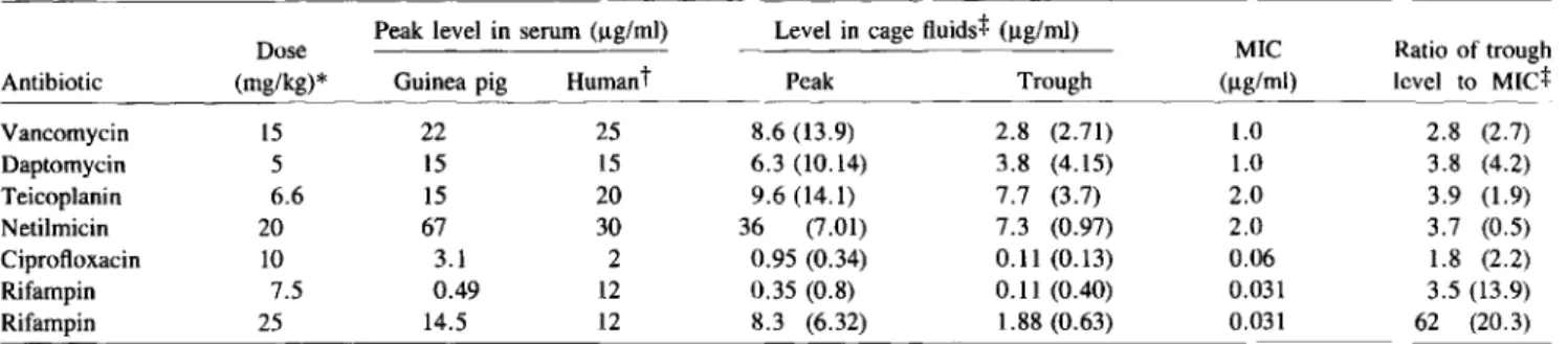
adher-Table 1. Pharmacokinetic studies of antibiotic levels and MIC of Staphylococcus epidermidis B3972. 2.8 (2.7) 3.8 (4.2) 3.9 (1.9) 3.7 (0.5) 1.8 (2.2) 3.5 (13.9) 62 (20.3) Ratio of trough level to MIC+ 1.0 1.0 2.0 2.0 0.06 0.031 0.031 MIC (ug/ml)
Guinea pig Humant Peak Trough
22 25 8.6 (13.9) 2.8 (2.71) 15 15 6.3 (10.14) 3.8 (4.15) 15 20 9.6 (14.1) 7.7 (3.7) 67 30 36 (7.01) 7.3 (0.97) 3.1 2 0.95 (0.34) 0.11 (0.13) 0.49 12 0.35 (0.8) 0.11 (0.40) 14.5 12 8.3 (6.32) 1.88 (0.63) 15 5 6.6 20 10 7.5 25
Dose Peak level in serum (I!g/ml) Level in cage fluids+ (I!g/ml)
Antibiotic (mg/kg)* Vancomycin Daptomycin Teicoplanin Netilmicin Ciprofloxacin Rifampin Rifampin
NOTE. MIC - minimum inhibitory concentration.
; Drugs were administered twice. daily.Netilmi~inwas administered intramuscularly; all others were administered intraperitoneally. Average of serum peak levelsInhumans provided by respective manufacturers.
:/: Ratios in parentheses calculated with the results of infected tissue-cage fluid.
ent bacteria of the controls were <102or >103cfu per slide so that each experiment had similar adherent inocula. The percentage kill rate was calculated as 100 - [(cfu per slide on drug-incubated slides)/(cfu per slide on control slides)] x 100.
Statistics. Fisher's exact test was performed for animal studies.
Variance component analysis was done for adherence studies using a statistical software (SAS Institute, Cary, NC).
Results
Pharmacokinetics.
Table 1 compares the pharmacokinetic studiesin
guinea pigs with the MIC of the corresponding drug. For each antibiotic a dosage was chosen that resulted in peak serum levels near those suggested by the drug's manufacturer. With these regimens,peak
levels in sterile cage fluids exceeded MBCs for every experimental drug. Trough levels in sterile fluids were below the MBC, but still above MICs for van-comycin, ciprofloxacin, and netilmicin (tables 1 and 2}. The ratio of the trough level in sterile cage fluid to MIC was simi-lar foralldrugs (1.8-3.9) except rifampin (table 1). The trough level in sterile tissue-cage fluid of the regular rifampin regi-men (25 mg/kg) exceeded the MIC 62 times. Thus, we also tested rifampin at a lower dose (7.5 mg/kg) , providing a trough level:MIC ratio in the same range as the other drugs. Antibi-otic levels in infected tissue-cage fluids (table 1, values in parentheses) were in the same range as in sterile fluids, ex-cept for netilmicin. As could be expected, these levels were lower in infected than in sterile tissue-cage fluid [19].TIssue-cage infection.
Infection of all cages was initiated by an initial inoculum of f\.IIQ4 cfu of S.epidermidis
B3972 or S.epidermidis
ATCC 35983. In control animals, bacterial counts in aspiration fluid fluctuated from 103to lOS cfu/mlregardless of the initial inoculum. '
Treatment results are summarized in figure1. No sponta-neous healing of implant infections occurred in 60 untreated cages, not even after a prolonged (3-month) observation(n =
4). The infection remained localized around the tissue cages. Infected tissue cages were expelled afterf\.I3months. Spon-taneous contamination of the cage fluid was observed during
the experiment in 5 %-10% , mostly due to gram-negative rods. The emergence of resistance was not observed with any an-tibiotic regimen.
Teicoplanin and ciprofloxacin, respectively, were the least efficient drugs; rifampin had the highest efficacy(P< .001).
Ciprofloxacin did not reduce bacterial counts during treat-ment (data not shown). The addition ofnetilmicin to daptomy-cin significantly improved the cure rate (P
<
.01). Synergy could not be demonstrated when netilmicin was combined with vancomycin. Rifampin was the only drug that sterilized any implant infection, mostly within 48 h after the start of treat-ment. With low-dose therapy (7.5 mg/kg), bacterial counts fell below the limit of detection, but in 25 % relapse with the same rifampin-sensitive strain occurred. This recurrence could be avoided by adding ciprofloxacin to low-dose rifampin. This combination failed to cure any implant (0 of 12) infected by the rifampin-resistant S.epidermidis
B3972 variant. In all ex-periments shown in figure 1, treatment was started 24 h after inoculation. For rifampin, the most efficient drug, this inter-val was prolonged to 1 week (3 and 7 days)..Rifampin (25mg!kg) sterilized all 12 implant infections, even when started 7 days after bacterial inoculation.
To check the importance of slime production on antibiotic efficacy, we performed two more series of experiments with
S. epidermidis
ATCC 35983, known to be a moderate to strongTable 2. Minimum bactericidal concentration (MBC) of
Staph-ylococcus epidermidis B3972.
Phases of bacterial growth (ug/ml)
Fold
Antibiotic Logarithmic Stationary increase
Vancomycin 4 50 12.5 Daptomycin 2 12.5 6 Teicoplanin 4 12.5 3 Ciprofloxacin 0.5 100 200 Rifampin 0.06 0.15 2.5 Netilrnicin 8 400 50
1101990;162 (July) Treatment of Foreign Body Infections 99 Antibiotic Regimen mg/kg bid Controls
~
• • • • • • • • • •1111• •'"
100% Ciprofloxacin 10+Rifampin 7.5~ 17% Figure 1. Cure rate of tissue-cageinfections with Staphylococcus epidermidis B397~. Guinea pigs with tissue-eage implants weretreated with antibiotics as indicated on the y axis. Numbers of infections: con-trols, 60; antibiotics, 12 each, ex-cept teicoplanin, 22, and rifampin (7.5 mg/kg), 24. Rifampin at 25 mg/kg was significantly more effi-cient than at 7.5 mg/kg (P
<
.01,Fisher~ exact tesQ.
Teicoplanin 6.6
Ciprofloxacin 10
17%
75%
0% 20% 40% 60% 80% 100%
% Cure Rate of Tissue Cages
slime producer [14]. With this strain also, any tissue cage(n =
8) in control animals remained infected during the experiment. Rifampin (25 mg/kg) still sterilized a1112 tissue cages, whereas ciprofloxacin did not cure any infection (0 of 12).
Characteristics oftissue-cage fluid. In view of the differ-ent cure rates by antibiotics that reached local levels exceed-ing MICs at any time, we analyzed some characteristics of the infected tissue-cage fluids that might influence the in vivo efficacy of antibiotics. A slight acidosis (pH 7.18
±
0.04,n =12) and a moderately decreased Po. (7.67
±
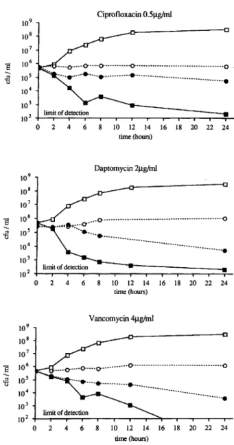
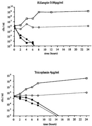
0.7 kPa) were measured.MBCs and time-kill curves in stationary and logarithmic phases of bacterial growth. Microorganisms involved in implant-associated infections may be in stationary growth phases. We thus sought in vitro tests with a better predictive value than the standard MIC and MBC, which are determined in the logarithmic phase of growth [9]. Table 2 shows that MBCs of S. epidermidisB3972 determined with bacteria in the stationary growth phase were up to 200 times higher (ciprofloxacin) than obtained with bacteria in the logarithmic phase of growth. Rifampin, teicoplanin, and daptomycin all had almost the same efficacy on nongrowing as on growing staphylococci. To further characterize these observations, we performed kill curves in stationary and logarithmic growth phases. The results of representative experiments are sum-marized in figures 2 and 3. Figure 2 shows drugs with dimin-ished activity on nongrowing bacteria. In contrast, rifampin and teicoplanin (figure 3) had a nearly identical killing rate in both systems. However, rifampin acted much faster than
Figure 2. Time-kill studies ofStaphylococcus epidermidisB3972. Concentration of antibiotic corresponds to minimum bactericidal concentration in logarithmic phase of growth. Controls and antibi-otics in logarithmic growth phases, respectively (D, _,); controls and antibiotics in stationary growth phases, respectively (0, .). 109 CiprofloxacinO.5Jlg/ml 108 107 E 106
--
105..a
<.> 104 103 102 limit of detection 0 2 4 6 8 10 12 14 16 18 20 22 24 time(hours) Oaptomycin2Jlg/ml 109 108 107e
106..a
o 105 ... 104...•
103 limit of detection 102 0 2 4 6 8 10 12 14 16 18 20 22 24 time (hours) Vancomycin4Jlg/ml 109 108 107e
106--
105..a
<.> 104 103 102 0 2 4 6 8 10 12 14 16 18 20 22 24 time (hours)lOW 109 108 8 10 7 ... 106 .E0 lOS 104 103 102 0 2 4 6 Rifampin O.06J.lg/ml 10 12 14 16 18 20 22 24 time (hours)
in device-related infections bacterial adhesion is crucial, we tested an additional parameter, drug efficacy on adherent bac-teria. With an initial inoculum of 8
x
1()3 cfu/ml, 330±
13 cfu per slide (mean±
SE,n
=
92) adhered to the glass slides. More than 103cfu per slide adhered with inocula exceeding 8x
1Q4cfu/ml. The variability among different assays with the same drug was always lower than among different drugs(F
=
11.86,P<
.01), confirming the reproducibility of the assay. Figure 4 summarizes the results of four or more ex-periments. Rifampin was the most efficient drug with a mean killing rate of 99.7%
by incubation of adherent bacteria at the doubled MBC. 109 Teicoplanin 4J.lg/ml 108 107 8 10 6 ... lOS .E 0 104 103 102 0 2 4 6 10 12 14 16 18 20 22 24 time (hours)Figure 3. Time kill studies ofStaphylococcal epidermidis B3972.
Concentration of antibiotic corresponds to minimum bacterical con-centration in logarithmic growth phase. Controls and antibiotics in logarithmic growth phase(0, _); controls and antibiotics in sta-tionary growth phases (0, .).
teicoplanin. In the time-kill curve, teicoplanin (4 p,g/ml) showed a killing rate of >99.9%, which contrasted with the stationary MBC of 12.5 p,g/ml; however, this may be due to the different techniques used. The antibiotic combinations used in the animal model did not improve the killing by the single agent (data not shown).
Drug efficacy on adherent S. epidermidis B3972. Since
Antibiotic
Discussion
We previously analyzed host defense mechanisms in the an-imal model used in this study [2, 3, 12]. Similar to humans, guinea pigs are highly susceptible to foreign-body infections [3]. Thus this model was used to test antibiotics for their ability to sterilize device-related infections. Clinical data concern-ing the antibiotic treatment of such infections are scarce and difficult to interpret. Therefore, criteria for surgical versus antibiotic treatment of the different implant infections are not well established[1].This reflects the difficulty in performing clinical studies under controlled conditions with unambigu-ous criteria to determine efficacy after prolonged follow-up periods.
To enable attachment and adherence of bacteria to the im-plant [5], we started the antimicrobial treatment not earlier than 24 h after inoculation. Short-term therapy was chosen for detecting antimicrobial agents with excellent efficacy. Ri-fampin was the most efficient drug; however, the extremely low MIC compared to the tissue-cage trough level could be a trivial explanation for its high efficacy. To correct for this, we used a low-dose regimen of rifampin that resulted in a ra-tio of trough level in tissue-cage fluid to MIC similar to that of the other drugs. At this low dose, rifampin was still the most efficient drug.
Ciprofloxacin11lg/ml Vancomycin81lglml Netilmicin 161lglml Teicoplanin81lg/ml Daptomycin 4Ilg/ml RifampinO.121lg/ml
Figure 4. Log killing of adherent Staph-ylococcus epidermidis B3972. Assay was
performed at drug concentrations twice the minimum bactericidal concentrations. Con-centrations are shown on y axis. Killing rate was determined as follows: 100 - [(cfu per slide on drug incubated slides)/(cfu per slide on control slides)]
x
100, expressed as 10glO killing.0.0 0.5 1.0 1.5
Log Killing
JID 1990;162 (July) Treatment of Foreign Body Infections 101
Ciprofloxacin had no detectable efficacy in the animal model. This failure could not be predicted by the standard MIC, which exceeded sterile and infected tissue-cage fluid trough levels twofold. The slightly decreased pH of the in-fected tissue-cage fluid could not explain the lack of an-timicrobial activity.
In contrast to regular MICs and MBCs, bacterial MBCs under stationary growth phase were predictive for the out-come of tissue-cage infections. The low performance of ci-profloxacin on nongrowing bacteria was surprising since Zeiler and Grohe [20] showed a high efficacy of this antibiotic on
Escherichiacoli
in1he stationary phase. However, this qual-ity of ciprofloxacin was later shown to be specific for some gram-negative rods [21, 22]. The low antistaphylococcal ac-tivity of cyprofloxacin has also been confirmedbyclinical data. Greenberg et al. [23] reported that none of six patients with osteomyelitis caused byStaphylococcus aureus
(monoculture) was cured with ciprofloxacin therapy.Inour experiments the stationary-phase MBC wassurpassed only by thetissue-cage peak and trough levels in animals receiving rifampin. This was the sole single-drug regimen with a high success rate. Several authors have reported the rapid emergence of rifampin-resistant staphylococci during exposure to this drug [24, 25]. Unlike Archer et al. [25], we did not detect the emergence of rifampin-resistant
S.· epidermidis
dur-ing treatment with rifampin, probably because the bacterial counts in tissue-cage fluid remained at a low level. Neverthe-less, in treatment of human infections, rifampin should al-ways be used in combination with other drugs. Vancomycin, which has a much lower volume of distribution than rifam-pin, is probably not the ideal drug to combine with rifampin [26, 27]. Thus, we tested the effect of ciprofloxacin in combina-tion with low-dose rifampin. The improved cure rate indicates in vivo synergism of this combination. We also sought a pos-sible nonantimicrobial effect of rifampin (e.g., antiadhesive potency) and repeated this experiment with the rifampin-resis-tant variant. This combination did not cure any tissue-cage infection, thus precluding a nonspecific effect of rifampin. As a further diagnostic parameter for the antimicrobial ac-tivity in foreign-body infections, we measured the killing of in vitro adherent bacteriabyantibiotics. Our assay proved sim-ple and reproducible because the variability among different assays with the same drug was always lower than between different drugs.Although the assay is fastidious, there are several possible pitfalls. The number of adherent bacteria is crucial. Also, poly-acetal dishes give better reproducible results than glass dishes (data not shown), and the adherent bacteria on each slide re-produce better if the dishes are gently agitated during the whole procedure. With this assay, rifampin again had excel-lent efficacy. The drug was the only one that eliminated >2.5 logs of the adherent bacteria after incubation for 24 h in a drug concentration corresponding to twofold MBC. The ex-cellent efficacy of rifampin in our in vitro assay and in the
animal model concurs with that found in arterial graft infec-tions in dogs [7].
Daptomycin and teicoplanin were the second best antibiot-ics, with a 1.7-10g killing of adherent S.
epidermidis.
This con-trasted with the poor in vivo results of teicoplanin. However, the drug concentration in tissue-cage fluid at trough levels did not exceed the stationary-phase MBC. Possibly the failure of teicoplanin was due to insufficient drug levels such as reported in clinical studies [28]. Daptomycin may also have been used at inadequate levels. All other antibiotics tested killed <1 log of adherent microorganisms. Ciprofloxacin again had the lowest activity. Gristina et al. [29] had similar findings with another method. They recently showed that adherent coagulase-negative staphylococci have generally higher MBCs than do planktonic bacteria.In conclusion, drug efficacy in the treatment of device-related experimental S.
epidermidis
infections may be pre-dicted by the described in vitro assays. In view of our experimental data, we treated some staphylococcal infec-tionsassociated with orthopedic implants with an antibiotic combination containing rifampin. With follow-up observa-tions of >1 year, disappearance of signs of inflammation could be observed (unpublished data). These preliminary data should be confirmed in a controlled clinical trial with the described assays as a guide for the choice of antibiotics.Acknowledgment
We thank: Peter Schumacher, Biocenter Basel, Switzerland, for assistance in statistical analyses.
References
1. Dougherty SH, Simmons RL. Infections in bionic man: the pathobiol-ogy of infections in prosthetic devices (parts I and II). Curr Probl Surg 1982;19:219-264, 268-319
2. Zimmerli W, Lew PO, Waldvogel FA. Pathogenesis of foreign body in-fection. Evidence for a local granulocyte defect. J Clin Invest 1984; 73:1191-1200
3. Zimmerli W, Waldvogel FA, Vaudaux P, NydeggerVE. Pathogenesis of foreign body infection: description and characteristics of an ani-mal model. J Infect Dis 1982;146:487-497
4. Vaudaux PE, Zulian G, Huggler E, Waldvogel FA. Attachment of
Staph-ylococcus aureusto polymethylmethacrylate increases its resistance to phagocytosis in foreign body infection. Infect Immun 1985;50: 472-477
5. Gristina AG. Biomaterial-centered infection: microbial adhesion versus tissue infiltration. Science 1987;237:1588-1595
6. Gristina AG, CostertonJW.Bacterial adherence to biomaterials and tissue. J Bone Joint Surg [Am] 1985;67A:264-273
7. Bergamini TM, Bandyk OF, Govostis OF, Kaebnick HW, TowneJB. Infection of vascular prostheses caused by bacterial biofilms. J Vase Surg 1988;7:21-30
8. National Committee for Clinical Laboratory Standards. Methods for dilution antimicrobial susceptibility tests for bacteria that grow aero-bically. 2nded. Tentative standard. NCCLS document M7-T2. Vil-lanova, PA: NCCLS, 1988
spe-cia! tests. In: Lennette EH, Balows A, Hausler WJ Jr, Shadomy HJ, eds. Manual of clinical microbiology. 4th ed. Washington, DC: Amer-ican Society for Microbiology, 1985;1000-1008
10. Tuomanen E, Cozens R, Tosch W, Zak0,Tomasz A. The rate of killing ofEscherichiacoliby (3-lactamantibiotics is strictly proportional to the rate of bacterial growth. J Gen MicrobiolI986;132:l297-1304 11. Blaser J, Weinmann0,Luethy R. Poor antibiotic activity against adher-ent bacteria [abstract 782]. In: Program and abstracts of the 28th In-terscience Conference on Antimicrobial Agents and Chemotherapy (Los Angeles). Washington, DC: American Society for Microbiol-ogy, 1988
12. Zimmerli W, WaldvogelFA. Models of foreign-body infections. In:Zak
0,Sande MA. Experimental models in antimicrobial chemotherapy. Vol I. London: Academic Press, 1986:295-317
13. Hartstein AI, ValvanoMA, Morthland VH, Fuchs PC, Potter SA, Crosa JH. Antimicrobic susceptibility and plasmid profile analysis as iden-tity tests for multiple blood isolates of coagulase-negative staphy-lococci. J Clin Microbiol 1987;25:589-593
14. Christensen GD, Simpson WA, Bisno AL, Beachey EH. Adherence of slime-producing strains ofStaphylococcus epidermidisto smooth sur-faces. Infect Immun 1982;37:318-326
15. Christensen GD, Simpson WA, Younger Il, Baddour LM, Barrett FF, Melton DM, Beachey EH. Adherence of coagulase-negative staphylococci to plastic tissue culture plates: a quantitative model for the adherence of staphylococci to medical devices. J Clin Microbiol 1985 ;22 :996-1006
16. Maki DG, Weise CE, Sarafin HW. A semiquantitative culture method for identifying intravenous-catheter-related infection. N Engl J Moo 1977;296:1305-1309
17. Edberg SC, Barry AL, Young LS. Therapeutic drug monitoring: an-timicrobial agents. In: Morello JA, ed. Cumulative techniques and procedures in clinical microbiology, Cumitech 20. Washington, DC: American Society for Microbiology, 1984:1-8
18. Andrew JR, Wale MCJ, Wale U, Greenwood D. The effect of cultural conditions on the activity of LYl46032 against staphylococciand strep-tococci. J Antimicrob Chemother 1987;20:213-221
19. Vaudaux P. Peripheral inactivation of gentamicin. J Antimicrob Che-mother 1981;8(suppl A):17-25
20. Zeiler HJ, Grohe K. The in vitro and in vivo activity of ciprofloxacin. Eur J Clin Microbiol Infect Dis 1984;3:339-343
21. Zeiler HJ. Evaluation of the in vitro bactericidal action of ciprofloxacin on cells ofEscherichiacoliin the logarithmic and stationary phases of growth. Antimicrob Agents Chemother 1985;28:524-527 22. Chalkley U, KoornhofHJ. Antimicrobial activity of ciprofloxacin against
Pseudomonas aeruginosa, Escherichia coli,andStaphylococcus aureus
determined by the killing curve method: antibiotic comparisons and synergistic interactions. Antimicrob Agents Chemother 1985; 28:331-342
23. Greenberg RN, Kennedy DJ, Reilly PM, Luppen KL, Weinandt WJ, Bollinger MR, Aguirre F, Kodesch F, Saeed AMK. Treatment of bone, joint, and soft-tissue infections with oral ciprofloxacin. Antimicrob Agents Chemother 1987;31:151-155
24. TshefuK, Zimmerli W, Waldvogel FA. Short-term administration of ri-fampin in the prevention or eradication of infection due to foreign bodies. Rev Infect Dis 1983;5(suppl 3):S474-S480
25. Archer GL, Johnston JL, Vazquez GJ, Haywood HB III. Efficacy of an-tibiotic combinations including rifampin against methicillin-resistant
Staphylococcus epidermidis:in vitro and in vivo studies. Rev Infect Dis 1983;5(suppl 3):S538-S542
26. Brumfitt W, Hamilton-Miller J. Methicillin-resistantStaphylococcus aureus.N Engl J Med 1989;320:1188-1196
27. Eng RHK, Smith SM, Tillem M, Cherubin C. Rifampin resistance. De-velopment during the therapy of methicillin-resistant Staphylococ-cus aureusinfection. Arch Intern Med 1985;145:146-148 28. Calain P, Krause KH, Vaudaux P, Auckenthaler R, Lew D, Waldvogel
F, Hirschel B. Early termination of a prospective, randomized trial comparing teicoplanin and f1ucloxacillin for treating severe staph-ylococcal infections. J Infect Dis 1987;155:187-191
29. Gristina AG, Jennings RA, Naylor PT, Myrvik QN, Webb LX. Com-parative in vitro antibiotic resistance of surface-colonizing coagulase-negative staphylococci. Antimicrob Agents Chemother 1989;33: 813-816