Hernia (2007) 11:261–263 DOI 10.1007/s10029-006-0172-5
123
C A S E R E P O R TSubcutaneous emphysema: a rare manifestation of a perforated
diverticulitis in a patent inguinal canal
Lambert de Vries · Anne-Sophie KnoepXi · Philippe Konstantinidis · Emmanuel Charbonney
Received: 29 August 2006 / Accepted: 9 November 2006 / Published online: 29 November 2006
© Springer-Verlag 2006
Abstract Patients with complicated diverticulitis rarely present with extraperitoneal manifestations but the manifestation of subcutaneous emphysema appears even more seldom. We present the case of a patient with a history of diabetes and immunosuppression, who was admitted with sepsis in association with cellu-litis and subcutaneous emphysema of the left groin. The absence of peritonism due to corticosteroid treat-ment, a history of a recent fall with an ilio- and ischio-pubic fracture and subcutaneous emphysema led to a delay in the diagnosis. The Wnal diagnosis was a perfo-rated diverticulitis in a patent inguinal canal, which was only revealed after surgery. The various complications of diverticulitis, including extraperitoneal manifesta-tions, and associated microorganisms implicated in cel-lulitis and subcutaneous emphysema are brieXy reviewed.
Keywords Diverticulitis · Inguinal canal · Subcutaneous emphysema
Introduction
One-third of patients with diverticulosis will develop diverticulitis. Intra-abdominal perforation, abscess, hemorrhage, intestinal stricture or obstruction and Wstula are the most common complications. These patients frequently present with abdominal symptoms and, on occasion, with peritonism. However, in rare cases, complications outside of the peritoneal cavity have been reported [1].
Case report
An 82-year-old institutionalized female patient with a history of corticosteroid (2 £ 40 mg/day) treatment for rheumatoid arthritis was admitted to our emergency department for dyspnea and left groin pain. These symptoms had appeared a week before admission, fol-lowing a fall onto the left hip. Her medical history included corticosteroid-induced diabetes, arterial hypertension, ischemic cardiopathy and peritonitis.
Physical examination revealed a patient in a mark-edly diminished general condition, afebrile, tachyp-neic, tachycardic and hypotensive. The abdominal examination showed a previous laparotomy scar and predominant pain in the left lower quadrant on palpa-tion, without signs of peritonism. A digital rectal exam-ination could not be carried out due to pain.
The left groin showed an inXammatory swelling with crepitations upon palpation. The surrounding skin was ischemic with a beginning of central necrosis. Blood tests conWrmed an inXammatory syndrome with high non-segmented neutrophiles and high C-reactive pro-tein in addition to a discrete rhabdomyolysis, diabetes L. de Vries
Internal Medicine Department, University Hospital Geneva, Rue Micheli-Du-Crest 24, 1211 Geneva 14, Switzerland A.-S. KnoepXi
Radiology Department, University Hospital Geneva, Rue Micheli-Du-Crest 24, 1211 Geneva 14, Switzerland
P. Konstantinidis
Surgery Department, University Hospital Geneva, Rue Micheli-Du-Crest 24, 1211 Geneva 14, Switzerland
E. Charbonney (&)
Intensive Care Department, University Hospital Geneva, Rue Micheli-Du-Crest 24, 1211 Geneva 14, Switzerland e-mail: emmanuel.charbonney@hcuge.ch
262 Hernia (2007) 11:261–263
123
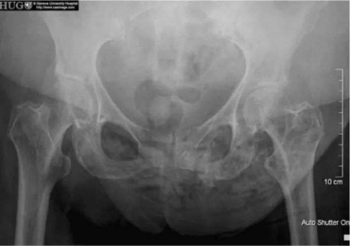
decompensation and acute renal failure. A pelvic X-ray showed subcutaneous air and a left ilio- and ischio-pubic fracture (Fig.1).
Systemic inXammation response syndrome (SIRS) along with the physical examination and pelvic X-ray described above led to the following diVerential diag-noses: rupture of a hollow pelvic organ consecutive to pelvic fracture, cutaneous infection in relation to the fall (with no evidence of portal of entry) or an incarcer-ated inguinal hernia with secondary rupture.
In addition to initiating resuscitation and antibiotic treatment, we performed an unenhanced – due to acute renal failure – abdominal-pelvic computed tomography scan (CT) with contrast media in the blad-der to investigate the possibility of a Wstula or a rup-tured bladder. The CT scan showed thickened intestinal loops in the lower left quadrant with a suspi-cion of communication with the subcutaneous air
associated with inWltration of the surrounding fat (Fig.2). Radiological Wndings were consistent with an old left minimally displaced ilio- and ischio-pubic frac-tures due to the presence of a callus. There was neither a bladder breach nor an intestinal lesion; there was also no free air or free Xuid in the peritoneal cavity.
An emergency surgical exploration of the groin region was decided upon. This showed cutaneous necrosis with an underlying abscess in an opening (con-taining no intestinal loop) leading to the peritoneal cavity. That opening was lateral, above the inguinal lig-ament through the direct Xoor. The laparotomy revealed a sigmoiditis, perforated along a length of 1 cm, with an important inXammatory adhesion against the patent inguinal canal, which was observed from the outside; there was no other extension of the diverticuli-tis inside the peritoneal cavity. An Hartmann’s sigmoi-dal resection with terminal colostomie was carried out. The hernia defect was closed with Wve separated stitches of 2.0 thread. Skin and tissue necrosis was extensively resected, the skin was closed with four loose separated stitches and a drain was left in place.
The surgical resection specimen conWrmed divertic-ulosis associated with a perforated diverticular abscess. The Wnal diagnosis was a perforated diverticulitis in a patent inguinal canal leading to subcutaneous absceda-tion and gas gangrene. In the postoperative period, our patient developed a progressive necrosis of the abdom-inal wall with septic shock and multiple organ failure which unfortunately led to her death.
Discussion
To our knowledge, this particular type of presentation of a perforated diverticulitis has never been described. The most common intra-peritoneal complications of diverticulitis are abscess formation (46%), frank perfo-ration (21%), diverticular hemorrhage (13%), intesti-nal stricture and occlusion (10%) and Wstula (10%) [2]. Among the latter, colo-vesical Wstulas are the most fre-quently encountered, followed by vaginal, colo-uterine and Wnally colo-cutaneous Wstula (1–2% of all Wstula cases) in decreasing order of prevalence.
Fistulas occur in more than 90% of cases after resec-tion surgery for acute diverticulitis [3] or after percuta-neous catheter drainage of an intra-abdominal abscess. Only a minority of the Wstulas occur spontaneously, without any abdominal pathology, with corticosteroid treatment being a risk factor [4]. However extraperito-neal manifestations of diverticulitis are rare and result from Wstulizations outside of the peritoneal cavity. Most reported cases are retroperitoneal Wstulas
Fig. 1 Pelvic X-ray of the 82-year-old female patient showing
subcutaneous air and a left ilio- and ischio-pubic fracture
Fig. 2 Computed tomography scan of abdomen and pelvis of the
82-year-old patient. # Foley catheter, X air containing collection, O subcutaneous emphysema
Hernia (2007) 11:261–263 263
123
presenting as thigh cellulites [5] that can also extend tothe mediastinum and present thoracic or cervical sub-cutaneous emphysema [6]. Very few cases have been reported of a communication with mesenteric or portal veins involving the liver [7], the area of the appendix or epidural space. Subcutaneous emphysema can also result from a remote infection, and some dermatologi-cal manifestations are also described in the case of diverticulitis [8]. Our patient, who was on corticoste-roid treatment for rheumatic arthritis [9], presented a clinically silent diverticulitis with this rare extra-perito-neal manifestation. The lower resistance of the patent's inguinal canal possibly allowed a perforation of the diverticular inXammation at this location.
Following pelvic trauma, one should suspect cutane-ous infection secondary to local bacterial contamina-tion. Once these microorganisms, typically Clostridium perfringens, have passed the skin barrier and are in anaerobic conditions [10], they can develop and pro-duce toxins. The incubation period ranges from several hours to a few days. Cellulitis or necrosis then pro-gresses to form an abscess, which ultimately develops into a severe systemic disease. Apart from Clostridium perfringens, Streptococcus group A or group B and Staphylococcus are frequently found in soft tissue infections, the former commonly encountered in dia-betic patients. However, Enterobacteria, Bacteroides fragilis and Enterococcus, which are most commonly found in the peritoneal cavity, can also be involved. Nevertheless, gas gangrene essentially occurs as a result of infection by Clostridium species or other fer-mentation bacteria able to produce gas. No cutaneous portal of entry was found in our patient. The groin abscess was positive for Bacteroides fragilis and Clos-tridium perfringens, and the blood cultures were posi-tive for Escherichia coli and Enterobacter, all of which are intestinal microorganisms.
Conclusion
Extra-abdominal rupture or Wstulas are uncommon and atypical presentations of diverticulitis. Our patient
showed very few speciWc symptoms in relation to the important immunosuppression by corticosteroids, making the diagnosis all the more diYcult.
Immunosuppressed patients presenting with mini-mal digestive symptoms should receivecomplete radiological examinations to exclude the most proba-ble diagnoses and extend potential diagnoses to the more unexpected possibilities. However, in the pres-ent case, the Wnal diagnosis was only revealed after surgery.
References
1. Rothenbuehler JM, Oertli D, Harder F (1993) Extraperitone-al manifestation of perforated diverticulitis. Dig Dis Sci 38:1985–1988
2. McConnell EJ, Tessier DJ, WolV BG (2003) Population-based incidence of complicated diverticular disease of the sig-moid colon based on gender and age. Dis Colon Rectum 46:1110–1114
3. Fazio VW, Church JM, Jagelman DG et al. (1987) Colocuta-neous Wstulas complicating diverticulitis. Dis Colon Rectum 30:89–94
4. Finkelstein JA, Jamieson CG (1987) An association between anti-inXammatory medication and internal pelvic Wstulas. Dis Colon Rectum 30:168–170
5. Edwards JD, Eckhauser FE (1986) Retroperitoneal perfora-tion of the appendix presenting as subcutaneous emphysema of the thigh. Dis Colon Rectum 29:456–458
6. Cifuentes Tebar J, Aguayo Albasini JL, Robles Campos R et al. (1990) Subcutaneous emphysema as the initial manifes-tation of perforation of a hollow abdominal viscus. Rev Esp Enferm Dig 78:38–40
7. Sonnenshein MA, Cone LA, Alexander RM (1986) Divertic-ulitis with colovenous Wstula and portal venous gas. Report of two cases. J Clin Gastroenterol 8:195–198
8. Kurgansky D, Foxwell MM JR (1993) Pyoderma gangreno-sum as a cutaneous manifestation of diverticular disease. South Med J 86:581–584
9. Mpofu S, Mpofu CM, Hutchinson D et al. (2004) Steroids, non-steroidal anti-inXammatory drugs, and sigmoid divertic-ular abscess perforation in rheumatic conditions. Ann Rheum Dis 63:588–590
10. Mourvillier B, Bedos JP (2001) Soft tissue infections by anaerobic bacteria. Etiology, diagnosis, treatment. Rev Prat 51:319–324