COMMENTARY
Is Positron Emission Tomography an Accurate
Non-invasive Alternative to Sentinel Lymph Node Biopsy
in Breast Cancer Patients?
Ulrich Guller, Egbert Nitzsche, Holger Moch, Markus Zuber
Breast cancer is the most common malignancy among women, leading to approximately 45 000 deaths per annum in the United States (1). The presence of axillary lymph node me-tastases has major prognostic implications in breast cancer pa-tients (2,3), and it is an important criterion in determining the need for adjuvant chemotherapy (4). Sentinel lymph node (SLN) biopsy has become routine practice in the surgical treatment of patients with breast cancer because the disease status of the SLNs accurately reflects the status of the remaining axillary lymph nodes (5–7). Hence, patients who present with a negative SLN (no metastases) can be spared from having a more trau-matic axillary lymph node dissection (ALND), which has been shown to be associated with substantial short- and long-term sequelae (8,9). Despite the enormous advantages of SLN biopsy over ALND in regard to post-operative complications in node-negative patients, it would be of great clinical benefit if a reliable non-invasive method to assess lymph node status in breast can-cer patients could be found.
Positron emission tomography (PET) reflects the biochemical and physiologic processes occurring in the tissues being imaged and has been used in diagnosing a variety of malignancies (10,
11). The most frequently used positron emitting
radiopharma-ceutical is 18-fluor labeled 2-deoxy-D-glucose (18F-FDG), a ra-dioactively labeled glucose analog. The clinical use of18F-FDG–PET is based on the premise that cancer cells exhibit a higher glyco-lytic rate than do non-neoplastic cells. Thus,18F-FDG accumu-lates predominantly in the tumor tissue and can be visualized by a PET camera. Since the first reported visualization of lymph node metastases with 18F-FDG–PET in a preclinical animal study in 1990 (12), several investigations (13–19) have assessed the accuracy of PET in evaluating the nodal status of patients with breast cancer. These investigations have yielded conflicting results, with some investigators doubting that18F-FDG–PET is capable of accurately assessing the nodal status of breast cancer patients (13–16) and others believing that a noninvasive PET scan could replace SLN biopsy at predicting the disease status of the axillary lymph nodes (17–19).
In a recent investigation by an Italian group (18), pre-operative18F-FDG–PET was compared with the histologic find-ings of ALND in 167 clinically node-negative T1 (tumor size 艋2cm) and T2 (tumor size >2cm to 艋5cm) breast cancer pa-tients, the largest patient collective published. 18F-FDG–PET
detected 68 of 72 patients with axillary metastases, resulting in an overall sensitivity for PET of 94%. In the subset of T2 pa-tients, the sensitivity was even higher (98%; 48/49), with only one false-negative finding. Based on these results, the authors claimed that 18F-FDG–PET can safely predict axillary lymph node status in patients with breast cancer and is a reliable and
accurate method to identify patients who can avoid ALND. In the second largest published patient collective (19), pre-operative18F-FDG–PET was compared with the histologic
find-ings of ALND in 124 pT1–T3 (T3 tumor size >5cm) breast cancer patients. PET scanning correctly identified all 44 patients with axillary lymph node metastases, resulting in an overall sensitivity for PET of 100%. These findings led the investigators of that study to suggest that18F-FDG–PET should be considered as the initial test in evaluating axillary lymph nodes in breast cancer patients and that those patients without increased axillary
18F-FDG-uptake may not require ALND. Other investigations
(17,20–23) with smaller patient numbers have reported similar
findings, with sensitivities of pre-operative 18F-FDG–PET at detecting axillary lymph-node metastases ranging from 90% to 100%.
In contrast to the above-mentioned investigations, several studies have found that18F-FDG–PET has a low sensitivity at detecting SLN or axillary lymph node metastases. Indeed, Avril et al. (14) suggest that18F-FDG–PET scanning cannot substitute for histologic analyses of axillary lymph nodes. Their study compared the diagnostic potential of18F-FDG–PET with ALND in 41 breast cancer patients and reported an overall sensitivity for PET of 79% (19/24). PET sensitivity in the subset of patients with pT1 tumors was, however, only 33%; four of six patients had false-negative results, and the largest metastasis undetect-ed by 18F-FDG–PET measured 12 mm in diameter. Kelemen et al. (15) also found low PET sensitivity when they compared
18
F-FDG–PET scanning with the histolopathologic findings of SLN biopsies in clinically node-negative T1 and T2 breast can-cer patients. If the SLN biopsies were negative by hematoxylin and eosin (H&E) staining, further biopsy sections were obtained for immunohistochemical (IHC) analyses using polyclonal anti-cytokeratin (CK) antibodies. Four of five patients with SLN metasta-ses had false-negative 18F-FDG–PET scans (i.e., 18F-FDG–PET
sensitivity was only 20%), and the missed metastases ranged from a micro-metastasis (defined as a cohesive cluster of ma-lignant cells >0.2 mm to 艋2.0 mm in diameter), which was identified by IHC only, to macro-metastases up to 11 mm in
Affiliations of authors: U. Guller, Dept. of Surgery, University of Basel, Basel,
Switzerland and Department of Surgery, Duke University Medical Center, Durham, NC; E. Nitzsche, Department of Radiology, Institute of Nuclear Medi-cine, University of Basel, Basel, Switzerland; H. Moch, Institute of Pathology, University of Basel, Basel, Switzerland; M. Zuber, Department of Surgery, Kantonsspital Olten, Olten, Switzerland.
Correspondence to: Markus Zuber, M.D., Dept. of Surgery, Kantonsspital
Olten, CH-4600 Olten, Switzerland (e-mail: [email protected]).
See “Notes” following “References.”
Journal of the National Cancer Institute, Vol. 95, No. 14, © Oxford University
Press 2003, all rights reserved.
diameter. Furthermore, a Canadian study (24) also found low PET sensitivity when evaluating the accuracy of18F-FDG–PET at detecting axillary lymph node metastases in 41 T1–3 breast cancer patients. All patients underwent pre-operative18F-FDG–PET scan, SLN biopsy, and ALND. The SLN was examined using serial sectioning and IHC using a monoclonal anti-CK antibody (CAM 5.2 clone) in addition to standard H&E staining. They reported that 18F-FDG–PET was unable to identify SLNs that
were found to be node positive by IHC only and that PET had a sensitivity of 27% at detecting axillary lymph node metastases. We found similar PET sensitivity results in our own investi-gation (13), in which we compared pre-operative18F-FDG–PET with the histopathologic analysis of SLN biopsies in 31 clini-cally node-negative pT1 and pT2 breast cancer patients. SLNs were analyzed by standard H&E staining, IHC (CK anti-body cocktail CK22 and monoclonal anti-CK antianti-body Lu-5), and step sectioning. The step sectioning technique examines adjacent serial sections of the SLN, which are cut at different intervals, making them different thicknesses. Each SLN was analyzed intra-operatively, and lymph nodes that were more than or equal to 5 mm in diameter were bisected along their longi-tudinal axis into sections of 3–4 mm and snap frozen. The SLNs were intra-operatively examined at three levels with H&E stained sections (cutting interval of 150 m). If no metastases were diagnosed in the frozen sections, the remaining tissue of the lymph node was formalin-fixed and embedded in paraffin for histologic analysis. This residual tissue was then examined using serial sectioning (cutting interval of 150m; at least six levels), and the permanent H&E stained slides were screened for tumor cells. Of the 14 patients with SLN metastases, eight had false-negative results by18F-FDG–PET scan, including three patients with macro-metastases (4 mm, 4 mm, and 13 mm, respectively), two patients with micro-metastases (0.8 mm and 1 mm, respec-tively), and three patients with sub-micro-metastases that re-mained unidentified. Hence,18F-FDG–PET had an overall
sen-sitivity of detecting SLN metastases of 43% (6/14).
A study from The Netherlands (16) that evaluated 70 patients with predominantly early-stage breast cancer who underwent
18F-FDG–PET followed by SLN biopsy and/or ALND has
con-firmed our finding of low PET sensitivity in detecting axillary lymph node metastases. If the first slice of the SLN was negative for the presence of metastases, an additional four to six sections were examined by H&E staining and IHC (using monoclonal anti-CK antibody CAM5.2). For patients undergoing ALND, up to two slices per lymph node were examined by IHC. The over-all sensitivity of18F-FDG–PET at detecting lymph node metas-tases was 25% (8/32), and sensitivity was dependent on the tumor load in the axilla and the18F-FDG avidity of the primary
tumor. The authors also found that 18F-FDG–PET was
unreli-able in the detection of axillary lymph node micro-metastases, regardless of the 18F-FDG avidity of the primary tumor.
Our investigation (13) as well as other studies (2,16,25–27) indicate that the percentage of detected metastases in lymph nodes increases when using step sectioning and IHC as com-pared with standard pathology protocols. Most institutions make only one H&E-stained slice from each node obtained from an ALND, or they divide the node in half along its longitudinal axis and make one section from each tissue slice. In addition, serial or step sectioning of lymph nodes is usually not required as a standard procedure. In response to the false-negative rates asso-ciated with the SLN biopsy procedure, new pathology protocols
have emerged, including intra-operative serial frozen sections, cytokeratin IHC, and molecular biology techniques such as re-verse transcription polymerase chain reaction. IHC and step sec-tioning techniques are particularly useful in the detection of small tumor infiltrates (i.e., micro-metastases), which often re-main undetected using standard histopathologic analyses. Al-though large retrospective studies (2) have shown disease-free and overall survival disadvantages for breast cancer patients with micro-metastases, therapeutic consequences and the prog-nostic importance of micro-metastases remain a matter of sci-entific debate. It should be emphasized, however, that both mi-cro- and small mami-cro-metastases cannot be identified by PET scanning, because their diameters (i.e., <3 mm) are below the current spatial resolution of PET imaging, which is currently reported to be between 3 mm and 10 mm (28–30). Hence, in-vestigations comparing pre-operative 18F-FDG–PET with stan-dard histologic analyses of ALND could miss micro-metastases and small macro-metastases by both histologic analysis and PET scanning. Thus, patients with small tumor infiltrates that are undetectable by PET scan (i.e., false-negatives) might be wrongly classified as “true-negatives” by histologic analysis, resulting in incorrectly high sensitivities for PET scanning.
Although step sectioning and IHC would increase the rate of detection of small tumor infiltrates in axillary lymph nodes, the systematic use of these techniques is not feasible in the assess-ment of ALND specimens because these procedures are prohibi-tively time consuming and costly (2). In contrast, SLN biopsy offers the advantage that these techniques can be applied on a small number of lymph nodes. Interestingly, all four reported studies (13,15,16,24) that compared18F-FDG–PET with
histo-pathologic analyses of axillary lymph nodes yielded poor sen-sitivities for PET at detecting metastases (20%–43%, Table 1). The use of step sectioning and IHC in our investigation (13) allowed the detection of sub-metastases, all micro-metastases, and one macro-metastasis, all of which were unde-tected in the intra-operative frozen section analysis and in the initial H&E staining. The failure to detect tiny metastatic foci in large lymph nodes represents a limitation of the frozen section technique used in standard pathology protocols. Indeed, in our study (13), the sensitivity of18
F-FDG–PET at detecting sentinel lymph node metastases, which was initially 88% using standard H&E staining only, dropped to 43% after step sectioning and IHC were also used. It should be emphasized that none of the studies (17–19) that suggest that18F-FDG–PET is a reliable and
non-invasive alternative to ALND/SLN biopsy used techniques that would allow the detection of small metastases. Therefore, it is reasonable to assume that the reported values for sensitivities of 18F-FDG–PET in those studies—most of them close to
100%—are overly optimistic and do not accurately reflect the limitations of this diagnostic tool. Therefore, it might be mis-leading to report the diagnostic potential of18F-FDG–PET with-out systematically performing histologic analyses that allow the detection of small tumor infiltrates.
In contrast to the limited sensitivity of PET scanning in the detection of small axillary lymph node metastases, most studies report high specificity (13,16,20–22,24,32) (Table 1). Although inflammatory processes, such as abscesses and sarcoidosis, can result in increased 18F-FDG uptake (33,34), the rate of
false-positive results is usually low (0–6%) (13,16,20–22,24,32). Due to high accuracy, pre-operative PET scanning has been sug-gested as a tool for tumor staging of patients with suspected
breast cancer (14,23). Previous studies (14,22) have found that PET scanning is able to provide additional and useful informa-tion about the extent of the disease. For example, the identifi-cation of distant metastases or tumor involvement of level III axillary lymph nodes, supra-clavicular, or internal mammary lymph nodes might have important implications in regard to the choice of local radiotherapy and systemic chemotherapy (14). However, it is currently premature to recommend a pre-operative PET scan for all patients with suspicious breast masses because the diagnostic, quality of life, and economic implica-tions of this diagnostic tool need further elucidation.
In summary, based on the present literature (14–16) and on own experience (13), we suggest that PET scanning does not currently have the adequate spatial resolution to detect both micro- and small macro-metastatic disease in axillary lymph nodes of patients with breast cancer. Therefore, PET scanning cannot serve at this time as a non-invasive alternative to SLN biopsy. It is possible, however, that with the advent of new detector materials in clinical practice (e.g., lithium, silicon, curved crystals), the sensitivity of 18F-FDG–PET will further improve (35) and could eventually replace invasive procedures in the evaluation of the nodal status of patients with breast cancer.
R
EFERENCES(1) Landis SH, Murray T, Bolden S, Wingo PA. Cancer statistics, 1999. CA
Cancer J Clin 1999;49:8–31.
(2) Dowlatshahi K, Fan M, Snider HC, Habib FA. Lymph node
micrometas-tases from breast carcinoma: reviewing the dilemma. Cancer 1997;80: 1188–97.
(3) Fisher B, Bauer M, Wickerham DL, Redmond CK, Fisher ER, Cruz AB,
et al. Relation of number of positive axillary nodes to the prognosis of patients with primary breast cancer. An NSABP update. Cancer 1983;52: 1551–7.
(4) Goldhirsch A, Glick JH, Gelber RD, Coates AS, Senn HJ. Meeting
high-lights: International Consensus Panel on the Treatment of Primary Breast
Cancer. Seventh International Conference on Adjuvant Therapy of Primary Breast Cancer. J Clin Oncol 2001;19:3817–27.
(5) Giuliano AE, Jones RC, Brennan M, Statman R. Sentinel
lymphadenecto-my in breast cancer. J Clin Oncol 1997;15:2345–50.
(6) Bass SS, Cox CE, Ku NN, Berman C, Reintgen DS. The role of sentinel
lymph node biopsy in breast cancer. J Am Coll Surg 1999;189:183–94.
(7) Veronesi U, Galimberti V, Zurrida S, Pigatto F, Veronesi P, Robertson C,
et al. Sentinel lymph node biopsy as an indicator for axillary dissection in early breast cancer. Eur J Cancer 2001;37:454–8.
(8) Warmuth MA, Bowen G, Prosnitz LR, Chu L, Broadwater G, Peterson B,
et al. Complications of axillary lymph node dissection for carcinoma of the breast: a report based on a patient survey. Cancer 1998;83:1362–8.
(9) Dayalan C, Arnold G, Antony J, Simon H. Short term morbidity from
axillary surgery in breast cancer. [Abstract]. Breast Cancer Res Treat 2001; 69:323.
(10) Bombardieri E, Aliberti G, de Graaf C, Pauwels E, Crippa F. Positron
emission tomography (PET) and other nuclear medicine modalities in stag-ing gastrointestinal cancer. Semin Surg Oncol 2001;20:134–46. Review.
(11) Delbeke D. Oncological applications of FDG PET imaging: brain tumors,
colorectal cancer, lymphoma and melanoma. J Nucl Med 1999;40: 591–603.
(12) Wahl RL, Kaminski MS, Ethier SP, Hutchins GD. The potential of
2-de-oxy-2[18F]fluoro-D-glucose (FDG) for the detection of tumor involvement in lymph nodes. J Nucl Med 1990;31:1831–5.
(13) Guller U, Nitzsche EU, Schirp U, Viehl CT, Torhorst J, Moch H, et al.
Selective axillary surgery in breast cancer patients based on positron emis-sion tomography with 18F-fluoro-2-deoxy-D-glucose: not yet! Breast Can-cer Res Treat 2002;71:171–3.
(14) Avril N, Dose J, Janicke F, Ziegler S, Romer W, Weber W, et al.
Assess-ment of axillary lymph node involveAssess-ment in breast cancer patients with positron emission tomography using radiolabeled 2-(fluorine-18)-fluoro-2-deoxy-D-glucose. J Natl Cancer Inst 1996;88:1204–9.
(15) Kelemen PR, Lowe V, Phillips N. Positron emission tomography and
sen-tinel lymph node dissection in breast cancer. Clin Breast Cancer 2002;3: 73–7.
(16) van der Hoeven JJ, Hoekstra OS, Comans EF, Pijpers R, Boom RP, van
Geldere D, et al. Determinants of diagnostic performance of [F-18]fluoro-deoxyglucose positron emission tomography for axillary staging in breast cancer. Ann Surg 2002;236:619–24.
(17) Adler LP, Faulhaber PF, Schnur KC, Al-Kasi NL, Shenk RR. Axillary
lymph node metastases: screening with [F-18]2-deoxy-2-fluoro-D-glucose (FDG) PET. Radiology 1997;203:323–7.
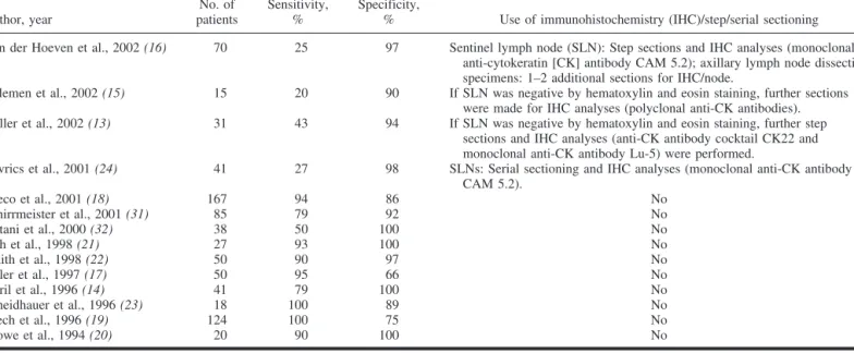
Table 1. Sensitivity and specificity of18F-FDG-PET scanning at detecting sentinel lymph node (SLN) and axillary lymph node metastases
in patients with breast cancer*
Author, year No. of patients Sensitivity, % Specificity,
% Use of immunohistochemistry (IHC)/step/serial sectioning Van der Hoeven et al., 2002 (16) 70 25 97 Sentinel lymph node (SLN): Step sections and IHC analyses (monoclonal
anti-cytokeratin [CK] antibody CAM 5.2); axillary lymph node dissection specimens: 1–2 additional sections for IHC/node.
Kelemen et al., 2002 (15) 15 20 90 If SLN was negative by hematoxylin and eosin staining, further sections were made for IHC analyses (polyclonal anti-CK antibodies). Guller et al., 2002 (13) 31 43 94 If SLN was negative by hematoxylin and eosin staining, further step
sections and IHC analyses (anti-CK antibody cocktail CK22 and monoclonal anti-CK antibody Lu-5) were performed.
Lovrics et al., 2001 (24) 41 27 98 SLNs: Serial sectioning and IHC analyses (monoclonal anti-CK antibody CAM 5.2). Greco et al., 2001 (18) 167 94 86 No Schirrmeister et al., 2001 (31) 85 79 92 No Yutani et al., 2000 (32) 38 50 100 No Noh et al., 1998 (21) 27 93 100 No Smith et al., 1998 (22) 50 90 97 No Adler et al., 1997 (17) 50 95 66 No Avril et al., 1996 (14) 41 79 100 No Scheidhauer et al., 1996 (23) 18 100 89 No Utech et al., 1996 (19) 124 100 75 No Crowe et al., 1994 (20) 20 90 100 No *18F-FDG-PET⳱ 2-[18
F]fluoro-2-deoxy-D-glucose positron emission tomography.
(18) Greco M, Crippa F, Agresti R, Seregni E, Gerali A, Giovanazzi R, et al.
Axillary lymph node staging in breast cancer by 2-fluoro-2-deoxy-D -glucose-positron emission tomography: clinical evaluation and alternative management. J Natl Cancer Inst 2001;93:630–5.
(19) Utech CI, Young CS, Winter PF. Prospective evaluation of fluorine-18
fluorodeoxyclucose positron emission tomography in breast cancer for staging of the axilla related to surgery and immunocytochemistry. Eur J Nucl Med 1996;23:1588–93.
(20) Crowe JP, Jr., Adler LP, Shenk RR, Sunshine J. Positron emission
tomog-raphy and breast masses: comparison with clinical, mammographic, and pathological findings. Ann Surg Oncol 1994;1:132–40.
(21) Noh DY, Yun IJ, Kim JS, Kang HS, Lee DS, Chung JK, et al. Diagnostic
value of positron emission tomography for detecting breast cancer. World J Surg 1998;22:223–7.
(22) Smith IC, Ogston KN, Whitford P, Smith FW, Sharp P, Norton M, et al.
Staging of the axilla in breast cancer: accurate in vivo assessment using positron emission tomography with 2-(fluorine-18)-fluoro-2-deoxy-D -glucose. Ann Surg 1998;228:220–7.
(23) Scheidhauer K, Scharl A, Pietrzyk U, Wagner R, Gohring UJ, Schomacker
K, et al. Qualitative [18F]FDG positron emission tomography in primary breast cancer: clinical relevance and practicability. Eur J Nucl Med 1996; 23:618–23.
(24) Lovrics P, Chen V, Coates G, Cornacchi S, Goldsmith C, Law C, et al.
A prospective study of PET scanning, sentinel lymph node biopsy, and standard axillary dissection for axillary staging in patients with early stage breast cancer. Abstract book 24thAnnual Cancer Symposium, Society of
Surgical Oncology. [Abstract]. March 2001.
(25) Giuliano AE, Dale PS, Turner RR, Morton DL, Evans SW, Krasne DL.
Improved axillary staging of breast cancer with sentinel lymphadenectomy. Ann Surg 1995;222:394–9; discussion 9–401.
(26) Wong SL, Chao C, Edwards MJ, Simpson D, McMasters KM. The use of
cytokeratin staining in sentinel lymph node biopsy for breast cancer. Am J Surg 2001;182:330–4.
(27) Cserni G. Metastases in axillary sentinel lymph nodes in breast cancer as
detected by intensive histopathological work up. J Clin Pathol 1999;52: 922–4.
(28) Avril N, Rose CA, Schelling M, Dose J, Kuhn W, Bense S, et al. Breast
imaging with positron emission tomography and fluorine-18 fluorodeoxy-glucose: use and limitations. J Clin Oncol 2000;18:3495–502.
(29) Grosev D, Loncarics S, Vandenberghe S, Dodig D. Triple-head gamma
camera PET: system overview and performance characteristics. Nucl Med Commun 2002;23:809–14.
(30) Hoffman EJ, Huang SC, Phelps ME. Quantitation in positron emission
computed tomography: 1. Effect of object size. J Comput Assist Tomogr 1979;3:299–308.
(31) Schirrmeister H, Kuhn T, Guhlmann A, Santjohanser C, Horster T, Nussle
K, et al. Fluorine-18 2-deoxy-2-fluoro-D-glucose PET in the preoperative staging of breast cancer: comparison with the standard staging procedures. Eur J Nucl Med 2001;28:351–8.
(32) Yutani K, Shiba E, Kusuoka H, Tatsumi M, Uehara T, Taguchi T, et al.
Comparison of FDG–PET with MIBI-SPECT in the detection of breast cancer and axillary lymph node metastasis. J Comput Assist Tomogr 2000; 24:274–80.
(33) Lewis PJ, Salama A. Uptake of fluorine-18-fluorodeoxyglucose in
sarcoid-osis. J Nucl Med 1994;35:1647–9.
(34) Tahara T, Ichiya Y, Kuwabara Y, Otsuka M, Miyake Y, Gunasekera R,
et al. High [18F]-fluorodeoxyglucose uptake in abdominal abscesses: a PET
study. J Comput Assist Tomogr 1989;13:829–31.
(35) Chatziioannou A, Tai YC, Doshi N, Cherry SR. Detector development for
microPET II: a 1 microl resolution PET scanner for small animal imaging. Phys Med Biol 2001;46:2899–910.
N
OTESWe thank the Swiss National Foundation, Bern/Switzerland, Krebsliga beider Basel, Basel/Switzerland, Freiwillige Akademische Gesellschaft, Basel/ Switzerland, and Fondazione Gustav und Ruth Jacob, Aranno/Switzerland for their financial support of Dr. Guller’s research fellowship. We also thank Mr. Jonathan McCall for carefully reading the manuscript and making many valuable suggestions.
Manuscript received November 7, 2002; revised April 25, 2003; accepted May 12, 2003.