Case report
Congenital subaortic stenosis by accessory mitral valve tissue,
recognition and management
Andreas C. Schmid, Gregor Zund*, Paul Vogt, Marko Turina
Clinic for Cardiovascular Surgery, University Hospital, Ramistrasse 100, CH 8091 Zurich, SwitzerlandReceived 7 September 1998; received in revised form 11 January 1999; accepted 12 January 1999
Abstract
Accessory mitral valve tissue as the single cause for left ventricular outflow tract obstruction is a very rare cardiac malformation in normally connected hearts. We report a case in which this condition was present as single cause for left ventricular outflow tract obstruction. The surgical technique is described and a review of the literature presented.1999 Elsevier Science B.V. All rights reserved. Keywords: Subaortic; Stenosis; Accessory tissue; Surgery; Recognition
1. Introduction
Accessory mitral valve tissue causing left ventricular out-flow tract obstruction is a rare pathologic finding in nor-mally connected hearts. It is of great importance to repair the valve without valve replacement. A high incidence of reoperations have been reported in cases where it is not recognized or is incompletely removed [1]. When this pathology is associated with a classic fibro-muscular sub-aortic stenosis, it may remain unrecognized pre-operatively and result in an incomplete surgical resection. We report such a case and the tactics we employed to completely relieve the obstruction without causing valve dysfunction.
2. Case report
A 17-month-old child was referred due to progressive development of a left ventricular outflow tract obstruction. The diagnosis of subaortic stenosis had been established by echocardiography shortly after birth and the child was fol-lowed regularly. The child had a normal growth and never
presented signs of heart failure. On admission, the transthor-acic echocardiography revealed a hypertrophied left ventri-cle and a severe subaortic stenosis with an estimated systolic gradient of 104 mmHg. No other cardiac anomaly was found. The analysis of the left ventricular outflow tract pointed out a subaortic ring-like membrane with a diameter of 9.7 mm and a normal mitral valve without a systolic anterior motion of the septal leaflet. The insertion of the mitral papillary muscles was normal. At that point, no abnormal valvular tissue was detected. Electrocardiography showed normal sinus rhythm and signs of right atrial over-load. There were no clinical findings indicating congestive heart failure. The child was presented at our clinic for sur-gical treatment at 17 months of age, 9.4 kg in weight and 78 cm in height.
The child underwent surgery using cardiopulmonary bypass under moderate hypothermia. After aortic cross-clamping and delivery of cold-blood cardioplegia into the aortic root, the ascending aorta was opened. The aortic valve and the annulus appeared normal. A subaortic mem-brane was at the usual position and created a moderate stenosis. This membrane was resected en bloc from annular and subannular tissues using sharp and blunt dissection. After the resection the left ventricular outflow tract appeared to be wide open and the subvalvular mitral appa-ratus did not appear pathologic. Therefore, the aorta was European Journal of Cardio-thoracic Surgery 15 (1999) 542–544
1010-7940/99/$ - see front matter 1999 Elsevier Science B.V. All rights reserved. P I I : S 1 0 1 0 - 7 9 4 0 ( 9 9 ) 0 0 0 2 3 - 8
* Corresponding author. Tel.: +41-1-255-1111; fax: +41-1-255-4369; e-mail: [email protected]
closed, the left ventricle de-aired and the aortic clamp removed. Upon resumption of heart activity, the transeso-phageal echocardiography still revealed turbulent flow underneath the aortic valve. Direct measurement revealed a systolic pressure gradient of 70 mmHg between the left ventricle and the aorta, indicating a remaining stenosis. Car-diopulmonary bypass and aortic cross-clamping were rein-stituted and a vertical left atriotomy was performed to inspect the mitral valve apparatus. A fibrotic mass attached to the mitral valve tissue was found close to the antero– lateral commissure. The tissue, including its subvalvular attachment was resected. Competence of the mitral valve was tested by injection of saline solution into the left ven-tricle. The left atrium was closed, the heart de-aired and the aortic clamp removed. Weaning from cardiopulmonary bypass occurred without difficulties. The TEE showed a non-turbulent flow between the left ventricle and the aorta and a competent mitral valve. Direct pressure measurement showed complete disappearance of the systolic gradient across the outflow tract.
The patients recovery was uneventful and she could be dismissed from the intensive care unit 2 days after surgery. After 11 months she remains in good general condition and shows normal development.
3. Discussion
To our knowledge, there are few such cases having been described until now. Usually there are coexisting cardiac malformations which may greatly influence the diagnostic pathway and the surgical approach. Most commonly this malformation is associated with ventricular septal defect (VSD) [2], transposition of great arteries (TGA) [3], coarc-tation, partial atrioventricular canal, double outlet right ven-tricle (DORV), membranous septal aneurysm (MSA) [4] and Noonan syndrome [5]. In this case, the accessory mitral valve leaflet was only associated with a small subaortic membrane.
The correct preoperative diagnosis is extremely difficult to establish and is often ignored by conventional diagnostic methods. Echocardiography precisely delineates the left ventricular outflow tract obstruction and is frequently used as a sole diagnostic tool to assess subaortic stenosis [6,7]. Assessing the anatomic and dynamic contributions of the mitral valve in the obstructive process is also possible by echocardiography, although this analysis requires expertise and may be difficult to obtain preoperatively. Rare causes of left ventricular outflow tract obstruction arising from the mitral valve may remain unrecognized preoperatively, espe-cially when a typical cause of obstruction, like a subaortic membrane is present. Detection of turbulent flow in post-repair transesophageal echocardiography pointed out the persistence of a significant outflow tract obstruction in this case. The fine analysis of the outflow tract revealed abnor-mal tissue in contact to the anterior mitral valve leaflet. The decision to approach this pathology through left atriotomy was made for two reasons.
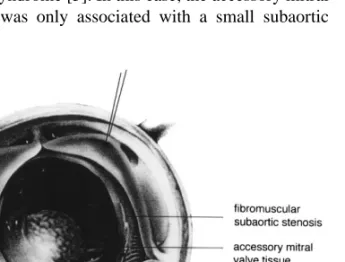
One, remaining transvalvular pressure gradient after resection of the subaortic membrane with no abnormal tis-sue having been seen through the small aortic annulus after resection of this membrane, and two, competence of the mitral valve could be tested immediately and a repair pro-cedure performed if necessary. The additional mitral valve tissue could not be properly identified at first, it appeared only as a mild valvular thickening (Fig. 1). The final patho-logic appearance is shown in Fig. 2. Additionally, through left atriotomy the inspection of the mitral valve and it’s accessory tissue is easier, the resection of the accessory tissue, the corresponding chordae and papillary muscle can be accomplished with great precision and without pos-sibly damaging other structures. Ascuitto et al. [8] describe a similar case in which the initial transaortic approach was unsuccessful and left atriotomy had to be performed. Yasui et al. [2] describe the approach through left atriotomy, but their cases all had coexisting cardiac malformations which Fig. 1. Intraoperative transaortic view.
Fig. 2. Final appearance of fibrous obstruction as seen and resected through the mitral valve.
543 A.C. Schmid et al. / European Journal of Cardio-thoracic Surgery 15 (1999) 542–544
enabled them to approach through an enlarged ventricle septal defect. Sono et al. [3] report a case where initial aortotomy followed by left ventriculotomy was used and Meldrum et al. [9] report a case where transaortic approach was successful. Szedo et al. [1] report a case where trans-aortic approach was successful upon reoperation. These cases stress the importance of controlling the relief of obstruction even in apparently straightforward cases. In this regard direct measurement of the gradient and operative TEE are most important in assessing the residual stenosis. TEE, furthermore, provides an anatomic definition of the outflow tract and may point out rare anomalies. A second look through left atriotomy appears advantageous because a wider view of the mitral subvalvular apparatus is obtained, allowing precise excision of the abnormal tissue. Although it requires expertise, direct cardioscopy through the aortic annulus may provide an alternative to this approach.
References
[1] Szedo¨ F, Thomka I, Arvay A. Accessory mitral valve tissue causing left ventricular outflow tract obstruction. J Cardiovasc Surg Torino 1987;28 (4 ):388–390.
[2] Yasui H, Kado H, Tokunaga S, Kanegae Y, Fukae K, Masuda M, Tokunaga K. Trans-ventricular septal defect approach for resection of accessory mitral valve tissue. Ann Thorac Surg 1993;55:950– 953.
[3] Sono J, McKay R, Arnold RM. Accessory mitral valve leaflet caus-ing aortic regurgitation and left ventricular outflow tract obstruction. Br Heart J 1988;59(4):491–497.
[4] Tokunaga S, Yasui H, Kado H, Yonenaga K, Nakamura Y, Shiokawa Y, Nakano E, Andou H, Tokunaga K. Diagnosis and surgical treat-ment of accessory mitral valve with associated congenital heart disease. Nippon Kyobu Geka Gakkai Zasshi 1991;39(11):2034– 2040.
[5] Marino B, Gagliardi MG, Diglio MC, Polletta B, Grazioli S, Agostino D, Giannotti A, Dallapiccola B. Noonan syndrome: struc-tural abnormalities of the mitral valve causing subaortic obstruction. Eur J Pediatr 1995;154:949–952.
[6] Baneriee A, Kohl T, Silverman NH. Echocardiographic evaluation of congenital mitral valve anomalies in children. Am J Cardiol 1995;76(17):1284–1291.
[7] Eiriksson H, Midgley FM, Karr SS, Martin GR. Role of echocardio-graphy in the diagnosis and surgical management of accessory mitral valve tissue causing left ventricular outflow tract obstruction. Ann Soc Echocardiogr 1995;8(1):105–107.
[8] Ascuitto RJ, Ross NT, Kopf GS, Kleinman CS, Talner NS. Acces-sory mitral valve tissue causing left ventricular outflow obstruction. Ann Thorac Surg 1986;42:58–64.
[9] Meldrum WG, Cartmill TB, Hawker RE, Celermajer JM, Wright CM. Accessory mitral valve tissue causing left ventricular outflow tract obstruction. Br Heart J 1986;55:376–380.