Treatment of gastrointestinal hemorrhage
P. Charbonnet,
2* J. Toman,
1* L. Bu¨hler,
2B. Vermeulen,
3P. Morel,
2C. D. Becker,
1F. Terrier
11
Division de Radiodiagnostic et de Radiologie interventionnelle, Hoˆpital Universitaire de Gene`ve 24, rue Micheli-du-Crest, 1211 Gene`ve 14, Switzerland
2Clinique et Policlinique de Chirurgie digestive, Hoˆpital Universitaire de Gene`ve, 24, rue Micheli-du-Crest, 1211 Gene`ve 14,
Switzerland
3Division des Urgences me´dico-chirurgicales, Hoˆpital Universitaire de Gene`ve, 24, rue Micheli-du-Crest, 1211 Gene`ve 14,
Switzerland
Abstract
Background: We assessed the value of selective arteri-ography in the diagnosis and management of acute gas-trointestinal hemorrhage.
Methods: We reviewed the records of 107 consecutive patients who had gastrointestinal hemorrhage and underwent selective arteriography between January 1992 and October 2003: 10 had upper gastrointestinal bleed-ing, 79 had lower gastrointestinal bleedbleed-ing, and 18 had varicose bleeding with portal hypertension. Selective embolization was attempted in 15 patients to obtain hemostasis. Angiographic findings were reviewed and prospective reports were compared with the final diag-nosis and outcome.
Results: Of 129 angiographic studies, 36 correctly re-vealed the bleeding site and 93 were negative. Extrava-sation was seen in 24 cases at the level of stomach (n = 2), duodenum (n = 1), small bowel (n = 5), or colon (n = 16). Indirect signs of bleeding sources were identified in 12 patients (stomach in one, small bowel in four, large bowel in four, liver in three). Transcatheter embolization induced definitive hemostasis in 11 of 15 patients (73%), namely in the stomach (n = 2), small bowel (n = 3), colon (n = 7), and liver (n = 3). Three patients required surgery after embolization.
Conclusion:Abdominal arteriography may localize gas-trointestinal bleeding sources in approximately one-third of cases. Selective embolization may provide definitive hemostasis in most instances.
Key words: Gastrointestinal hemorrhage—Selective embolization—Surgery
The diagnosis of the etiology and localization of acute gastrointestinal bleeding is challenging for several rea-sons: the possible sites of origin involve the digestive tract in its entirety and the bleeding may be intermittent with brisk recurrence and may, notably in case of upper hemorrhage, threaten the patient’s life.
In acute upper gastrointestinal bleeding, the bleeding site is by definition proximal to the ligament of Treitz: the patient presents with hematemesis or melena [1]. Its main clinical manifestation is hematemesis. A bleeding site distal to the ligament of Treitz is defined as lower intestinal tract bleeding. Its main clinical manifestations are melena or hematochezia.
The incidence of acute upper gastrointestinal bleeding is 50 to 150 per 100,000 of the population each year [2]. It arises at all ages, generally after age 60 years and concerns mostly the males (two males for each female until age of 75 years) [1]. The incidence of acute lower gastrointestinal bleeding is estimated to be 20 to 30 per 1000,000 persons [3], also with a male predominance in those older than 60 years [4]. The Overall mortality rate of upper gastroin-testinal bleeding is approximately 14% [1] and that of lower gastrointestinal bleeding lower than 5% [4].
A multidisciplinary approach improves the efficiency of patient care for acute upper or lower gastrointestinal bleeding [5]. Emergency physicians, gastroenterologists, and surgeons have a well-defined role in these situations. Radiologists have been involved since the 1960s, when selective arterial catheterization to identify bleeding sites in the gastrointestinal tract was first described by Nus-baum and Baum [6]. Rosch et al. [7] managed to control an acute gastric hemorrhage by embolization of the
*Both authors contributed equally to this work.
Correspondence to:P. Charbonnet; email: pierre.charbonnet@hcuge.ch
gastroepiploic artery using an autologous clot. During the 1970s, developments in catheter technology and the emergence of newmaterials for embolization (e.g., Gel-foam [8] and steel spiral coils [9]) boosted interest in embolization.
The place of arteriography in the management algo-rithm of gastrointestinal bleeding depends on the bleed-ing site: whereas endoscopy is always the initial investigative option for upper gastrointestinal bleeding after hemodynamic stabilization, mesenteric angiogra-phy is often the first step in the management of massive lower gastrointestinal bleeding [10].
This present study evaluated the efficiency and the place of emergency angiography in the management of patients who have acute gastrointestinal bleeding. For this purpose, we reviewed retrospectively all clinical histories and angiographies performed for acute gastro-intestinal bleeding in our institution from January 1992 to October 2003.
Materials and methods
The clinical records of all consecutive patients who had confirmed acute upper and/or lower gastrointestinal bleeding and were admitted for an emergency angiogra-phy over 11 years (from January 1992 to October 2003) were studied retrospectively. Main indications for arte-riography were localization of the bleeding site before surgery and/or embolization.
Arteriography was ordered by abdominal surgeons and performed by a senior radiologist. Embolization was performed during the same examination if the radiologist and the surgeon agreed to do so.
Two radiologists and one surgeon reviewed the an-giographies and were blinded to a patient’s outcome and they compared retrospectively their conclusions with those of the initial reports.
Our study group included 107 patients (69 men and 38 women). Average age was 56 years (range 17–98 years). At least one significant comorbid condition was found in 75% of the admissions: 23 patients (21%) had cirrhosis, 39 patients (36%) had cardiovascular disease, and 43 patients (40%) were using antiagreggant or anti-coagulation medication.
Fourteen patients (13%) presented with hematemesis, suggesting an upper bleeding site; 91 (85%) with melena or hematochezia, suggesting an upper or a lower bleeding site; and two with hematemesis and hematochezia, sug-gesting severe upper gastrointestinal bleeding with rapid transit of blood.
Thirty patients (28%) had an endoscopic diagnosis before angiography to definitely localize the ongoing bleeding and/or control by embolization (poor surgical candidates). In 44 patients (41%), the bleeding site re-mained undiagnosed despite endoscopic investigation. Ten patients (9%) were directly referred to arteriography.
For five patients (5%), no data regarding endoscopic examination were retrospectively found. Eighteen pa-tients (17%) had a hemorrhage related to their portal hypertension and arteriography was done to define the bleeding site and assess the possibility of embolization, to check the permeability of a portal shunt, or to determine vascular anatomy before surgery.
Six patients underwent two angiographies during different hospitalization periods. Hemoglobin levels be-fore angiography were higher than 8 g/L (the value at which blood transfusions are generally required) in 66 patients (62%), lower than 8 g/L in 33 patients (31%) and unknown in eight patients. Urea creatinine dissociation (a serum urea nitrogen level disproportionately high in relation to the creatinine level as a sign of upper gas-trointestinal bleeding) was observed in only nine pa-tients. More than 90% of patients received more than 2 hemoglobin units (275 ± 75 mL of erythrocytic con-centrate). Thirty-six patients (34%) were admitted to the intensive care unit.
All vascular punctures were performed at the levels of the right common femoral artery. After the examination, compression at the puncture site was maintained with the patient at rest in a supine position.
Angiographic criteria to diagnose active bleeding or define the bleeding site were separated into direct and indirect signs. Contrast extravasation is the only direct sign, whereas vascular tuft, arteriovenous fistula, early filling vein, or a hypervascular mass represent an indirect sign.
Table 1. Sources of gastrointestinal bleeding Final diagnosis No. of patients (%) Gastrointestinal hemorrhage of unknown origin 28 (26) Colonic diverticulum 20 (19) Esophageal varices 15 (14) Angiodysplasia 9 (8)
Small bowel tumor 6 (6)
Duodenal ulcer 3 (3)
Anastomotic bleeding 3 (3) Small bowel varices 3 (3) Small bowel ischemia 2 (2) Dieulafoy gastric ulcer 2 (2) Meckel diverticulum 1 (1)
Colonic carcinoma 1
Status post rectal polypectomy 1
Large bowel ulcer 1
Arterioportal fistula 1 Multiple organ failure 1 Duodenal diverticulum 1
Gastric metastasis 1
Portal cavernous angioma 1
Colonic amyloidosis 1
Colonic ischemia 1
Inflammatory bowel disease 1
Rectal hemorrhoids 1
Rectal ulcer 1
Small bowel diverticulum 1
For embolizations, silk particles, coils, and polyvinyl alcohol particles were employed.
All possible complications (local: hematoma, pseud-oaneurysm, infection, or arterial dissection; systemic: acute renal insufficiency, allergic reactions, digestive necrosis, or ischemia) were carefully sought and checked by the radiologist who visited the patient in the hours after the examination.
No provocative angiography (provocation of bleed-ing with vasodilatators, anticoagulants, and/or throm-bolytics) was performed (departmental decision).
Radionuclide scanning (commonly the technetium 99m pertechnetate-labeled red blood cell scan) was
al-ways performed after a negative angiogram, owing to the time required to perform the radioisotopic scan in our institution. Ten patients had a radionuclide scan after a negative angiogram.
Table 2. Radiological signs for positive angiographie result
Radiologic signs No. of patients Localization (cases) Direct sign Contrast medium
extravasation
24 Stomach (2), duodenum (1), small bowel (5), large bowel (16) Indirect signs Vascular spot and
drainage vein
3 Large bowel
Arteriovenous fistula 3 Liver
Hypervascular mass 6 Stomach (1), small bowel (4), large bowel (1)
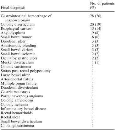
Fig. 1. Patient with a Dieulafoy gastric lesion. (A) Selective and (B) superselective celiac angiograms show an extrava-sation of contrast from a branch of the left gastric artery, indicating continuous bleeding.
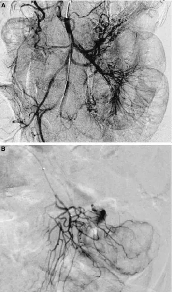
Fig. 2. Bleeding of the small bowel. A No extravasation of contrast medium is seen at the first selective superior mesenteric arteriogram. B The next day, a superselective jejunal arteriogram clearly shows active extravasation of contrast medium.
Most patients (82%) underwent one arteriography, and 16 patients (15%) underwent two arteriographies, and three patients (3%) underwent three. In total 129 arteriographies were performed.
Results
Ten patients (9%) had upper gastrointestinal bleeding, 79 (74%) had lower gastrointestinal bleeding, 15 (14%) had upper varicose bleeding, and three (3%) had lower vari-cose bleeding, all with known portal hypertension.
Table 1 summarizes all the final diagnoses, based on the final clinical reports. Angiography was positive in 31% (36 of 118; 11 examinations were performed in pa-tients who had portal hypertension to check shunt per-meability or determine vascular anatomy were subtracted from the total number of positive arteriog-raphies; Table 2). Direct or indirect signs were observed in the stomach in three cases (Fig. 1), the liver in three cases, the duodenum in one case, the small bowel in nine cases (Fig. 2), and the large bowel in 20 cases (Fig. 3).
The most frequent disease associated with a positive arteriogram was diverticulosis. All 24 patients (22%) w ho had vascular extravasation of contrast medium at angi-ography had hypotension corresponding to hypovolemic shock. Sixteen embolizations in 15 patients (14%) were performed: twice at the level of the stomach, three times at the level of the small bowel, eight at the level of the large bowel (Fig. 4), and three at the level of the liver. Embolization was the definitive treatment in 11 patients (10%). After embolization, three patients underwent surgery for recurrent bleeding in the stomach (n = 1) or small bowel (n = 2). In total, 30 patients (28%) were operated upon, 17 after a positive arteriogram that
Fig. 3. Angiodysplasia of the cecum. A Superior mesenteric artery angiogram shows a hypervascular area in the cecum containing a dense tangle of vessels. B Early draining vein, a characteristic finding of angiodysplasia.
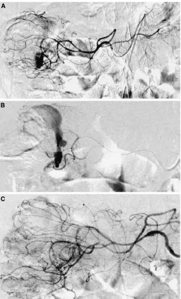
Fig. 4. Embolization of large right colon bleeding from diverticular disease. A Selective superior mesenteric arterio-gram demonstrates extravasation in the lumen at the right angle of the large bowel. B Superselective middle colic arte-riogram confirms the intraluminal extravasation at the bleed-ing site. C Result after silk particles placed in the distal middle colic artery by a microcatheter: Extravasation has ceased, without bleeding from other branches.
localized the site, five after a negative arteriogram and a positive radionuclide scan (Fig. 5), and eight after a negative arteriogram and no other radiographic exami-nation (cf. Table 3, Figs. 6, 7)
Twelve patients died consecutively due to hemorrhage (global mortality rate of 11%). Mortality rate was higher in patients who had upper gastrointestinal bleeding (five of 25 compared with seven of 82 patients who had slower gastrointestinal bleeding).
No patient died as a consequence of complications caused by the procedure. Three patients (2%) had local complications related to the catheter manipulation: one distal artery rupture and two pseudoaneurysms. No bo-wel ischemic event was noticed in this series.
Arteriograms were reviewed, with concordant results in 96%. There were five discordant results: in four cases, the retrospective evaluation demonstrated contrast medium extravasation not observed at the initial inter-pretation (locations were confirmed according to the fi-nal diagnosis); in one case, the retrospective evaluation considered contrast medium extravasation at the level of the left colon, although the interpretation was negative.
The final diagnosis was cecal angiodysplasia and we concluded that the erroneous retrospective evaluation was due to motion artifacts.
Discussion
Angiography can localize the site of active bleeding to 0.5–1 ml/min [11, 12], which corresponds approximately to 3 U of blood per day [13]. Identification of a focal bleeding point depends on the hemodynamic condition
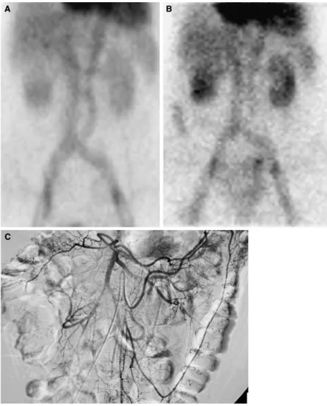
Fig. 5. Immediate and delayed images from a large bowel bleeding demonstrated by technetium 99m–labeled erythrocyte scan. A An early image from the scan shows no abnormal extraluminal radioactivity. B A late image from the same study displays a focus of increased radioactive uptake in the right midabdomen at the level of the ascending colon. C A superior mesenteric arteriogram from the same patient demonstrates no abnormal extravasation of contrast medium.
Table 3. Surgical interventions for acute bleeding
Interventions No. of patients
Right colectomy 9
Small bowel resection 7
Portocaval shunt 4 Left hemicolectomy 3 Subtotal colectomy 2 Anastomosis resection 1 Gastrectomy 1 Meckel resection 1 Duodenotomy 1
of the patient at the time of angiography [14]. Clinically, an initial heart rate of 100 beats/min or higher, an initial systolic blood pressure of 115 mmHg or lower, and a history of syncope are some of the independent signs of severity in lower gastrointestinal bleeding [3] and signs of shock (class III or IV hemorrhages) [15]. The fact that all patients with a direct angiographic sign had hypotension illustrates this point. In patients with such clinical con-ditions, the probability that angiography will be positive is the highest. These clinical manifestations are essential considerations in the assessment of patients as candidates for angiography.
The most frequent sign for positive arteriography is contrast medium extravasation (direct sign). This sign was mainly associated with diverticular disease, owing to the prevalence of this disease in the studied population (nearly 50% of the patients who are hospitalized with acute gastrointestinal hemorrhage have bleeding related to diverticulosis [16]). In our study, similar to the indirect sign of an early filling vein, extravasation was most fre-quently present at the level of the right colon, where angiodysplasia and diverticular bleeding are often localized.
Endoscopy is the first step in the management of upper gastrointestinal bleeding. For lower gastrointesti-nal bleeding, angiography may be the first diagnostic study in patients with massive bleeding [10]. Although angiography was the first diagnostic method in only 1% of patients in the study by Brackman et al. [17], in our series angiography was selected as a first diagnostic choice in nearly 12% of patients with lower gastrointes-tinal bleeding (10 of 82). Emergency endoscopy for lower gastrointestinal bleeding may be difficult because stool
Fig. 6. Treatment modalities for patients with positive arteriogram.
Fig. 7. Treatment modalities for patients with negative arteriogram.
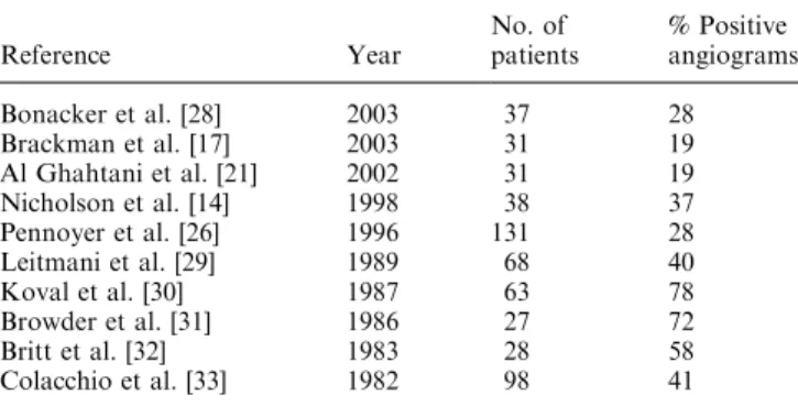
Table 4. Previous series of angiographies in management of acute gastrointestinal bleeding Reference Year No. of patients % Positive angiograms Bonacker et al. [28] 2003 37 28 Brackman et al. [17] 2003 31 19 Al Ghahtani et al. [21] 2002 31 19 Nicholson et al. [14] 1998 38 37 Pennoyer et al. [26] 1996 131 28 Leitmani et al. [29] 1989 68 40 Koval et al. [30] 1987 63 78 Browder et al. [31] 1986 27 72 Britt et al. [32] 1983 28 58 Colacchio et al. [33] 1982 98 41
or blood in the colon may preclude adequate inspection of the mucosa. Although purging may clear retained blood and clots, active bleeding frequently quickly fills up the lumen and handicaps the examination [18]. Moreover, colonoscopy has to be deferred until patients are hemodynamically stable and have adequate colonic preparation [18]. Finally, a source of bleeding in the small bowel (in all cases of gastrointestinal hemorrhage, 3–5% originate from the small intestine [19]) is poorly accessible to endoscopic investigations. Therefore, pa-tients with massive lower gastrointestinal bleeding gen-erally require emergency mesenteric angiography to localize and/or control the bleeding site [19]. Accurate presurgical localization of the bleeding site decreases postoperative rates of morbidity and mortality [20]. With lower gastrointestinal bleeding, 10% to 15% of patients require surgery according to the literature [21]. In our series, 28% who had lower gastrointestinal bleeding (23 of 82) underwent surgery.
We performed 16 embolizations, which were the definitive treatments for 11 patients. Only selective and superselective embolizations were performed (we did not use infusion of intra-arterial vasopressin as an alterna-tive). Three patients underwent surgery for recurrent bleeding after the site of hemorrhage had been localized. In upper gastrointestinal bleeding, endoscopic therapy will achieve hemostasis in 80% of patients [19]. Patients who have upper gastrointestinal bleeding, who are not surgical candidates (comorbid illness), and in whom endoscopic techniques have failed are candidates for angiographic control of bleeding [22, 23]. In these cases, angiography is often the last therapeutic option. Very fewembolizations have been conducted with this indication in our series. Most of our upper hemorrhages were related to varicose bleeding without extravasation at arteriography and the results of Ljungdal et al. [24] (embolization and permanent hemostasis achieved in all but one patient who had ulcerative gastric or duodenal disease) are not applicable to our group. In another study [25], arterial embolization for upper gastrointesti-nal bleeding was effective in 99%, with a periprocedural mortality rate of 35%.
There was no mortality related to arteriography and embolization. The complication rate was 2% and limited to local complications. In the literature, the reported complication rate for diagnostic arteriography in acute gastrointestinal bleeding is approximately 2–4% [20, 26, 27].
Thirty-one percent of angiographies were positive. Although the reviewby Zuckermann and Prakash [11] reported that the rate of positive angiograms varied from 27% to 77%, our results (concerning a large number of patients) are comparable to those of recent series (Ta-ble 4). To increase the positive rate, we could improve the indications by selecting patients who have massive ongoing bleeding early in the course of management.
In conclusion, abdominal arteriography may localize gastrointestinal bleeding sources in approximately one-third of cases. Selective embolization, when indicated, may provide definitive hemostasis in most instances. References
1. Rockall TA, Logan RFA, Devlin HB, Northfield TC (1995) Inci-dence of, mortality from acute upper gastrointestinal haemorrhage in the United Kingdom. BMJ 311:22–226
2. British Society of Gastroenterology Endoscopy Committee (2002) Non-variceal upper gastrointestinal haemorrhage: guidelines. Gut 51:iv1–iv6
3. Strate LL, Orav EJ, Syngal S (2003) Early predictors of severity in acute lower intestinal tract bleeding. Arch Intern Med 63:838– 843
4. Longstreth GF (1997) Epidemiology and outcome of patients hospitalized with acute lower gastrointestinal haemorrhage: a population-based study. Am J Gastroenterol 92:419–424 5. Podila PV, Ben-Menachem T, Batra SK, et al. (2001) Managing
patients with acute, nonvariceal gastrointestinal haemorrhage: development and effectiveness of a clinical care pathway. Am J Gastoenterol 96:208–219
6. Nusbaum M, Baum S (1963) Radiographic demonstration of un-known sites of gastrointestinal bleeding. Surg Forum 14:374–375 7. Rosch J, Dotter CT, Brown MJ (1972) Selective arterial
emboli-sation: a newmethod for control of acute gastrointestinal bleeding. Radiology 102:303–306
8. Carey LS, Grace DM (1974) The brisk bleed: control by arterial catheterization and gelfoam plug. J Can Assoc Radiol 25:113–115 9. Gianturco C, Anderson JH, Wallace S (1975) Mechanical devices
for arterial occlusion. AJR 124:428–435
10. Bar AH, DeLaurentis DA, Parry CE, Keohane RB (1980) Angi-ography in the management of massive lower gastrointestinal tract haemorrhage. Surg Gynecol Obstet 150:226–228
11. Zuckerman GR, Prakash C (1998) Acute lower intestinal bleeding. Part one: clinical presentation and diagnosis. Gastrointest Endosc 48:606–615
12. Bloomfeld RS, Rockey DC (2000) Diagnosis and management of lower gastrointestinal bleeding. Curr Opin Gastroenterol 16:89–97 13. Porter DH, Kim D (1997) Angiographic intervention in upper gastrointestinal bleeding. In: Taylor MB, Gollan JL, Steer ML, (eds). Gastrointestinal emergencies. Baltimore: Williams & Wilkins, 163–180
14. Nicholson AA, Ettles DF, Hartley JE, et al. (1998) Transcatheter coil embolotherapy: a safe and effective option for major colonic haemorrhage. Gut 43:79–84
15. American College of Surgeons Committee on Trauma (1997) Ad-vanced Trauma Life SupportÒ for doctors (ALSÒ) Chicago: ATLS, 94–95
16. Gostout CJ (2000) The role of endoscopy in managing acute lower gastrointestinal bleeding. N Engl J Med 342:125–127
17. Brackman MR, Gushchin V, Smith L, et al. (2003) Acute lower gastroenteric bleeding retrospective analysis (the ALGEBRA study): an analysis of the triage, management and outcomes of patients with acute lower gastrointestinal bleeding. Am Surg 69:145–149
18. Angtuaco TL, Reddy SK, Drapkin S, et al. (2001) The utility of urgent colonoscopy in the evaluation of acute lower gastrointestinal tract bleeding: a 2-year experience from a single center. Am J Gastroenterol 96:1782–1785
19. Ghosh S, Watts D, Kinnear M (2002) Management of gastroin-testinal haemorrhage. Postgrad Med J 78:4–14
20. Zuccaro G (1998) Management of the adult patient with acute lower gastrointestinal bleeding. Am J Gastrointest 93:1202–1208 21. Al Gahtani AR, Sathi R, Stern J, Gordon PH (2002) Investigative
modalities for massive lower gastrointestinal bleeding. World J Surg 26:620–625
22. Gostout CJ (2000) Gastrointestinal bleeding in the elderly patient. Am J Gastrointes 95:590–595
23. Lefkovitz Z, Cappell MS, Lookstein R, et al. (2002) Radiologic diagnosis and treatment of gastrointestinal haemorrhage and ischemia. Med Clin North Am 86:1357–1399
24. Ljungdahl M, Eriksson LG, Nyman R, Gustavsson S (2002) Arterial embolisation in management of massive bleeding from gastric and duodenal ulcers. Eur J Surg 168:384–390
25. Aina R, Oliva VL, Therasse E, et al. (2001) Arterial embolotherapy for upper gastrointestinal haemorrhage: outcome assessement. J Vasc Interv Radiol 12:195–200
26. Pennoyer WP, Vignati PV, Cohen JL (1996) Management of angio-gram positive lower gastrointestinal haemorrhage: long term follow-up of non-operative treatments. Int J Colorect Dis 11:279–282 27. Vernava AM, Moore BA, Longo WE, Johnson FE (1997) Lower
gastrointestinal bleeding. Dis Colon Rectum 40:846–858
28. Bonacker MJ, Begemannn PGC, Dieckmann C, et al. (2003) Stel-lenwert der Angiography in der Diagnose und Therapie gastroin-testinaler Blutungen. Fortschr Roentgenstr 175:524–531
29. Leitman IM, Paull DE, Shires T (1989) Evaluation and manage-ment of massive lower gastrointestinal haemorrhage. Ann Surg 209:175–180
30. Koval G, Benner KG, Rosch J, Kozak BE (1987) Aggressive angiographic diagnosis in acute lower gastrointestinal haemor-rhage. Dig Dis Sci 32:248–253
31. Browder W, Cerise EJ, Litwin MS (1986) Impact of emergency angiography in massive lower gastrointestinal bleeding. Ann Surg 204:530–536
32. Britt LG, Warren L, Moore OF (1983) Selective management of lower gastrointestinal bleeding. Am Surg 49:121–125
33. Colacchio TA, Forde KA, Patsos TJ, Nunez D (1982) Impact of modern diagnostic methods on the management of active rectal bleeding. Am J Surg 143:607–510