Review
Protein phosphatases and their potential implications
in neuroprotective processes
C. E. Gee and I. M. Mansuy*
Brain Research Institute, University of Zürich, Department of Biology, Swiss Federal Institute of Technology, Winterthurerstrasse 190, 8057 Zürich (Switzerland), Fax: + 41 44 6353303, e-mail: [email protected] Received 8 January 2005; received after revision 3 March 2005; accepted 14 March 2005
Abstract. Several neurological disorders such as stroke,
amyotrophic lateral sclerosis and epilepsy result from excitotoxic events and are accompanied by neuronal cell death. These processes engage multiple signalling path-ways and recruit numerous molecular components, in particular several families of protein kinases and protein phosphatases. While many investigations have examined the importance of protein kinases in excitotoxicity, protein phosphatases have not been well studied in this context. DOI 10.1007/s00018-005-5008-4
© Birkhäuser Verlag, Basel, 2005
CMLS
Cellular and Molecular Life SciencesHowever, recent advances in understanding the functions of protein phosphatases have suggested that they may play a neuroprotective role. In this review, we summarize some of the recent findings that illustrate the pleiotropic and complex functions of tyrosine and serine/threonine pro-tein phosphatases in the cascade of events leading to neu-ronal cell death, and highlight their potential intervention in limiting the extent of neuronal death.
Key words. Tyrosine phosphatase; serine/threonine phosphatase; excitotoxicity; neuroprotection; NMDA receptor.
Introduction
Neurological and cognitive disorders associated with de-generation of brain tissue represent a major challenge for clinicians and research scientists because of the limited potential for de novo production of nerve cells in the ma-ture organism. Although the mechanisms of degeneration and death of brain cells are not fully understood, one of their features appears to be linked to aberrant phosphor-ylation of proteins in these cells. Abnormal phosphoryla-tion has been associated with excitotoxicity, a process that generally results from the excessive stimulation of calcium-permeable glutamate receptors followed by mas-sive calcium influx into cells, inappropriate activation of calcium-dependent enzymes, production of free radicals, alteration of mitochondrial functions and ultimately initi-ation of cell death. Neuronal loss due to excitotoxicity may contribute to the pathophysiology of chronic neu-rodegenerative illnesses such as Alzheimer’s disease,
*Corresponding author.
amyotrophic lateral sclerosis, Parkinson’s disease and Huntington’s disease [1–4].
Clearly, multiple pathways are affected in response to perturbed calcium homeostasis. This represents a major complication for the development of potential therapeutic intervention. For instance, interfering with one of the most upstream molecular components of the cascade such as glutamate receptors in an attempt to hinder the whole cascade has proven unsuccessful. The develop-ment of specific antagonists acting on neuronal gluta-mate receptors has not provided the expected beneficial effect for treatment because of the multiplicity and sever-ity of their side effects. Advances in the identification and understanding of pathways implicated in excitotoxicity and neuronal cell death have, however, raised the hope that targeting intracellular transduction elements down-stream from glutamate receptors may prove a useful strategy for alleviating or attenuating neuronal injury. Of particular interest in this respect are protein phos-phatases, which despite their demonstrated implication in excitotoxicity may play an important role in the
mech-anisms that limit brain damage and mediate neuropro-tection. In this review, we summarize recent findings suggesting the involvement of tyrosine and serine/threo-nine protein phosphatases in neuroprotection, with a fo-cus on experimental findings derived from models of brain injury such as transient or global cerebral ischemia, glutamate excitotoxicity and hypoxia. We try to illustrate the pleiotropic functions and complexity of the mecha-nisms that these molecules are involved in during these processes (see table 1).
Tyrosine and serine/threonine protein phosphatases
Tyrosine and serine/threonine protein phosphatases are highly abundant proteins present in many cellular com-partments of mammalian cells. Together with protein kinases, they set the phosphorylation state of signalling and effector proteins and thereby play a large role in con-trolling cellular responses. The balance between protein phosphatases and kinases is essential for the regulation of cellular signalling, and inappropriate or defective phos-phatase or kinase activity leads to aberrant patterns of
phosphorylation. Dysregulated phosphorylation/dephos-phorylation underlies numerous human diseases, including but not restricted to diabetes [5], cancer [6–8], autism [9], Alzheimer’s disease [10], Lafora’s disease [11] and Parkinson’s disease [12].
Tyrosine phosphatases
To date, 107 genes encoding tyrosine phosphatases have been identified in the human genome [13, 14]. All share the ability to hydrolyze p-nitrophenyl phosphate, are in-hibited by vanadate and are insensitive to okadaic acid [15, 16]. In addition to their ability to dephosphorylate phosphotyrosine residues, a subset of tyrosine phos-phatases can also dephosphorylate phosphoserine or phosphothreonine residues. These dual-specificity phatases may also target messenger RNA (mRNA), phos-pholipids and phosphoinositides in addition to phospho-proteins. Membrane-spanning phosphotyrosine-specific phosphatases are known collectively as receptor-like protein tyrosine phosphatases, whereas others lack mem-brane-spanning domains and are found in the cytoplasm. Except for the Eya (eyes absent) tyrosine phosphatases,
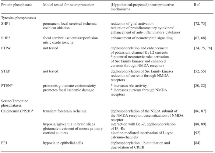
Table 1. Overview of potential neuroprotection conferred by Tyr and Ser/Thr protein phosphatases.
Protein phosphatase Model tested for neuroprotection (Hypothetical/proposed) neuroprotective Ref. mechanisms
Tyrosine phosphatases
SHP1 permanent focal cerebral ischemia reduction of glial activation [72, 73] cochlear ablation reduction of proinflammatory cytokines/
enhancement of anti-inflammatory cytokines
SHP2 focal cerebral ischemia/reperfusion enhancement of neurotrophin signalling [67, 68] nitric oxide toxicity
PTPa* not tested dephosphorylation and enhancement [74, 75, 78]
of potassium channel Kv1.2 currents * potential neurotoxic role: activation of Src family kinases and enhanced currents through NMDA receptors
STEP not tested dephosphorylation of Src family kinases [52, 55]
reduction of currents through NMDA receptors
PTEN* promotes glutamate excitotoxicity * increases Akt activity, [80, 82] promotes focal ischemic damage * increases currents through NMDA
receptors Serine/Threonine
phosphatases
Calcineurin (PP2B)* transient forebrain ischemia dephosphorylation of the NR2A subunit of [86, 87] the NMDA receptor, desensitization of NMDA
receptor
hypoxia/aglycemia in brain slices interaction with Bcl-2, dephosphorylation [88, 89] glutamate treatment of mouse primary of IP3-Rs
cortical cultures nicotine-mediated inactivation of L-type [92] calcium-channels
PP1 hypoxia in epithelial cells dephosphorylation, ubiquitination and [84] degradation of CREB
which use a different catalytic process requiring an aspar-tic acid [17], dephosphorylation generally begins when a protein with a phosphorylated tyrosine residue enters the active site of the phosphatase. Then the phosphoryl group is transferred to a key cysteine residue by nucleophilic attack, after which the tyrosine is protonated and ejected from the active site. The thiol intermediate on the cysteine residue is then hydrolysed, and the phosphatase is re-turned to its resting state [18, 19].
Target specificity of tyrosine phosphatases is ensured by multiple molecular strategies. One mechanism relies on the property that different tyrosine phosphatases prefer-entially recognize certain phosphopeptides. Specificity is also achieved by cell-type and organelle-specific ex-pression of individual PTPs (protein tyrosine phos-phatases). In addition, most tyrosine phosphatases have multiple domains, which may be involved in target recognition and in localization to particular cellular compartments and complexes of protein assemblies. For example, SHP-1 and SHP-2 possess Src homology 2 (SH2) domains, LAR and RPTP have immunoglobulin-and fibronectin-like domains, immunoglobulin-and the Eya members con-tain DNA recognition sequences. The presence or exclu-sion of particular tyrosine phosphatases in protein com-plexes is critical for their signalling specificity. For ex-ample, CD45, which is found in all hematopoetic cells, can activate Src family tyrosine kinases by dephosphor-ylating the inhibitory site or can inhibit them by dephos-phorylating the activation site depending on the cell type.
The mechanism whereby pathway specificity is deter-mined appears to be the co-localization of or exclusion of CD45 from the receptor/Src signalling complex [20].
Serine/threonine phosphatases
Like PTPs, Ser/Thr protein phosphatases represent a di-verse family, are expressed in many cell types and cellular compartments, and are regulated via several mechanisms [21, 22]. They are classified into PPP and PPM families defined by distinct amino acid sequence and 3-dimen-sional structure. The major phosphatases in the PPP family are PP1, PP2A and PP2B (calcineurin), and the PPM family includes PP2C. PP1, PP2A and PP2B are composed of catalytic and regulatory subunits, with each subunit type being expressed in several isoforms by dis-tinct genes and/or alternative splicing. By contrast, PP2C exists as a monomer devoid of regulatory subunits. Regulatory subunits generally have multiple functions, including controlling catalytic activity, subcellular lo-calization and substrate-specificity of the protein phos-phatase. The marked molecular diversity brought about by the varied subunit composition is further enhanced by the requirement of covalent modifications on some sub-units and/or the dependence on specific ions. For in-stance, PP2B requires Ca2+and calmodulin for full
ac-tivity, whereas PP2C needs Mg2+or Mn2+[21, 22].
The specificity and activity of Ser/Thr protein phos-phatases are largely controlled by interacting partners and Figure 1. Schematic representation of pathways associated with excitotoxic events. Excessive glutamate overactivates glutamate receptors leading to increased intracellular Ca2+([Ca2+i]) and the activation of multiple downstream pathways. Protein phosphatases may intervene
in several parts of these pathways to activate or inhibit different substrates. See text for more detail. NOS nitric oxide synthase, VDCC voltage-dependent Ca2+channel, ASIC acid sensing ion channel, GP G protein, PLC phospholipase C.
scaffolding proteins. These proteins act as regulatory subunits that compartmentalize phosphatase subunits in discrete subcellular locations and bring them into close proximity with target substrates [23]. They thereby con-fer spatial control to their catalytic activity. In the brain, localization of protein phosphatases to synaptic struc-tures such as the postsynaptic density (PSD) is critical for neuronal signalling. For example, the g1 subunit of PP1
is specifically concentrated in dendrites and presynaptic boutons in part through attachment to the anchoring pro-tein yotiao localized in these compartments. In addition to compartmentalizing protein phosphatases, anchoring proteins also have inhibitory or activating actions towards different substrates. For instance, A-kinase anchoring proteins (AKAPs) belong to a family of proteins that can anchor PP2B and protein kinase A (PKA) to N-methyl-D-aspartate (NMDA) receptors, and therefore control the activity of this complex. Likewise, spinophilin is a binding protein containing a PDZ domain (postsynaptic-density-protein/disc-large/zo-1 domain) that mediates the associa-tion of PP1 with actin and neurotransmitter receptors such as the D2 dopamine (DA) receptor [24], the alpha adrener-gic receptor [25] and p70S6 kinase (p70S6K) [26]. Other interacting proteins have also been found to have dual functions depending on their partner. Inhibitor 1/PP2A and inhibitor 2/PP2A are two proteins known to inhibit PP2A that can also stimulate PP1 activity in vitro [unlike in-hibitor-1 (I1), which is a PP1 inhibitor] [27].
In addition to anchoring proteins, specific endogenous activators/inhibitors exist that contribute to regulating phosphatase activity. Their nature and mode of action may vary depending on cell type and brain structure. I1 and dopamine and cyclic AMP (cAMP)-regulated phos-phoprotein Mr 32000 (DARPP-32) are two PP1 inhibitors (different from inhibitor 1/PP2A above) with similar se-quences that are selectively expressed in cortical/hippo-campal neurons or striatal neurons, respectively. Finally, phosphatase activity can be controlled by redox mecha-nisms such as demonstrated for PP2B. Overall, these mul-tiple mechanisms represent an intricate means of tightly regulating the activity, substrate specificity and intracel-lular localization of protein phosphatases, making them highly selective.
The role of protein phosphatases in the cascade of events triggered during excitotoxic cell death has not been ex-tensively studied, but some protein phosphatases, such as Ca2+-dependent calcineurin, were found to contribute to
excitotoxicity (because its inhibition is neuroprotective [28]). However, recent findings have indicated that pro-tein phosphatases are implicated in both excitotoxic and cell survival mechanisms, depending on the timing of their activation, the nature and extent of the insult etc. These findings have prompted interest in studying their role as endogenous neuroprotective molecules and, in turn, considering their use as potential drug targets for
the treatment of disorders involving excitotoxicity and neuronal cell death [8, 29, 30].
Events underlying excitotoxicity
Excitotoxicity is a complex process initiated when neu-ronal receptors for excitatory neurotransmitters are ex-cessively activated during traumatic events such as cere-bral ischemia, brain injury or seizure [31, 32]. Since sev-eral of the protein phosphatases discussed here might be neuroprotective by decreasing processes leading to exci-totoxic cell death, the main aspects of this process will be reviewed. Excitotoxicity is triggered by energy and oxygen deprivation, and disruption of ionic homeostasis [33]. Under these deleterious conditions, neurons strongly depolarize, which leads to an excessive entry of calcium through calcium-permeant channels such as volt-age-dependent calcium channels, channels associated with ionotropic glutamate receptors such as the NMDA receptor [34], transient receptor potential channels [35] and acid-sensing channels [36]. The NMDA receptor is recognized as one of the major mediators of calcium in-flux, triggering excitotoxic cell death in numerous neuro-logical disorders [1, 37, 38]. As following hypoxia [39], oxygen-glucose deprivation enhances currents through NMDA receptors in hippocampal CA1 pyramidal neu-rons, but this does not occur in CA3 pyramidal neurons [P. Benquet, C. E. Gee and U. Gerber, unpublished obser-vations]. Transient forebrain ischemia also increases NMDA receptor currents in CA1, leading to selective cell death of these neurons [40]. NMDA receptors are regu-lated by protein phosphorylation and are thus a central target of kinases and phosphatases during both excitotox-icity and cell survival [41]. Further to extracellular cal-cium entry, calcal-cium release from endoplasmic reticulum stores contributes to massively increasing the level of intracellular calcium during excitotoxicity. Excessive activation of 1,4,5-inositol-trisphosphate receptors (IP3-Rs) by IP3leads to depletion of these Ca2+stores and
triggers neuronal death through both necrosis and apop-tosis [42].
In addition to calcium entry, the production of reactive oxygen species exacerbates excitotoxic cell damage [32, 38], possibly by enhancing the activity of protein kinases or decreasing the activity of protein phosphatases [43]. The NMDA receptor is closely linked to the neuronal en-zyme nitric oxide synthase (NOS) via PSD-95 interac-tions. Calcium entering through NMDA receptors stimu-lates NO production by NOS, which is neurotoxic at high levels [44, 45]. This observation may explain why calcium entering through NMDA receptors is more neurotoxic than calcium entering through voltage-dependent calcium channels or non-NMDA ionotropic glutamate receptors [46].
The induction of ischemic insult is accompanied by an impairment of mitochondrial functions, a decrease in the concentration of ATP and glucose, an accumulation of lactate and often also the initiation of inflammatory re-sponses. Neurons within the injury core may then un-dergo apoptotic cell death or die as a result of necrosis [33]. A substantial number of neurons in regions adjacent to the injury core (the penumbra) may also be affected and die by programmed cell death, which contributes to a significant loss of neurological functions. Nevertheless, the viability of neurons in the penumbra is generally bet-ter than in the core, and therefore these neurons have a higher potential for preservation by appropriate interven-tion. Importantly, the occurrence of an excitotoxic event is accompanied by the activation of endogenous cell sur-vival pathways that counteract cell death pathways. Mechanistically, excitotoxic events engage many of the intracellular molecules that mediate neuronal signalling in physiological conditions. These molecules intervene in two different ways: either they contribute to damage and promote neuronal death, or they counteract damage and sustain cell survival. Clearly, several individual mole-cules can take part in both processes depending on the type and level of injury, the homeostatic state of the in-jured cell and the timing after injury. Tyrosine and Ser/Thr protein phosphatases in particular play an active role in both processes and can have excitotoxic and neu-roprotective functions. For instance, dramatic changes in phosphorylation/dephosphorylation of many proteins have been demonstrated during global ischemia and sub-sequent brain reperfusion. We will focus in the following section primarily on protein phosphatases with potential neuroprotective functions.
Protein phosphatases that may be neuroprotective
Tyrosine phosphatases
Several protein phosphatases have been found to take part in processes that counteract excitotoxic or other insults and therefore may function as neuroprotectants. Possible neu-roprotective candidates include the tyrosine phosphatases striatal-enriched phosphatase (STEP), the SH2-containing tyrosine phosphatases SHP1, SHP2 and protein tyrosine phosphatase alpha (PTPa). PTPa may also promote
neu-rotoxicity as has been shown for the tyrosine phosphatase phosphatase and tensin homolog deleted from chromo-some 10 (PTEN). In trauma or disorders such as cerebral ischemia [47, 48], epileptiform activity [49, 50] or Alz-heimer’s disease [51], increased tyrosine phosphorylation mediated by Src or Fyn kinases is suggested to promote neuronal cell death.
Endogenous pathways and mechanisms involving protein phosphatases have evolved to counteract the negative ef-fects of hyperphosphorylation. Because of its association
with the NMDA receptor complex, the tyrosine phos-phatase STEP, especially its large isoform STEP61, is
poised to fulfill such a function by opposing the potenti-ating effects of Src family kinase phosphorylation on NMDA receptor function [52]. In spite of its name, STEP is expressed in many regions of the brain, including the basal ganglia, striatum, cortex and hippocampus. It is composed of four isoforms, a large membrane-bound form (STEP61) and three lower molecular weight
cytoso-lic isoforms [53]. Following transient global ischemia, STEP immunoreactivity increases in glia, specifically in regions where neurons undergo cell death [54]. In neu-rons, STEP dephosphorylates and inactivates several tar-gets, including the Src family kinase Fyn [55] and ERK (extracellular signal-related kinase) [56]. These targets of STEP have also been implicated in neuronal death. Dur-ing forebrain hypoxia/ischemia, STEP61is cleaved and its
catalytic domain is released into the cytosol as a small molecular weight isoform (STEP33) [57]. It is not clear
whether STEP33 maintains its catalytic activity and
whether its separation from the NMDA receptor complex changes its availability; however, STEP appears to be a good candidate for reversing Src-dependent enhancement of NMDA currents. It is possible that its cleavage is pre-vented in ischemia-resistant neurons and may contribute to the tyrosine phosphatase-dependent maintenance of NMDA currents in protected CA3 cells. Interestingly, STEP is activated by dephosphorylation by the calcium-dependent Ser/Thr protein phosphatase calcineurin [56], which is more abundant in CA3 neurons, and therefore STEP may be part of a complex mechanism counteract-ing Src-dependent enhancement of NMDA responses [52].
Besides STEP, several other protein phosphatases also control Src activity and Src-mediated phosphorylation of targets involved in excitotoxicity. Src activation depends on the phosphorylation/dephosphorylation of two tyrosine residues, Y527 and Y416, with Y527 phosphorylation be-ing inhibitory and Y416 phosphorylation bebe-ing excitatory. The tyrosine phosphatases SHP2 and PTPa increase Src
activity by promoting Y527 dephosphorylation either in-directly for SHP2 by reducing the interaction of Src with the tyrosine kinase Csk [58] or directly for PTPa [59].
De-spite this function, however, SHP2 was found to promote cell survival by mechanisms that are not well understood but that may involve enhancement of neurotrophin sig-nalling [60–62]. SHP2 is expressed in various neurons in human and mouse adult brain [63, 64] and is localized in pre- and postsynaptic membranes [65] and mitochondria [66]. It is essential during development and its loss by knockout is lethal. Its downregulation, achieved by expres-sion of a dominant negative in the adult brain, increases the sensitivity to focal cerebral ischemia/reperfusion injury [67]. Consistently, in a model of CNS injury using nitric oxide, SHP2 inhibition was found to reduce survival of
primary cultured neurons and increase caspase activation, a marker of programmed cell death [68]. It may have a neuroprotective role as its level of activity increases in reactive glia following permanent focal ischemia [64]. The targets of SHP2 have not all been identified, but tyrosine kinase-dependent signalling pathways are posi-tively regulated by SHP2 through, for instance, dephos-phorylation and inactivation of the receptor tyrosine ki-nase inhibitor Sprouty [69]. SHP2 itself is also activated by tyrosine phosphorylation [70] and therefore appears as a central component of feedback and crosstalk mecha-nisms between multiple signalling pathways.
SHP1 is a tyrosine phosphatase with SH2 domains that is upregulated in rodent brain following insult. SHP1 is found in some neurons but is primarily expressed in glial cells such that loss of SHP1 leads to dysmyelination in the central nervous system (CNS) [71]. In young mice, SHP1 is upregulated by permanent focal cerebral ischemia in non-dividing astrocytes and microglia and appears to limit their activation, thereby reducing the potential for glial scar formation [72]. The production of some anti- and pro-inflammatory cytokines following deafferentation is also decreased and increased, respectively, in SHP1 mutants, suggesting that a further neuroprotective action of SHP1 is the reduction of the inflammatory response triggered by glial activation [73].
PTPa activity regulates the activity of neuronal Kv1.2
potassium channels and NMDA receptors. Carbachol ac-tivates m1 muscarinic receptors, resulting in tyrosine phosphorylation that correlates with the suppression of Kv1.2 channel currents [74]. PTPa activity is increased
by carbachol via tyrosine phosphorylation. Carbachol stimulation additionally promotes the association of phos-phorylated PTPa with phosphorylated Kv1.2 channels,
resulting in dephosphorylation, which reverses the sup-pression of currents [75]. Increasing conductance of potassium channels to hyperpolarize neurons is an impor-tant mechanism to limit calcium entry during excitotoxic-ity. Whether PTPa activity plays a role in maintaining
potassium channel activity under excitotoxic conditions has not been explored. However, it is known that oxida-tive stress promotes a conformational change in PTPa
dimers that reduces its activity and would be maladaptive in this context [76, 77]. Another effect of PTPa activity is
to activate Src family kinases and increase currents through NMDA receptors [78]. An increase in this prop-erty of PTPa during insults would be expected to
exacer-bate injury; thus it is unclear whether promoting PTPa
activity would be neuroprotective or whether the reduc-tion of PTPa activity by reactive oxygen species would
instead be neuroprotective.
PTEN is a dual-specificity kinase that dephosphorylates phosphotyrosine and phosphoserine/threonine-contain-ing substrates [79]. In contrast to most of the other phos-phatases mentioned in this review, downregulation of
PTEN is neuroprotective against glutamate excitotox-icity [80] and transient focal ischemia [81]. PTEN inter-acts with NR1/NR2B-containing NMDA receptors [82]. Knock-down of PTEN with small interfering RNA (siRNA) decreases currents through the extrasynaptic NR1/NR2B-containing NMDA receptors by decreasing both the channel-open probability and the surface expres-sion of the channels. Neuroprotection against transient global ischemia-induced neuronal death conferred by PTEN downregulation depends on the resulting increase in Akt activity and the decrease in NMDA receptor cur-rents [82].
Ser/Thr phosphatases
In parallel to Tyr protein kinases and protein phos-phatases, numerous Ser/Thr kinases and phosphatases are activated during excitotoxic and cell survival events and participate in controlling the outcome of insults to the brain. Protein kinases such as the Ca2+
/calmodulin-de-pendent protein kinase CaMKII, protein kinase C (PKC) and A (PKA), and Cdk5 are recruited. For instance, phos-phorylation by Cdk5 is activated in CA1 hippocampal neurons after transient forebrain ischemia and targets, in particular, the NR2A subunit of the NMDA receptor [40]. Such phosphorylation is detrimental to tissue because its prevention by inhibition of endogenous Cdk5 or by block-ade of its interaction with NR2A protects CA1 pyramidal neurons from ischemic insult. This mechanism may con-tribute to the selective vulnerability of CA1 neurons to ischemia. Concomitantly to kinases, several Ser/Thr pro-tein phosphatases, such as PP1, calcineurin and PP2A are activated to counteract the effect of kinases. In the rat hippocampus in vivo, inhibition of PP1 by okadaic acid increases NMDA receptor phosphorylation and induces a marked degeneration of CA1 hippocampal neurons, sug-gesting a positive effect of PP1 on cell survival [83]. Likewise, in vitro, PP1 inhibition mimics the physiologi-cal effects of hypoxia and severe hypoxia in epithelial cell culture and is accompanied by downregulation of PP1g
expression. The drop in PP1 leads to hyperphosphoryla-tion of several PP1 targets, including the transcriphyperphosphoryla-tion factor CREB [84], followed by ubiquitination and pro-teosomal degradation of CREB, a final phase in the ex-pression of hypoxic damage. During ischemia, PP1 may also control the formation of clusters of CaMKII in synap-tic terminals, which represents a cellular strategy to pre-vent CaMKII-mediated phosphorylation during episodes of Ca2+overload [85].
Despite its recognized role as a major mediator of exci-totoxicity and neuronal cell death, calcineurin is also in-volved in mechanisms of neuroprotection [86]. Following transient forebrain ischemia, calcineurin activity de-creases in the hippocampus, but interestingly not to the same extent in CA1 and CA3 neurons. While it decreases
only transiently in the CA3 region, it does so persistently in CA1 neurons. This may result from its larger concen-tration in CA3 versus CA1 neurons under normal condi-tions, a difference that may maintain homeostatic levels of NMDA receptor phosphorylation. It may also help reverse ischemia-induced NMDA hyperactivation to prevent exci-totoxicity. Calcineurin can efficiently downregulate NMDA receptor activity by dephosphorylating NR2A and desensitizing the receptor, a mechanism known to be neu-roprotective [87]. It can also inactivate L-type Ca2+
chan-nels and thereby diminish Ca2+entry [21, 22].
Another target of calcineurin involved in neuroprotection is Bcl-2, a critical regulator of apoptosis. During the early phase of hypoxia/aglycia in brain slices, calcineurin and Bcl-2 interact, and this interaction triggers the shuttling of calcineurin to intracellular substrates, in particular the IP3-Rs [88, 89]. Shuttling of cellular proteins to their
sub-strates by Bcl-2 is actually a general anti-apoptotic mech-anism [90]. By dephosphorylating IP3-Rs, calcineurin
in-hibits excitation-induced calcium release from internal stores, thereby preventing further increase in calcium in the cell and the depletion of calcium stores [91]. Cal-cineurin-induced desensitization of IP3-Rs may be an
im-portant neuronal defense system against excitotoxicity. Calcineurin is involved in specific mechanisms of neuro-protection such as those induced by nicotine. Nicotine pretreatment is effective in reducing cell death in cortical neurons subjected to glutamate overactivation. This pro-tective function is mediated by activation of alpha7- and beta2-containing nicotinic acetylcholine receptors fol-lowed by Ca2+influx and recruitment of calcineurin [92].
Nicotine-mediated effects are abolished when cal-cineurin is inhibited before glutamate excitotoxicity. However, when inhibited only after glutamate overload, the neurons are preserved. This observation is consistent with the demonstrated neuroprotective effect of the inhi-bition of calcineurin by immunosuppressants such as cy-closporin A and FK506 [86]. It illustrates the dual role of calcineurin in excitotoxic cell death, depending on whether it is activated or inhibited, and whether this oc-curs before, during or after insult. Thus, calcineurin acti-vation before excitotoxicity is neuroprotective, whereas its activation after excitotoxicity promotes damage, in which case calcineurin blockade is neuroprotective. This duality is reminiscent of the binary nature of the activa-tion of the NMDA receptor itself that, depending on its level, either mediates excitotoxic cell death or promotes activity-dependent cell survival. Thus, strong NMDA re-ceptor activation by high concentrations of NMDA in-duces excitotoxicity accompanied by calcineurin activa-tion and transient increase in the phosphorylaactiva-tion of the transcription factor CREB. By contrast, non-toxic levels of NMDA mimic endogenous synaptic activity without activating calcineurin but with sustained CREB phospho-rylation [93]. The mechanisms and targets involved in
calcineurin signalling during excitotoxic/neuroprotective events are thus variable, depending on the nature and ex-tent of the injury, the temporal stage, the affected brain area and the cell type.
Finally, the apparent discrepancy between the demon-strated neuroprotective property of calcineurin inhibitors such as the immunosuppressants CsA and FK506, and the potential neuroprotective function of calcineurin itself, may be accounted for by indirect mechanisms of drug ac-tion. For instance, CsA acts through inhibition of the im-munophilin cyclophilin D, which itself can prevent mito-chondrial damage [94]. Also FK506, in addition to inhibit-ing calcineurin, activates heat shock proteins and is therefore not specific [95]. Another possible mechanism of immunosuppressant-mediated neuroprotection may be via inhibition of astrocyte and microglial activation and prolif-eration, and reduction of the inflammatory response [28].
Search for endogenous mechanisms of neuroprotection
As CA1 pyramidal neurons are more sensitive to excito-toxic injury than CA3 pyramidal neurons, it is of interest to exploit this property to study the mechanisms underly-ing excitotoxicity and endogenous neuroprotective mech-anisms. In an attempt to identify the molecular targets potentially involved in such mechanisms, a recent study compared the profile of gene expression in microdis-sected human CA1 versus CA3 pyramidal neurons. At least 60 genes were found to be differentially expressed at the mRNA level in these neurons (a minimum 1.7-fold enrichment) [96]. Among them, two Ser/Thr kinases, JNK and JNK2, involved in apoptotic cell death were more highly expressed in CA1 pyramidal neurons. Block-ade of JNK activation after cerebral ischemia was found to be neuroprotective, suggesting that JNK promotes exci-totoxicity [97, 98], which is consistent with the increased level of JNK expression observed after ischemia [99, 100]. However, this increase may be counteracted by tein phosphatases, in particular the dual-specificity pro-tein phosphatase MKP (MAP kinase phosphatase), which was found to dephosphorylate and inactivate JNK [101]. However, MKP activity was found to decrease with hy-poxia. Ultimately, the net balance between this kinase/ phosphatase interplay determines whether cell death or survival is favoured.
Another molecule more predominantly expressed in CA1 than in CA3 is the receptor-like tyrosine phosphatase PTPro. In brain, PTPro is highest during development, where it is postulated to play a role in axogenesis [102, 103]. In adult brain, its levels remain high in CA1 neu-rons and certain other cell types [104]. The function of PTPro has not been explored in the adult brain or during excitotoxic injury, but it may be important.
As previously mentioned, currents through NMDA recep-tors are enhanced in several models of ischemic insult. In vitro oxygen-glucose deprivation rapidly enhances NMDA currents in CA1 pyramidal neurons, but not in the less vul-nerable CA3 pyramidal neurons [P. Benquet, C. E. Gee and U. Gerber, unpublished observations]. In CA3 pyra-midal neurons, inhibiting tyrosine phosphatases unmasks a similar tyrosine kinase-dependent enhancement of NMDA currents and exacerbates ensuing cell death, sug-gesting that tyrosine phosphatase activity contributes to protection of these neurons. Whether enhancing this phos-phatase activity in more vulnerable neurons will be neuro-protective warrants further investigation.
Conclusions and perspectives
At present the possible role of protein phosphatases in either preventing or promoting excitotoxic mechanisms is not fully understood. As their modes of action are com-plex, their effects may be diametrically opposed, depend-ing on the type and extent of injury, the area of the brain and cell type injured, and the physiological state of the cell. Concerted efforts will be required to elucidate the mechanisms through which they act. In addition to the excitatory mechanisms delineated above, non-excitatory processes may also mediate glutamate excitotoxicity and cell death, as recently shown in retinal ganglion cells [105]. These mechanisms involve kainate-type ionotropic glutamate receptors and calcium influx but no depo-larization. Protein phosphatases are also implicated in these processes. For instance, cellular damage induced by kainate is reduced by the calcineurin inhibitor CsA. How-ever, here again little is known about the mechanisms involved. Thus, overall, better knowledge of the mecha-nisms of excitotoxicity will be indispensable in develop-ing new approaches for the treatment of brain trauma and disorders associated with neuronal cell death, and should allow the design of novel neuroprotective agents [106].
Acknowledgements. We thank Eva Hochreutener for graphical
work and Urs Gerber and the anonymous reviewers for their critical reading. C.G. is supported by the National Center for Competence in Research ‘Neural Plasticity and Repair’. The lab of I.M.M. is funded by the Swiss Federal Institute of Technology, the University of Zurich, the National Center for Competence in Research ‘Neural Plasticity and Repair’, the Swiss National Science Foundation, the Human Frontier Science Program, EMBO, Novartis Research Foundation, Roche Research Foundation and UBS bank.
1 Lipton S. A. (2004) Paradigm shift in NMDA receptor antag-onist drug development: molecular mechanism of uncompeti-tive inhibition by memantine in the treatment of Alzheimer’s disease and other neurologic disorders. J. Alzheimers Dis. 6: S61–S74
2 Koller W. C. and Cersosimo M. G. (2004) Neuroprotection in Parkinson’s disease: an elusive goal. Curr. Neurol. Neurosci. Rep. 4: 277–283
3 Facheris M., Beretta S. and Ferrarese C. (2004) Peripheral markers of oxidative stress and excitotoxicity in neurodegener-ative disorders: tools for diagnosis and therapy? J. Alzheimers Dis. 6: 177–184
4 Gardian G. and Vecsei L. (2004) Huntington’s disease: patho-mechanism and therapeutic perspectives. J. Neural Transm. 111: 1485–1494
5 Hooft v. H., Sauer W. H., Bombrun A. and Swinnen D. (2004) Prospects for inhibitors of protein tyrosine phosphatase 1B as antidiabetic drugs. J. Med. Chem. 47: 4142–4146
6 Smukste I. and Stockwell B. R. (2003) Restoring functions of tumor suppressors with small molecules. Cancer Cell 4: 419–420
7 Tartaglia M., Niemeyer C. M., Shannon K. M. and Loh M. L. (2004) SHP-2 and myeloid malignancies. Curr. Opin. Hema-tol. 11: 44–50
8 Stiles B., Groszer M., Wang S., Jiao J. and Wu H. (2004) PTENless means more. Dev. Biol. 273: 175–184
9 Smith M., Filipek P. A., Wu C., Bocian M., Hakim S., Modahl C. et al. (2000) Analysis of a 1-megabase deletion in 15q22-q23 in an autistic patient: identification of candidate genes for autism and of homologous DNA segments in 15q22-q23 and 15q11-q13. Am. J. Med. Genet. 96: 765–770
10 Bottini N., Bottini E., Gloria-Bottini F. and Mustelin T. (2002) Low-molecular-weight protein tyrosine phosphatase and human disease: in search of biochemical mechanisms. Arch. Immunol. Ther. Exp. (Warsz. ) 50: 95–104
11 Minassian B. A. (2001) Lafora’s disease: towards a clinical, pathologic and molecular synthesis. Pediatr. Neurol. 25: 21– 29
12 Zhen X., Torres C., Cai G. and Friedman E. (2002) Inhibition of protein tyrosine/mitogen-activated protein kinase phosphatase activity is associated with D2 dopamine receptor supersensitiv-ity in a rat model of Parkinson’s disease. Mol. Pharmacol. 62: 1356–1363
13 Alonso A., Sasin J., Bottini N., Friedberg I., Friedberg I., Osterman A. et al. (2004) Protein tyrosine phosphatases in the human genome. Cell 117: 699–711
14 Andersen J. N., Jansen P. G., Echwald S. M., Mortensen O. H., Fukada T., Del Vecchio R. et al. (2004) A genomic perspective on protein tyrosine phosphatases: gene structure, pseudo-genes, and genetic disease linkage. FASEB J. 18: 8–30 15 Zhang Z. Y. (2002) Protein tyrosine phosphatases: structure
and function, substrate specificity and inhibitor development. Annu. Rev. Pharmacol. Toxicol. 42: 209–234
16 Zhang Z. Y. (2003) Mechanistic studies on protein tyrosine phosphatases. Prog. Nucleic Acid Res. Mol. Biol. 73: 171–220 17 Li X., Oghi K. A., Zhang J., Krones A., Bush K. T., Glass C. K. et al. (2003) Eya protein phosphatase activity regulates Six1-Dach-Eya transcriptional effects in mammalian organo-genesis. Nature 426: 247–254
18 Denu J. M. and Dixon J. E. (1998) Protein tyrosine phos-phatases: mechanisms of catalysis and regulation. Curr. Opin. Chem. Biol. 2: 633–641
19 Zhang Z. Y. (1998) Protein-tyrosine phosphatases: biological function, structural characteristics and mechanism of cataly-sis. Crit Rev. Biochem. Mol. Biol. 33: 1–52
20 Thomas M. L. and Brown E. J. (1999) Positive and negative regulation of Src-family membrane kinases by CD45. Im-munol. Today 20: 406–411
21 Price N. E. and Mumby M. C. (1999) Brain protein serine/ threonine phosphatases. Curr. Opin. Neurobiol. 9: 336–342 22 Rusnak F. and Mertz P. (2000) Calcineurin: form and function.
Physiol Rev. 80: 1483–1521
23 Wong W. and Scott J. D. (2004) AKAP signalling complexes: focal points in space and time. Nat. Rev. Mol. Cell Biol. 5: 959–970
24 Smith F. D., Oxford G. S. and Milgram S. L. (1999) Associa-tion of the D2 dopamine receptor third cytoplasmic loop with
spinophilin, a protein phosphatase-1-interacting protein. J. Biol. Chem. 274: 19894–19900
25 Richman J. G., Brady A. E., Wang Q., Hensel J. L., Colbran R. J. and Limbird L. E. (2001) Agonist-regulated interaction between alpha2-adrenergic receptors and spinophilin. J. Biol. Chem. 276: 15003–15008
26 Burnett P. E., Blackshaw S., Lai M. M., Qureshi I. A., Burnett A. F., Sabatini D. M. et al. (1998) Neurabin is a synaptic pro-tein linking p70 S6 kinase and the neuronal cytoskeleton. Proc. Natl. Acad. Sci. USA 95: 8351–8356
27 Katayose Y., Li M., Al Murrani S. W., Shenolikar S. and Damuni Z. (2000) Protein phosphatase 2A inhibitors, I(1)-(PP2A) and I(2)I(1)-(PP2A), associate with and modify the sub-strate specificity of protein phosphatase 1. J. Biol. Chem. 275: 9209–9214
28 Kaminska B., Gaweda-Walerych K. and Zawadzka M. (2004) Molecular mechanisms of neuroprotective action of immuno-suppressants – facts and hypotheses. J. Cell Mol. Med. 8: 45–58 29 Hoffman B. T., Nelson M. R., Burdick K. and Baxter S. M. (2004) Protein tyrosine phosphatases: strategies for distin-guishing proteins in a family containing multiple drug targets and anti-targets. Curr. Pharm. Des. 10: 1161–1181
30 Umezawa K., Kawakami M. and Watanabe T. (2003) Molecu-lar design and biological activities of protein-tyrosine phos-phatase inhibitors. Pharmacol. Ther. 99: 15–24
31 Liou A. K., Clark R. S., Henshall D. C., Yin X. M. and Chen J. (2003) To die or not to die for neurons in ischemia, traumatic brain injury and epilepsy: a review on the stress-activated signaling pathways and apoptotic pathways. Prog. Neurobiol. 69: 103–142
32 Sapolsky R. M. (2001) Cellular defenses against excitotoxic insults. J. Neurochem. 76: 1601–1611
33 Vajda F. J. (2002) Neuroprotection and neurodegenerative disease. J. Clin. Neurosci. 9: 4–8
34 Lipton P. (1999) Ischemic cell death in brain neurons. Physiol. Rev. 79: 1431–1568
35 Aarts M., Iihara K., Wei W. L., Xiong Z. G., Arundine M., Cerwinski W. et al. (2003) A key role for TRPM7 channels in anoxic neuronal death. Cell 115: 863–877
36 Xiong Z. G., Zhu X. M., Chu X. P., Minami M., Hey J., Wei W. L. et al. (2004) Neuroprotection in ischemia: blocking cal-cium-permeable acid-sensing ion channels. Cell 118: 687–698 37 Waxman E. A. and Lynch D. R. (2005) N-methyl-D-aspartate receptor subtypes: multiple roles in excitotoxicity and neuro-logical disease. Neuroscientist 11: 37–49
38 Arundine M. and Tymianski M. (2004) Molecular mecha-nisms of glutamate-dependent neurodegeneration in ischemia and traumatic brain injury. Cell. Mol. Life Sci. 61: 657–668 39 Crepel V., Hammond C., Chinestra P., Diabira D. and Ben Ari
Y. (1993) A selective LTP of NMDA receptor-mediated currents induced by anoxia in CA1 hippocampal neurons. J. Neurophysiol. 70: 2045–2055
40 Wang J., Liu S., Fu Y., Wang J. H. and Lu Y. (2003) Cdk5 ac-tivation induces hippocampal CA1 cell death by directly phos-phorylating NMDA receptors. Nat. Neurosci. 6: 1039–1047 41 Salter M. W. and Kalia L. V. (2004) Src kinases: a hub for
NMDA receptor regulation. Nat. Rev. Neurosci. 5: 317–328 42 Patterson R. L., Boehning D. and Snyder S. H. (2004) Inositol
1,4,5-trisphosphate receptors as signal integrators. Annu. Rev. Biochem. 73: 437–465
43 Klann E. and Thiels E. (1999) Modulation of protein kinases and protein phosphatases by reactive oxygen species: implica-tions for hippocampal synaptic plasticity. Prog. Neuropsy-chopharmacol. Biol. Psychiatry 23: 359–376
44 Lipton S. A. (1999) Neuronal protection and destruction by NO. Cell Death. Differ. 6: 943–951
45 Nelson E. J., Connolly J. and McArthur P. (2003) Nitric oxide and S-nitrosylation: excitotoxic and cell signaling mecha-nism. Biol. Cell 95: 3–8
46 Sattler R., Charlton M. P., Hafner M. and Tymianski M. (1998) Distinct influx pathways, not calcium load, determine neu-ronal vulnerability to calcium neurotoxicity. J. Neurochem. 71: 2349–2364
47 Ohtsuki T., Matsumoto M., Kitagawa K., Mabuchi T., Mandai K., Matsushita K. et al. (1996) Delayed neuronal death in ischemic hippocampus involves stimulation of protein tyro-sine phosphorylation. Am. J. Physiol. 271: C1085–C1097 48 Paul R., Zhang Z. G., Eliceiri B. P., Jiang Q., Boccia A. D.,
Zhang R. L. et al. (2001) Src deficiency or blockade of Src activity in mice provides cerebral protection following stroke. Nat. Med. 7: 222–227
49 Chun J. T., Crispino M. and Tocco G. (2004) The dual re-sponse of protein kinase Fyn to neural trauma: early induction in neurons and delayed induction in reactive astrocytes. Exp. Neurol. 185: 109–119
50 Sanna P. P., Berton F., Cammalleri M., Tallent M. K., Siggins G. R., Bloom F. E. et al. (2000) A role for Src kinase in spon-taneous epileptiform activity in the CA3 region of the hippo-campus. Proc. Natl. Acad. Sci. USA 97: 8653–8657 51 Lee G., Thangavel R., Sharma V. M., Litersky J. M., Bhaskar
K., Fang S. M. et al. (2004) Phosphorylation of tau by fyn: im-plications for Alzheimer’s disease. J. Neurosci. 24: 2304–2312 52 Pelkey K. A., Askalan R., Paul S., Kalia L. V., Nguyen T. H., Pitcher G. M. et al. (2002) Tyrosine phosphatase STEP is a tonic brake on induction of long-term potentiation. Neuron 34: 127–138
53 Boulanger L. M., Lombroso P. J., Raghunathan A., During M. J., Wahle P. and Naegele J. R. (1995) Cellular and molecular characterization of a brain-enriched protein tyrosine phos-phatase. J. Neurosci. 15: 1532–1544
54 Hasegawa S., Morioka M., Goto S., Korematsu K., Okamura A., Yano S. et al. (2000) Expression of neuron specific phos-phatase, striatal enriched phosphatase (STEP) in reactive astrocytes after transient forebrain ischemia. Glia 29: 316–329 55 Nguyen T. H., Liu J. and Lombroso P. J. (2002) Striatal
enriched phosphatase 61 dephosphorylates Fyn at phosphoty-rosine 420. J. Biol. Chem. 277: 24274–24279
56 Paul S., Nairn A. C., Wang P. and Lombroso P. J. (2003) NMDA-mediated activation of the tyrosine phosphatase STEP regulates the duration of ERK signaling. Nat. Neurosci. 6: 34–42
57 Gurd J. W., Bissoon N., Nguyen T. H., Lombroso P. J., Rider C. C., Beesley P. W. et al. (1999) Hypoxia-ischemia in perina-tal rat brain induces the formation of a low molecular weight isoform of striatal enriched tyrosine phosphatase (STEP). J. Neurochem. 73: 1990–1994
58 Zhang S. Q., Yang W., Kontaridis M. I., Bivona T. G., Wen G., Araki T. et al. (2004) Shp2 regulates SRC family kinase activ-ity and Ras/Erk activation by controlling Csk recruitment. Mol. Cell 13: 341–355
59 Brandt D. T., Goerke A., Heuer M., Gimona M., Leitges M., Kremmer E. et al. (2003) Protein kinase C delta induces Src kinase activity via activation of the protein tyrosine phos-phatase PTP alpha. J. Biol. Chem. 278: 34073–34078 60 Okada N., Wada K., Goldsmith B. A. and Koizumi S. (1996)
SHP-2 is involved in neurotrophin signaling. Biochem. Bio-phys. Res. Commun. 229: 607–611
61 Takai S., Yamada M., Araki T., Koshimizu H., Nawa H. and Hatanaka H. (2002) Shp-2 positively regulates brain-derived neurotrophic factor-promoted survival of cultured ventral mesencephalic dopaminergic neurons through a brain im-munoglobulin-like molecule with tyrosine-based activation motifs/Shp substrate-1. J. Neurochem. 82: 353–364 62 Araki T., Yamada M., Ohnishi H., Sano S., Uetsuki T. and
Hatanaka H. (2000) Shp-2 specifically regulates several tyro-sine-phosphorylated proteins in brain-derived neurotrophic factor signaling in cultured cerebral cortical neurons. J. Neu-rochem. 74: 659–668
63 Reeves S. A., Ueki K., Sinha B., Difiglia M. and Louis D. N. (1996) Regional expression and subcellular localization of the tyrosine-specific phosphatase SH-PTP2 in the adult human nervous system. Neuroscience 71: 1037–1042
64 Servidei T., Bhide P. G., Huang Z., Moskowitz M. A., Harsh G. and Reeves S. A. (1998) The protein tyrosine phosphatase SHP-2 is expressed in glial and neuronal progenitor cells, postmitotic neurons and reactive astrocytes. Neuroscience 82: 529–543
65 Suzuki T., Matozaki T., Mizoguchi A. and Kasuga M. (1995) Localization and subcellular distribution of SH-PTP2, a pro-tein-tyrosine phosphatase with Src homology-2 domains, in rat brain. Biochem. Biophys. Res. Commun. 211: 950–959 66 Salvi M., Stringaro A., Brunati A. M., Agostinelli E., Arancia
G., Clari G. et al. (2004) Tyrosine phosphatase activity in mi-tochondria: presence of Shp-2 phosphatase in mitochondria. Cell Mol. Life Sci. 61: 2393–2404
67 Aoki Y., Huang Z., Thomas S. S., Bhide P. G., Huang I., Moskowitz M. A. et al. (2000) Increased susceptibility to ischemia-induced brain damage in transgenic mice overex-pressing a dominant negative form of SHP2. FASEB J. 14: 1965–1973
68 Chong Z. Z., Lin S. H., Kang J. Q. and Maiese K. (2003) The tyrosine phosphatase SHP2 modulates MAP kinase p38 and caspase 1 and 3 to foster neuronal survival. Cell. Mol. Neuro-biol. 23: 561–578
69 Hanafusa H., Torii S., Yasunaga T., Matsumoto K. and Nishida E. (2004) Shp2, an SH2-containing protein-tyrosine phos-phatase, positively regulates receptor tyrosine kinase signaling by dephosphorylating and inactivating the inhibitor Sprouty. J. Biol. Chem. 279: 22992–22995
70 Vogel W., Lammers R., Huang J. and Ullrich A. (1993) Acti-vation of a phosphotyrosine phosphatase by tyrosine phos-phorylation. Science 259: 1611–1614
71 Massa P. T., Wu C. and Fecenko-Tacka K. (2004) Dysmyeli-nation and reduced myelin basic protein gene expression by oligodendrocytes of SHP-1-deficient mice. J. Neurosci. Res. 77: 15–25
72 Wishcamper C. A., Brooks D. M., Douglas C. J. and Lurie D. I. (2003) Focal cerebral ischemia upregulates SHP-1 in reactive astrocytes in juvenile mice. Brain Res. 974: 88–98
73 Zhao J. and Lurie D. I. (2004) Loss of SHP-1 phosphatase alters cytokine expression in the mouse hindbrain following cochlear ablation. Cytokine 28: 1–9
74 Huang X. Y., Morielli A. D. and Peralta E. G. (1993) Tyrosine kinase-dependent suppression of a potassium channel by the G protein-coupled m1 muscarinic acetylcholine receptor. Cell 75: 1145–1156
75 Tsai W., Morielli A. D., Cachero T. G. and Peralta E. G. (1999) Receptor protein tyrosine phosphatase alpha participates in the m1 muscarinic acetylcholine receptor-dependent regula-tion of Kv1.2 channel activity. EMBO J. 18: 109–118 76 Blanchetot C., Tertoolen L. G. and den Hertog J. (2002)
Reg-ulation of receptor protein-tyrosine phosphatase alpha by oxidative stress. EMBO J. 21: 493–503
77 van der Wijk T., Blanchetot C., Overvoorde J. and den Hertog J. (2003) Redox-regulated rotational coupling of receptor protein-tyrosine phosphatase alpha dimers. J. Biol. Chem. 278: 13968–13974
78 Lei G., Xue S., Chery N., Liu Q., Xu J., Kwan C. L. et al. (2002) Gain control of N-methyl-D-aspartate receptor activity by receptor-like protein tyrosine phosphatase alpha. EMBO J. 21: 2977–2989
79 Myers M. P., Stolarov J. P., Eng C., Li J., Wang S. I., Wigler M. H. et al. (1997) P-TEN, the tumor suppressor from human chromosome 10q23, is a dual-specificity phosphatase. Proc. Natl. Acad. Sci. USA 94: 9052–9057
80 Gary D. S. and Mattson M. P. (2002) PTEN regulates Akt kinase activity in hippocampal neurons and increases their
sensitivity to glutamate and apoptosis. Neuromolecular Med. 2: 261–269
81 Lee J. H., Kim K. Y., Lee Y. K., Park S. Y., Kim C. D., Lee W. S. et al. (2004) Cilostazol prevents focal cerebral ischemic injury by enhancing casein kinase 2 phosphorylation and sup-pression of phosphatase and tensin homolog deleted from chromosome 10 phosphorylation in rats. J. Pharmacol. Exp. Ther. 308: 896–903
82 Ning K., Pei L., Liao M., Liu B., Zhang Y., Jiang W. et al. (2004) Dual neuroprotective signaling mediated by downreg-ulating two distinct phosphatase activities of PTEN. J. Neu-rosci. 24: 4052–4060
83 Arias C., Montiel T., Pena F., Ferrera P. and Tapia R. (2002) Okadaic acid induces epileptic seizures and hyperphosphory-lation of the NR2B subunit of the NMDA receptor in rat hippocampus in vivo. Exp. Neurol. 177: 284–291
84 Taylor C. T., Furuta G. T., Synnestvedt K. and Colgan S. P. (2000) Phosphorylation-dependent targeting of cAMP re-sponse element binding protein to the ubiquitin/proteasome pathway in hypoxia. Proc. Natl. Acad. Sci. USA 97: 12091– 12096
85 Tao-Cheng J. H., Vinade L., Winters C. A., Reese T. S. and Dosemeci A. (2005) Inhibition of phosphatase activity facil-itates the formation and maintenance of NMDA-induced calcium/calmodulin-dependent protein kinase II clusters in hippocampal neurons. Neuroscience 130: 651–656
86 Morioka M., Fukunaga K., Hasegawa S., Okamura A., Kore-matsu K., Kai Y. et al. (1999) Activities of calcineurin and phosphatase 2A in the hippocampus after transient forebrain ischemia. Brain Res. 828: 135–144
87 Wood A. M. and Bristow D. R. (1998) N-methyl-D-aspartate receptor desensitisation is neuroprotective by inhibiting gluta-mate-induced apoptotic-like death. J. Neurochem. 70: 677–687 88 Erin N., Lehman R. A., Boyer P. J. and Billingsley M. L. (2003) In vitro hypoxia and excitotoxicity in human brain induce calcineurin-Bcl-2 interactions. Neuroscience 117: 557–565 89 Erin N., Bronson S. K. and Billingsley M. L. (2003)
Calcium-dependent interaction of calcineurin with Bcl-2 in neuronal tissue. Neuroscience 117: 541–555
90 Reed J. C. (1997) Double identity for proteins of the Bcl-2 family. Nature 387: 773–776
91 Cameron A. M., Steiner J. P., Roskams A. J., Ali S. M., Ron-nett G. V. and Snyder S. H. (1995) Calcineurin associated with the inositol 1,4,5-trisphosphate receptor-FKBP12 complex modulates Ca2+ flux. Cell 83: 463–472
92 Stevens T. R., Krueger S. R., Fitzsimonds R. M. and Picciotto M. R. (2003) Neuroprotection by nicotine in mouse primary cortical cultures involves activation of calcineurin and L-type calcium channel inactivation. J. Neurosci. 23: 10093–10099 93 Lee B., Butcher G. Q., Hoyt K. R., Impey S. and Obrietan K.
(2005) Activity-dependent neuroprotection and cAMP re-sponse element-binding protein (CREB): kinase coupling, stimulus intensity and temporal regulation of CREB phospho-rylation at serine 133. J. Neurosci. 25: 1137–1148
94 Uchino H., Ishii N. and Shibasaki F. (2003) Calcineurin and cyclophilin D are differential targets of neuroprotection by immunosuppressants CsA and FK506 in ischemic brain dam-age. Acta Neurochir. Suppl. 86: 105–111
95 Klettner A. and Herdegen T. (2003) FK506 and its analogs – therapeutic potential for neurological disorders. Curr. Drug Targets CNS Neurol. Disord. 2: 153–162
96 Torres-Munoz J. E., Van Waveren C., Keegan M. G., Bookman R. J. and Petito C. K. (2004) Gene expression profiles in mi-crodissected neurons from human hippocampal subregions. Brain Res. Mol. Brain Res. 127: 105–114
97 Borsello T., Clarke P. G., Hirt L., Vercelli A., Repici M., Schorderet D. F. et al. (2003) A peptide inhibitor of c-Jun N-terminal kinase protects against excitotoxicity and cerebral ischemia. Nat. Med. 9: 1180–1186
98 Hirt L., Badaut J., Thevenet J., Granziera C., Regli L., Maurer F. et al. (2004) D-JNKI1, a cell-penetrating c-Jun-N-terminal kinase inhibitor, protects against cell death in severe cerebral ischemia. Stroke 35: 1738–1743
99 Seta K. A., Kim R., Kim H. W., Millhorn D. E. and Beitner-Johnson D. (2001) Hypoxia-induced regulation of MAPK phosphatase-1 as identified by subtractive suppression hy-bridization and cDNA microarray analysis. J. Biol. Chem. 276: 44405–44412
100 Bernaudin M., Tang Y., Reilly M., Petit E. and Sharp F. R. (2002) Brain genomic response following hypoxia and re-oxygenation in the neonatal rat. Identification of genes that might contribute to hypoxia-induced ischemic tolerance. J. Biol. Chem. 277: 39728–39738
101 Hirsch D. D. and Stork P. J. (1997) Mitogen-activated protein kinase phosphatases inactivate stress-activated protein kinase pathways in vivo. J. Biol. Chem. 272: 4568–4575
102 Beltran P. J., Bixby J. L. and Masters B. A. (2003) Expression of PTPRO during mouse development suggests involvement in axonogenesis and differentiation of NT-3 and NGF-depen-dent neurons. J. Comp Neurol. 456: 384–395
103 Stepanek L., Sun Q. L., Wang J., Wang C. and Bixby J. L. (2001) CRYP-2/cPTPRO is a neurite inhibitory repulsive guid-ance cue for retinal neurons in vitro. J. Cell Biol. 154: 867–878 104 Cheng J., Wu K., Armanini M., O’Rourke N., Dowbenko D. and Lasky L. A. (1997) A novel protein-tyrosine phosphatase related to the homotypically adhering kappa and mu receptors. J. Biol. Chem. 272: 7264–7277
105 Shen W. and Slaughter M. M. (2002) A non-excitatory para-digm of glutamate toxicity. J. Neurophysiol. 87: 1629–1634 106 Levi M. S. and Brimble M. A. (2004) A review of
![Figure 1. Schematic representation of pathways associated with excitotoxic events. Excessive glutamate overactivates glutamate receptors leading to increased intracellular Ca 2+ ([Ca 2+ i]) and the activation of multiple downstream pathways](https://thumb-eu.123doks.com/thumbv2/123doknet/14862446.635842/3.892.114.806.93.453/schematic-representation-associated-excitotoxic-excessive-overactivates-intracellular-activation.webp)
