DOI 10.1007/s12306-009-0010-x
Non-purulent low-grade infection as cause of pain following
shoulder surgery: preliminary results
Alberto G. Schneeberger · Michael K. Gilbart · Ralph Sheikh · Christian Gerber · Christian Ruef
Published online: 16 March 2009 © Springer-Verlag 2009
A.G. Schneeberger (쾷) Shoulder & Elbow Surgery
Seefeldstr. 27, 8008 Zurich, Switzerland e-mail: [email protected] A.G. Schneeberger
University of Zurich Zurich, Switzerland
M.K. Gilbart · R. Sheikh · C. Gerber Department of Orthopaedic Surgery University of Zurich
Zurich, Switzerland C. Ruef
Division of Infectious Diseases and Hospital Epidemiology University Hospital of Zurich
Zurich, Switzerland
cases that had a repeated biopsy. However, nine out of 10 patients continued to suffer from moderate to severe pain. Low-grade infection of the shoulder can be a cause of per-sistent pain and stiffness. The results of antibiotic treatment are disappointing. Further studies are necessary to analyse this difficult pathology.
Introduction
Various causes may lead to painful failures after shoulder surgery. Infection is one and needs to be ruled out in patients who present with pain and/or decreased range of motion after shoulder surgery. Purulent infections of the shoulder have been described [1–9]. Scarce data exist about non-puru-lent, non-draining, low-grade infections of the shoulder.
The purpose of this report is to describe our experi-ence with 10 patients who presented with pain after shoulder surgery and who had synovial tissue cultures harvested at the time of revision surgery that were posi-tive for bacteria with low pathogenicity.
Materials and methods
Patients
Since 2001, tissue samples have been regularly harvested to search for low-grade infection during all shoulder revi-sion surgeries. Ten consecutive patients were identified between June 2001 and December 2002 with a low-grade infection of the shoulder joint following shoulder sur-gery. Three had been operated on at our institution and seven elsewhere. Patients with prior shoulder replace-ment were excluded from this study.
Abstract Low-grade infection was systematically searched
for in all revision shoulder surgeries by harvesting tissue samples. Ten consecutive patients were identified with a non-purulent low-grade infection of the shoulder. All of these patients suffered from pain and eight were stiff. Preoperative aspiration in eight patients yielded bacterial growth in only one case. Serum C-reactive protein levels were normal in seven out of 10 cases. Propionibacterium acnes was identified in seven, coagulase-negative Staphylococcus in two and Staphylococcus saccharolyticus in one case. The delay between harvesting the tissue sam-ples and detection of bacterial growth averaged eight days (range, 2–17). After debridement and antibiotic treatment for a mean of 4.5 months, tissue samples were repeatedly harvested in nine patients due to persistent pain. The infec-tion was microbiologically eradicated in six out of nine
There were nine males and one female with a median age at the time of the index operation of 50 years (32–59). In nine cases the dominant arm was involved. Six patients were smokers (mean 18 cigarettes per day, range 10–30). One patient suffered from type II diabetes mellitus. No other risk factors for infection were identified. All patients were off work due to symptoms related to their affected shoulder.
The revision surgery at which the low-grade infection was detected was defined as the index operation. Before index surgery, the patients had an average of two (one to five) prior procedures including open or arthroscopic rotator cuff repairs (6), acromioplasties (6), open reduc-tion and internal fixareduc-tion (1), stabilisareduc-tion (1) or other procedures (6). The mean duration between the first prior operation and the index procedure was 10 months (5–24). The index procedures with harvesting of tissue samples were arthroscopic acromioplasties in five cases, arthro-scopic capsulotomies in four cases, and open debride-ment and bone anchor removal in one case.
Before the index operation, all patients had a clinical examination with determination of the Constant-Murley score [10], laboratory studies (including C-reactive pro-tein) and plain radiographs. Five patients had recently had a MR arthrography. Eight patients had a fluoroscop-ically guided aspiration of the glenohumeral joint for bacterial cultures before the index operation. This aspira-tion was performed by a radiologist at our instituaspira-tion. To perform this aspiration, a small amount of local anaes-thetic (0.5–1.0 ml of Scandicain 2%, Mepivacain, AstraZeneca, Zug, Switzerland) was first administered intracutaneously and also before penetration of the joint capsule. Special care was taken to avoid injection of local anaesthetic into the joint. After aspiration of gleno-humeral joint fluid, several millilitres of iopamidol con-trast medium (Iopamiro 300, Bracco, Milan, Italy) was injected to ensure the correct intraarticular, glenohumer-al position of the needle using fluoroscopy.
Detection and definition of low grade infection
Low-grade infection was determined by harvesting tissue samples at the index operation. In nine cases the samples for cultures were harvested by arthroscopy and in one case by open surgery. Two patients received 2 g of cefa-zolin (Kefzol, Eli Lilly, Vernier, Switzerland) intra-venously half an hour before the index procedure. All other patients received no antibiotics preoperatively in order to avoid a negative influence on the detection of bacteria. On average, four tissue samples were harvested (two to seven). In most cases, two samples were obtained from the glenohumeral joint and two from the subacromi-al space. Arthroscopic harvesting of the samples was
obtained with a shaver (5.0 mm full radius resector). One layer of 10×10 cm large sterile gauze placed at the out-flow of the shaver (instead of the outout-flow tube) served as a filter, which allowed relatively large amounts of tissue probes to be collected. Synovium, capsule and/or sub-acromial bursa were harvested. The gauzes with the fil-tered tissue samples or the tissue probes from the open biopsies were placed into sterile containers and then sent to the Institute for Medical Microbiology of the University of Zurich of microbiological workup. Microorganisms were identified using standard methods. In particular, culture and identification of Propionibacterium acnes was performed as described by Jakab and coauthors [11]. The cultures were incubated for at least 10 days. Minimal inhibitory concentrations (MIC) of relevant antibiotics against identified bacteria were determined by standard E-test.
Low-grade infection of the shoulder joint was defined by the presence of at least two positive cultures identify-ing the same bacteria from the biopsies and the absence of pus and fistulas. If only one culture was positive, the result was arbitrarily interpreted as a contamination.
Follow-up
The data of this study were retrospectively collected from the charts. Nine out of 10 patients had a follow-up examination at our outpatient clinic after a mean of 18 months (9–40) after the index procedure with determina-tion of the Constant-Murley score and plain radiographs. The degree of stiffness was estimated according to the notes in the chart. Stiffness was defined as mild for a pas-sive limitation of the shoulder joint up to 25°, as moder-ate for 25–50º and as severe for more than 50º. One patient had a cardiac infarction two months after the index procedure. He refused further treatment. For this case, there was only a telephone follow-up nine months after the index procedure.
Statistical analysis
The relationship between discrete variables was deter-mined using the Wilcoxon signed rank test.
Results
Clinical and radiological presentation before index procedure The study population consisted of 10 patients with infect-ed shoulder. Eight patients had undergone joint
aspira-tion prior to the diagnosis of infecaspira-tion. The sample was positive in only one patient, yielding growth of coagu-lase-negative Staphylococcus (coagucoagu-lase-negative S.).
Patient characteristics prior to the index procedure are shown in Table 1. All patients had painful shoulders and eight had mild to moderate stiffness. All patients were afebrile with normal leukocyte counts. C-reactive protein was normal in seven cases and mildly to moderately ele-vated in three patients (6, 7.5 and 33.4 mg/l).
Nine patients had no radiographic signs of arthrosis. One patient had a moderate arthrosis on the preoperative radiographs according to the classification of Samilson and Prieto [12]. There were five preoperative MR arthro-graphies that did not show signs of bacterial infection such as joint effusion, bone marrow oedema, cortical ero-sions, focal bone destruction or periostal reaction.
Findings at index procedure
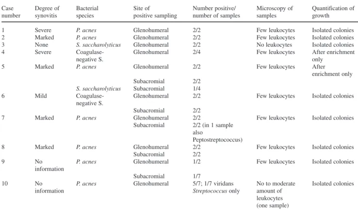
The intraoperative findings during the index procedure showed synovitis in most patients (Table 2). In two patients, more than one bacterial species was cultured from one or more samples. P. acnes was the most com-monly isolated pathogen (seven of 10 patients), while coagulase-negative S. was found in two patients, and Staphylococcus saccharolyticus in one case.
The inflammatory response in the involved joints was absent to minimal in most patients, as the samples con-tained no or few leukocytes. This microscopic finding was in contrast to the finding of synovitis in most patients.
Antibiotic treatment and clinical presentation at latest follow-up
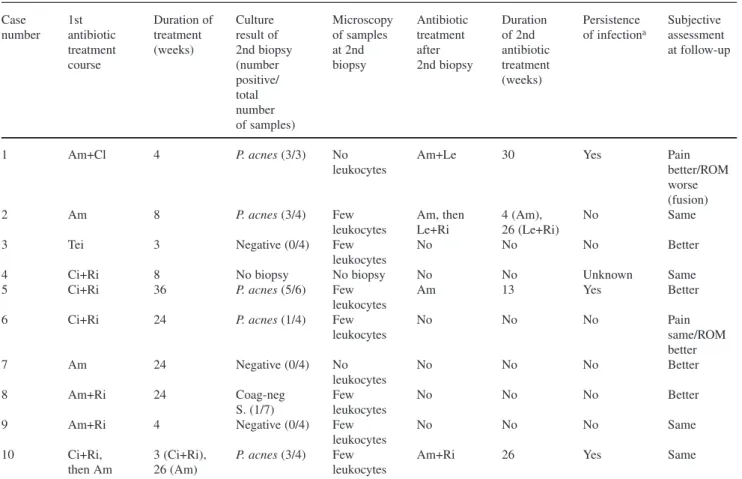
In this series therapy of the low-grade infections mainly consisted of debridement and oral antibiotics (Tables 1 and 3). Antibiotic treatment was based on the MICs of different antibiotics for the identified pathogens. The fol-lowing antibiotics were used: amoxicillin and clavulanic acid (Augmentin, GlaxoSmithKline, Münchenbuchsee, Switzerland), ciprofloxacin (Ciproxin, Bayer, Zurich, Switzerland), clindamycin (Dalacin C, Pfizer, Zurich, Switzerland), levofloxacin (Tavanic, Aventis Pharma, Zurich, Switzerland), rifampicin (Rimactan, Medika, Aesch, Switzerland) and teicoplanin (Targocid, Aventis Pharma, Zurich, Switzerland). The duration of antibiotic treatment varied between 3 and 36 weeks (Table 3). The antibiotics were administered orally except for teicoplanin (intravenous treatment). Due to persistent symptoms after completion of antibiotic therapy and in
order to determine whether the infection was eradicated, a second arthroscopic biopsy was performed in all cases except for the patient with the cardiac infarction. In five of these patients the culture of the biopsies yielded growth of P. acnes. In a sixth patient coagulase-negative S. were identified. The patients with positive cultures underwent a second cycle of antibiotic treatment. However, the clinical results remained unsatisfactory (Tables 1 and 3). Additional cycles of antibiotic treat-ment with durations ranging between 13 and 30 weeks were administered following positive cultures of repeat-ed biopsies in patients 1, 5 and 10. Despite these pro-longed courses of treatment, cultures of a third set of biopsies were again positive and the clinical situation remained unchanged with persistent pain and decreased range of motion of the affected joint for two of these patients.
Overall, at latest follow-up, nine out of 10 patients had severe to moderate pain. Stiffness improved in one patient only. Subjectively, half of the patients felt a slight improvement of their symptoms, but only one described the condition of his shoulder as satisfactory whereas the other nine were not satisfied with the outcome.
One patient with persistent P. acnes infection was finally treated with a fusion of the shoulder due to severe pain despite a relatively intact joint (moderate chondro-malacia). The fusion healed uneventfully. The patient’s pain improved somewhat, especially at rest and at night, but the position of the fusion was not satisfactory. A cor-rective osteotomy was performed. The intraoperative cul-tures revealed coagulase-negative S. as well as P. acnes. After corrective osteotomy, the symptoms worsened to the level before fusion.
Complications
Several patients had gastrointestinal symptoms associat-ed with oral antibiotic therapy, but no other adverse events were noted in the charts.
Discussion
In this report we describe 10 patients with chronic shoul-der pain who meet standard microbiological criteria for the diagnosis of low-grade bacterial infection, namely the iso-lation of the same pathogen in at least two samples from a normally sterile site. However, the diagnosis of infection can be challenged by two aspects that do not fit the classi-cal presentation of a bacterial infection. First, the patients did not present with typical clinical signs of infection (swelling, fever, pus, draining, etc.) and had only minimal
symptomatic. These findings are, however, preliminary and definitely require further investigation.
Oral antibiotic treatment, which is considered to be appropriate by current standards, appears relatively ineffi-cient as such low-grade infections, whether eradicated or not, seem to be associated with persistent clinical symptoms. to moderate intraarticular inflammation. Microscopy of
tissue samples revealed no microorganisms on gram stain and no or only a few leukocytes. Second, the expected response to antibiotic treatment – eradication of the pathogen and clinical improvement – did essentially not occur. On the contrary, most patients continued to be
Table 2 Intraoperative findings at index procedure and microbiological results at the time of diagnosis
Case Degree of Bacterial Site of Number positive/ Microscopy of Quantification of
number synovitis species positive sampling number of samples samples growth
1 Severe P. acnes Glenohumeral 2/2 Few leukocytes Isolated colonies
2 Marked P. acnes Glenohumeral 2/2 Few leukocytes Isolated colonies
3 None S. saccharolyticus Glenohumeral 2/2 No leukocytes Isolated colonies
4 Severe Coagulase- Glenohumeral 2/4 Few leukocytes After enrichment
negative S. only
5 Marked P. acnes Glenohumeral 2/2 Few leukocytes After
enrichment only
Subacromial 2/2
S. saccharolyticus Subacromial 1/4
6 Mild Coagulase- Glenohumeral 2/2 Few leukocytes Isolated colonies
negative S.
Subacromial 2/2
7 Marked P. acnes Glenohumeral 2/2 Few leukocytes Isolated colonies
Subacromial 2/2 (in 1 sample also
Peptostreptococcus)
8 Marked P. acnes Glenohumeral 2/2 Few leukocytes Isolated colonies
Subacromial 2/2
9 No P. acnes Glenohumeral 1/2 Few leukocytes Isolated colonies information
Subacromial 1/7
10 No P. acnes Glenohumeral 5/7; 1/7 viridans No to moderate Isolated colonies
information Streptococcus only amount of
leukocytes (one sample)
P Propionibacterium, S Staphylococcus
Table 1 Characteristics of the 10 patients prior to diagnostic surgery (index procedure) and at latest follow-up
Case Pain Active flexion Stiffness Arthrosis Subjective Subjective
Constant-number (Pre/f-up) (°) (Pre/f-up) (Pre/f-up) (Pre/f-up) assessment result at f-up Murley score
at f-up (%) (Pre/f-up)
1 Sev/mod 80/50 Mod/na None/na Pain same/ Unsat 29/10
ROM worse (fusion, see text)
2 Sev/sev 120/80 Mod/mod None/none Same Unsat 28/14
3 Sev/mod 150/160 Mild/none None/none Better Sat 70/83
4 Sev/sev 75/na Mild/na None/none Same Unsat 18/na
5 Mod/mild 150/140 Mild/mild None/none Better Unsat 60/na
6 Sev/mod 80/100 Mild/mild None/none Pain same/ Unsat 24/49
ROM better
7 Sev/sev 150/160 None/none None/none Better Unsat 53/56
8 Mod/mod 80/90 Mod/mod None/none Better Unsat 20/39
9 Mod/sev 110/100 None/none None/none Same Unsat 60/33
10 Mod/sev 50/80 Mod/mod Mod/mod Same Unsat 18/6
Mean 105/107* 38/36*
Pre/f-up before index procedure/at latest follow-up, sev severe, mod moderate, ROM range of motion, Unsat unsatisfactory, Sat satisfactory, na
not available
Most of the infections in this study were caused by P. acnes and coagulase-negative S. These bacteria have been recognised as typical skin flora, located particular-ly in moist areas such as the axilla [13, 14]. In various studies, high rates of contamination with such bacteria during surgery have been identified probably because these bacteria are located deep within the skin, thus not being fully accessible to antiseptic solutions that are used preoperatively for skin preparation [15–17].
Infections with P. acnes such as endocarditis, meningitis, brain abscess, septic arthritis, osteomyelitis and implant-related infections have been described [11, 13, 18–29]. At the shoulder, several studies reported P. acnes infections, most of them with the typical symptoms of infection such as wound erythema, purulent drainage or chronic sinus tracts as well as systemic symptoms such as fever and elevated C-reactive protein levels [1, 2, 6, 8]. In contrast to these stud-ies, in our study these typical local and systemic symptoms of infection (except of pain) were absent, making detection of P. acnes infection difficult [30–32].
The diagnosis of P. acnes infection and other low-grade infections in this series was only made if cultures
of at least two tissue samples were positive. These slow-growing bacteria may require prolonged incubation. Therefore, the recommendation is to monitor cultures for at least 10 days before declaring a result as negative. Similar conclusions have previously been made by other authors [1, 33, 34].
One shoulder in the current series was infected by S. saccharolyticus. It is part of the typical skin flora [35, 36]. In a literature search we found only two reported cases of endocarditis and one case of spondylodiscitis [37–39].
Therapy of low-grade infections mainly consisted in this series of oral antibiotics for a duration of 3–36 weeks. As harvesting of the tissue samples was predom-inantly done by arthroscopic synovectomies and subacro-mial bursal resection including removal of foreign bodies such as sutures or anchors, debridement was considered also part of the treatment. In three out of 10 cases the infection persisted. Due to the small number of cases and the lack of a prospective, randomised study design, we cannot make a recommendation regarding a successful treatment protocol. In some reported series, debridement
Table 3 Antibiotic treatment and microbiological outcome
Case 1st Duration of Culture Microscopy Antibiotic Duration Persistence Subjective
number antibiotic treatment result of of samples treatment of 2nd of infectiona assessment
treatment (weeks) 2nd biopsy at 2nd after antibiotic at follow-up
course (number biopsy 2nd biopsy treatment
positive/ (weeks)
total number of samples)
1 Am+Cl 4 P. acnes (3/3) No Am+Le 30 Yes Pain
leukocytes better/ROM
worse (fusion) 2 Am 8 P. acnes (3/4) Few Am, then 4 (Am), No Same
leukocytes Le+Ri 26 (Le+Ri)
3 Tei 3 Negative (0/4) Few No No No Better
leukocytes
4 Ci+Ri 8 No biopsy No biopsy No No Unknown Same
5 Ci+Ri 36 P. acnes (5/6) Few Am 13 Yes Better
leukocytes
6 Ci+Ri 24 P. acnes (1/4) Few No No No Pain
leukocytes same/ROM
better
7 Am 24 Negative (0/4) No No No No Better
leukocytes
8 Am+Ri 24 Coag-neg Few No No No Better
S. (1/7) leukocytes
9 Am+Ri 4 Negative (0/4) Few No No No Same
leukocytes
10 Ci+Ri, 3 (Ci+Ri), P. acnes (3/4) Few Am+Ri 26 Yes Same
then Am 26 (Am) leukocytes
Am amoxicillin and clavulanic acid (2–3 g/day), Cl clindamycin (1.8 g/day), Le levofloxacin (1 g/day), Ri rifampicin (0.9 g/day), Tei teicoplanin
(0.4 g/day), Ci ciprofloxacin (1–1.5 g/day), P Propionibacterium, Coag-neg S. coagulase-negative Staphylococcus
followed by intravenous or oral antibiotics resulted in clinical eradication of the infection in all cases [1–3, 6, 8]. Whether intravenous treatment would have changed the outcome in this study cannot be answered with the current data. Oral instead of intravenous antibiotic treat-ment was chosen because the MIC of the selected antibi-otics were very low. Local tissue and intraarticular con-centrations, although not measured, probably exceeded the MIC against the microorganisms by far. Another obvious advantage of oral treatment is the feasibility of prolonged treatment in the outpatient setting.
Limitations of this study are the retrospective study design and the small number of patients. Therefore, the findings of this study should be considered as prelimi-nary, and further investigations are needed.
In conclusion, pain after shoulder surgery can be caused by low-grade infection without the typical symp-toms of infection such as erythema, purulent draining or fever. The diagnosis can only be made by tissue harvest-ing for cultures and a prolonged incubation time of about 10 days. Risk factors for failure of treatment with persistence of bacteria despite prolonged courses of antibiotic therapy might need a prospective study design.
Acknowledgement This study was supported by the ResOrtho Foundation.
Conflict of interest The authors declare that they have no conflict of interest related to the publication of this manuscript.
References
1. Athwal GS, Sperling JW, Rispoli DM, Cofield RH (2007) Deep infection after rotator cuff repair. J Shoulder Elbow Surg 16:306–311
2. Herrera MF, Bauer G, Reynolds F et al (2002) Infection after mini-open rotator cuff repair. J Shoulder Elbow Surg 11:605–608 3. Kwon YW, Kalainov DM, Rose HA et al (2005) Management of early deep infection after rotator cuff repair surgery. J Shoulder Elbow Surg 14:1–5
4. Leslie BM, Harris JM 3rd, Driscoll D (1989) Septic arthritis of the shoulder in adults. J Bone Joint Surg 71:1516–1522 5. Mansat P, Cofield RH, Kersten TE, Rowland CM (1997)
Complications of rotator cuff repair. Orthop Clin N Am 28:205–213 6. Mirzayan R, Itamura JM, Vangsness CT Jr et al (2000) Management of chronic deep infection following rotator cuff repair. J Bone Joint Surg 82-A:1115–1121
7. Pfeiffenberger J, Meiss L (1996) Septic conditions of the shoul-der – an up-dating of treatment strategies. Arch Orthop Trauma Surg 115:325–331
8. Settecerri JJ, Pitner MA, Rock MG et al (1999) Infection after rotator cuff repair. J Shoulder Elbow Surg 8:1–5
9. Ward WG, Goldner RD (1994) Shoulder pyarthrosis: a concomi-tant process. Orthopedics 17:591–595
10. Constant CR, Murley AH (1987) A clinical method of functional assessment of the shoulder. Clin Orthop Relat Res (214):160–164 11. Jakab E, Zbinden R, Gubler J et al (1996) Severe infections
cau-sed by Propionibacterium acnes: an underestimated pathogen in late postoperative infections. Yale J Biol Med 69:477–482 12. Samilson RL, Prieto V (1983) Dislocation arthropathy of the
shoulder. J Bone Joint Surg 65:456–460
13. Brook I, Frazier EH (1991) Infections caused by Propionibacterium species. Rev Infect Dis 13:819–822
14. Swartz MN, Gibbons R, Socransky S (eds) (1990) Indigenous bac-teria: oral microbiology. JB Lippincott Company, Philadelphia 15. Dietz FR, Koontz FP, Found EM, Marsh JL (1991) The
importan-ce of positive bacterial cultures of specimens obtained during clean orthopaedic operations. J Bone Joint Surg 73:1200–1207 16. McLorinan GC, Glenn JV, McMullan MG, Patrick S (2005)
Propionibacterium acnes wound contamination at the time of spi-nal surgery. Clin Orthop Relat Res 437:67–73
17. Padgett DE, Silverman A, Sachjowicz F et al (1995) Efficacy of intraoperative cultures obtained during revision total hip arthro-plasty. J Arthroplasty 10:420–426
18. Barazi SA, Gnanalingham KK, Chopra I, van Dellen JR (2003) Delayed postoperative intracerebral abscess caused by Proprionibacterium acnes: case report and review of the literatu-re. Br J Neurosurg 17:336–339
19. Brook I (2002) Meningitis and shunt infection caused by anaero-bic bacteria in children. Pediatr Neurol 26:99–105
20. Dupont JA (1986) Significance of operative cultures in total hip arthroplasty. Clin Orthop Relat Res 211:122–127
21. Esteban J, Ramos JM, Soriano F (1998) Clinical spectrum of infections due to Propionibacterium acnes. Clin Microbiol Infect 4:48–49
22. Estoppey O, Rivier G, Blanc CH et al (1997) Propionibacterium avidum sacroiliitis and osteomyelitis. Rev Rhum Engl Ed 64:54–56
23. Heggeness MH, Esses SI, Errico T, Yuan HA (1993) Late infec-tion of spinal instrumentainfec-tion by hematogenous seeding. Spine 18:492–496
24. Maderazo EG, Judson S, Pasternak H (1988) Late infections of total joint prostheses. A review and recommendations for preven-tion. Clin Orthop Relat Res 229:131–142
25. Noble RC, Overman SB (1987) Propionibacterium acnes osteom-yelitis: case report and review of the literature. J Clin Microbiol 25:251–254
26. Pan SC, Wang JT, Hsueh PR, Chang SC (2005) Endocarditis cau-sed by Propionibacterium acnes: an easily ignored pathogen. J Infect 51:e229–e231
27. Richards BS, Herring JA, Johnston CE et al (1994) Treatment of adolescent idiopathic scoliosis using Texas Scottish Rite Hospital instrumentation. Spine 19:1598–1605
28. Schofferman L, Zucherman J, Schofferman J et al (1991) Diptheroids and associated infections as a cause of failed instru-ment stabilization procedures in the lumbar spine. Spine 16:356–358
29. Yocum RC, McArthur J, Petty BG et al (1982) Septic arthritis caused by Propionibacterium acnes. JAMA 248:1740–1741 30. Do TT, Strub WM, Witte D (2003) Subacute Propionibacterium
acnes osteomyelitis of the spine in an adolescent. J Pediatr Orthop B 12:284–287
31. Marculescu CE, Berbari EF, Hanssen AD et al (2005) Prosthetic joint infection diagnosed postoperatively by intraoperative cultu-re. Clin Orthop Relat Res 439:38–42
32. Sulkowski MS, Abolnik IZ, Morris EI, Granger DL (1994) Infectious arthritis due to Propionibacterium acnes in a prosthetic joint. Clin Infect Dis 19:224–225
33. Richards BS (1995) Delayed infections following posterior spinal instrumentation for the treatment of idiopathic scoliosis. J Bone Joint Surg 77:524–529
34. Wilkins J, Patzakis MJ (1990) Peripheral teflon catheters. Potential source for bacterial contamination of orthopedic
spondylodiscitis due to Staphylococcus saccharolyticus. Joint Bone Spine 72:91–93
38. Krishnan S, Haglund L, Ashfaq A et al (1996) Prosthetic valve endocarditis due to Staphylococcus saccharolyticus. Clin Infect Dis 22:722–723
39. Westblom TU, Gorse GJ, Milligan TW, Schindzielorz AH (1990) Anaerobic endocarditis caused by Staphylococcus saccharolyti-cus. J Clin Microbiol 28:2818–2819
implants? Clin Orthop Relat Res 254:251–254
35. Evans CA, Mattern KL (1978) Individual differences in the bac-terial flora of the skin of the forehead: Peptococcus saccharolyti-cus. J Invest Dermatol 71:152–153
36. Evans CA, Mattern KL, Hallam SL (1978) Isolation and identifi-cation of Peptococcus saccharolyticus from human skin. J Clin Microbiol 7:261–264