Predictive Value of Cerebrospinal Fluid (CSF) Lactate Level Versus CSF/Blood
Glucose Ratio for the Diagnosis of Bacterial Meningitis Following Neurosurgery
Stephen L. Leib, Remy Boscacci, Othmar Gratzl, and Werner Zimmerli
From the Division of Infectious Diseases and the Clinic of Neurosurgery, University Hospitals Basel, Basel, Switzerland
The value of cerebrospinal fluid (CSF) lactate level and CSF/blood glucose ratio for the identifi-cation of bacterial meningitis following neurosurgery was assessed in a retrospective study. During a 3-year period, 73 patients fulfilled the inclusion criteria and could be grouped by preset criteria in one of three categories: proven bacterial meningitis (n5 12), presumed bacterial meningitis (n 5 14), and nonbacterial meningeal syndrome (n5 47). Of 73 patients analyzed, 45% were treated with antibiotics and 33% with steroids at the time of first lumbar puncture. CSF lactate values (cutoff, 4 mmol/L), in comparison with CSF/blood glucose ratios (cutoff, 0.4), were associated with higher sensitivity (0.88 vs. 0.77), specificity (0.98 vs. 0.87), and positive (0.96 vs. 0.77) and negative (0.94 vs. 0.87) predictive values. In conclusion, determination of the CSF lactate value is a quick, sensitive, and specific test to identify patients with bacterial meningitis after neurosurgery.
The diagnosis of bacterial meningitis (BM) is difficult in patients after neurosurgery for several reasons. Meningeal syn-drome with stiff neck, fever, and headache occurs frequently in patients following craniotomy [1–3]. Up to 50% of patients with clinical signs of meningitis have already been treated with steroids or antibiotics, at the time of lumbar puncture [4, 5]. Standard CSF studies (i.e., gram staining and determinations of leukocyte count and glucose and protein concentration) are unreliable for the diagnosis of BM after neurosurgery [4]. Therefore, additional CSF parameters that allow differentiation of BM from nonbacterial meningeal syndrome (NBMS) are needed.
CSF lactate levels and CSF/blood glucose ratios have re-ceived increasing attention because of the ease and precision with which they are measured in the clinical laboratory. Al-though the CSF lactate level and, to a lesser degree, the CSF/blood glucose ratio do differentiate BM from aseptic meningitis in spontaneously occurring cases, there is an ongo-ing controversy about the usefulness of determinongo-ing these values rather than performing standard CSF studies [6 –15].
CSF lactate in BM originates from different sources. Bacte-rial pathogens themselves produce varying amounts of lactate, accounting for;10% of total CSF lactate in patients with BM [6]. The main source of lactate in BM is brain tissue, including neurons and glia cells, which produce lactate by distinct mech-anisms [16]. First BM is associated with generalized brain
edema, causing a reduction of global cerebral blood flow and inflammatory involvement of the vasculature, with loss of autoregulatory mechanisms, vasospasms, and thrombosis [17– 19]. This leads to cerebral ischemia and consequently to gly-colysis by means of anaerobic metabolism.
In addition, cytokines that flood the brain in meningitis reduce tissue oxygen uptake and cause a shift toward anaerobic metabolism, thus increasing lactate production [16, 20]. Be-cause lactate penetrates the blood-brain barrier at a very low rate, measurement of CSF lactate is a useful index of cerebral metabolism [7]. In addition, cytokines also mediate invasion of neutrophils into the subarachnoid space, which may also con-tribute to the rise in CSF lactate level by glycolysis [21].
Standard CSF tests for the diagnosis of BM in neurosurgical patients have been evaluated in only one retrospective study [4]. The authors found that the examined parameters (total and differential leukocyte counts, gram stains, and values for glu-cose and total protein) are either not sensitive or not specific enough to reliably distinguish BM from NBMS. In spontane-ously occurring meningitis, CSF lactate level has proved to be a more reliable discriminatory factor than the CSF/blood glu-cose ratio [6, 9 –11, 22, 23]. The present study was performed to evaluate the value of CSF lactate level and CSF/blood glucose ratio for the diagnosis of BM in patients with clinically suspected BM following neurosurgery.
Patients and Methods
Data from all consecutive lumbar punctures performed over a 3-year period, from 1 December 1993 to 1 December 1996, in the neurosurgical ward and the surgical intensive care unit at the University Hospitals of Basel, Switzerland, were collected by review of the logbook from the CSF analysis laboratory. Data were screened by review of the patients’ charts for eligi-bility. Inclusion criteria were as follows: (1) the first lumbar puncture per patient was performed within 40 days after neu-Received 22 June 1998; revised 27 October 1998.
Data from this study were presented in part at the 37th Interscience Confer-ence on Antimicrobial Agents and Chemotherapy, held 28 September through 1 October 1997 in Toronto.
Reprints or correspondence (current address): Dr. Stephen Leib, Institute for Medical Microbiology, University of Bern, Friedbu¨hlstrasse 51, CH-3010 Bern, Switzerland (sleib@imm.unibe.ch).
Clinical Infectious Diseases 1999;29:69 –74
© 1999 by the Infectious Diseases Society of America. All rights reserved. 1058 – 4838/99/2901– 0011$03.00
rosurgery; (2) there was complete documentation of bacterial gram staining and culture results and of the total and differen-tial leukocyte count, CSF/blood glucose ratio, and CSF lactate level; and (3) lumbar puncture was performed when BM was clinically suspected. Patients with sepsis syndrome due to causes other than BM were excluded from the analysis.
Patients with clinically suspected BM were categorized ac-cording to the following preset criteria: (1) for proven BM, a positive bacterial CSF culture and a leukocyte count of .250/mL; (2) for presumed BM, .1,000 WBCs/mL with .50% neutrophils or (if the patient was treated with steroids and/or antibiotics at the time of the first lumbar puncture).250 WBCs/mL with .50% neutrophils; and (3) for non-BM, a negative CSF culture and ,250 WBCs/mL with ,50% neu-trophils. Criteria for the clinical categories were established prior to the evaluation.
Clinical parameters examined were age, sex, type of surgery, presence and type of a foreign device, postoperative day on which lumbar puncture was performed, amount of blood in the CSF sample, the antibiotic and/or steroid regimen initiated before and at the time of lumbar puncture, and organism(s) identified by CSF culture.
All patients received as antibiotic prophylaxis either fusidic acid (500 mg iv for craniotomy) or cefamandole (2 g iv for spinal cord surgery), as a single dose at induction of anesthesia. CSF culture and laboratory analysis of CSF and blood were performed within 2 hours of lumbar puncture by the central laboratory for bacteriology and chemistry. Leukocytes, neutro-phils, and erythrocytes were assessed by cell counting, and lactate and glucose were measured with use of a commercially available test (Dimension; DuPont, Wilmington, DE) accord-ing to the manufacturer’s instructions.
Published guidelines for differentiation between spontane-ously occurring BM and nonbacterial meningitis involve the use of discriminatory limits with ranges of 3.5– 4.2 mmol/L for CSF lactate and 0.4 – 0.5 for CSF/blood glucose ratio [6 –15]. In this study we chose a cutoff of 4 mmol/L for the CSF lactate level and 0.4 for the CSF/blood glucose ratio. These were chosen to maximize discrimination between proven or pre-sumed BM and NBMS and to be simple enough to be remem-bered in clinical practice.
Continuous data were compared by the Student’s t test. The association between continuous variables was assessed by the Pearson correlation coefficient. Fisher’s exact test was used to evaluate categorical data. Sensitivity, specificity, and predic-tive values were calculated by standard formulas.
Results
Over the 3-year period, 477 consecutive lumbar punctures were performed, because of a clinical suspicion of BM, in the neurosurgical ward and the surgical intensive care unit. For 164 samples, the results of bacterial gram staining and culture as
well as the total and differential leukocyte counts, CSF/blood glucose ratio, and CSF lactate level were documented. After screening of these 164 samples by review of the patients’ charts, the findings of 77 first diagnostic lumbar punctures performed after neurosurgery were analyzed.
We excluded 75 other lumbar punctures because they were performed as follow-up punctures, 9 others because of the lack of a neurosurgical intervention, 1 other because the patient had a concurrent sepsis syndrome other than BM, and 2 others because of insufficient clinical documentation. Eighteen pa-tients had a positive bacterial CSF culture. All positive culture results were reviewed for relevance with regard to the patient’s clinical course, treatment received, subsequent culture results, and laboratory data; this review was done by an investigator (W.Z.) who was unaware of the CSF lactate level and CSF/blood glucose ratio at first lumbar puncture.
Propionibacterium acnes in CSF culture was considered relevant in two patients with foreign devices. Six patients were categorized as having NBMS despite the positivity of the CSF culture, which was considered to be due to contamination. In all six patients’ cultures, bacterial growth was detected only after .5 days, and the cultures of subsequent CSF samples remained negative despite the fact that no antibiotic therapy was initiated. Microorganisms identified in these 6 CSF sam-ples were P. acnes (n5 3), coagulase-negative staphylococci (n 5 2), and mixed isolates of Enterobacter cloacae and Citrobacter diversus (n5 1).
The prevalence of BM was high (34%), since only patients with clinically suspected BM were included. Twelve patients with microbiologically proven BM (table 1), 14 with presumed BM, and 47 patients with NBMS were analyzed. Four patients did not fulfill the criteria for inclusion in any of the above groups. No statistically significant differences between groups were found for age, sex, or contamination of CSF samples with blood.
In this study, 33 patients (45%) received antibiotics and 24 (33%) received steroids at the time of the first lumbar puncture. The rate of treatment in the combined (presumed or proven) BM group vs. that in the NBMS group was significantly higher (P, .002) with steroids (58% [15 of 26] vs. 19% [9 of 47]) but not with antibiotics (46% [12 of 26] vs. 45% [21 of 47]). In 25 patients (34%) an intraventricular shunt was present at the time of lumbar puncture, but there was no significant association with a specific group.
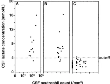
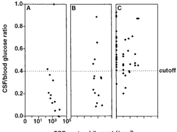
CSF lactate values were significantly higher for patients with proven BM than for patients with NBMS (mean6 SD, 7.8 6 3.6 mmol/L vs. 2.36 0.8 mmol/L; P , .0001), but they did not differ markedly from those for patients with presumed BM (6.76 3.3; P 5 NS vs. proven BM and P , .0001 vs. NBMS) (figure 1). The CSF/blood glucose ratio was significantly lower in patients with proven BM (median [range], 0.17 [0 –1]) and presumed BM (0.34 [0.1– 0.9]) than for patients with NBMS (0.54 [0.9 – 0.2]; P, .0001 vs. proven BM and P , .0005 vs.
presumed BM) (figure 2). For the differentiation of spontane-ously occurring BM from aseptic meningitis, discriminatory levels of CSF lactate and ratios of CSF/blood glucose have been postulated as 3.5– 4.2 mmol/L and ,0.4–0.5, respec-tively [6, 9 –11, 22, 23].
In this study, we chose a CSF lactate concentration of.4.0 mmol/L and a CSF/blood glucose ratio of,0.4 as the critical values for the diagnosis of BM. To evaluate the diagnostic performance of the tests, we combined proven and presumed BM. As presented in figure 1, CSF lactate concentration at a cutoff of 4 mmol/L did differentiate proven and presumed BM from NBMS. There was an overlap in 4 cases, with 3 false-negatives in the combined BM group and 1 false-positive in the NBMS group. The CSF lactate cutoff level of.4 mmol/L as a discriminant factor for BM had a sensitivity of 88%, a speci-ficity of 98%, a positive predictive value of 96%, and a nega-tive predicnega-tive value of 94% (table 2).
The CSF/blood glucose ratio cutoff of 0.4 discriminated between patients with culturally proven BM and patients with NBMS, with two false-negatives in the proven BM group and six false-positives in the NBMS group (figure 2). However, in the group with presumed BM, where additional information would be most desirable, the values showed considerable over-lap, with four false-negatives (30%). Therefore, determination of the ratio was not helpful in this group of patients. For the CSF/blood glucose ratio, in a comparison of the combined BM group and the NBMS group, sensitivity was 77%, specificity
was 87%, positive predictive value was 77%, and negative predictive value was 87% (table 2).
To evaluate specificity, we analyzed our data for a possible effect of CSF contamination with blood on lactate concentra-tion. CSF lactate levels in samples with.500 erythrocytes/mL
were compared to those in samples with ,500
erythrocytes/mL, in patients with and without BM. In accor-dance with previous observations, lactate values were not sig-nificantly affected by the presence of red cells [7, 8]. In addition, we assessed the correlation between the lactate con-centration in CSF and the number of neutrophils and leuko-cytes in CSF. In the NBMS group, the two variables were not correlated (r5 .1 and P 5 NS for neutrophils; r 5 . 17 and P 5 NS for leukocytes). However, in the BM group the correlation was significant (r5 .72 and P , .001 for neutro-phils; r5 .83 and P , .001 for leukocytes), probably because inflammatory cytokines and chemokines released in BM cause neutrophil pleocytosis, metabolic changes, and cerebral hyp-oxia [16 –18].
The postoperative day on which the first lumbar puncture was performed because of the clinical suspicion of BM was significantly later (P, .001) for patients in the NBMS group (10 6 7.6) than for patients in the BM group (5 6 2.2). Subgroup analysis of the data showed no correlation between the number of days after neurosurgery and the level of CSF lactate in the BM and the NBMS groups and showed no significant difference in CSF lactate level over time in patients
Figure 1. Concentration of lactate in CSF from patients with men-ingeal syndrome after neurosurgery. CSF lactate values for patients with proven bacterial meningitis (BM) (A) were significantly higher than those for patients without BM (C) (mean6 SD, 7.8 6 3.6 [n 5 12] vs. 2.36 0.8 mmol/L [n 5 47]; P , .0001), but they did not differ markedly from those for patients with presumed BM (B) (6.76 3.3 [n5 14]; P 5 NS vs. proven BM and P , .0001 vs. patients without BM). A cutoff lactate concentration of 4 mmol/L (dotted line) differ-entiated proven and presumed BM from nonbacterial meningitis with a sensitivity of 88%, specificity of 98%, positive predictive value of 96%, and negative predictive value of 94%.
Table 1. Characteristics of patients with culturally proven bacterial meningitis following neurosurgery.
Age (y)/sex Indication or type of surgery Day of LP after surgery Foreign
device Microorganism(s) isolated
62/F Myelography 1 2 Escherichia coli
64/M Shunt revision 4 1 Streptococcus oralis 44/M Intracerebral hematoma 6 2 Klebsiella pneumoniae 72/F Vascular malformation 2 1 Staphylococcus aureus
79/F Brain tumor 15 1 Peptostreptococcus magnus
54/M Brain tumor 2 1 Klebsiella pneumoniae,
Klebsiella oxytoca 39/M Vascular
malformation
6 1 Corynebacterium jeikeium
50/M Brain tumor 4 2 Streptococcus salivarius,
Streptococcus mitis, Peptostreptococcus micros 72/F Vascular malformation 6 1 Coagulase-negative Staphylococcus
65/F Brain tumor 0 1 Coagulase-negative
Staphylococcus
28/M Brain tumor 9 1 Propionibacterium acnes
64/F Brain tumor 5 1 Propionibacterium acnes
without BM, indicating that neurosurgery by itself did not significantly alter CSF lactate level.
Discussion
The occurrence of bacterial meningitis after neurosurgery has been documented in up to 4% of patients [24, 25]. The diagnosis of BM relies on the isolation of bacteria from CSF samples. However, in up to 70% of clinically suspected cases, bacterial CSF cultures remain negative [3, 4]. In a recent study, CSF samples were obtained from patients who had undergone neurosurgery and had a meningeal syndrome and from clini-cally negative controls. The samples were submitted to con-ventional culture and to PCR with use of primers encoding for a highly conserved region of eubacterial ribosomal DNA [26]. With this technique, the investigators obtained a positive amplification result not only for CSF from patients whose bacterial cultures of CSF were positive but also for CSF from the majority of patients with clinical signs and laboratory values suggestive of bacterial meningitis. In contrast, CSF from control patients yielded no amplification products. The authors concluded that many cases of culture-negative meningitis fol-lowing neurosurgery are probably BM and that antibiotic treat-ment is therefore justified.
The severe consequences of delayed or untreated BM, in conjunction with the lack of clear diagnostic criteria for BM occurring after neurosurgery, explain the prevailing practice of empirically treating all suspected cases with high-dose broad-spectrum intravenous antibiotics. The expense and risk of un-necessary treatment of uninfected patients call for a more specific test for the diagnosis of BM after neurosurgery.
Inflammation of the meninges in reaction to surgical proce-dures is usually treated with high-dose steroids to prevent adhesive arachnoiditis and hydrocephalus by their immunosup-pressive action [4]. In cases of postoperative BM, however, treatment with steroids and antibiotics further impairs the di-agnostic value of CSF studies by decreasing inflammatory parameters and the yield of bacterial cultures, respectively. In this study, 45% of patients with a meningeal syndrome had already been empirically treated with antibiotics, and 33% with steroids, at the time of the first lumbar puncture.
The empirical use of antibiotics was equally high for patients with BM and patients with NBMS, reflecting the difficulties in distinguishing the diseases on clinical grounds. Steroids were used more frequently in the group with later-confirmed BM than in the group with NBMS. The higher proportion of steroid treatment in the BM group might be explained by the fact that symptoms of meningeal inflammation after neurosurgery are frequently treated with steroids based on the assumption that the condition is caused by NBMS. Only when symptoms worsen or there is no clinical response to steroids, as in BM, is a lumbar puncture performed to establish a diagnosis [4].
In 1917 Levinson [27] observed that spinal fluid from pa-tients with meningococcal meningitis had a low pH. Killian
Table 2. Summary and predictive values of CSF lactate levels and CSF/blood glucose ratios in patients after neurosurgery.
Variable CSF lactate level: mean6 SD CSF/blood glucose ratio: median (range) Proven bacterial meningitis (n5 12) 7.86 3.6 mmol/L 0.17 (0–1) Presumed bacterial meningitis (n5 14) 6.76 3.3 mmol/L* 0.34 (0.1–0.9)† Nonbacterial meningitis syndrome (n5 47) 2.36 0.8 mmol/L 0.54 (0.9–0.2) Sensitivity 88% 77% Specificity 98% 87% Positive predictive value 98% 77% Negative predictive value 94% 87%
NOTE. Cutoff values were 4 mmol/L for lactate level and 0.4 for CSF/blood glucose ratio.
* P5 NS vs. proven bacterial meningitis; P , .0001 vs. nonbacterial men-ingitis syndrome.
†P5 NS vs. proven bacterial meningitis; P , .0003 vs. nonbacterial meningitis syndrome.
Figure 2. Ratio of CSF/blood glucose concentration for patients with meningeal syndrome after neurosurgery. CSF/blood glucose ratios for patients with proven bacterial meningitis (A) were signifi-cantly lower than those for patients with nonbacterial meningitis (C) (median [range], 0.17 [0 –1] [n5 12] vs. 0.54 [0.9–0.2] [n 5 47];
P, .0001) but did not differ markedly from those for patients with
presumed bacterial meningitis (B) (0.34 [0.1– 0.9] [n5 14]; P 5 NS). A cutoff CSF/blood glucose ratio of 0.4 (dotted line) discriminated between patients with culturally proven meningitis and patients with nonbacterial meningitis but showed a considerable overlap of 30% in the groups with presumed and nonbacterial meningitis. The CSF/blood glucose ratio cutoff of 0.4 differentiated proven and pre-sumed bacterial meningitis from nonbacterial meningitis with a sen-sitivity of 77%, specificity of 87%, positive predictive value of 77%, and negative predictive value of 87%.
[28] recognized in 1925 that BM lowered the CSF glucose concentration and elevated the CSF lactate level, and in 1933 De Sanctis et al. [29] reported that for the diagnosis of bacterial meningitis, an increased CSF lactate level is more reliable than a decreased CSF glucose level. Since then, many studies on spontaneously occurring BM have confirmed and refined these initial observations [6, 9 –11, 22, 23]. However, the diagnostic value of the CSF lactate level for patients after neurosurgery has not been tested.
CSF lactate measurement has not found widespread accep-tance as a discriminant CSF parameter for the diagnosis of spontaneously occurring BM. Estimates of sensitivity and specificity of the test vary, depending on the discriminant value chosen. We chose 4.0 mmol/L as a cutoff CSF lactate value for diagnosis of BM after neurosurgery. This value was slightly modified from established discriminatory values for spontane-ous meningitis [6, 9 –11, 22, 23] in order to maximize the diagnostic value for neurosurgical patients and to keep it sim-ple enough to be remembered in clinical practice.
Inconsistencies in the reported diagnostic power of CSF lactate measurement for spontaneous BM also depend upon the patient populations in which the test has been applied. As for any diagnostic procedure, the value of the test is diminished when applied indiscriminately to CSF samples from patients in whom BM is not suspected. In this study, the test was exclu-sively applied to patients with clinically suspected BM.
The 34% prevalence of BM reflects the fact that this study was performed in patients after neurosurgery in whom a bac-terial cause of BM could not be ruled out on clinical grounds, necessitating a diagnostic lumbar puncture. This prevalence is comparable to that in two similar studies comparing culturally proven BM with “aseptic” meningitis, in which BM occurred after neurosurgery in 26%– 42% of the patients [4, 26]. Thus, the 34% prevalence of the disease in the tested population led to a high predictive value.
Some authors have argued that despite its diagnostic value, determination of the CSF lactate level does not offer more information than standard CSF tests for the diagnosis of spon-taneously occurring BM. For neurosurgical patients, however, it has been shown that CSF studies (including gram staining and determination of the total and differential leukocyte counts and of glucose and total protein levels), neither alone nor in combination, did not reliably distinguish BM from NBMS [4]. Therefore, determination of the CSF lactate level may contrib-ute to the accurate diagnosis of BM in this population.
A positive correlation between the number of neutrophils and the concentration of lactate in CSF has led some authors to suggest that neutrophils are responsible for the rise in lactate level in cases of BM and that the lactate level would therefore not be more useful than the neutrophil count [30, 31]. In the NBMS group, we found no significant correlation between the CSF lactate concentration and the number of neutrophils or leukocytes in CSF. However, in the BM group these
parame-ters were correlated, possibly as a reflection of the effect of cytokines and chemokines released into the CSF, leading to WBC invasion and a shift toward anaerobic metabolism by the brain parenchymal cells.
Several lines of evidence argue against neutrophils as the main source of CSF lactate. High concentrations of CSF lactate have been found in patients with BM despite a low neutrophil count [32]. This clinical observation has also been made in experimental BM where high levels of CSF lactate were doc-umented in neutropenic and normal control animals, indepen-dent of the presence of neutrophils in CSF [21]. Moreover, production of lactate during experimental BM has been local-ized to brain tissue by in vivo microdialysis [33], and in vitro experiments with neutrophils incubated in CSF showed very low lactate production [32].
The high prevalence of a meningeal syndrome in neurosur-gical patients underscores the need of a rapid and reliable test to diagnose BM. Several properties of CSF lactate make de-termination of such levels a valid ancillary test to use in addition to standard biochemical and microbiological CSF analysis. It is an inexpensive, easy, and rapidly performed analysis that is already widely used for other clinical questions in hospital-affiliated laboratories. As shown by other authors and in this study, the CSF lactate level is not affected by the presence of RBCs in the CSF [7, 8].
The slow clearance of lactate from CSF keeps high levels elevated for up to 4 days, and determining these levels has been particularly useful in identifying the significant proportion of patients previously treated with antibiotics [34, 35]. In our own clinical practice, determination of the CSF lactate level is now routinely performed when postoperative meningitis is sus-pected, and it provides useful additional information for the difficult decision about whether to start empirical antibiotic therapy.
In conclusion, the CSF lactate level (cutoff, 4 mmol/L) is superior to the CSF/blood glucose ratio (cutoff, 0.4) for diag-nosis of BM in neurosurgical patients (table 2). For patients with a meningeal syndrome after neurosurgery, determination of CSF lactate level is a quick, sensitive, and specific ancillary test to identify the need for antimicrobial therapy.
Acknowledgments
The authors appreciate the assistance of Dr. Reno Frei and Dr. Axel Regeniter of the Central Laboratory of Bacteriology and Chemistry, University Hospitals Basel, and the helpful discussion with Dr. Kathrin Mu¨hlemann, University of Bern.
References
1. Kaufman HH, Carmel PW. Aseptic meningitis and hydrocephalus after posterior fossa surgery. Acta Neurochir (Wien) 1978;44:179 –96. 2. Carmel PW, Greif LK. The aseptic meningitis syndrome: a complication
3. Blomstedt GC. Infections in neurosurgery: a retrospective study of 1143 patients and 1517 operations. Acta Neurochir (Wien) 1985;78:81–90. 4. Ross D, Rosegay H, Pons V. Differentiation of aseptic and bacterial
meningitis in postoperative neurosurgical patients. J Neurosurg 1988; 69:669 –74.
5. Roland PS, Meyerhoff WL, Balcombe KL, Mickey BE. Spinal fluid profile following surgery in the subarachnoid space. Otolaryngol Head Neck Surg 1989;101:445– 8.
6. Salord F, Boussaid O, Eynard N, Perret C, Grando J, Chacornac R. Value of D(-) lactate determination for the fast diagnosis of meningitis after craniotomy: an initial study. Ann Fr Anesth Reanim 1994;13:647–53. 7. Cameron PD, Boyce JM, Ansari BM. Cerebrospinal fluid lactate in
men-ingitis and meningococcaemia. J Infect 1993;26:245–52.
8. Begovac J, Bace A, Soldo I, Lehpamer B. Lactate and glucose in cere-brospinal fluid heavily contaminated with blood. Acta Med Croatica 1991;45:341–5.
9. Genton B, Berger JP. Cerebrospinal fluid lactate in 78 cases of adult meningitis. Intensive Care Med 1990;16:196 –200.
10. Bailey EM, Domenico P, Cunha BA. Bacterial or viral meningitis? Mea-suring lactate in CSF can help you know quickly. Postgrad Med 1990; 88:217–9, 223.
11. Nelson N, Eeg-Olofsson O, Larsson L, Ohman S. The diagnostic and predictive value of cerebrospinal fluid lactate in children with menin-gitis: its relation to current diagnostic methods. Acta Paediatr Scand 1986;75:52–7.
12. Komorowski RA, Farmer SG, Knox KK. Comparison of cerebrospinal fluid C-reactive protein and lactate for diagnosis of meningitis. J Clin Microbiol 1986;24:982–5.
13. Briem H. Comparison between cerebrospinal fluid concentrations of glu-cose, total protein, chloride, lactate, and total amino acids for the differential diagnosis of patients with meningitis. Scand J Infect Dis 1983;15:277– 84.
14. Berg B, Gardsell P, Skansberg P. Cerebrospinal fluid lactate in the diag-nosis of meningitis: diagnostic value compared to standard biochemical methods. Scand J Infect Dis 1982;14:111–5.
15. Knight JA, Dudek SM, Haymond RE. Early (chemical) diagnosis of bacterial meningitis— cerebrospinal fluid glucose, lactate, and lactate dehydrogenase compared. Clin Chem 1981;27:1431– 4.
16. Tureen J. Effect of recombinant human tumor necrosis factor-a on cerebral oxygen uptake, cerebrospinal fluid lactate, and cerebral blood flow in the rabbit: role of nitric oxide. J Clin Invest 1995;95:1086 –91. 17. Leib SL, Kim YS, Chow LL, Sheldon RA, Ta¨uber MG. Reactive oxygen
intermediates contribute to necrotic and apoptotic neuronal injury in an infant rat model of bacterial meningitis due to group B streptococci. J Clin Invest 1996;98:2632–9.
18. Ta¨uber MG, Kim YS, Leib SL. Neuronal injury in meningitis. In: Peterson PK, Remington JS, eds. In defense of the brain. Malden, Massachusetts: Blackwell Science, 1997:124 – 43.
19. Leib SL, Kim YS, Black SM, Tureen JH, Ta¨uber MG. Inducible nitric oxide synthase and the effect of aminoguanidine in experimental neo-natal meningitis. J Infect Dis 1998;177:692–700.
20. Bogdan I, Leib SL, Bergeron M, Chow L, Ta¨uber MG. Tumor necrosis factor-a contributes to apoptosis in hippocampal neurons during experimental group B streptococcal meningitis. J Infect Dis 1997; 176:693–7.
21. Ta¨uber MG, Borschberg U, Sande MA. Influence of granulocytes on brain edema, intracranial pressure, and cerebrospinal fluid concentrations of lactate and protein in experimental meningitis. J Infect Dis 1988;157: 456 – 64.
22. Imuekehme S, Obi J, Alakija W. Cerebro-spinal lactate status in childhood pyogenic meningitis in Nigeria. J Trop Pediatr 1997;43:361–3. 23. Pavese P, Francois P, Lafond JL, Kayemba Kay SS, Bosson JL. Assay of
lactic acid in the cerebrospinal fluid for the diagnosis of bacterial meningitis: strategies for the choice of discriminatory threshold. Presse Med 1997;26:551– 4.
24. Narotam PK, van Dellen JR, du Trevou MD, Gouws E. Operative sepsis in neurosurgery: a method of classifying surgical cases. Neurosurgery 1994;34:409 –15.
25. Haines SJ. Antibiotic prophylaxis in neurosurgery: the controlled trials. Neurosurg Clin North Am 1992;3:355– 8.
26. Salord F, Druel B, Grando J, et al. Aseptic meningitis: demonstration of bacterial DNA in cerebrospinal fluid by gene amplification. Ann Fr Anesth Reanim 1995;14:320 –5.
27. Levinson A. The hydrogen-ion concentration of cerebrospinal fluid: stud-ies in meningitis. J Infect Dis 1917;21:556 –70.
28. Killian JA. Lactic acid of normal and pathological spinal fluids. Proc Soc Exp Biol Med 1925;23:255–7.
29. De Sanctis AG, Killian JA, Garcia T. Lactic acid of spinal fluid in meningitis. Am J Dis Child 1933;46:239 – 49.
30. Kolmel HW, von Maravic M. Correlation of lactic acid level, cell count and cytology in cerebrospinal fluid of patients with bacterial and non-bacterial meningitis. Acta Neurol Scand 1988;78:6 –9.
31. Jordan GW, Statland B, Halsted C. CSF lactate in diseases of the CNS. Arch Intern Med 1983;143:85–7.
32. Bland RD, Lister RC, Ries JP. Cerebrospinal fluid lactic acid level and pH in meningitis: aids in differential diagnosis. Am J Dis Child 1974;128: 151– 6.
33. Guerra-Romero L, Ta¨uber MG, Fournier MA, Tureen JH. Lactate and glucose concentrations in brain interstitial fluid, cerebrospinal fluid, and serum during experimental pneumococcal meningitis. J Infect Dis 1992; 166:546 –50.
34. Posner JB, Plum F. Independence of blood and cerebrospinal fluid lactate. Arch Neurol 1967;16:492– 6.
35. Eross J, Silink M, Dorman D. Cerebrospinal fluid lactic acidosis in bacterial meningitis. Arch Dis Child 1981;56:692– 8.