HAL Id: hal-02988620
https://hal.archives-ouvertes.fr/hal-02988620
Submitted on 19 Nov 2020HAL is a multi-disciplinary open access archive for the deposit and dissemination of sci-entific research documents, whether they are pub-lished or not. The documents may come from teaching and research institutions in France or abroad, or from public or private research centers.
L’archive ouverte pluridisciplinaire HAL, est destinée au dépôt et à la diffusion de documents scientifiques de niveau recherche, publiés ou non, émanant des établissements d’enseignement et de recherche français ou étrangers, des laboratoires publics ou privés.
Small interfering RNA from the lab discovery to
patients’ recovery
Marie Caillaud, Mévidette El Madani, Liliane Massaad-Massade
To cite this version:
Marie Caillaud, Mévidette El Madani, Liliane Massaad-Massade. Small interfering RNA from the lab discovery to patients’ recovery. Journal of Controlled Release, Elsevier, 2020, 321, pp.616-628. �10.1016/j.jconrel.2020.02.032�. �hal-02988620�
Small interfering RNA from the lab discovery to patients’ recovery
1
2
Marie Caillaud1, Mévidette El Madani1 and, Liliane Massaad-Massade1* 3
4
1 Université Paris-Saclay, Inserm 1195, Bâtiment Gregory Pincus, 80 rue du Général Leclerc, 5
94276 Le Kremlin-Bicêtre, France
6
7
8
1 *: Correspondance to Liliane MASSADE, PhD, Université Paris-Saclay, Inserm 1195, 9
Bâtiment Gregory Pincus, 80 rue du Général Leclerc, 94276 Le Kremlin-Bicêtre, France
10
11
12
Key words : siRNA, delivery, nanotechnology, formulation, galenic, clinical studies
13
Abstract
1
In 1998, the RNA interference discovery by Fire and Mello revolutionized the scientific and
2
therapeutic world. They showed that small double-stranded RNAs, the siRNAs, were capable
3
of selectively silencing the expression of a targeted gene by degrading its mRNA. Very quickly,
4
it appeared that the use of this natural mechanism was an excellent way to develop new
5
therapeutics, due to its specificity at low doses. However, one major hurdle lies in the delivery
6
into the targeted cells, given that the different extracellular and intracellular barriers of the
7
organism coupled with the physico-chemical characteristics of siRNA do not allow an efficient
8
and safe administration. The development of nanotechnologies has made it possible to
9
counteract these hurdles by vectorizing the siRNA in a vector composed of cationic lipids or
10
polymers, or to chemically modify it by conjugation to a molecule. This has enabled the first
11
clinical developments of siRNAs to begin very quickly after their discovery, for the treatment
12
of various acquired or hereditary pathologies. In 2018, the first siRNA-containing drug was
13
approved by the FDA and the EMA for the treatment of an inherited metabolic disease, the
14
hereditary transthyretin amyloidosis. In this review, we discuss the different barriers to the
15
siRNA after systemic administration and how vectorization or chemical modifications lead to
16
avoid it. We describe some interesting clinical developments and finally, we present the future
17
perspectives.
18
19
Table of content
1 2
Introduction ... 4
3
I. Hurdles for systemic siRNA-based therapeutics ... 4
4
A. Obstacles due to intrinsic siRNA properties ... 4
5
B. Obstacles due to administration mode ... 6
6
II. siRNA vectorization for systemic delivery ... 9
7 A. Physical vectorization ... 9 8 a) Lipid nanoparticles (LNP) ... 9 9 b) Polymeric nanocarriers ... 14 10
c) Common limits of lipidic- and polymeric- siRNA nanocarriers ... 17
11
B. Chemical modification : siRNA conjugates ... 19
12
a) Conjugation to cholesterol ... 20
13
b) Conjugation to galactose derivative ... 21
14
c) Conjugation to aptamer ... 24
15
d) Common limits of siRNA bioconjugates ... 24
16
III. Therapeutic applications – overview of some clinical trials ... 26
17 A. Lipid nanoparticles ... 28 18 a) Lipoplex ... 28 19 b) SNALP ... 28 20 B. Polyplex ... 30 21 C. siRNA conjugates ... 31 22
IV. What is next? ... 33
23 Conclusion ... 35 24 Bibliography ... 36 25 26 27
Introduction
1
Since the RNA interference (RNAi) discovery, a new specific therapeutic approach for severe
2
diseases has emerged and brings new hope for patients [1, 2]. In 2001, Elbashir and Tushl have
3
already mentioned the possible use of small interfering RNA (siRNA) as “a new alternative to
4
antisense or ribozyme therapeutics” [3]. The design and synthesis facilities lead to several
5
research and development of siRNA drug [4].
6
Indeed, nine years after Elbashir and Tushl discovery, the first RNAi evidence in human was
7
done by Davis et al. [5]. The authors reported that the systemic administration of targeted
8
nanoparticles of siRNA against M2 subunit of ribonucleotide reductase (RRM2) silence
9
efficiently the targeted gene [5]. In 2018, the first-ever siRNA has been approved by the FDA
10
(Food and Drug Administration) for the treatment of a severe neuropathy, the hereditary
11
transthyretin-mediated amyloidosis [6, 7]. Very recently, a second siRNA has been marketed
12
to cure an inherited rare hepatic disease after significant results in phase III clinical trial [8].
13
Despite the fact that siRNA are specific and efficient at low doses, their highly hydrophilicity
14
and their short plasmatic half-life constitute the main hurdles for their systemic administration.
15
Therapeutic applications of siRNA require development of nanosized delivery carriers, named
16
as nanovector or nanoparticle (NPs), to protect oligonucleotides and improve pharmacokinetic
17
parameters. In this review, we describe the siRNA hurdles that must be overcome to exert its
18
effects. Then, different vectorization approaches and their limits to counteract the siRNA
19
barriers will be presented. Finally, we depict some interesting clinical studies and the future
20
challenges of the RNAi therapy.
21
I. Hurdles for systemic siRNA-based therapeutics
22
A. Obstacles due to intrinsic siRNA properties
23
The siRNA is a double-stranded RNA composed of 19-21 base pairs with 3’-cohesive ends [3,
24
9]. Antisense strand, also called guide strand, is fully complementary to a mRNA sequence of
a targeted gene and induces its specific degradation. The synthetic siRNA bypasses through
1
Dicer cleavage and is taken directly by the RNA-Induced Silencing Complex (RISC). The RISC
2
nuclease, Ago2, unwinds siRNA strands and degrades the sense strand. The guide strand
3
anneals with its complementary mRNA leading to its degradation and the silencing of the
4
targeted gene [1, 10]. The activated RISC complex (with antisense strand included) can move
5
on, to degrade additional targeted mRNA, allowing a transient gene silencing for 3-7 days in
6
rapidly dividing cells and, for several weeks, in slowly dividing cells [11, 12].
7
The siRNA administration aims to deliver it from the injection point to the cytosol of targeted
8
cells in enough quantities, to be able to inhibit the targeted gene expression. Unfortunately,
9
some intrinsic characteristics do not help to reach this aim. Indeed once in the blood, naked
10
siRNA are degraded by nucleases into shortened oligonucleotides then, eliminated by
11
glomerular filtration, leading to a very short blood half-life (less than 15mins) [13-15].
12
Moreover, the siRNA is of high molecular weight (13kDa), hydrophilic and polyanionic thus
13
preventing a good cellular uptake. It can also stimulate the immune system by
sequence-14
dependent manner TLR-activation [1]. Several immune-stimulatory sequence patterns have
15
been described, in general a U-rich sequence correlates with TLR-7 and 8 activation, which
16
transmembrane receptors present in endosome of dendritic and monocytic cells [1].
17
To overcome these disadvantages, siRNA can be protected by a vector that should be non-toxic,
18
biocompatible, biodegradable and non-immunostimulatory. However, each nanovector has its
19
own toxicity, tissue biodistribution and internalization according to its physico-chemical
20
characteristics [13].
21
We will focus on two siRNA vectorization strategies: the physical one (encapsulation in lipidic
22
and polymeric nanovector) and the chemical one (modification on the siRNA).
B. Obstacles due to administration mode
1
The aim of siRNA encapsulation or modification is to deliver this biologic molecule in the
2
targeted tissue and cells. However, several extracellular and intracellular barriers obstruct this
3
route depending on the administration mode.
4
Indeed, local injection corresponds to the direct administration of the drug inside the tissue. It
5
limits systemic toxicity of siRNA and nanovector but only few tissues are candidates to
6
localized therapy, including eye, skin, lung (by direct instillation) and local tumors. For deep
7
tissues, systemic administration in the bloodstream via intravenous and subcutaneous injections
8
is mandatory. Intravenous injections allow distribution through the whole bloodstream and
9
avoid rapid hepatic clearance by the first-pass effect. The major disadvantage is the occurrence
10
of frequent hypersensitivity reactions, that can be prevented by prophylactic antihistaminic
11
treatment. Subcutaneous injections into adipose tissue are faster and easier to administrate, even
12
by patients themselves. Molecules administered subcutaneously have a slower release into
13
blood and can access circulation via capillaries or lymphatic drainage from interstitial space
14
[10, 16].
15
After systemic administration, active molecules disappear from the bloodstream by several
16
ways. The most common one is through kidney clearance. Glomerular filtration allows water
17
and small molecules (size < 8nm) to pass into urine. This process of excretion concerns only
18
naked siRNA because of its small size (less than 10nm). On the other hand, siRNA vectorized
19
into nanocarrier avoids renal clearance because of changes in its intrinsic properties. This would
20
allow its retention in the blood circulation. The siRNA-nanovector can be uptaken by tissues of
21
the mononuclear phagocyte system (MPS) after recognition by the monocytes and
22
macrophages. These cells can phagocyte and degrade foreign molecules after their opsonization
23
[17].
siRNA-loaded nanocarriers have to cross the bloodstream to reach the target tissue and achieve
1
their inhibitory effect. This step is called extravasation. Knowledge and exploitation of the
2
vascular characteristics of the target tissue are fundamental in the design and development of
3
the vector that will carry the siRNA. Not all organs have the same accessibility, particularly
4
depending on the endothelium surrounding them. For example, it is easier to reach the liver or
5
some solid tumors, because there are surrounded by disjointed endothelial cells [18]. This
6
endothelial specificity leads to passive accumulation of NPs >100 nm in size. For tumor, this
7
phenomenon, called "Enhanced Permeability and Retention (EPR) effect" is due to uncontrolled
8
angiogenesis and strong inflammatory reaction [19]. For other tissues/organs, passive exit from
9
the bloodstream is very limited due to continuous capillaries with constant endothelial cells and
10
basal lamina. Only water and small molecules can easily diffuse [17]. In the nervous system,
11
both blood-brain barrier (BBB) and the blood-nerve barrier (BNB) constitute an obstacle for
12
drug delivery. In the brain, it is due to the continuous endothelium with tight junctions.
13
However, transporters in the cerebral endothelial cells for hormones, amino acids and others,
14
could facilitate the internalization of drugs [20]. In the peripheral nervous system, the
15
perineurium and its projections into the endoneurium are responsible of the blood nerve barrier
16
[21].
17
Once siRNA nanocarriers reach the targeted organs, a second limiting step appears, concerned
18
with the uptake by the targeted cells within the tissues and endosomal escape to provide a
19
silencing effect. Nanoparticles can enter the cells by different endocytosis mechanisms
20
depending on their chemical structure, surface modification, surface charge and size.
21
Macropinocytosis is a non-specific internalization mechanism, characterized by vesicles size
22
from 100 nm to 5 µm. The mechanism is similar to phagocytosis, except that it can occur in all
23
cells [22]. Clathrin- and caveolae-mediated endocytosis are both receptor-mediated endocytosis
24
and are defined by internalization vesicles size of respectively, 120 and 80 nm [23]. The
clathrin-endocytic pathway is used by several biological molecules, as lipoproteins and
1
transferrin. Once the ligand attaches to its receptor, it triggers uptake inside the cells and
2
endosome formation. The maturation of this internalization vesicle is correlated with a decrease
3
of pH, the activation of enzymes involved in siRNA degradation and finally fusion with
4
lysosome [24, 25]. The ability of NPs to deliver their cargo in the cytoplasm is strongly
5
determined by the mechanism through which they are taken up and how the siRNA goes from
6
the endosome to the cytosol (named as endosomal escape) [26].
7
Finally, to elicit a biological response (Figure 1), siRNA chemically modified or encapsulated
8
in a cationic vector, have to circulate into the bloodstream and leave it, be internalized by the
9
targeted cells, and escape from endosome before fusion with lysosome to bind to RISC
10
complex.
11
12
Figure 1: Barriers to the delivery of siRNAs after systemic administration. After systemic
13
injection, encapsulated siRNA (by physical or chemical interactions) has to avoid glomerular
14
filtration (a) and phagocytosis by MPS cells (b). Once in the targeted tissue (c), it has to be
15
internalized into the cell (d) and escape from endosome before degradation (e).
II. siRNA vectorization for systemic delivery
1
A. Physical vectorization
2
Polyanionic siRNA is complexed with a nanomaterial as cationic lipid or cationic polymer via
3
electrostatic interactions. Different generations of nanocarrier have been developed. The first
4
generation is composed of simple nanovector that rapidly reach the MPS tissues mainly in the
5
liver and spleen. The fast distribution into theses tissues can be beneficial for the targeting of
6
MPS organs. Otherwise, it can cause toxicity and treatment inefficiency [27]. To address this
7
issue, these nanocarriers can be coated with highly flexible, hydrophilic and neutral
8
polyethylene glycol (PEG) chains. This second generation vector, is also called “stealth
9
nanovector”, because this coating permits to avoid opsonization by MPS cells, reducing
non-10
specific interaction and therefore, improve their circulation time. The challenge here is to
11
control the concentration of PEG surrounding the nanoparticle. A high concentration of
12
polymer chains can reduce cellular uptake and siRNA delivery. Additionally, it causes a size
13
increase of the siRNA nanocarrier, preventing uptake in some organs surrounded by continuous
14
endothelium but allowing accumulation in sites where the endothelial barrier is fenestrated as
15
liver, spleen and tumors with high EPR [15]. The distribution profile of siRNA nanocarriers
16
could be improved by chemically modifying PEG chains through the addition of targeting
17
ligands for specific interactions with receptor expressed at the targeted cells surface (third
18
nanocarrier generation) [27, 28].
19
a) Lipid nanoparticles (LNP)
20
Lipid NPs are one of the leading non-viral siRNA delivery nanosystems. There are composed
21
of different lipid types, able to cross the lipid bilayer of the cell membrane. We describe here
22
the two main groups: the lipoplex or cationic liposome and the stable nucleic acid lipid particle
23
(SNALP).
i. Liposome
1
Liposome are unilamellar or multilamellar vehicles composed of lipid bilayer, entrapping
2
hydrophilic and lipophilic drugs [29, 30]. Some liposomal formulations of different active
3
molecules have been FDA-approved and show to be effective in maintaining high plasma
4
concentrations of low bioavailable drugs with hydrophilic feature or to reduce toxicity, like the
5
antineoplastic doxorubicin molecule (Doxil®, PEGylated liposome) [31].
6
Cationic lipids interact via electrostatic interactions between their positive charges carried by
7
the polar head and polyanionic siRNA, leading to “lipoplex” formation. Positive surface
8
charges are advantageous for siRNA encapsulation, cell uptake and endosomal escape by
9
binding to negatively charged cell membrane. Two basic categories of cationic lipids exist: the
10
monovalent as 1,2-dioleoyloxy-3-trimethylammoniumpropane (DOTAP) and multivalent as
11
2,3-dioleyloxy-N-[2(sperminecarboxamido) ethyl]-N,N-dimethyl-1-propanaminium
12
trifluoroacetate (DOSPA), a lipid present in the commercial lipoplex Lipofectamine (ratio
13
DOSPA/DOPE 3/1). Lipid structure also has significant influence on lipoplex efficiency and
14
toxicity which could be prevented by lipid library construction approaches, developed to select
15
efficient cationic lipid characterized by enhanced gene silencing and reduced toxicity [32].
16
Other lipid types are also incorporated into lipoplexes. The “helper lipids” such as
1,2-dioleoyl-17
sn-glycero-3-phosphoethanolamine (DOPE) or cholesterol, facilitate siRNA complexation,
18
increase liposome stability and decrease their toxicity. The fusogenic characteristic of DOPE
19
explains its common use in lipoplex formulation as its “fusogenicity” contributes to cytoplasmic
20
delivery of nucleic acids by breakdown of endosomal membrane [32-34].
21
PEGylated lipid can also be incorporated. The benefit of this incorporation has been
22
demonstrated several times. Recently, Lee and Ahn [35] observed a significant difference in
23
tumor accumulation of a PEGylated and non-PEGylated formulation. This cationic liposome is
24
composed of 3ß-[N-(N′,N′,N′-diméthylaminoéthane)-carbamoyl]cholesterol (DC-chol) as
cationic lipid and DOPE as helper. It encapsulates a siRNA against kinesin spindle protein
1
(KSP), involved in cell cycle (formation of bipolar mitotic spindle during centrosome
2
separation). Its inhibition induces mitotic arrest during cell cycle followed by apoptosis. The
3
results were interpreted in three points. Firstly, intravenous injection of PEGylated KSP
siRNA-4
lipoplexes did not activate the immune system in immunocompetent mice, confirming the
non-5
immunostimulatory effects of this formulation. Secondly, biodistribution differences were
6
observed between PEGylated and non-PEGylated form: the PEGylation of the lipoplex
7
increased its circulation time, by reducing renal excretion and scavenging in liver and led to
8
higher tumor accumulation. Thirdly, systemic administration of PEGylated KSP
siRNA-9
lipoplexes caused gene silencing, correlated with substantial tumor growth inhibition [35].
10
PEGylation improves significantly pharmacokinetic parameters but does not prevent siRNA
11
nanocarrier accumulation in MPS organs (mainly liver). Therefore, adding a ligand at the
12
nanoparticle surface allows to target other tissues such as placenta and glioma [36, 37].
13
Placenta connects the embryo to the uterine wall ensuring its nutrition and respiration. It can be
14
the cause of dysfunction like pre-eclampsia, characterized by hypertension and proteinuria after
15
20 weeks of gestation. The lack of preclinical models leads Yu et al, to develop a relevant model
16
based on siRNA anti-H19X vectorized in arginine-glycine-aspartic acid (RGD)-modified
17
PEGylated liposome [36]. The efficient delivery of this formulation is possible because placenta
18
possesses similar features to tumors including rich blood flow, rapid proliferation and
19
overexpressed αvβ3 integrin receptor, which could be targeted by RGD peptide.
20
Glioma is one of the most aggressive and lethal cancer with a 5-years survival rate less than
21
10%. To have an antineoplastic effect, the drug must cross the BBB to reach the brain and the
22
cancerous cells. In 2016, Wei et al, were able to formulate a third generation liposome with
23
specific ligand of transferrin receptor and encapsulated a siRNA to silence epidermal growth
24
factor receptor (EGFR) expression [37]. The transferrin receptor (TfR) is expressed on brain
endothelial cells and cerebral glioma cells, so the conjugation of a peptide binding TfR leads to
1
a dual targeting: BBB and glioma. Therefore, a T7 peptide was designed to bind TfR with high
2
affinity and has been added to cationic liposome surface. This active targeting shows high
3
capacity in vitro to cross the BBB and to be uptaked by glioma cells. In vivo, intravenous
4
injections of this nanoformulation lead to higher accumulation in the brain tumor, associated
5
with a longer survival without side effect [37].
6
The active targeting permits also to reach a specific cell type in a tissue. A good example is
7
Vitamin A-coupled liposome at the nanovector surface to target specific cells in the liver, the
8
hepatic stellate cells, responsible for liver fibrosis. The intravenous injection with a siRNA
9
against HSP47 (heat shock protein 47; collagen-specific chaperone), completely resolved
10
hepatic fibrosis and prolonged survival in rat using three models of liver cirrhosis (with
11
dimethylnitrosamine, CCl4 or bile duct ligation) [38].
12
ii. Stable nucleic acid lipid particle (SNALP)
13
The positive charge explains the success of lipoplex in delivering siRNA to cells, but it is also
14
a cause of adverse events, such as non-specific interactions with negatively charged serum
15
components [39]. Nowadays, new formulation are developed based on nanocarriers with
16
distinct physico-chemical properties from lipoplex [30]. The stable nucleic acid lipids particles
17
(SNALP) are composed of a lipid bilayer containing a lipids mixture of: i) pH sensitive
18
ionizable lipids that are optimized in order to have a pKa polar head of around 7, allowing a
19
neutral charge at physiological pH and, a cationic part at low pH allowing better siRNA
20
condensation during nanocarrier formation and allowing a good siRNA release of the
21
endosome. In blood circulation, the SNALP is uncharged, stable with an increased plasmatic
22
half-life and a reduced toxicity, compared to lipoplexes [15]; ii) Helper lipids that contribute to
23
the stability and delivery efficiency of the particle, enabling cellular uptake and endosomal
24
release of siRNA [40] and, iii) Shielding lipids, PEGylated lipids used to stabilize particle and
to provide a neutral hydrophilic surface. It increases circulation time into the blood and
1
decreases uptake by MPS cells. Shielding lipid can be served as an anchor for a targeting ligand
2
[30].
3
SNALP formation procedure is divided in three steps. Firstly, liposomes are prepared by
4
injection of 40% ethanol solution containing lipid, into an acidic buffer (pH=4). The resulting
5
liposomes have a positive charge and a high membrane permeability, allowing siRNA to bind
6
on the inner cationic membrane. The last step consists of removing the siRNA by dialysis
7
against neutral buffer [32, 40, 41].
8
Different ionizable lipid exists. Alnylam Pharmaceuticals developed a novel type named
9
“lipidoid”. This “lipid-like material” is composed of a polar core surrounded by short
10
hydrophobic carbon tails. The link between the different chemical groups is easily biodegraded
11
by esterases [42].
12
Another interesting ionizable lipid –named malate deshydrogenase- contains a nitroimidazole
13
chemical function linking the hydrophobic tails (whose nitro group from is converted to amino
14
group in hypoxic conditions) and the polar head containing tertiary amines function protonable
15
at low pH [43]. Tumor environment is hypoxic (partial pressure of oxygen near 0 mmHg) and
16
acidic (pH<7). Thus, the combination of this lipid with
1,2-distearoyl-sn-glycero-3-17
phosphoethanolamine-N-[amino(polyethylene glycol)-2000] (DSPE-PEG2000) and
18
cholesterol leads to liposome formation named MLP and has the unique feature to be
19
“doubling” positive in hypoxic tumor microenvironment [43]. Polo-like kinase 1 (PLK1) is a
20
serine/threonine protein kinase, highly expressed in glioma tissues. PLK1-targeted siRNA has
21
been encapsulated in MLP and injected intravenously in vivo. The safety of the formulation is
22
demonstrated on heart, liver, lung and kidney with no-noticeable tissue damages. MLP siRNA
23
PLK1 shows also an effective inhibition of tumor growth in vitro and in vivo and seems to be a
24
promising siRNA delivery for cancer treatment [43].
Other monogenic pathologies can be cured by siRNA such as thrombotic and hemostatic
1
disorders. The coagulation factors are synthetized by the liver. SNALP was prepared by
2
dissolving ionizable amino lipid, cholesterol, DSPE and PEG2000 and then assembled with
3
different siRNA duplexes depending on animal models. Indeed, two common models for
4
thrombosis and hemostasis study were used: rat model with siRNA anti-kallikrein and rabbit
5
model with siRNA anti-factor X [44]. In both species, the lipidic formulation was well tolerated
6
after single intravenous injection and exerted selective mRNA expression inhibition of over
7
90% until 7 days in the liver [44].
8
b) Polymeric nanocarriers
9
For siRNA vectorization, few polymers have been used and even less have reached clinical
10
stages. Contrary to LNP, polymeric nanocarriers contain only cationic polymers with no
11
hydrophobic moiety and are fully soluble in water. Two polymer categories can be
12
distinguished: synthetic (polyethyleneimine (PEI) or dendrimers) and natural (cyclodextrin,
13
chitosan) [45, 46].
14
i. Synthetic polymer
15
PEI is the most successful and widely studied polymer for oligonucleotide delivery due to its
16
good cell membrane interaction, high cellular uptake and endosomal escape rate by the “proton
17
sponge effect” [47]. The efficiency of different types of PEI depends on their chemical
18
characteristics in terms of molecular weight and structure. Branched PEI in low molecular
19
weight (25 kDa) is usually used in vitro for nucleic acid transfection. When PEI is
20
bioconjugated to siRNA, delayed cytotoxicity was noticed after their cell internalization. This
21
could be explained by the fact that after siRNA release, cationic charges of free PEI can disturb
22
cellular process, leading to several changes such as reduced cell size, decrease of mitosis
23
number and of cytoplasm vacuolization [39, 41]. In vivo, PEI cannot be used because of its
non-24
specific interaction with serum protein leading to aggregation and toxicity [39].
However, strategies to reduce toxicity and improve pharmacokinetic parameters, while
1
retaining its potent transfection ability, are in development. They mainly concern surface
2
modification. Administration of mixed micelles composed of methotrexate conjugated to PEI
3
linked to a fatty acid, the linolenic acid, and siRNA against survivin (an apoptotic inhibitor)
4
showed elevated tumor uptake and significant tumor growth inhibition. This co-delivery
5
formulation may be a model for future anticancer bi-therapy in order to overcome some drug
6
resistance and increases antineoplastic efficiency [48]. Moreover, another study proved that
7
lipidation of PEI-based polyplex improved serum stability of siRNA, leading to an efficient
8
delivery into orthotopic hepatocellular carcinoma xenograft tumors [49].
9
Apart from PEI, dendrimers, which are well-defined globular structures with low polydispersity
10
index, are also frequently studied for nucleic acid delivery. The most commonly used,
11
polyaminated dendrimers (PANAMs), can interact with siRNA to form dendriplexes, leading
12
to efficient transfection in vitro and in vivo [50]. Despite their biocompatibility, they lack
13
biodegradability and interact with blood components, resulting in toxicity when injected in vivo.
14
Adding PEG chains to dendriplexes is one possibility to improve pharmacokinetic parameters
15
and reduce toxicity [50]. This concept was successfully applied to target BACE1 (β-site APP
16
cleavage enzyme 1) responsible for the accumulation of age-related amyloid-beta aβ peptide
17
characterizing Alzheimer’s disease [51]. It has been shown that inhibition of this enzyme
18
reduced the amyloid-beta aβ peptide deposition and improved neurodegenerative and
19
behavioral deficits in transgenic mice [52]. However, intracerebral administration of small or
20
large molecules is very invasive. Therefore, a recent study was conducted to specifically
21
deliver, by intravenous injection, a siRNA anti-BACE1 to neuronal cells [51]. The PEGylated
22
polymer Poly(2-(N,N-dimethylamino)ethyl methacrylate) (PDMAEMA) is known for its low
23
toxicity. This nanomaterial has been coupled with cingulin peptide, a brain targeting ligand, to
24
cross the BBB and, Tet-1 to target neuronal cells by binding on GT1B gangliosides. The
functionnalized polyplex was injected intravenously and inhibited BACE1 expression inside
1
the neurons. Moreover, the four injections of siRNA at 240 µg cumulative dose succeeded in
2
restoring cognitive performance of transgenic mice, similarly to wild-type. These results are
3
encouraging and demonstrate the capacity of polymer to cross the BBB and achieve silencing
4
to treat neurodegenerative disorders [51].
5
Nowadays, a commercially available lipid polymers was also used for in vivo siRNA delivery
6
in the peripheral nervous system to treat the Charcot-Marie-Tooth (CMT) disease [53]. CMT is
7
the most common hereditary neurogenic disorders with a prevalence of 1:2500 worldwide. This
8
pathology is no curable, only supportive care are available for the patients [54, 55]. The cause
9
is mutations in Schwann cells genes leading to demyelination of peripheral nerves [56] [57].
10
Several genes are found to be involved in the CMT development. The most common subtype
11
is CMT1A, caused by duplication of peripheral myelin protein 22 (PMP22) gene, responsible
12
for formation and maintenance of compact myelin [54, 55]. Moreover, a mutation within this
13
gene (Leu16Pro) is reliable to CMT1E development, therefore, a specific siRNA against one
14
mutant allele of PMP22 gene, encapsulated in a commercial lipid polymers showed notable
15
results. This formulation restored peripheral nerves myelination by the Schwann cells and
16
improved the motor activity in the treated mice compared to the control [53]. These studies are
17
promising and highlight the potentiality of RNAi molecules to treat non-hepatic or tumoral
18
pathologies.
19
ii. Natural polymer
20
Natural cationic polymers as chitosan and cyclodextrin are considered safer for
21
oligonucleotides delivery. The polysaccharide chitosan is derived from chitin, usually found in
22
crustacean exoskeleton. Its biocompatibility and biodegradability explain the study of this
23
polycationic polymer for siRNA delivery. Compared to PEI, in vitro transfection efficiency is
24
lower but it can to be modified in order to optimize in vivo siRNA delivery. The most widely
used approach is the PEGylation. The advantage of PEGylated chitosan is its high solubility,
1
increased stability in the biological environment and reduced non-specific interaction [58].
2
Recently, survivin targeted siRNA loaded in PEGylated chitosan, showed higher antineoplastic
3
activity in vitro and in vivo in a breast cancer model compared to naked siRNA [59].
4
Cyclodextrins are neutral cyclic oligosaccharides. Accordingly, the team of Pr. Davis did
5
chemical modifications to make them cationic [60-62]. Then, the nanoparticle surface has been
6
modified by PEGylation and conjugated to transferrin to target specifically tumor cells [63, 64].
7
A siRNA targeting the fusion oncogene, EWS-FLI1, present in 85% of Ewing's sarcomas, has
8
been encapsulated in this functionalized polyplex. The formulation showed a strong anti-tumor
9
activity in vivo, without any observations of toxicity or immune system stimulation [65].
10
c) Common limits of lipidic- and polymeric- siRNA nanocarriers
11
All of these formulations are the most commonly used to encapsulate siRNA in the past
12
decades. However, they shared some disadvantages (Table 1) that can explain their
non-13
approval by the health authorities until recently (Onpattro®, first siRNA drug encapsulated in
14
a SNALP and approved in August 2018) [6].
15
Firstly, electrostatic interactions between the polyanionic siRNA and cationic carrier are
16
considered weak and the risk of siRNA “burst release” just after systemic administration exists
17
which can induce non-specific side effects and no siRNA efficiency [28].
18
Secondly, PEGylation is a strategy to escape MPS system and to extend plasmatic half-life of
19
siRNA nanocarrier. Additionally to the size increase induced by PEG chains, Ishida et al,
20
observed an “accelerated blood clearance” after repeated injections of PEGylated liposome
21
[66]. Indeed, due to anti-PEG antibody synthesis after two injections, PEGylated liposomes
22
were rapidly cleared from the blood circulation [67]. This resulted in the loss of long-circulating
23
characteristic of the 2nd generation vector and faster excretion. Some hypersensitivity reactions 24
related to complement activation have also been observed after repeated injection of a
PEG-1
liposomal doxorubicin [66-69].
2
Thirdly, high numbers of cationic nanomaterials (lipidic or polymeric) have to be used to
3
entirely encapsulate the siRNA and this is considered to be the main cause of toxicity. For
4
example, PEI exhibits a dose-dependent toxicity, which may explain why it has not been used
5
yet in clinical trials. Its toxicity depends on its physico-chemical characteristics. Indeed,
high-6
molecular weight PEI is more toxic and even more if branched because of a high cell
7
internalization [41]. Cationic lipids are divided in three parts: polar head-group, a linker and
8
hydrophobic tails (Figure 2). All can affect the cytotoxicity in a dose-dependent manner but
9
toxic effects are mainly determined by the head-group chemical structure. Positive charges are
10
responsible for non-specific interaction with anionic serum proteins, leading to unfavorable
11
aggregation, clearance through the MPS system - mainly the liver - and of lower efficiency
12
compared to in vitro results. Cationic nature of SNALP depends on the biological environment.
13
However, the toxicity related to cationic charges (at acidic pH) is not negligible and remains a
14
significant hurdle [39, 45].
15
Figure 2: Schematic representation of cationic lipid and its associated toxicity. DOTAP is
1,2-16
dioleoyloxy-3-trimethylammoniumpropane, a common cationic lipid used for siRNA delivery.
17
Most of the cationic NPs contain several excipients as polymer or lipid, PEG chains, ligand.
1
Each additional component is another toxic risk and adds a difficulty to characterize precisely
2
the nanovector [16, 41]. Obviously, systemic and non-specific toxicity is reduced when
3
nanoparticle surface is modified by the conjugation of a ligand targeting a specific receptor.
4
Nevertheless, it is important to keep in mind that this ligand targets a receptor overexpressed in
5
specific cells and therefore can also be uptaken by non-targeted cells expressing the same
6
receptor but at another level [27].
7
Nanocarrier toxicity is not limited to the cytoplasm or biological fluids. It can also occur in the
8
nucleus, where the modulation of gene expression alteration by chemical compounds is called
9
as toxicogenomic. Akhtar and his colleagues demonstrated that polymeric and lipidic
10
nanocarriers are able to alter cell gene expression, with consequences on cell phenotype and
11
siRNA efficiency [70].
12
B. Chemical modification : siRNA conjugates
13
Even if the first therapeutic approved siRNA are encapsulated in a SNALP, their development
14
is declining, because of the disadvantages cited above (Table 1). In order to avoid them,
15
chemists and galenists searched for a novel way to deliver siRNA [71]. They modified the
16
siRNA by linking it covalently to different molecules as lipid, aptamer, carbohydrate, and others
17
[72, 73]. As for physical encapsulation, siRNA bioconjugation changes its intrinsic
18
characteristics and decreases its hydrophilicity in favor to the ligand lipophilicity, which
19
changes some pharmacokinetics parameters. It improves its plasmatic half-life and its
20
biodistribution depending on the linked molecule. Indeed, it is possible to link ligands targeting
21
specific cells as hepatocytes, cancer cells. The link occurs usually on the sense strand to enhance
22
RISC attachment and the keeping of the guide strand (the siRNA active part). Thus, the
23
bioconjugate can be injected by themselves and so no additive cationic molecules will be
administered. Compared to cationic nanocarriers, these formulations decrease the siRNA
1
toxicity in vivo but render it more vulnerable and exposed to serum nucleases [72].
2
a) Conjugation to cholesterol
3
Different lipid types have been linked to siRNA to improve plasmatic half-life and intracellular
4
uptake. Cholesterol is one of the major component of cell membrane and precursor of steroid
5
hormones such as testosterone or cortisol. As mentioned above, it is usually used as a helper
6
lipid in lipoplex to facilitate siRNA condensation, increase liposome stability and decrease
7
cationic toxicity. In blood, lipoprotein (mainly low density lipoprotein LDL and high density
8
lipoprotein HDL) carry the cholesterol to the different organs. Cholesterol-siRNA
9
bioconjugates bind to these lipoproteins and are delivered to tissues expressing lipoprotein
10
receptor, mainly the liver and cancer cells [74]. The first reported systemic administration of
11
cholesterol-siRNA conjugate was at a higher dose (50 mg/kg) compared to liposomal
12
nanosystems (less than 10 mg/kg) [75]. Efforts have been exerted to improve siRNA potency
13
and delivery efficiency. Chernikov et al. [76] developed an anti-MDR1 siRNA-cholesterol NPs,
14
that showed efficient silencing for 8 days before recovery of initial expression level, in cancer
15
xenograft overexpressing the MDR1 gene. After intravenous injection, siRNA-cholesterol
16
bioconjugates spread throughout all mice bloodstream and accumulated mainly in the liver and
17
kidney (but to a lesser extent than with non-conjugated siRNA) in healthy mice. In mice bearing
18
xenograft tumor, the lower renal clearance induced a higher drug accumulation within the tumor
19
thanks to the EPR effect. However, when the siRNA-cholesterol bioconjugates were injected
20
subcutaneously, the biodistribution of the formulation showed that it remained at the injection
21
point and was not distributed in the mice organs [76].
22
The biotech company Arrowhead Pharmaceuticals, developed formulations with siRNA
23
conjugated to cholesterol. The first one, named DynamicPolyConjugates (DPC), was composed
24
of cholesterol-siRNA against Apolipoprotein B or factor VII genes. The cholesterol-siRNA
bioconjugates were co-injected with a hepatocyte-targeted ligand (N-acetyl-galactosamine)
1
linked to endosomolytic polymer (poly(butyl-aminovinyl ether or PBAVE) [77]. This
2
formulation allowed an efficient endosomal escape via polymer protonation, resulting in a
3
significant decrease of siRNA dose injected in mice (500-fold less than that usually used) and
4
a high gene silencing effect [77].
5
The second DPC formulation is constituted by cholesterol-conjugated siRNA but co-injected
6
with the hepatocyte-targeted ligand N-acetyl galactosamine linked to melitin-like peptide
7
(NAG-MLP). Melitin is a peptide component of bee venom, frequently used as endosomolytic
8
agent and fully biodegradable. Two hepatitis B virus (HBV) targeted siRNA have been
9
designed, conjugated to cholesterol and co-injected with 6mg/kg of NAG-MLP to HBV
10
transgenic mice. It leads to silencing more than 85% of targeted HBV mRNA, decreased
11
significantly viral protein production and inhibited viral replication. This co-injection is well
12
tolerated, all serum parameters are normal and no cytokine/chemokine secretions are detected
13
[78, 79].
14
b) Conjugation to galactose derivative
15
Nowadays, most of siRNA drug-candidates in clinical development are oligonucleotides
16
modified by conjugation to N-acetylgalactosamine (GalNAc). This carbohydrate is a galactose
17
derivative, which has an enhanced binding affinity on the asiaglycoprotein-receptor. This
18
receptor is a C-type lectin receptor allowing the clearance of circulating glycoprotein via
19
clathrin-mediated endocytosis followed by receptor recycling. It is highly expressed in the
20
hepatocyte membrane, some subunits have been found in human thyroid, large intestine, renal
21
epithelium, testis and blood monocytes [80].
22
The most successful formulation has been developed by Alnylam Pharmaceuticals in various
23
range of hepatic disorders [80-84].
Small interfering RNA have been covalently conjugated to a triantennary GalNAc sugar and
1
successfully delivered to the liver [82]. In contrast to all previous presented siRNA
2
nanocarriers, these molecules are injected subcutaneously. The link between chemically
3
modified siRNA to GalNAc results in bioconjugates with high systemic stability and improved
4
pharmacokinetics. Moreover, a higher liver accumulation was detected when it is injected
5
subcutaneously compared to intravenous route that paralleled with robust and durable targeted
6
gene silencing [82]. For example, siRNA targeted rodent transthyretin (TTR) has been
7
successfully linked to triantennary GalNAc and demonstrated a relevant gene silencing after a
8
single dose administration from 1 to 5 mg/kg. The chronic injections of an effective dose of 1
9
mg/kg over 280 days resulted in sustained pharmacological effect without toxicity, indicating
10
the possibility to treat chronic disease [81, 82].
11
The same formulation was also applied to treat primary hyperoxaluria, an autosomic recessive
12
inborn error of metabolism, caused by mutation on alanine glyoxylate aminotransferase (AGT)
13
gene leading to deficiency of this hepatic enzyme (Figure 3). It induces high oxalate
14
production, which will form calcium oxalate crystals in kidney and urinary tract, blocks urine
15
elimination and leads to a progressive kidney disease that could be extended to other organs.
16
No cure is available except liver transplantation after years of dialysis and vitamin B6
17
supplementation [85]. Herein, the authors inhibit by siRNA conjugated to GalNAc (named
18
ALN-GO1) the expression of hepatic enzyme upstream of AGT, the glycolate oxidase (GO).
19
Four weekly subcutaneous injections of ALN-GO1 (from 0,3 to 3 mg/k for 14 days) in rodent
20
with primary hyperoxaluria, silenced more than 95% of GO mRNA and inhibited urinary
21
oxalate accumulation. In healthy non-human primates, subcutaneous injection of ALN-GO1
22
(single injection at 4 mg/kg) completely abolished GO mRNA up to 15 days [83, 85].
23
24
Figure 3: Endogenous oxalate pathway in hepatocytes and primary hyperoxaluria type 1. AGT,
1
alanine glyoxylate aminotransferase; GO, glycolate oxidase; LDH, lactate deshydrogenase; GR
2
glyoxylate reductase.
3
General toxicology studies have also been performed for all siRNA-GalNAc bioconjugates, in
4
two species: one rodent and one non-human primate. Rats are known to be more sensitive to
5
detect hepatic toxicity. At high doses, asiaglycoprotein receptor is saturated and the
siRNA-6
GalNAc excess is eliminated by the kidney. Histologically, hepatocellular vacuolation has been
7
observed after one injection per week during 3 weeks in rats at doses ≥ 30mg/kg (30- to
300-8
fold greater than efficient pharmacological doses) but without significant clinical changes.
9
Interestingly, this observation has not been detected in non-human primate even at the highest
10
repeated dose tested [80].
c) Conjugation to aptamer
1
Aptamer is a short single-strand oligonucleotide with a unique three-dimensional structure, that
2
allows binding and uptake by specific cells when the targeted receptor is expressed. It has low
3
production cost, minimal nonspecific toxicity and immunogenicity[86]. It has already been
4
shown that aptamer conjugates have intracellular uptake via receptor-mediated endocytosis and
5
efficient gene silencing in vitro and in vivo [72, 86]. Aptamer-conjugated siRNA were first
6
described in 2006 [87]. Passenger strand of siRNA is covalently linked to A10 aptamer
7
targeting prostate-specific membrane antigen (PSMA) expressed in LNCaP prostate cancer
8
cells. Cell internalization of this formulation and efficient anti-cancer activity correlated with
9
gene silencing were observed [87, 88]. More recently in another study, bivalent PSMA aptamer
10
allows the delivery of two different siRNAs silencing EGFR whose expression is associated
11
with bone metastasis formation, and survivin which is an inhibitor of apoptosis protein family.
12
This bi-combinatorial formulation leads to effective delivery to xenograft mice model, where it
13
significantly suppressed tumor growth and angiogenesis [89]. Despite promising results in
14
different diseases, no aptamers-mediated siRNA deliveries are under clinical trials. It can be
15
explained by the lack of in-depth pharmacokinetic/pharmacodynamic and toxicities studies [86,
16
90].
17
d) Common limits of siRNA bioconjugates
18
These bioconjugate formulations have been developed to overcome some undesirable effects
19
of cationic lipid and polymer, such as the burst release and toxicity risk associated with cationic
20
charges in order to have easier formulation (with only one component). Nevertheless, as
21
physically encapsulated siRNA, conjugates may also have some disadvantages that can limit
22
their clinical translation (Table 1) [91]. Indeed, compared to the “physical” encapsulation,
23
siRNA conjugates have poorer silencing effects in vitro that can impede translation to develop
24
preclinical and clinical studies [72]. Another limit, is the cost of the “scale-up” to have enough
quantities of siRNA bioconjugates for clinical development.. The “scale-up” nanoformulation
1
has to respond to good manufacture’s practice and to the same quality control than the
non-2
“scale-up” siRNA conjugate, meaning same size distribution, morphology and efficiency. The
3
translation to clinics will depend on this step and on cost of the final product [91, 92]. We
4
should also keep in mind that the conjugated siRNA may be more exposed to serum nucleases
5
than encapsulated siRNA and therefore have a lowest bioavailability due to unfavorable
6
pharmacokinetic, leading to higher dose injected [72].
7
Table 1: Advantages and disadvantages of siRNA physical and chemical encapsulation
8
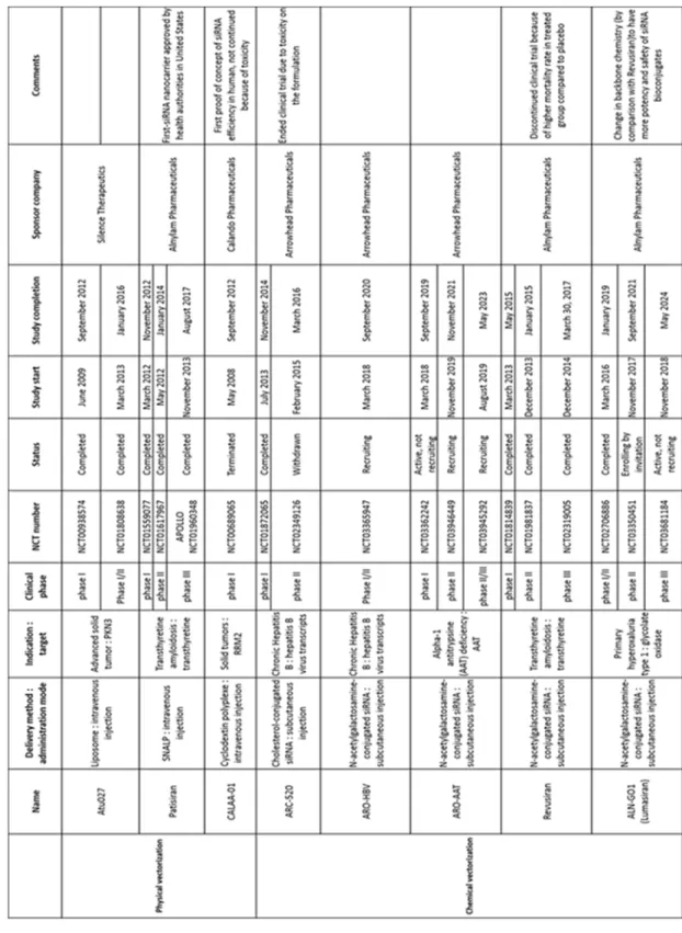
III. Therapeutic applications – overview of some clinical trials
1
Some of the formulations passed successfully all preclinical development steps, proving that
2
they were found safe enough and efficient enough to be administered to patients whether they
3
have been successfully translated to clinical phases or not. According to FDA website, the aim
4
of clinical trials is to “test the potential treatments in human volunteers” in terms of safety,
5
tolerance and efficiency1. 6
We focused on different interesting drug candidates, which were or are currently in phase I, II
7
or III clinical trials (Table 2).
8
9
1 https://www.fda.gov/drugs/development-approval-process-drugs/conducting-clinical-trials,
Table 2: Selected siRNA-based drug candidates in clinical trials
1
2
Abbreviations: siRNA target PKN3 protein kinase 3, RRM2 ribonucleotide reductase subunit
3
M2, AAT alpha-1 antitrypsine
4
A. Lipid nanoparticles
1
a) Lipoplex
2
Atu027 is a liposomal siRNA formulation, composed of an anti-protein kinase 3 (PKN3;
3
downstream effector of phosphatidylinositol 3 kinase) siRNA encapsulated with a new cationic
4
lipid AtuFECT01 in combination with helper and PEG-modified lipids. It proved its efficiency
5
in different cancer models (orthopic mouse models for prostate and pancreatic tumors) with
6
significant tumor growth inhibition and changes in tumor lymphatic vasculature [93] without
7
elevated systemic levels of immune cytokines (IFN-alpha or IL12) [94]. These results led to the
8
clinical development of this novel lipoplex in oncology. The first-in-human phase I study on
9
Atu027 started in 2009 on thirty-four patients with the aim to evaluate the safety, the
10
tolerability, as well as the pharmacokinetic and therapeutic effects on primary tumors and
11
metastatic lesions. Atu027 was considered to be well tolerated with only low-grade toxicities
12
(1 and 2) and safe for patients with advanced solid tumors. Tumor growth was even stable in
13
41% patients [95]. A new phase I/II (NCT01808638) started in 2013. During this stage, Atu027
14
has been co-administered (one or two times per week) with the chemotherapeutic drug,
15
gemcitabine. Twice weekly injection of Atu027 at a dose of 0.253mg/kg with gemcitabine
16
significantly increased patient survival compared to the single injection per week. In addition
17
to this antineoplastic efficacy, patients' quality of life improved [96]. However, no follow-up
18
was available since 2016.
19
b) SNALP
20
Transthyretin amyloidosis is a rare progressive disease caused by the abnormal deposition of
21
transthyretin amyloid in various organs, including peripheral nerves. Patients suffer mainly
22
from progressive neuropathy and cardiomyopathy with median survival less than 15 years after
23
diagnosis. TTR is a circulating protein synthetized by the liver, its role is to carry mainly retinol
24
(vitamin A) and in a lower proportion, thyroid hormone (thyroxine). The gene encoding for
TTR can be mutated, resulting in protein misfolding and aggregation to form amyloid fibrils
1
that will deposit in tissues. No curative treatment existed before the Onpattro® approval.
2
Onpattro®, commercialized by Alnylam Pharmaceuticals, is composed of patisiran, siRNA
3
targeted mutant and wild-type TTR in the 3’-untranslated region of the mRNA and vectorized
4
in a SNALP. The lipid components of this formulation include the ionizable amino lipid
DLin-5
MC3-DMA, PEGylated lipid 1,2-dimyristoyl-rac-glycero-3-methoxypolyethylene glycol-2000
6
(PEG2000-C-DMG), a polar lipid 1,2-distearoyl-sn-glycero-3-phosphocholine (DSPC) and
7
cholesterol. In the bloodstream, PEGylated lipids are removed and replaced by serum protein
8
such as apolipoprotein E, inducing hepatocyte internalization [16, 97]. The receptor-mediated
9
endocytosis leads to endosome acidification, inducing re-ionization of the main ionizable lipid
10
and liberation of the siRNA in the cytoplasm. In non-human primate, which has the same TTR
11
sequence as human, injection of patisiran induced efficient knockdown after single dose of
0.1-12
0.3 mg/kg that prevents protein deposition [98]. Besides, TTR silencing was more effective
13
than tafamidis, the gold treatment used to inhibit products of the Val30Met mutation of TTR
14
gene [98, 99]. Taking together these results, the patisiran passed successfully phase II [99, 100]
15
then phase III [6, 7]. During these studies, it showed its non-toxicity and its efficiency in
16
decreasing circulating TTR and improving patients’ quality of life [7, 100]. Six years after
17
clinical studies beginning, drug-candidate was approved by the European Medicine Agency
18
(EMA) and FDA bringing new hope for patients with hereditary transthyretin amyloidosis [6].
19
Another formulation has been also developed by Anylam Pharmaceuticals but instead of being
20
complexed in a LNP, the TTR-targeted siRNA is conjugated to triantennary GalNAc. This
21
nanovector is amenable to less permissive administration mode (subcutaneous) and has been
22
named revusiran. Preclinical studies demonstrated efficiency and non-toxicity in rodent and
23
non-human primate species [98, 99]. Phase I clinical trial (NCT01814839) did not display
24
severe adverse effects, except those caused by the injection (mostly mild to moderate injection
site reactions and one severe with erythema >10cm) [101]. Phase II (NCT01981837) confirmed
1
the previous results but phase III (ENDEAVOUR; NCT02319005) was dismissed before the
2
end due to report of peripheral neuropathy. Moreover, an increase in lactate concentration and
3
mortality imbalance were observed between treated patients and placebo group [102, 103].
4
Post-trial investigations did not link the higher mortality in the treatment arm at the revusiran
5
but did not totally exclude that this molecule have a toxicity2 [16]. Formulation has been 6
improved by chemical modifications on siRNA sequence and is currently in phase III clinical
7
trial (Vutrisiran; NCT03759379).
8
B. Polyplex
9
Cyclodextrin is a nontoxic and non-immunostimulantcyclic oligomer of glucose. Formulation
10
with linear cationic cyclodextrin-based polymer, PEG chains and transferrin receptor ligand has
11
been developed for siRNA delivery [104]. Encapsulation in this polyplex of siRNA targeted
12
ribonucleotide reductase subunit M2 (RRM2) leads to reduced tumor growth and gene
13
expression silencing [104]. Preclinical toxicity studies in non-human primate displayed, at the
14
highest dose (27mg/kg), mild increase in transaminases and elevated blood urea nitrogen and
15
creatinine, correlated with hepatic- and renal-toxicity respectively. Mild immune stimulation
16
has also been detected at this dose (increased IL-6 levels in all animals). However, no clinical
17
signs of toxicity have been observed; therefore it is used in clinical studies at lowest doses
18
(efficient dose in non-human primates, 0.6-1.2 mg/kg) to avoid biological toxicity [105]. The
19
phase I study conducted by Dr. Heidel at Calando Pharmaceuticals started in May of 2008
20
(NCT00689065), was the first to show that multi-siRNA injections could be safely and
21
successfully accomplished in a non-rodent species and after then in human [5, 105]. Indeed, the
22
targeted nanovector denoted as CALAA-01, accumulated preferentially in human metastatic
23
2
melanoma tumors in a dose-dependent manner due to its targeted ligand and inhibited RRM2
1
expression [106]. Unfortunately, phase IB was ended prematurely because of toxicity due to
2
the long-term drug instability [106, 107].
3
C. siRNA conjugates
4
These formulations aim to reduce toxicity due to cationic charges and are actually used to treat
5
several hepatic disorders.
6
The eradication of hepatitis B virus was found to be unachievable in the case of chronic
7
infection because the virus integrates the genome [108]. ARC-520 is developed by Arrowhead
8
Pharmaceuticals and is composed of two HBV targeted siRNA conjugated to cholesterol and
9
co-injected with 6mg/kg of endosomolytic agent (NAG-MLP) to provide effective endosomal
10
escape [78]. This promising formulation went into phase I clinical trial and was injected to
11
healthy volunteers to assess its safety, tolerability, pharmacokinetics and pharmacodynamics.
12
No serious side effects were found [109]. The phase IIb clinical trial started in 2015 and showed
13
a significant efficiency of ARC-520 with a reduction of Hepatitis B antigen up to 57 days [110].
14
However, the clinical phases were stopped prematurely, due to toxicity concerns, after
non-15
human primate death in preclinical studies3 [16, 111]. 16
The company, Arrowhead Pharmaceuticals, developed a new bioconjugation method, named
17
“Targeted RNAi Molecules (TRiM) technology” that can be injected subcutaneously. This
18
time, the siRNA is directly linked to N-acetylgalactosamine but the specific structure is not yet
19
published. ARO-HBV is one of the first molecule derived from “TRiM technology” and as for
20
ARC-520, is composed of two siRNA silencing HBV transcript and its integrated form, leading
21
to reduce virus resistance to treatments and cure from the infection. Phase I/II of this drug
22
candidate started in 2018 and study completion normally occurs in January 2020
23
3
(NCT03365947) [112]. It should be noted that another formulation is under development for
1
deficiency in alpha-1 antitrypsin (OMIN code 613490) which is an inherited and well-defined
2
disease that can trigger pulmonary and liver disorders [113, 114].
3
After revusiran failure, Alnylam Pharmaceuticals continued the research and focused on
4
changing the backbone chemistry (from standard template chemistry to enhanced stabilization
5
chemistry platform by adding phosphorothioates linkages at the 5’-end of both strands) [82].
6
This leads to a more stable and safe GalNAc-conjugate siRNA [111]. Five drug-candidates are
7
accounted currently in clinical studies: i) vutrisiran (phase III ; NCT03759379) for the treatment
8
of hereditary transthyretin amyloidosis, ii) lumasiran (phase III ; NCT03681184) for the
9
treatment of primary hyperoxaluria type 1 (PH1), iii) fitusiran (phase III ; NCT03754790) for
10
the treatment of hemophilia, iv) inclisiran (phase III ; NCT03814187) for the treatment of
11
hypercholesterolemia and, v) cemdisiran (phase II ; NCT03999840) for the treatment of
12
atypical hemolytic uremic syndrome.
13
ALN-GO1, also called lumasiran, is a siRNA-bioconjugated to triantennary GalNAc to treat
14
the PH1 pathology (Figure 3), which is a rare autosomic recessive disease (OMIN code
15
259900) [115, 116]. The impressive efficiency results in animal models (mice, rat and
non-16
human primate) opened the door for clinical trials [83]. Phases I/II started in 2016. Urinary
17
oxalate level decreased substantially after the first dose. The results showed that the treatment
18
was well-tolerated among the patients with PH1. No drug-related severe adverse effects were
19
observed. The majority of side effects were mild to moderate and unrelated to the drug
20
candidate 4. The same patients were enrolled into phase II open-label extension and then in 21
4
phase III (ILLUMINATE-A) which is currently ongoing in adult and children
1
(NCT03681184)5. 2
On November 20, 2019, the first-ever GalNAc-conjugate siRNA (givosiran, commercial name
3
Givlaari®) has been approved by the FDA (followed in January 2020 by the EMA) for the
4
treatment of a rare inherited hepatic pathology, the acute hepatic porphyria [8]6. 5
IV. What is next?
6
Therapeutic RNAi history spans over different eras. In 1998, its discovery paved the way for
7
a new therapeutic field, which started development in the beginning of the 2000’s (2005 to
8
2008). It was a lavish period for RNAi research, notably thanks to the Nobel Prize in 2006.
9
Unfortunately, uncertainties about the real therapeutic effect of siRNA in human (more due to
10
immunostimulatory effects than silencing) accompanied by bad investments by pharmaceutical
11
companies, led to a funding crisis in early 2010’s. Development of new nanomaterial less toxic,
12
more efficient and RNAi efficiency demonstration in silencing targeted genes (proof of concept
13
by CALAA-01) allowed a recovery of the pharmaceutical industries in this field [117-119].
14
Twenty years after RNAi discovery by Fire and his colleagues, the first siRNA drug has been
15
approved by the FDA and opens a new era for RNAi research and development. However, some
16
challenges still need to be addressed related to: 1) the lifelong administration of encapsulated
17
siRNA in term of toxicity and 2) the enlargement of the treatment for non-hepatic or tumor
18 pathologies. 19 5 https://www.alnylam.com/wp-content/uploads/2019/04/Lumasiran-Phase-2-OLE-ISN-2019.pdf, accessed 30 December 2019 6