HAL Id: tel-02887569
https://tel.archives-ouvertes.fr/tel-02887569
Submitted on 2 Jul 2020HAL is a multi-disciplinary open access archive for the deposit and dissemination of sci-entific research documents, whether they are pub-lished or not. The documents may come from teaching and research institutions in France or abroad, or from public or private research centers.
L’archive ouverte pluridisciplinaire HAL, est destinée au dépôt et à la diffusion de documents scientifiques de niveau recherche, publiés ou non, émanant des établissements d’enseignement et de recherche français ou étrangers, des laboratoires publics ou privés.
mechanical properties on podocyte behaviors
Maya Abdallah
To cite this version:
Maya Abdallah. Development of hydrogels and study the effect of their mechanical properties
on podocyte behaviors. Material chemistry. Université Montpellier, 2019. English. �NNT :
RAPPORT DE GESTION
2015
THÈSE POUR OBTENIR LE GRADE DE DOCTEUR
DE L’UNIVERSITÉ DE MONTPELLIER
En Chimie et Physicochimie Des Matériaux École doctorale SCIENCE CHIMIQUES BALARD (ED459)
Unité de recherche Institut Européen Des Membranes (UMR 5635, CNRS, UM, ENSCM)
Présentée par Maya ABDALLAH
Le 10 Décembre 2019
Sous la direction de Sébastien BALME, La codirection de Maria BASSIL, L’encadrement de Mikhael BECHELANY
Devant le jury composé de
Mme Patricia BASSEREAU, Directrice de Recherche, CNRS, Institut Curie M. Hassane OUDADESSE, Professeur, Université de Rennes I
M. Xavier GARRIC, Professeur, Université de Montpellier
M. Michel BOISSIERE, Maître de Conférences, Université de Cergy-Pontoise Mme Maria BASSIL, Professeur, Université Libanaise
M. Sébastien BALME, Maître de Conférences, Université de Montpellier M. Mikhael BECHELANY, Chargé de Recherches, CNRS
Rapporteur Rapporteur Examinateur Examinateur Co-Directeur Directeur Invité
DEVELOPPER DES HYDROGELS ET ETUDIER LES EFFETS DES
PROPRIETES MECANIQUES SUR LES ACTIVITES BIOLOGIQUES DES
Thank you God for giving me the strength, the support, the opportunities and helping me to exceed all the difficulties and the barriers to pursue and complete the research work.
I would like to extend my deepest sincerest gratitude to all the people who helped me and have shared with me the knowledge to achieve this work.
My deepest appreciation belongs to Mr. Ramy Adwan, Mr. Anthony Awad and Ms. Sonia Abou Azar. You are the greatest and kindest people I have ever met. Thank you for your guidance and your valuable support. It was a great pleasure and honor to meet you.
I would like to acknowledge the attendance and the helpful suggestions from my committee members.
For my director “Dr. Sebastien Balme”: I would like to express my sincere gratitude for his continuous support, for his patience, his motivation, his enthusiasm and his immense knowledge. It was a great honor to work under his guidance. Thank you for teaching me the research methodology in order to present my work in a good way.
My deepest gratitude goes to my co-director “Dr. Maria Bassil” for helping me to pursue my PhD study in a very important and interesting field. Thank you for her continuous guidance, her expert advices and ideas that helped me to accomplish this research project. I want to express my sincere gratitude to “Dr. Mikhael Bechelany”, my supervisor, for giving me the opportunity to work on my thesis within his research group and for providing me invaluable guidance during this period. His dynamism and motivation have deeply inspired me and I am really grateful for everything he offered to me. I would also thank him for his empathy and great sense of humor.
I would like to give my sincere gratitude to Dr. Frederic Cuisinier, Dr. Csilla Gergely, Dr. Marta Martin and Dr. Bela Varga for their expertise, their time, their suggestions and their encouragement. Thank you for allowing me to have an appropriate platform to work on the biological part and to get all the necessary information I need to make this thesis possible. These acknowledgements would not complete without mentioning my research lab colleagues: “Thianji, Nicoletta, Socrates, Octavio, Habib and Amr”. It was a great pleasure
friends: “Zaineb, Soumaya, Ahmed and Wassim”. I really appreciate your true friendship, your guidance and your positive support. It was a great pleasure to meet you. I would like to express my sincere gratitude to my Lebanese friends: “Maryline, Loraine, Ghady, Ghenwa, Joelle, George, Marleine, and Petros” for having great time together within the same Institute. Thank you my special girls, my sisters “Sarah and Syrena” for having great moments, for spending the last working nights together and for providing such a great support. For my best friends, my supports, my sisters “Catherine and Rasha”, I would like to thank you for everything you have done for me. I love you so much!!
I would like to express my gratitude to my dearest friend, to my brother and to my love “Omar“, Thank you for your continuous support , your advices, your patience and your faith. You have always been encouraging me and caring throughout this period. I LOVE YOU.
Very special thanks to my lovely family and especially to my mother for their unconditional love, their continuous and spiritual support given to me throughout the time and their moral encouragement. I could not have done without them. I LOVE YOU. Thank Dr. Wissam for believing on me and helping me to start my PhD studies. You were always a great supporter for me and a big brother who I can count on. Thank you.
Dedication
This humble piece of work is lovingly dedicated to my angel, to my FOTHER, who was my source of strength and my inspiration. You were always by my side.
List of Figures
... iList of Tables
... iiiList of Abbreviations
... ivGeneral Introduction
... 1Chapter I: Literature Review
... 7I. Introduction ... 9
II. Hydrogels ... 10
1. Definition ... 10
2. Hydrogels as scaffolding materials: ... 12
3. Natural and Synthetic Hydrogels ... 13
4. Preparation methods of hydrogels ... 23
5. Hydrogels propertie ... 30
6. Hydrogels For Tissue Engineering Applications ... 36
III. Cell-ECM interaction ... 37
1. Mechanotransduction ... 37
2. Effect of stiffness on tissue functions ... 41
IV. Podocytes ... 44
1. Definition ... 44
2. Podocyte Slit Diaphragm ... 46
V. Conclusion ... 50
VI. Thesis Objectives ... 51
VII. References ... 53
Chapter II: Materials and Methods
... 67I. Materials ... 69
1. Fabrication of Hydrolyzed Polyacrylamide Hydrogels ... 69
2. Synthesis of Gelatin Methacrylamide(GelMA) ... 71
3. Preparation of GelMA-AAm hydrogels... 72
4. Swelling Measurement ... 72
5. Cell Culture ... 72
II. Experimental Methods ... 75
1. Conventional Scanning Electron Microscopy (CSEM) ... 75
2. Environmental Scanning Electron Microscopy (ESEM) ... 76
3. Atomic Force Microscopy (AFM) ... 77
4. Fluorescence Microscopy and Immunocytochemical characterization... 81
5. Multiphoton Microscopy (MPM) ... 84
6. Rheology ... 86
7. Differential Scanning Calorimetry (DSC) ... 88
8. Infrared Spectroscopy ... 89
9. Statistical Analysis... 90
III. Conclusion ... 91
IV. References ... 92
Chapter III: Influence of hydrolyzed polyacrylamide hydrogel stiffness on
podocyte morphology, phenotype and mechanical properties
... 95I. Abstract ... 97
II. Introduction ... 99
III. Materials and Methods ... 102
1. Fabrication of Hydrolyzed Polyacrylamide Hydrogels ... 102
2. Swelling Measurement ... 103
3. Rheology of PAAm Hydrogel ... 103
4. Differential Scanning Calorimetry (DSC) ... 104
5. Scanning Electron Microscopy (SEM) ... 104
6. Atomic Force Microscopy (AFM) ... 104
7. Cell Culture ... 105
8. Immunocytochemical characterization ... 106
9. Multiphoton Microscopy (MPM)... 106
10. Cell Proliferation Assay ... 107
11. Statistical Analysis ... 107
IV. Results and Discussion ... 108
1. PAAm hydrogels properties ... 108
Chapter IV: Enhancement of Podocyte Attachment on Polyacrylamide
Hydrogel with Gelatin-based polymers
... 130I. Introduction ... 131
II. Materials and Methods ... 133
1. Synthesis of GelMA ... 133
2. Preparation of GelMA-PAAm hydrogels ... 134
3. Swelling Measurements ... 134
4. Fourier Transform Infrared Spectroscopy (FT-IR) ... 135
5. Mechanical Characterization ... 135
6. Cell culture... 136
III. Results ... 137
1. H-NMR of Gelatin Methacrylamide ... 137
2. FT-IR of hydrogel material ... 138
3. Swelling Characterization ... 139
4. Mechanical Properties ... 142
5. Podocytes Cells Culture ... 145
6. Cells Elasticity ... 147
IV. Conclusion ... 149
V. References ... 151
i
Chapter I:
Figure 1: Picture of swelled polyacrylamide hydrogel ... 11
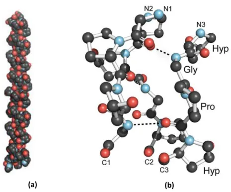
Figure 2: (a) Collagen Triple-Helix Structure (b) Ball-and-stick image of a segment of collagen triple helix ... 15
Figure 3: Basic Chemical Structure of Gelatin ... 17
Figure 4: Gelatin methacrylate (GelMA) synthesis scheme ... 18
Figure 5: Polyacrylamide Matrix Structure ... 19
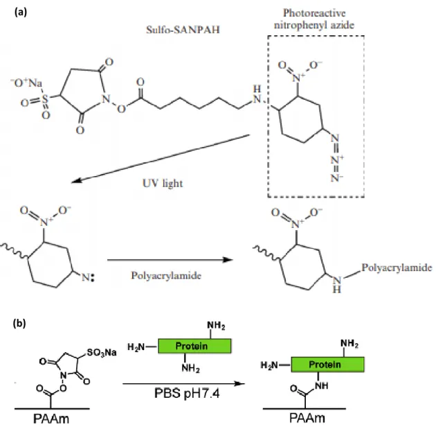
Figure 6: (a) The heterobifunctional photoreactive reagent sulfo-SANPAH. (b) A scheme is shown for ECM protein conjugation using sulfo-SANPAH. ... 20
Figure 7: Hydrolyzed PAAm hydrogels functionalized with EDC and NHS. ... 22
Figure 8: Types of crosslinking: Physical and Chemical Hydrogels... 24
Figure 9: Representative reactions during the photocrosslinking of GelMA to form hydrogel networks. ... 26
Figure 10: (a) Addition of radical to acrylamide monomer. (b) Propagation of PAAm polymerization. ... 27
Figure 11: Hydrogel network formation due to intermolecular H-bonding in CMC at low pH ... 28
Figure 12: Ionotropic gelation by interaction between anionic groups on alginate (COO-) with divalent metal ions (Ca2+) ... 29
Figure 13: Swelling of hydrophilic polymers ... 32
Figure 14: Cells are tuned to the materials properties of their matrix ... 33
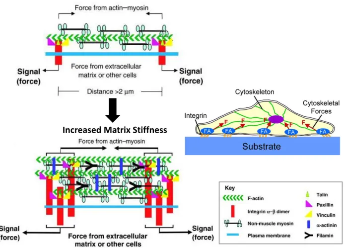
Figure 15: Sensation of, and responses to, matrix-generated mechanical signals ... 40
Figure 16: Effect of matrix mechanics on cell behavior. Schematic of general changes in cell behavior observed as matrix stiffness increases ... 40
Figure 17: Podocytes Imaging... 46
Figure 18: The glomerulus and slit diaphragm ... 49
Chapter II:
Figure 1: PAAm hydrogels polymerization... 70Figure 2: Reversible network swelling and shrinking with solvent... 71
Figure 3: Synthesis of GelMA ... 71
Figure 4: Conventional Scanning Electron Microscopy ... 76
Figure 5: Environmental Scanning Electron Microscopy ... 77
Figure 6: Tip (A) and cantilever (B) schematics of the MLCT tips (noted as C and D) used in our work (© Bruker AFM Probe). ... 78
Figure 7: Principle of the AFM imaging modes ... 78
Figure 8: Typical force-curve as a function of sample indentation showing the contact point between the tip and the sample and the fit performed to calculate the Young’s modulus. ... 79
Figure 9: Energy diagram describing the excitation and emission states ... 83
Figure 10: Epi- fluorescence microscope set-up ... 84
Figure 11: Mechanisms of two-photon excited fluorescence (Fluo-2P). ... 85
Figure 12: Confinement of the 2P excitation to a point like volume (© Cornell University) ... 85
Figure 13: Basic modular components of the multiphoton microscope. ... 86
ii
Figure 1: (A) Representative curve of swelling degree as a function of Bis-acrylamide
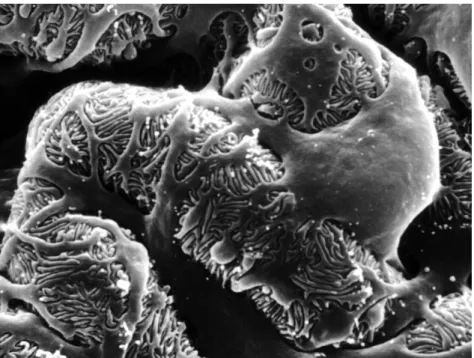
concentrations (0.5, 1, 2, 5, 10 and 30 µl). (B) Scanning Electron Microscopy images of dehydrated PAAm hydrogels ... 109 Figure 2: DSC curves for PAAm hydrogels having different concentrations of crosslinker. Curve (1) corresponds to 1 µl of Bis-acrylamide and curve (2) corresponds to 30 µl of Bis-acrylamide ... 111 Figure 3: (A) Polyacrylamide hydrogels Young's modulus maps measured by AFM ... 113 Figure 4: A) Human Podocyte Cells Line cultured on PAAm hydrogels substrates having different properties (2 and 10 µl Bis-acrylamide respectively). (B) Podocyte proliferation: % of podocytes cover surface as a function of substrate’s elastic modulus ... 115 Figure 5: Immortalized podocytes showed an arborized morphology and interdigitating foot
processes with adjacent cells (yellow arrow). These images were taken using multiphoton
microscopy (A) and environmental SEM (B) for substrate of 2.1 kPa ... 116 Figure 6: (A) (B) Representative immunofluorescence images of podocytes cultured on PAAm substrates with an elasticity ranging from 0.6 to 44 kPa ... 119 Figure 7: Western Blot for the detection of podocin expression on PAAm hydrogels with various elasticity ... 120 Figure 8: Elasticity of podocytes cultured on PAAm membranes.(B) AFM elasticity distribution indicating podocyte elasticity for cells cultured on different samples. (C) Evolution of the cells’ Young’s modulus (E) as a function of hydrogel stiffness ... 121
Chapitre IV:
Figure 1: Synthesis of GelMA ... 134 Figure 2: 1H-NMR of gelatin and modified gelatin (methacrylamide gelatin GelMA). ... 138 Figure 3: FT-IR of pure gelatin and GelMA-AAm interpenetrated polymer network ... 139 Figure 4: Representative histogram of swelling degree as a function of acrylamide and GelMA concentrations ... 140 Figure 5: Schematic simplified view of GELMA-PAAm sequences: (a) GELMA-GELMA
macromolecules; (b) GELMA-AAm; (c) AAm-AAm ... 141 Figure 6: Scanning Electron Microscopy for GelMA-AAm based hydrogel having different polymers concentrations ... 142 Figure 7: (A) Elasticity distribution of GelMA-PAAm hydrogels swelled in cell medium. Young’s moduli were fitted with Gaussian distributions. (B) Evolution of the Young’s modulus “E” as a function of GelMA and AAm concentrations ... 143 Figure 8: The storage modulus of GelMA-PAAm membranes fully swelled in water was determined as function GelMA and AAm concentrations ... 144 Figure 9: (A) Podocyte cells stained with calcein for the determination of cells viability. (B)
Representative immunofluorescence images of podocytes cultured on GelMA-PAAm substrates having various mechanical properties ... 147 Figure 10: Evolution of the Young’s modulus (E) as a function of AMM and GelMA concentrations ... 149
iii
Table 2: List of natural and synthetic hydrogels ... 14
Table 3: The characteristics of: Collagen, Gelatin and PAAm ... 23
Table 4: Physical and chemical hydrogels properties ... 30
Table 5: Hydrogels: Preparation and Mechanical Properties ... 35
iv
CKD: Chronic Kidney Disease GFB: Glomerular Filtration barrier GBM: Glomerular Basement Membrane ECM: Extracellular Matrix
AAm: Acrylamide PAAm: Polyacrylamide GelMA: Gelatin Methacrylate
TEMED: Tetramethylethylendiamine APS: Ammonium Persulfate
ESRD: End Stage Renal Disease
1
3
Regenerative medicine and tissue engineering are defined as an expanding interdisciplinary field implicated in the restriction of donor shortage and immunological rejection evaluated as a major public health issue. The concept of tissue engineering consists to develop a functional scaffold in order to regenerate and enhance the functions of damaged or diseased tissues and organs. The strategies of tissue engineering requires the following factors: (1) the isolation of cells from target tissue, (2) the design of biomaterial scaffold providing an appropriate mechanical environment for cell functions in order to mimic the extracellular matrix, (3) the use of molecules such as proteins and growth factors for cells survival. Nowadays, hydrogels have shown a great advancement and progression as scaffolding materials in tissue engineering. They are defined as three-dimensional hydrophilic polymers network and characterized by their high water content. Various natural and synthetic polymers have been employed to generate physical and chemical crosslinked hydrogels. These hydrogels provide mechanical and physical support similar to the natural extracellular matrix microenvironment of targeted tissue and allow the cell growth and tissue regeneration. The main question is how to design a suitable hydrogel scaffold for tissue engineering. The design of these scaffolds requires various criteria such as biocompatibility, mechanical strength and biofunctionality indispensable in the regulation of cellular behaviors. The purpose of this manuscript is focused on the synthesis of two polymers based hydrogels: Hydrolyzed polyacrylamide as a synthetic hydrogel and GelMA-AAm as a natural/synthetic hydrogel able to: (i) promote the cell adhesion, (ii) ensure the transport of nutrients and growth factors for cell survival and proliferation, (iii) have a negligible immune response and toxicity, (iv) deliver the seeded cells towards the desired damaged site and (v) control and regulate the structure and functions of engineered tissue. Chronic kidney disease (CKD) affects 10% of the population worldwide. CKD is the consequence of
4
glomerular filtration barrier (GFB) dysfunction. The GFB is composed of endothelial cells which are involved in the maintenance of the filtration barrier. Podocyte provide a barrier composed of filtration slits between foot processes that prevent the passage of plasma proteins into the urinary filtrate. The progression of glomerulopathies is the consequence of podocyte injury. These scaffolds materials were developed to mimic the kidney glomerular basement membrane in order to restore the main function of the glomerulus kidney.
This thesis manuscript is composed of four chapters:
1. The first chapter is an overview of various categories of hydrogels, the preparation methods and the main properties of hydrogels. The application of hydrogels as a scaffold in tissue engineering and the criteria implicated in the development of efficient hydrogel scaffold were studied. Moreover, since the extracellular matrix (ECM) provides mechanical and biological environment involved in the regulation of cells behaviors such as proliferation and differentiation, the mechanisms of ECM-cells interaction were developed to better understand the effect of polymer substrates on cells. Furthermore, the characteristics of the in vivo kidney podocytes and their role on maintaining the glomerular filtration barrier were investigated as the work consists to develop a scaffold material having properties similar to the physiological glomerular basement membrane of the kidney.
2. The second chapter will describe on details the materials and the methods used to synthesize the hydrogels scaffolds and to characterize their properties such as the swelling and the mechanical properties. Furthermore, the effect of substrate mechanical properties on the podocyte behaviors was investigated by evaluating the morphology, the proliferation rate, the cytoskeleton organization and the differentiation of these cells.
5
3. The third chapter will be focus on the development of hydrolyzed PAAm hydrogel as a new scaffolding material which is a key step toward the development of adaptive implant materials. First, hydrolyzed PAAm hydrogels were synthesized and covalently crosslinked with various concentrations of Bis-acrylamide that contribute to obtain various crosslinking polymer network densities and mechanical strengths. The influence of hydrolyzed PAAm hydrogels mechanical properties on podocyte morphology, cytoskeleton organization, differentiation and mechanical properties was also evaluated. The hydrolyzed PAAm substrates, on which the physiological activities of podocytes were preserved, were determined. These substrates have shown elasticity within the range of the physiological stiffness.
4. The fourth chapter will present a novel stable hybrid hydrogels based scaffold composed of gelatin methacrylate and acrylamide (GelMA-AAm). The combination of biological and synthetic materials imitates the heterogeneity of native ECM due to the presence of bioactive cell-binding domain and tunable mechanical properties. The properties of GelMA-AAm hydrogels and the effect of scaffolds mechanical properties on the podocyte behaviors were investigated.
Finally, the conclusion part will summarize the main findings of this thesis. Moreover, perspectives will be proposed as possible for future work.
7
9
Hydrogels: Promising Scaffold for Tissue Engineering
I. Introduction:
Extracellular matrix (ECM) is a complex and organized network having a wide variety of macromolecules. Therefore, each tissue is characterized by their specific ECM structure and composition which are altered by the diseases. The major components of ECMs are proteins including laminin, fibronectin, elastin and collagen. These proteins present specific binding sites that recognize and interact with cell surface receptors and matrix constituents that form a three dimensional network. ECM regulates the cells behaviors and promotes the main biological activities such as tissue homeostasis, cellular growth, survival and differentiation1,2,3. The regulation of cells functions is maintained by ECM signals transmitted to the cells. Cells sense the signals, interpret and respond to the different parameters such as ECM structure, components and mechanical properties as stiffness and viscoelasticity4,5. The loss of ECM functions characterized by the modification of its composition and physical properties induce an alteration of ECM-cells interactions and thus create a microenvironment leading to the development and progression of several diseases5. Among these diseases, autoimmune and inflammatory diseases have shown the presence of a modified ECM. However, the understanding of ECM properties and characteristics helps to develop and optimize an in vitro cell culture environment to generate a three-dimensional (3D) model such as biomaterials having requirements close to the in vivo milieu. The 3D in
vitro system can be applied in various biomedical applications such as drug delivery system
and tissue engineering helping to elaborate a new therapeutic target for disease treatment. Nowadays, tissue engineering strategies consist to develop in vitro biological or synthetic materials mimicking the native ECM and able to replace and recover the functions of injured
10
tissues or organs. Tissue engineering should follow different criteria to be achieved. In fact, a functional engineered tissue should be characterized by appropriateness mechanical properties of native tissues and should be incorporated within the physiological environment. The concept of tissue engineering aims to comprehend the in vivo mechanical properties of tissues and to study the biophysical environment of cells. The characterization of ECM properties has permitted the advancement and the progression of substrate used as an engineered construct. In this chapter, the development of hydrogels study and the comprehension of ECM mechanical properties and their influence on cells behaviour such as cell proliferation and differentiation will be discussed and developed. The first part of this chapter consists to define the concept of hydrogels and to develop their preparation methods and their properties. Moreover, the application of hydrogels in tissue engineering field will be elaborated. Then, the chapter will be based on the mechanisms of cells - ECM interactions and the effect of ECM mechanical properties on the cellular behaviors. The third part of this chapter will be focused on introducing the podocyte kidney cells. This work will lead to establish a biomaterial based hydrogel to study the effect of kidney glomerulus extracellular matrix on podocyte behaviors.
II. Hydrogels:
1. Definition:
Gels are defined as semi-solid material composed of hydrophilic polymers and dispersed in a large quantity of fluid. Different types of gels have been identified and classified depending on the type of the swelling factor employed such as hydrogels, organogels and xerogels. In this work, hydrogels are the gels of study. They are characterized by their three-dimensional
11
crosslinked hydrophilic polymer network holding a large amount of water within their network without dissolving (Fig.1). Recently, hydrogels have been progressed and advanced as biomaterials in biomedical engineering such as tissue engineering and drug delivery.
Figure 1: Picture of swelled polyacrylamide hydrogel.
“Hydrogel” term was started since 1894 and was previously widely used in many researches papers, according to Lee, Kwon and Park6. In 1960, polyhydroxyethylmethacrylate (PHEMA) was considered as a first material crosslinked network having a high water affinity and the development of this material has been used in many research projects of permanent contact lens production.“Hydrogel” was considered as a soft material advanced to be used in vivo as a biomaterial. The history classification of hydrogels was identified by Buwalda et al.7:
Hydrogel history was started by the development of materials having a high swelling degree and tunable mechanical properties. These materials were characterized by random crosslinking polymer network using initiators to induce the chemical modifications.
Then, these materials were developed and optimized to respond to the variations of specific stimuli such as temperature, pH and polymers concentrations. These stimuli
12
were utilized to induce specific phenomenon including the polymer polymerization and the drug delivery systems7.
Finally, the researchers were focused on the development of stereo-complexed materials such as PEG-PLA interactions8 and this category of hydrogels has shown a promising advancement.
2. Hydrogels as scaffolding materials:
Research studies have investigated the role and the effect of substrate stiffness on cells behaviour. Generally, the study of cellular processes is directed on non-physiological support as polystyrene which exhibit a high stiffness (1GPa)9,10. With such environment, unconventional cells behaviors were developed corresponding to a loss of phenotype differentiation. Thus, these traditional cell culture techniques fail to ensure a milieu to recapitulate the functional characteristics of cells11. Recently, experiment researches have exhibited the significant role of cell’s microenvironment mechanical properties on cellular behaviors12,13,14. Then, new in vitro culture systems were advanced and progressed in order to establish an appropriate microenvironment for cells and so to mimic a functional unit of human living organ in vitro environment. Biomaterials, and particularly hydrogels, have recently shown a great potential in several biomedical fields such as drug delivery applications and tissue engineering15,16. Moreover, since the mechanical properties have exhibited a great impact on cells behaviour, these polymer materials have permitted to develop 3D in vitro cell culture model with tunable elasticity in order to study the effect of the substrate stiffness on cells17.
Hydrogels materials are viscoelastic crosslinked polymer networks. They are characterized by their hydrophilic structure which allows the absorption of high amount of water and
13
biological fluid, their biocompatibility and their robust mechanical properties similar to living tissues15,18,19. These hydrogels characteristics permit to imitate the properties of native ECM and then to act as an artificial ECM. Therefore, these materials are considered as a promising candidate for tissue engineering due to the similarities of native ECM structure and composition20. Since each tissue requires specific necessities, hydrogels properties can be modified and adjusted in order to match the features of an organ for tissue engineering applications. Over the past decade, hydrogels used as biomaterials have been progressed and have been applied in several applications. Table 1 shows the main properties of hydrogels as biomaterials.
Table 1: Properties of hydrogels as biomaterials21 Properties
Biocompatible
Ease of handling
Injectable
High water content
Mechanical strength
Chemical modification (e.g. having attached cell adhesion ligand)
Addition of cells/drugs
The following sections will be focused on a detailed description of some hydrogels widely used in tissue engineering. Afterward, the methods of preparation and the properties of hydrogels will be talked over.
3. Natural and Synthetic Hydrogels:
Hydrogels were classified into natural and synthetic categories depending on polymer network nature22,23,24 (Table 2).
14
Table 2: List of natural and synthetic hydrogels21
Natural Polymers Synthetic Hydrogels
Collagen Gelatin Alginate Hyaluronic acid Fibrin Poly(ethylene glycol) Poly(acrylamide) Poly(vinyl alcohol) Poly(acrylic acid) Poly(hydroxyethyl methacrylate)
Natural polymers are biological systems characterized by their intrinsic biocompatibility, their biodegradability and their specific interactions with the cells due to their bioactive properties. However, these hydrogels present many disadvantages such as the difficulty of purification, the immunogenicity and the pathogen transmission. Collagen, gelatin, chitosan, fibrin and alginate are considered as natural hydrogels and they represent the most essential components of the extracellular matrix in vivo25. The reproducibility of the structure and the mechanical properties of this category remains a problem poorly understood26,27. These drawbacks can be limited and controlled by using hydrogels based on synthetic polymers. The synthetic hydrogels including polyethylene glycol (PEG), polyacrylamide (PAA) and polyvinyl Alcohol (PVA) are more reproducible and can be controlled and modified for tuning the desired mechanical properties. However, many important factors are investigated as polymer composition, biocompatibility and mechanical properties to design a material suitable to be applied in tissue engineering23,26.
Natural hydrogels such as collagen and gelatin and synthetic hydrogels such as polyacrylamide will be described in this section. In this thesis, these hydrogels will be studied and investigated as scaffold materials for cell culture.
15
a. Collagen:
Type I collagen is the most abundant protein present in human connective tissues accompanied by a significant amount of collagen type II, III and IV. The structure of native collagen structure is characterized by its triple-helical α1- α2 chains having a molecular weight of 300kDa28. Glycine, proline and hydroxylproline constitute the major amino acid sequences of collagen involved in the establishment of triple helix (Fig.2). Collagen protein plays a crucial role in performing numerous biological activities. These proteins provide physical anchorage sites inducing the cells attachment and the cell signalling pathway through the cell-surface receptors such as integrin29.
Figure 2: (a) Collagen Triple-Helix Structure (b) Ball-and-stick image of a segment of collagen triple helix Collagen Structure and Stability28 .
Collagen is considered as a natural polymer widely studied for the development of hydrogels scaffolds. Collagen based hydrogels have been used to mimic the extracellular matrix and have shown advancement as three-dimensional scaffold for cell culture and tissue
16
engineering due to their biomimetic properties such as biocompatibility and similarity to native extracellular matrix30. Collagen scaffold have been used as biomaterials for numerous applications such as wound cover dressings, osteogenic and bone filling materials31,32. However, one of the main inconveniences of using hydrogels based on collagen type I as a substrate for tissue engineering is the difficulty to optimize the properties of these materials. Therefore, the ability to reproduce a scaffold having properties similar to physiological tissues and thus to establish physiological factors for cell regulation remain a problem. This problem depends on the variability of collagen I hydrogel origin. Moreover, collagen type I hydrogels are characterized by their low mechanical strength and low stability. It has been shown that in the absence of chemical crosslinker agent which may damage the collagen structure, there is a difficulty to generate a collagen scaffold with high stiffness (> 1 kPa)11. Collagen type I hydrogels have been used in several applications use for regenerative medicine. Eric Hess et al. have studied the influence of collagen derived hydrogel on Bone Marrow Stromal Cells behaviors. It has been shown the ability of these cells to migrate and to proliferate on these hydrogels. In addition, the osteogenic differentiation was detected on these biomaterials and this study was elaborated to develop a novel bone graft based on collagen type I hydrogels. Moreover, Wong et al. have investigated the collagen based scaffolds properties for brain tissue engineering. Researchers have shown the potential role of collagen in repairing and regenerating the injured brain by providing a supportive matrix for cells33.
b. Gelatin:
Gelatin is a natural denatured form of collagen while maintaining the bioactive properties such as RGD sequences (Arg-Gly-Asp). Gelatin is the result of collagen hydrolysis and is
17
considered as a reversible gel dissolving at body temperature. Figure 3 represents the basic chemical structure of gelatin.
Figure 3: Basic Chemical Structure of Gelatin34.
Gelatin hydrogels have been widely used and applied as suitable scaffolding biomaterials in tissue engineering such as cardiovascular, bone and hepatic tissue engineering due to their significant characteristics such as biocompatibility, biodegradability and poor immunogenicity and cytotoxicity35. Nevertheless, gelatin hydrogels represent many disadvantages recognized by the mechanical properties and their unstable structure which lead to a quick enzymatic degradation. These drawbacks contribute to restrict the gelatin hydrogel applications in tissue engineering field. To improve the mechanical properties and the stability of these hydrogels, gelatin based hydrogels can be covalently crosslinked with carbodiimide and glutaraldehyde36,37. However, these chemical crosslinking agents are cytotoxic and elicit immunological responses. Gelatin methacrylate (GelMA), derivative form of gelatin, has shown a potential attention as implantable materials in tissue engineering applications. The preparation of GelMA is based on the reaction of methacrylic anhydride with the hydroxyl and the amino groups presenting on the side chains of gelatin. This reaction is carried out in the phosphate buffer at 50°C (Fig. 4).
18
Figure 4: Gelatin methacrylate (GelMA) synthesis scheme38
The polymerization of GelMA is performed by UV irradiation using a water soluble photoinitiator such as Irgacure 295939. The photopolymerization reaction contributes to the development of a covalently crosslinked GelMA polymer network. The photocrosslinked GelMA hydrogels are characterized by their biocompatibility, their inherent biological activity due to the presence of cell-attachment sequence and their tunable mechanical properties. These characteristics provide a platform to study the cellular behaviors and therefore GelMA hydrogels will be the choice of study as they represent good candidates to mimic the native extracellular matrix for cell culture40,41.
c. Polyacrylamide:
Polyacrylamide (PAAm) hydrogel is a synthetic covalently crosslinked polymer network. PAA hydrogels have been used as a biomaterial for cell culture and have shown a wide progression in this field due to their several important advantages. These materials have formed an important platform to study the cell-substrate interactions depending on material stiffness. The preparation of PAAm hydrogels is simple and inexpensive. PAAm hydrogels are biocompatible and non-biodegradable polymers and can be exposed with broad range of elasticity (0.3 kPa-300 kPa) by changing the acrylamide and bis-acrylamide relative concentrations. The stiffness variations contribute to study and to more understand the
19
influence of substrate stiffness on cellular behaviors such as cell morphology, motility and differentiation42. PAAm hydrogels are synthesized by mixing various concentrations of acrylamide and bis-acrylamide crosslinker. This reaction is initiated and catalyzed using Ammonium Persulfate (APS) and N, N, N’, N’ Tetramethylethylenediamine respectively (Fig.5). APS serves as an inducer of a free radical polymerization43.
Figure 5:Polyacrylamide Matrix Structure44
Furthermore, ECM proteins comprising laminin, fibronectin and collagen type I, IV are covalently conjugated to PAAm hydrogels surface which serve as ligands for cell attachment. Sulfosuccinimidyl 6-(40-azido-20-nitrophenylamino) hexanoate, known as Sulfo-SANPAH, is a
20
photoactivatable heterobifunctional reagent holding two functional groups: Sulfosuccinimidyl
group reacts constitutively with the primary amine groups of proteins and phenylazide group forms a non-specific binding with polymerized PAAm hydrogel during the photoactivation. Thus, ECM proteins - PAAm hydrogels surface interactions is covalent using sulfo-SANPAH (Fig. 6).
Figure 6: (a) The heterobifunctional photoreactive reagent sulfo-SANPAH44. (b) A scheme is shown for ECM protein conjugation using sulfo-SANPAH45.
(a)
21
Since PAAm hydrogels furnish for cell culture a physiological environment, Yeung et al. have worked on PAAm substrates and have studied the effect of substrate stiffness on cells fate. They have concluded that the surface stiffness has an impact on cells morphology and proteins expression by showing that the morphology of fibroblast and endothelial cells, seeded on substrate having elasticity higher than 2kPa, is more spread and promotes the development of actin stress fibers. Furthermore, the regulation of cell locomotion has been studied on PAAm hydrogel with variable elasticity. Researchers have observed that the migration rate of fibroblasts is high on soft substrates comparing to stiff substrates. In addition, it has been shown that the epithelial cells on these substrates present a highly lamellipodial protrusion which is the consequence of destabilized adhesion46. Despite these advantages, PAAm hydrogels have a limited capacity to interact with the physiological environment and the presence of unreacted acrylamide within the PAAm hydrogels which is toxic for the cells will be remained44. Once placed in a basic solution, the PAAm hydrogel will turn into hydrolyzed PAAm form. The hydrolyzed PAAm hydrogel are defined as biocompatible polyelectrolyte holding a large amount of water and biological fluids within its interstitial space. During the swelling process, the unpolymerized acrylamide will be washed out from the polymer matrix. Hydrolyzed PAAm hydrogel are capable to exhibit morphological changes in response to the external stimuli such as pH, electrical fields and ionic strength47. In this thesis, hydrolyzed polyacrylamide hydrogel is investigated as a new scaffolding material which is an important step toward the development of adaptive implant materials.
The hydrolyzed PAAm hydrogel surface is functionalized with (3-dimethylaminopropyl) carbodiimide-HCL (EDC). EDC reacts with the carboxyl groups of hydrolyzed PAAm hydrogel and induce the appearance of an amino-reactive intermediate which contributes to the
22
protein conjugation. EDC is used in combination with N-hydroxysuccinimide (NHS) or sulfo-NHS in order to create a more stable intermediate by increasing the coupling efficiency48(Fig. 7).
Figure 7: Hydrolyzed PAAm hydrogels functionalized with EDC and NHS48.
EDC Carboxylate-containing molecule PA Unstable o-isoacylurea intermediate Sulfo-NHS Amine-reactive NHS ester PA PA Protein Polymer with available amine Protein PA Protein-polymer conjugate
23
Table 3 shows a summary of the main characteristics of the hydrogels mentioned above.
Table 3: The characteristics of: Collagen, Gelatin and PAAm.
Collagen Gelatin PAAm
Nature Natural Natural Synthetic
Bonding Physical Physical Chemical
Preparation
Methods Protein Self-Assembly Protein Self-Assembly Free Radical Polymerization
Advantages Biocompatible (Biological properties) Biodegradable Biocompatible (Biological properties) Biodegradable Poor immunogenicity Tunable mechanical properties
Durable and storage stability Possibility of control
Disadvantages
Weak mechanical properties
Limited long term stability Batch-to-batch variability Weak mechanical properties Non-stable Toxicity of unreacted monomers Tested on Cartilage Bone Tendons Cartilage Bone Podocyte Brain Fibroblast
4. Preparation methods of hydrogels:
The preparation methods of polymers based hydrogels are processed in different ways according to their applications. Depending on crosslinking types, hydrogels can be classified into two categories: physical and chemical crosslinked hydrogels. Chemical hydrogels require the development of covalent crosslinked polymer network. Different methods are applied to synthesize chemically crosslinked permanent hydrogels including photopolymerization and chemical polymerization. The crosslinking mechanisms are based on the chemical reaction of polymer having functional groups as OH, COOH and NH2 with the crosslinking agent. Physical
hydrogels do not require the use of crosslinker and are considered as reversible polymer network. The forces implicated in the development of physical hydrogels are hydrogen bonding, ionic bonding and hydrophobic interactions. These interactions can be disrupted with changes in the physical conditions49,50 . These chemical and physical methods are discussed and summarized below (Fig.1).
24
Figure 8: Types of crosslinking: Physical and Chemical Hydrogels51
a. Chemical crosslinked hydrogels:
Chemical crosslinked hydrogels are characterized by the presence of covalent interaction bonds. The polymerization of end-functional group in polymers chain is the common method to establish covalently crosslinked polymer network. The photochemical and chemical polymerization will be described below in details.
i. Photochemical polymerization:
Photoinitiated polymerization process is considered as an efficient method for hydrogels synthesis. This technique provides many advantages during the hydrogels preparation such as no need to use catalysts or additives to initiate the polymerization, the crosslinking density of the polymer network chain can be controlled by changing the irradiation dose and the presence of contamination will be eliminated. The photopolymerization method has been widely used for many biomedical applications due these benefits. Photopolymerization, known also as free radical polymerization technique, is based on the
25
presence of monomers holding appropriate photosensitive functional groups. Photopolymerization process follows up the subsequent steps: initiation, propagation and termination. The initiation step is activated by the use of a photoinitiator involved in free radical generation. These radicals initiate the polymerization by interacting with the monomer reagent in order to turn it into an active form. Then, the propagation step goes on to get a polymer radical chain. The termination step is thus accomplished through radical combination leading to the development of connected polymer network chain20. As example, many research studies have worked on the photopolymerization of PEG by using UV irradiations with specific photoinitiator in order to generate radicals which induce the polymerization. PEG photopolymerization contributes to the development of PEG hydrogels used as a scaffold for cells delivery involved in tissue regeneration. For example, PEG hydrogels have been used as a matrix for osteoblasts encapsulation to evaluate their applicability in promoting bone tissue engineering52,53,54. Moreover, GelMA based hydrogels have been widely used in biomedical research field due to their suitable biological properties and tunable mechanical characteristics. The photopolymerization is the method of GelMA hydrogels preparation. The initiation of GelMA polymerization is induced by the presence of free radicals generated by the photoinitiator. The propagation of GelMA chain is performed between the methacryloyl groups. Eventually, the termination step will be occurred between two propagating chains or between one propagating chain and a second radical55(Fig. 9).
26
Figure 9: Representative reactions during the photocrosslinking of GelMA to form hydrogel networks55.
ii. Chemical polymerization:
Chemical polymerization is considered as one of the main polymerization techniques for the development of natural and synthetic hydrophilic polymers based hydrogels. This technique is based on chemical reaction of bi-functional crosslinking agent with a hydrophilic polymer holding appropriate functional group such as COOH, OH and NH220. This technique has been
widely utilized to form various chitosan-based hydrogels. These hydrogels are covalently crosslinked polymer hydrogels and involved in many applications such as drug delivery systems by releasing bioactive materials and can be used as a scaffold for cell culture due to their biocompatibility56. For instance, gelatin and albumin based hydrogels were prepared using dialdehyde or formaldehyde as crosslinking agents. In this thesis, polyacrylamide
gelatin gelatin gelatin gelatin gelatin gelatin
gelatin gelatin
gelatin gelatin
gelatin gelatin gelatin gelatin
gelatin gelatin gelatin Initiation Propagation Initiator Termination
27
hydrogels are synthesized using acrylamide monomer and bis-acrylamide as crosslinking agent. This polymerization technique requires the use of Ammonium Persulfate (APS) and N,
N, N’, N’ Tetramethylethylendiamine (TEMED) to induce and initiate the polymerization
reaction of polyacrylamide. In fact, the following steps are involved in the synthesis of polyacrylamide: (1) The initiation of polyacrylamide polymerization is induced by the presence of free radicals generated and accelerated by APS and TEMED respectively; (2) The free radicals provoke the activation of the acrylamide monomers by converting them into acrylamide free radicals. The activated acrylamide monomers react with the unactivated one and induce the elongation of polymer chain. Meanwhile, the polyacrylamide chain is also covalently crosslinked with bis-acrylamide; (3) The termination of polyacrylamide propagation is achieved when the activated acrylamide monomers are totally consumed57 (Fig. 10).
Figure 10: (a) Addition of radical to acrylamide monomer. (b) Propagation of PAAm polymerization.
(a)
28
b. Physical crosslinked hydrogels:
The physical crosslinking of polymers represents a common technique for hydrogel synthesis. For instance, the physical interactions corresponding to hydrogen bonding and ionic interactions will be developed. These techniques are generally processed under mild conditions.
i. Hydrogen Bonding:
Hydrogen bonds mechanism contributes to obtain physically cross-linked hydrogels. H-bonded hydrogel is obtained with the presence of polymers containing carboxyl groups in a low pH solution as carboxymethyl cellulose (CMC) in HCl solution. The substitution of CMC sodium with hydrogen of acidic solution promotes the creation of hydrogen bonds within the polymer. These hydrogen bonds decrease the solubility of CMC in water which contribute to the development of an elastic physical hydrogel58(Fig. 11).
29
ii. Ionic Interaction:
Physical hydrogels can be developed as well through ionic interactions under physiological conditions. Alginate, a naturally derived polymer, is a linear polysaccharide copolymer composed of β-D-mannuronic acid and α-L-guluronic acid. Alginate hydrogel, used as a matrix for protein release and living cells incorporation, is formed in the presence of divalent cations such as calcium Ca2+ which interact with the carboxylic groups of alginate polymer chain (Fig.12). The mechanical strength of these ionic crosslinked hydrogels was shown a variation over time due to the exchange of Ca2+ ions with monovalent ions in surrounding solutions and thus this procedure is considered uncontrollable60. Likewise, chitosan polymer network is ionically crosslinked and mainly used for drug delivery56.
Figure 12: Ionotropic gelation by interaction between anionic groups on alginate (COO-) with divalent metal ions (Ca2+)59.
30
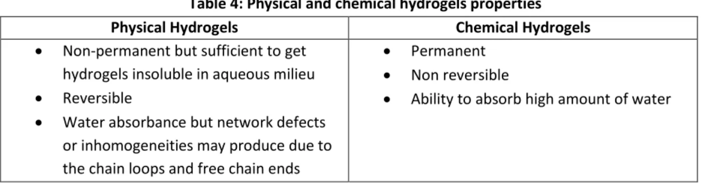
Table 4 shows the characteristics and properties of physical and chemical crosslinking hydrogels61.
Table 4: Physical and chemical hydrogels properties
Physical Hydrogels Chemical Hydrogels
Non-permanent but sufficient to get hydrogels insoluble in aqueous milieu
Reversible
Water absorbance but network defects or inhomogeneities may produce due to the chain loops and free chain ends
Permanent
Non reversible
Ability to absorb high amount of water
In our study, the chemical polymerization was focused on to synthesize the hydrogels scaffolds.
5. Hydrogels properties:
Hydrogels are characterized by numerous properties such as swelling, mechanical strength and biocompatibility. For a perfect scaffolding material, these physical properties should be as similar as in vivo for specific tissue engineering. These properties depend on hydrogel structure that can be controlled by modifying the synthesis mechanisms and the chemical compositions62. The characterization of these properties permits to understand their abilities to affect cells behaviors and therefore their influence on cells mechanotransduction.
a. Swelling:
Swelling property is the capacity of hydrogels, in an aqueous environment, to uptake a large amount of water without dissolution since hydrogels are defined as hydrophilic polymers and characterized by their high water affinity due to the presence of hydrophilic groups within the polymer network chains such as -OH, -COOH63,64. The water molecules are attracted toward these hydrophilic and polar groups and form primary bound water. The hydrophobic moieties also interact with the water molecules and form secondary bound
31
water. Moreover, additional water will be absorbed by the hydrogels under the influence of an osmotic driving force opposed to the network elastic force based on the polymer network crosslinks. This imbibed water, called free water, contributes to reach the equilibrium swelling of hydrogels. A balance between the osmotic pressure and the elastic force will be occurred at the equilibrium swelling. This concept is explained by the Flory-Huggens theory65. Swelling phenomenon is based on osmotic pressure due to the presence of unequal ions distribution in the polymer network and the aqueous environment20. Once hydrogels are placed in water, the osmotic pressure promotes the expansion of polymer network and the hydrogels start to be swelled. The equilibrium swelling degree will be reached when the osmotic forces and the elastic forces of polymer chains are balanced. Swelling ratio indicates the hydrophilicity and the crosslinking degree of polymer network. It was shown that the swelling ratio of hydrogels decreases with increasing the crosslinking density of polymer chains because the polymer structure will become more compacted limiting the network extent57,63. Moreover, Vishal et al. have studied the effect of the ionic strength on hydrogel swelling capacity. They have shown that hydrogels placed in a swelling medium, containing Na+ and Cl- ions, contributes to decrease the osmotic pressure and thus the swelling degree due to the presence of counter ions66. The water swelling capacity of polyacrylamide hydrogel is lower than the hydrolyzed form of polyacrylamide because of the electrostatic repulsion induced by the resultant anionic carboxyl groups. To obtain a hydrolyzed form, PAAm hydrogel is placed in a basic solution of sodium hydroxide (NaOH). The hydrolysis process induces the conversion of amine moieties (R-NH2) of PAAm network
chain into carboxylate (-COO-) moieties having a high water affinity. Furthermore, Na+ ions present in NaOH solution are attracted toward the COO- entrapped into the PAAm polymer chain. Afterwards, the hydrolyzed PAAm hydrogel is placed in deionized water. A counterion
32
osmotic pressure will be occurred since the concentration of Na+ free ions present into the polymer chain is not equivalent to the ions in the swelling solution. Na+ ions start to diffuse to the outer solution in order to decrease the concentration gradient and thus the swelling phenomenon of the PAAm hydrogel is occurred. The equilibrium state of water swelling degree becomes established where the osmotic pressure and the network elastic forces are balanced63. Experimentally, the swelling ratio is determined as the ratio of wet weight (WS)
to dry weight (Wd) and is expressed using this following equation:
S d d W W S W
Where Ws is obtained by measuring the weight of a fully swollen hydrogels (in equilibrium with water and cell media) and Wd is the measurement of a dehydrated hydrogel weight67
(Fig.13).
Figure 13: Swelling of hydrophilic polymers68.
b. Mechanical properties:
The mechanical properties, characteristics of cell microenvironment, have been shown critical role in the regulation and maintenance of cell behaviour. Matrix stiffness represents
PAAm gel (Dry State)
+ Liquid
PAAm hydrogel in Swollen State
33
a fundamental physical property and affects the cells by modifying their shape, size and cytoskeleton organization46. Cell-extracellular matrix (ECM) interactions induce the cells to respond to the physical properties of the membrane and to react by applying a force in order to control the cellular functions through actin dynamics and actomyosin contractility. The tension of this force applied depends on the matrix stiffness. Therefore, each type of cell has a specific mechanical microenvironment (Fig.14).
Figure 14: Cells are tuned to the materials properties of their matrix69.
Recent studies have shown that the mechanical properties of the substrate and particularly the stiffness have a great impact on cells functions including cells migration70,71, proliferation and differentiation72,73. Numerous studies have worked on various density of crosslinking hydrogel in order to clarify the effects of mechanotransduction on cells behaviors. Substrate stiffness can be assessed by measuring the Young’s Modulus defined as material resistance to deformation using different characterization methods such atomic force microscopy (AFM), dynamic mechanical analysis (DMA) and rheology by applying force on a definite material zone. Researches have used AFM to measure the hydrogels surface elasticity and
34
thus to evaluate the network crosslinking density10,74. Actually, the Young’s Modulus of a substrate increases with increasing the concentration of crosslinker and therefore the substrate is considered as a stiff material. Hydrogels, with tunable elastic properties, were developed so as to create in vitro cell culture model mimicking the extracellular environment. Notably, polyacrylamide hydrogels have been widely used as biomaterials for tissue engineering as they present a wide range of adjustable elasticity imitating several tissues stiffness as neurons ( ̴0.5kPa)75, fibroblast ( ̴10kPa)76 and osteoblast (>20kPa)77 stiffness and this stiffness variation depends on polymer network crosslinking density. Most studies have worked on the stiffness effect on mesenchymal stem cells (MSCs) differentiation. They have shown that MSCs upregulate the expression of neuronal markers on soft substrate having an elasticity close to brain tissue whereas these cells express myogenic and osteogenic markers on stiff substrate with elasticity similar to muscle and collagenous bone, respectively14. Moreover, the GelMA hydrogels have shown a great interest as scaffold materials for biomedical applications due to their widely biological properties and their tunable mechanical properties. Wang et al. have shown that the degree of methacrylation has a significant effect on the GelMA mechanical properties. The high methacrylation degree increases the stiffness of GelMA hydrogels due to the increase of methacrylic substitutions that induces the network crosslinking78. According to the mechanical property, hydrogels were used recently for several biomedical applications such as drug delivery, wound dressing and tissue engineering.
Table 5 includes different type of hydrogels using as scaffold materials in biomedical applications. The preparation methods and the mechanical properties of these hydrogels are also determined.
35
Table 5: Hydrogels: Preparation and Mechanical Properties
Hydrogels Method of Synthesis Method of
Characterization Elasticity References
Gelatin-mTG Gelatin-EDC Chemical Polymerization Chemical Polymerization Rheology Rheology Tensile Stress 0,6 kPa – 13 kPa 2 kPa 196 kPa Mufeng Hu et al.79 Qi Xing et al.80 Polyacrylamide Chemical Polymerization Photopolymerization Chemical Polymerization Atomic Force Microscopy Atomic Force Microscopy Rheology 2 kPa – 10 kPa 1 kPa – 240 kPa 0,18 kPa – 16 kPa
Jérôme Solon et al.10
Raimon Sunyer et al.81 Tony Yeung et al.76
PEG-GelMA Photopolymerization Tensile Stress 0.2 MPa – 0.6 MPa Jia Liang et al.82
PEGMA-PEGDMA Photopolymerization Rheology 10
6
Pa – 108 Pa Ji Won Hwang et al.83
PEG-PLLA-DA Photopolymerization Compression Test 1 kPa – 13 kPa Yu-Chieh Chiu et al.84
GelMA Photopolymerization Compression Test 1 kPa – 34 kPa Yupeng Sun et al.
c. Biocompatibility:
Tissue engineering concept requires the development of an appropriate biomaterial able to restore the main tissue functions and to obstruct the immune system by inhibiting the macrophage response which knocks down the functional properties of implantable materials. Biocompatibility is defined as the material ability to interact within the physiological environment without damaging the host tissues. Many studies have suggested the usage of appropriate extracellular matrix based polymer for cells transplantation. Therefore, biocompatibility is a main property in making polymer based hydrogels for tissue engineering applications85. Researchers have studied the central nervous system (CNS) properties to develop biocompatible hydrogels for neural tissue engineering. It was proved that the non-biocompatibility of hydrogels with neural tissue induces the neuroimmune
36
system to react toward the implanted polymer and thus damages will be produced. Recently, the development of biocompatible hydrogels for neural tissue engineering has advanced and has shown that neural cells transplantation encased within a hydrogel might be protected from acute inflammatory response and thus enhances the cells survival86. The evaluation of biological responses allows characterizing the materials biocompatibility. Inflammation diseases, mechanical damages and immunological rejections are assessed as side effects of synthetic materials. The toxicity of synthetic hydrogels remains a drawback for tissue engineering due to the initiators and organic solvents used and due to the presence of unreacted monomers during hydrogel polymerization which are harmful for tissue cells. Nowadays, researches start to focus on the development of advanced biomaterials by controlling their efficacy in order to reduce their side effects.
According to these properties, hydrogel has been used as a promising candidate in tissue engineering. The application of hydrogels as a scaffold for tissue engineering requires various main functions including the cell adhesion, the nutrients and growth factors transport ensuring the cell survival, the delivery of seeded cells toward the desired site and the absence of toxicity. Therefore, the concept of tissue engineering using hydrogels as matrix will be elaborated.
6. Hydrogels For Tissue Engineering Applications:
Tissue engineering is a promising process to regenerate and replace damaged tissues or organs. Tissue engineering concept was suggested by Langer et al. in 1990s as “the application of engineering and the life sciences principles toward the fundamental understanding of structure-function relationships in normal and pathological mammalian tissues and the development of biological substitutes that restore, maintain or improve tissue function”. Tissue engineering strategy consists to develop and design an appropriate
37
scaffold material serving as an extracellular matrix for appropriate tissue cells without inducing immune responses reactions. Biomaterials such as hydrogels are used as functional scaffolds for tissue engineering as they provide mechanical support for cells and suitable biophysical and biochemical signals implicated in the regulation of cell behaviour and function. Selection and design of the appropriate hydrogel scaffold material for tissue engineering requires fundamental characteristics such as hydrogels biocompatibility, mechanical strength and biofunctionality essential in the regulation of cellular behaviors. Table 6 shows the main characteristics of hydrogel to be designed as a scaffold material for tissue engineering.
Table 6: Design criteria for hydrogels as scaffolds for tissue engineering Hydrogels design criteria
Gel synthesis mechanisms
Appropriate mechanical properties
Appropriate diffusion of nutrients and metabolites
Biological properties
Biocompatibility
Promotion of cell adhesion, proliferation and differentiation
The following section will be focused on the mechanisms of interaction between the extracellular matrix and the cells and on the study of the effect of hydrogels mechanical properties on tissue functions.
III. Cell-ECM interaction:
1. Mechanotransduction:
The ECM mechanical characteristics have an important role in the regulation of cell fate and cell behaviour. Cells sense, integrate and interpret the physical and chemical signals of extracellular environment. Thus, cells feel the topography87,88 and the rigidity12,14 of ECM and therefore produce appropriate physiological responses regulating the cellular activities
38
such as proliferation, migration, differentiation and apoptosis89. Accordingly, every living tissue is determined by a specific elasticity. Previous studies have shown the significant role of ECM mechanical properties in the progression of breast cancer disease. They have proved that the stiffness of healthy mammary glands is approximately 150 Pa, while the breast tumor exhibits a high stiffness of 4 kPa. Moreover, the morphology and the growth rate of mammary epithelial cells have showed a significant difference depending on stiffness environment90. Regarding the liver tissue, the stiffness of healthy liver is around 300 - 600 kPa while the stiffness of liver diseases such as fibrosis and cirrhosis increases over 20 kPa91. Cellular adhesion machinery is composed of specialized transmembrane receptors associated via the cytoplasmic domain with the actin cytoskeleton. These sites enhance the ECM – cells and cells - cells interactions in a way to maintain the specific form and mechanical properties of tissues. Through these active sites, cells recognize the physical cues which are defined by the mechanical forces applied to cellular membrane. Several studies have investigated the importance of microenvironment mechanical properties in the regulation of cellular behaviors92,93,94,95. In fact, cells sense and respond to the mechanical properties generated by ECM through integrin. Integrin is a transmembrane cell adhesion protein characterized by the presence of two domains: an intracellular domain in interaction with the cell cytoskeleton and an extracellular domain binding to the substrate96,97. ECM mechanical signals will be transduced into intracellular signals and affect the cells properties as cells morphology, migration, cytoskeleton organization, differentiation and cells stiffness. This is called a mechanosensing phenomenon. The mechanosensing definition is the capability of a tissue or cell to detect the microenvironment applied forces such as contractile forces5,93. The transduction of ECM mechanical signals is established through focal adhesion complex characterized by the binding of ECM molecules to a group of