HAL Id: hal-03060153
https://hal.archives-ouvertes.fr/hal-03060153
Submitted on 21 Dec 2020
HAL is a multi-disciplinary open access
archive for the deposit and dissemination of
sci-entific research documents, whether they are
pub-lished or not. The documents may come from
teaching and research institutions in France or
abroad, or from public or private research centers.
L’archive ouverte pluridisciplinaire HAL, est
destinée au dépôt et à la diffusion de documents
scientifiques de niveau recherche, publiés ou non,
émanant des établissements d’enseignement et de
recherche français ou étrangers, des laboratoires
publics ou privés.
coactivators (CRTC) in transcriptional activation of
steroidogenic acute regulatory protein (Star) by ACTH
Lorna I.F. Smith, Victoria Huang, Mark Olah, Loc Trinh, Ying Liu, Georgina
Hazell, Becky Conway-Campbell, Zidong Zhao, Antoine Martinez,
Anne-Marie Lefrançois-Martinez, et al.
To cite this version:
Lorna I.F. Smith, Victoria Huang, Mark Olah, Loc Trinh, Ying Liu, et al.. Involvement of
CREB-regulated transcription coactivators (CRTC) in transcriptional activation of steroidogenic acute
regu-latory protein (Star) by ACTH. Molecular and Cellular Endocrinology, Elsevier, 2020, 499, pp.110612.
�10.1016/j.mce.2019.110612�. �hal-03060153�
Contents lists available atScienceDirect
Molecular and Cellular Endocrinology
journal homepage:www.elsevier.com/locate/mceInvolvement of CREB-regulated transcription coactivators (CRTC) in
transcriptional activation of steroidogenic acute regulatory protein (Star) by
ACTH
Lorna I.F. Smith
a,b,∗, Victoria Huang
a, Mark Olah
a, Loc Trinh
a, Ying Liu
a, Georgina Hazell
b,
Becky Conway-Campbell
b, Zidong Zhao
b, Antoine Martinez
c, Anne-Marie Lefrançois-Martinez
c,
Sta
fford Lightman
b, Francesca Spiga
b, Greti Aguilera
aaSection on Endocrine Physiology, Program on Developmental Endocrinology and Genetics, Eunice Kennedy Shriver National Institute of Child Health and Human
Development, NIH, Bethesda, MD, USA
bHenry Wellcome Laboratories for Integrative Neuroscience and Endocrinology, University of Bristol, Bristol, UK
cGénétique Reproduction & Développement, CNRS UMR 6293, Inserm U1103, Université Clermont Auvergne, 63001, Clermont-Ferrand, France
A R T I C L E I N F O Keywords: CRTC StAR CREB Steroidogenesis Adrenal cortex Transcription A B S T R A C T
Studies in vivo have suggested the involvement of CREB-regulated transcription coactivator (CRTC)2 on ACTH-induced transcription of the key steroidogenic protein, Steroidogenic Acute Regulatory (StAR). The present study uses two ACTH-responsive adrenocortical cell lines, to examine the role of CRTC on Star transcription. Here we show that ACTH-induced Star primary transcript, or heteronuclear RNA (hnRNA), parallels rapid increases in nuclear levels of the 3 isoforms of CRTC; CRTC1, CRTC2 and CRTC3. Furthermore, ACTH promotes recruitment of CRTC2 and CRTC3 by the Star promoter and siRNA knockdown of either CRTC3 or CRTC2 attenuates the increases in ACTH-induced Star hnRNA. Using pharmacological inhibitors of PKA, MAP kinase and calcineurin, we show that the effects of ACTH on Star transcription and CRTC nuclear translocation depend predominantly on the PKA pathway. The data provides evidence that CRTC2 and CRTC3, contribute to activation of Star tran-scription by ACTH, and that PKA/CRTC-dependent pathways are part of the multifactorial mechanisms reg-ulating Star transcription.
1. Introduction
Glucocorticoid hormone release from the adrenal gland is essential for maintaining normal metabolic function and survival during severe stress (Cole et al., 1995;Whitehead et al., 2013). In basal (unstressed) conditions, glucocorticoids are secreted rhythmically, with both circa-dian (daily) and ultracirca-dian (pulsatile) variations (Park et al., 2013;
Weitzman et al., 1971;Tapp et al., 1984). The episodic nature of glu-cocorticoid secretion is critical for homeostasis, and its alteration can result in changes in tissue steroid responsiveness, resulting in neu-roendocrine, behavioural and metabolic dysfunction (Deuschle et al., 1997; van den Berg et al., 1995; Qian et al., 2012). Glucocorticoid circadian and ultradian rhythmicity follow preceding changes in cir-culating ACTH. As with other steroid hormones, stimulation of gluco-corticoid secretion by ACTH requires de novo synthesis. These ACTH-dependent glucocorticoid secretory episodes in vivo, are associated with
rapid transcription of genes encoding steroidogenic proteins in the zona fasciculata of the adrenal cortex (Sewer and Waterman, 2002; Spiga et al., 2011a,b;Liu et al., 2013;Spiga et al., 2017), including the rate-limiting steroidogenic acute regulatory (StAR) protein (Stocco and Clark, 1996). Although initiation of steroidogenesis following ACTH stimulation depends on rapid post-translational modifications of StAR (Arakane et al., 1997), this protein is short-lived (Artemenko et al., 2001) and transcriptional episodes are essential for maintaining ade-quate mRNA and protein levels for subsequent secretory episodes (Ferguson, 1963;Garren et al., 1965;Clark et al., 1997).
The mechanism of action of ACTH involves cAMP/PKA-dependent mechanisms (Cammas et al., 1995;Schimmer, 1972;Clark et al., 2001), and, to a lesser extent, the Mitogen-activated protein kinase (MAPK) pathway (Gyles et al., 2001; Lotfi et al., 2000; Rocha et al., 2003;
Winnay and Hammer, 2006). This results in activation of transcription factors responsible for inducing Star transcription, including
https://doi.org/10.1016/j.mce.2019.110612
Received 3 June 2019; Received in revised form 6 September 2019; Accepted 4 October 2019
∗Corresponding author. Diabetes Research Group, School of Life Course Sciences, 2.28 Henriette Raphael, Guy's Campus, King's College London, London, SE1 1UL,
UK.
E-mail address:lorna.smith@kcl.ac.uk(L.I.F. Smith).
Molecular and Cellular Endocrinology 499 (2020) 110612
Available online 08 October 2019
0303-7207/ © 2019 The Authors. Published by Elsevier B.V. This is an open access article under the CC BY license (http://creativecommons.org/licenses/BY/4.0/).
steroidogenic factor 1 (SF-1) and cAMP response element binding protein (CREB) (Sandhoff et al., 1998;Aesoy et al., 2002;Manna et al., 2003;Lefrancois-Martinez et al., 2011). The transcription factor coac-tivator, CREB-regulated transcription coactivator (CRTC, previously known as Transducer of Regulated CREB activity; TORC), has been shown to enhance CREB binding to the RNA polymerase II pre-initiation complex at the promoter, through its binding to the CREB bZIP domain (Conkright et al., 2003; Orphanides et al., 1996). Three isoforms of CRTC have been identified, CRTC1, CRTC2 and CRTC3, (Conkright et al., 2003;Iourgenko et al., 2003), with Crtc1 mRNA being located predominantly in the brain, and Crtc2 and Crtc3 ubiquitously expressed (Conkright et al., 2003;Wu et al., 2006;Watts et al., 2011;Uebi et al., 2010). Whilst in basal conditions CRTC remains sequestered in the cytoplasm, activation by administration of cAMP or forskolin leads to dephosphorylation and subsequent translocation of CRTC into the nu-cleus (Bittinger et al., 2004;Lee et al., 2015;Takemori et al., 2007), where it is required for maximal CREB-mediated transcriptional activity (Conkright et al., 2003;Liu et al., 2010;Wang et al., 2010). CRTC2 has been implicated in the regulation of Star transcription in transfected adrenocortical cells (Lee et al., 2015;Takemori et al., 2007). In vivo, in the rat, both ACTH injection and stress rapidly (within 5–7 min) induce CRTC2 dephosphorylation and nuclear translocation in the adrenal zona fasciculata, and is followed by an increase in Star transcription by 15 min (Liu et al., 2013;Spiga et al., 2011a,b).
The above evidence strongly suggests that CRTC2 is involved in the regulation of Star transcription. The adrenal cortex also expresses CRTC1 and CRTC3, though levels of CRTC1 are much lower than those of CRTC2 and CRTC3 both in rat;Fig. S1, and in humans (Conkright et al., 2003). Thus, it is reasonable to hypothesize that, in addition to CRTC2 (Liu et al., 2013;Takemori et al., 2007;Spiga et al., 2011a,b), activation of the other isoforms, especially the highly expressed CRTC3, is also involved in the transcriptional regulation of Star. The objective of the present study was to elucidate the roles of different CRTC iso-forms on ACTH-regulated Star transcription and the signalling path-ways involved in this regulation. For this purpose, we investigated the dynamics of nuclear translocation of the three endogenous CRTC iso-forms in response to ACTH, in relationship with the time course of Star transcription and CREB phosphorylation in two murine adrenal cell lines, Y1-BS1 (Watt and Schimmer, 1981), and ATC7-L (Ragazzon et al., 2006). In contrast with the original cell line, Y1 (Yasumura et al., 1966;
Bloch and Cohen, 1960), widely used to study steroidogenesis (Whitehouse et al., 2002;Zaidi et al., 2014;Rainey et al., 2004), the sub-clone, Y1-BS1, is ACTH-responsive but as with the parent line, has the disadvantage of not producing corticosterone (Parker et al., 1985). The line ATC7-L, derived from an adrenal fasciculata tumour (induced by targeted expression of SV40 large T antigen (Sahut-Barnola et al., 2000)), produces corticosterone in response to physiological ACTH concentrations, as well as episodic secretion in response to ACTH pulses (Ragazzon et al., 2006; Hazell et al., 2019). Using these two ACTH-responsive cell lines, we examined the physiological role of CRTC2 and CRTC3 on Star transcription, employing siRNA knockdown, and
chromatin immunoprecipitation assays to investigate the recruitment of CRTCs by the Star promoter.
2. Materials and methods
All chemicals were purchased from Sigma Aldrich unless otherwise stated.
2.1. Cell cultures, transfections and treatments
Mouse adrenocortical Y1-BS1 cells (kindly provided by Dr Bernard Schimmer, University of Toronto, ON), were cultured in MEMα (Gibco) containing 2.5% heat-inactivated foetal bovine serum (Gibco), 15% heat-inactivated horse serum (Gibco), 1% penicillin/streptomycin (pen/strep). Mouse adrenocortical ATC7-L cells were cultured in 0.005% poly-L-lysine (P1399) pre-coated flasks, in DMEM/F12-GlutaMAX medium (Gibco) containing 2.5% heat-inactivated horse serum, 2.5% heat inactivated fetal bovine serum, 1% pen/strep and 1% insulin, transferrin and sodium selenite (ITS; Gibco) (Ragazzon et al., 2006). Both cell lines were cultured at 37 °C under a 5% CO2-95% air
atmosphere.
For CRTC silencing experiments, Y1-BS1 cells were transfected by electroporation (Nucleofector II, Amaxa, Lonza, Gaithersburg, MD, USA). Six million Y1-BS1 cells per cuvette in 100μl solution V, were combined with 600 nM siRNA oligonucleotides and transfected using program L-033, 600 nM of scrambled control #1 siRNA (Thermofisher Scientific) for control groups; 300 nM 5′-GGUCCUGGAUUUUUAG GGAtt-3′ Crtc2 siRNA (Thermofisher Scientific) plus 300 nM scrambled control #1 siRNA for the CRTC2 knockdown group; 300 nM 5′-GACC AAUUCUGAUUCUGCUtt-3′ Crtc3 siRNA (Thermofisher Scientific) plus 300 nM scrambled control #1 siRNA for the CRTC3 knockdown group; 300 nM each, Crtc2 and Crtc3 siRNA for combined CRTC2 and CRTC3. Following transfection, cells were cultured for 48 h in supplemented MEMα prior to changing to supplement-free media containing 0.1% BSA for 1 h (Y1-BS1) or 24 h (ATC7-L cells) before experimentation. Cells were stimulated with either 10 nM synthetic ACTH (ACTH-(1–39)), 1 mM 8-Br-cAMP, 100 nM of phorbol 12-myristate 13-acetate (PMA) or 3 nM epidermal growth factor (EGF) for the times indicated. In experiments involving inhibitors, cells were pre-incubated for 15 min with 10μM of the PKA inhibitor, H89, 1 μM the MEK1/MEK2 inhibitor, UO126 or 5μM the calcineurin inhibitor, cyclosporine A (CsA), or ve-hicle (final concentration 0.5% DMSO).
2.2. Rat adrenal cell isolation
Female Sprague Dawley rats were decapitated following CO2
seda-tion, adrenal glands removed, decapsulated and quartered, and then digested with collagenase Type II, 2 mg/ml in DMEM/HEPES (Gibco), containing 1% pen/strep, 1% BSA fraction V, 0.02% Deoxyribonuclease I, for 20 min at 37 °C, 95% air/5%CO2under agitation. Tissue was then
sedimented, washed and resuspended in DMEM/HEPES containing 1% Abbreviations
CBD CREB binding domain CBP CREB binding protein
ChIP chromatin immunoprecipitation CRE cyclic AMP response element
CREB cyclic AMP response element binding protein CRTC CREB-regulated transcription coactivator CsA cyclosporine A
EGF epidermal growth factor hnRNA heteronuclear RNA
MAPK mitogen-activated protein kinase
MC2R melanocortin 2 Receptor MRAP MC2R accessory protein mRNA messenger RNA
pCREB phosphorylated cyclic AMP response element binding protein
pen/strep penicillin and streptomycin PKA protein kinase A
PMA phorbol 12-myristate 13-acetate siRNA silencing RNA
StAR steroidogenic acute regulatory protein SF-1 steroidogenic factor 1
pen/strep, 1% BSA, 0.002% Deoxyribonuclease I and 0.01% Trypsin inhibitor, and mechanically dispersed by aspiration/release with a syringe attached to 3 mm tubing. The supernatant containing dispersed cells was filtered through a 100 μm nylon gauze, and the procedure repeated until the media became clear. A second collagenase incubation and dispersion was performed on undigested tissue. Pooled supernatant was centrifuged at 100×g, cell pellets resuspended and preincubated for 1 h in 20 ml DMEM/HEPES containing 0.1% BSA and 1% pen/strep, before resuspending in fresh medium at 250,000 cell/ml. Aliquots (1 ml) were incubated at 37 °C for the time periods and conditions in-dicated. Incubations were terminated by placing vials on ice, then cells pelleted by centrifugation for RNA isolation.
2.3. RNA isolation and RT-qPCR
Cells were harvested in TRIzol reagent (Thermofisher Scientific) and RNA extracted using phase separation with chloroform. Total RNA was purified from the aqueous phase using RNeasy mini kit reagents and column DNase digestion (Qiagen, Valencia, CA, USA), per manufacturer instructions. Complementary (c)DNA was reverse transcribed from 1000 ng of RNA as previously described (Liu et al., 2008). qPCR primers for murine Star primary transcript (prior to splicing to mRNA), or heteronuclear RNA (hnRNA), were designed spanning an intronic-exonic region: Forward TGTCTCGCTCGGGGTCACACA-3′, Reverse 5′-AGGCAGGGGCACCTCAAGCT-3′, Invitrogen). PCR reactions were per-formed using power SYBR green PCR mix (ThermoFisher Scientific), 166 nM of each primer and 2μl cDNA, final volume 15 μl, in a spec-trofluorometric thermal cycler 7900 HT Fast real-time PCR system (ThermoFisher Scientific) as previously described (Liu et al., 2010). Briefly, samples underwent denaturation at 50 °C for 2 min, 95 °C for 10 min, followed by 45 cycles at 95 °C for 15 s and 60 °C for 1 min hnRNA levels were calculated using relative quantification by standard curve, normalised to glyceraldehyde 3-phosphate dehydrogenase (GAPDH) mRNA (murine Gapdh mRNA: Forward 5′-CCATCACTGCCAC CCAGAAGA-3′, Reverse 5′-GACACATTGGGGGTAGGAACA-3′, In-vitrogen), as determined in separate qRT-PCR reactions. Absence of detection when omitting the reverse transcription enzyme Superscript III (Invitrogen) indicated a lack of genomic DNA contamination.
2.4. Western immunoblot analysis
Following treatment, cells were washed and collected in ice-cold PBS containing protease and phosphatase inhibitors (Pierce, Rockford, IL, USA). Nuclear and cytosolic proteins were extracted using the NE-PER Nuclear and Cytoplasmic Extraction Reagent kit (Pierce), whole cell protein extracts were obtained using Tissue Protein Extraction Reagent (T-PER; Pierce), per manufacturer instructions. Protein sam-ples (18μg) were separated in Tris-Glycine gels, transferred to PVDF membranes and incubated overnight at 4 °C with the following primary antibodies: rabbit anti-CRTC1 (1:1000 dilution; Cell Signalling, Danvers, MA, USA), rabbit anti-CRTC2 (1:2000; ST1099 EMD Millipore Billerica, MA, USA), rabbit anti-CRTC3 (1:1000; Cell Signalling), mouse anti-phospho-CREB (1:500; 10E9 EMD Millipore), goat anti-HDAC1 (1:1000; C-19, Santa Cruz Biotech., Dallas, TX, USA), goat anti-β-actin (1:1000; I-19, Santa Cruz Biotech.) or goat anti-vinculin (1:5000; N19, Santa Cruz Biotech). Membranes were washed and incubated for 1 h at room temperature with a horseradish peroxidase-conjugated secondary antibody at 1:10000 (donkey anti-rabbit) or 1:5000 (donkey anti-goat and goat anti-mouse) dilution. Detection of immunoreactive bands was performed using ECL Plus TM reagents (GE Amersham Biosciences, Pittsburgh, PA, USA) followed by exposure to BioMax MR film (Eastman Kodak, Rochester, NY, USA). Band intensity was semi-quan-tified using ImageJ (freely available https://imagej.nih.gov/ij/ download.html). Results are expressed as fold-change over the control values after correction for protein loading using HDAC1 for the nucleus, β-actin or Vinculin for cytoplasm and whole cell.
2.5. Chromatin immunoprecipitation assay
Following ACTH treatment, ATC7-L (1.7 × 10⁶) cells were fixed
with 1% formaldehyde, quenched with glycine (0.125M final con-centration), washed with PBS, collected into tubes and pelleted by centrifugation. For pCREB and CRTC2 immunoprecipitation, chromatin was sheared by sonication, adapted from work described previously (Evans et al., 2013). Briefly, cell pellets were resuspended in Lysis
buffer (0.5% SDS, Invitrogen; 1% Triton x-100; 10 mM KCl; 1.5 mM MgCl2; 1 mM EDTA pH 7.4, Ambion; 20 mM HEPES pH 7.3,
Thermo-Fisher Scientific) with protease/phosphatase inhibitors, and incubated on ice before Dounce homogenisation (Kontes, Kimble Chase, Vineland, NJ, USA). Following centrifugation, nuclear pellets were resuspended in Sonication buffer (0.2% SDS, 0.1M KCl, 1.5 mM MgCl2, 1 mM EDTA,
20 mM HEPES pH 7.3) with protease/phosphatase inhibitors, and then sonicated by Bioruptor (Diagenode, Liège, Belgium) to generate 0.25–1 kb chromatin fragments. In a different set of experiments for CRTC3 immunoprecipitation, conducted at the University of Bristol laboratory, chromatin fragmentation was achieved using micrococcal nuclease (MNase), as described previously (Stubbs et al., 2018).
Immunoprecipitation was performed as previously described (Grontved et al., 2013;Grontved et al., 2010). DNA concentration was determined by absorbance at 260 nm and samples were diluted in ChIP dilution buffer (0.01% SDS; 1.1% Triton x-100; 1.2 mM 0.5M EDTA; 17 mM Tris-HCl pH 8.1; 170 mM NaCl) at equal DNA amounts (max 0.1% SDS), then pre-cleared by incubation with Protein A/G plus agarose beads (Santa Cruz Biotech). Protein G Magnetic beads (Active Motif, Carlsbad, CA, USA) were linked to either anti-CRTC2 antibody (1.2μg; Bethyl, Mongomery, TX, USA), a 1:1 cocktail of anti-pCREB antibodies (6μg; 06–519 and ChIPAb+, Millipore) (Liu et al., 2011), anti-CRTC3 antibody (2μg; ab91654, AbCam, Cambridgeshire, UK) (Jurek et al., 2015), or ChIP-grade non-specific rabbit IgG (4 μg; Cell Signalling) in Low Salt buffer (0.1% SDS; 1% Triton x-100; 2 mM EDTA; 20 mM Tris HCl pH8.1; 150 mM NaCl). Pre-cleared chromatin (28–43 μg PCREB/CRTC2, 12–25 μg CRTC3) was incubated with anti-body-linked beads (50μl beads/ml chromatin) overnight at 4 °C, then washed in Low Salt buffer, High Salt buffer (0.1% SDS; 1% Triton x-100; 2 mM EDTA; 20 mM Tris HCl pH8.1; 500 mM NaCl), LiCl buffer (0.25M LiCl; 1% IGEPAL; 1% sodium deoxycholate; 1 mM EDTA; 10 mM Tris-HCl pH8.1) and Tris-EDTA (TE) buffer (10 mM Tris-HCl, 1 mM EDTA pH8.1). Samples were eluted in Elution buffer (10 mM Tris-HCl pH 8.0, 300 mM NaCl, 55 mM EDTA, 0.5% SDS) with Proteinase K at 65 °C overnight, then chromatin resuspended in water following phenol-chloroform-isoamyl alcohol extraction. Samples were stored at −80 °C prior to DNA quantification by qRT-PCR, as detailed above. Primers spanning the three putative CRE sites at−104 bp to −5 bp of the Star promoter (Forward 5′-TTCCATCCTTGACCCTCTGC-3′, Reverse 5′-AGATCAAGTGCGCTGCCTTA-3′, Invitrogen) were designed using NCBI Primer-BLAST, as determined via NCBI GenBank. Promoter pull-down was quantified using mouse genomic DNA, normalised to the total levels in the chromatin input (promoter content in chromatin not subjected to immunoprecipitation).
2.6. Statistical analyses
All data are expressed as mean ± SEM of values obtained from a minimum of three independent experiments. Normal distribution of the data and homogeneity of variance were verified using the Shapiro-Wilk test and Normal Q-Q plots, and the LEVENE test, respectively. Data were analysed using one-way ANOVA, Welch ANOVA or two-way ANOVA, as indicated in thefigure legend. When appropriate, one- or two-way ANOVA was followed by Fisher Least Significant Difference (Fisher LSD) post-hoc test, Welch ANOVA was followed by the Dunnett T3 post-hoc test. Statistical significance was set at P ≤ 0.05, with a trend defined as P ≤ 0.1.
3. Results
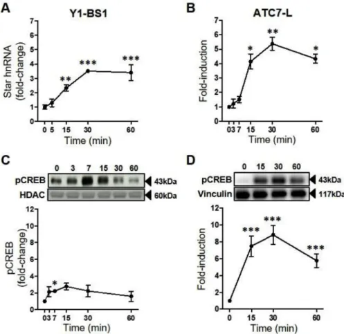
3.1. Time course of the effect of ACTH on Star transcription, CREB phosphorylation and CRTC translocation in Y1-BS1 and ATC7-L cells
The dynamics of Star transcription in response to ACTH were de-termined by measuring the levels of Star primary transcript (Star hnRNA). ACTH induced an overall increase in Star hnRNA levels both in Y1-BS1 and ATC7-L cells (P < 0.001;Fig. 1A and B), with no changes detected in thefirst 5–7 min, whereas a significant increase was ob-served at 15–60 min. Concomitant with the increase in Star hnRNA, ANOVA analysis revealed a significant effect of ACTH on pCREB in both cell lines (Y1-BS1: P = 0.02;Fig. 1C ATC7-L: P = 0.002;Fig. 1D). In Y1-BS1 cells, nuclear pCREB transiently increased 2.2-fold by 7 min, peaking at 15 min. In ATC7-L cells, pCREB increased by 7.5-fold at 15min and remained significantly elevated at 60 min (5.8-fold).
To investigate whether these changes in pCREB and Star transcrip-tion are associated with differential changes in CRTC subtype nuclear translocation, we measured the dynamics of cytosolic and nuclear levels of CRTC1, CRTC2 and CRTC3. ACTH had no significant effect on cy-tosolic CRTC1, CRTC2 or CRTC3 levels in Y1-BS1 (Fig. 2A; data not shown) or ATC7-L cells (Fig. 3A; data not shown). Y1-BS1 cells ex-hibited a significant overall increase in nuclear translocation of CRTC1 (P = 0.04), CRTC2 (P = 0.01) and CRTC3 (P = 0.04;Fig. 2A and B). Nuclear levels of CRTC3 were already significantly increased by 3 min, whilst nuclear levels of CRTC1 and CRTC2 were significantly increase at 7 min. A significant overall effect of ACTH on nuclear CRTC1 and CRTC3 was also detected in ATC7-L cells (CRTC1: P=0.05; CRTC3: P = 0.02;Fig. 3A and B), with nuclear levels of CRTC3 also significantly increased by 3 min. Despite an increase in nuclear levels of CRTC2 in each one of four experiments (range 1.5–5.3-fold at 3 min), the changes were not significant due to high variability in the magnitude of the
effect across experiments (P = 0.22).
3.2. ACTH induces rapid association of pCREB, CRTC2 and CRTC3 at the proximal Star promoter in ATC7-L cells
The increases in nuclear CRTCs, preceding increases in Star hnRNA, suggest an involvement of the coactivators in the initiation of Star transcription. To determine whether CRTC2 and CRTC3 can interact with the Star promoter, we used the adrenocortical cell line, ATC7-L, and ChIP assays to measure recruitment of pCREB and CRTC proteins by the Star promoter. Recruitment of CRTC1 could not be measured because of the lack of a suitable antibody for ChIP. ACTH significantly increased the binding of pCREB (P = 0.04; Fig. 4A) and CRTC2 (P = 0.04;Fig. 4B) at the Star promoter. No increases in binding were detected at 7 min, but by 30 min, there was a significant increase above basal levels for both pCREB (P = 0.02) and CRTC2 (P = 0.02). Im-munoprecipitation of the Star promoter by the CRTC3 antibody also revealed a significant effect of ACTH on CRTC3 binding (P < 0.001;
Fig. 4C). Binding of CRTC3 to the Star promoter was detected by 15 min (P = 0.001), the earlier point measured, and remained at similar levels at 30 min (P < 0.001).
3.3. Knockdown of CRTC2 and CRTC3 attenuates Star transcription
The involvement of the different CRTC subtypes on ACTH-induced Star transcription was investigated further using siRNA oligonucleo-tides to inhibit the expression of CRTC1-3 in the adrenocortical cell line, Y1-BS1. While technical difficulties impaired CRTC1 knockdown, transfection with Crtc2 and Crtc3 siRNA oligonucleotides, alone or in combination effectively reduced the respective CRTC in whole cell protein extracts, for CRTC2 (P = 0.01;Fig. 5B) and CRTC3 (P = 0.002;
Fig. 5C) protein levels. Transfection with Crtc2 siRNA decreased CRTC2
Fig. 1. Time course of the effect of ACTH on Star hnRNA and pCREB in Y1-BS1 and ATC7-L cells. Y1-BS1 (A and C) and ATC7-L (B and D) cells were treated with 10 nM ACTH. Data points represent the mean ± SEM of Star hnRNA (A-B) and pCREB (C-D). Star hnRNA was normalised to Gapdh mRNA and is expressed as fold-changes of time 0 (n = 3–6/ time point). InC, pCREB was measured in the nu-clear extract, was normalised to HDAC, and is ex-pressed as fold-changes of time 0 (n = 4–5/time point). InD, pCREB was measured in the whole cell extract, was normalised to vinculin, and is ex-pressed as fold-change of time 0 (n = 3/time point). Data in A were analysed using one-way ANOVA followed by Fisher LSD post-hoc test; data inB, C andD were analysed using Welch ANOVA followed by Dunnett T3 post-hoc test. *P≤ 0.05, **P ≤ 0.01, ***P≤ 0.001 vs time 0.
protein by 75.3% ± 8.4 (P = 0.02 vs non-coding siRNA sequence, siNC;Fig. 5B), whilst Crtc3 siRNA transfection decreased CRTC3 pro-tein by 49.2% ± 14.3 (P = 0.03 vs siNC;Fig. 5C). Furthermore, there was no significant difference in whole cell CRTC2 and CRTC3 protein levels when cells were transfected with the heterologous siRNA,
confirming specificity of the siRNA used. Knockdown of CRTC2 and CRTC3, alone or combined, did not affect CRTC1 protein levels (P = 0.49;Fig. 5A).
Star hnRNA levels were then measured either following 45 min treatment with 10 nM ACTH or vehicle (Fig. 5D). Two-way ANOVA
Fig. 2. Time course of the effects of ACTH on nuclear levels of CRTC in Y1-BS1 cells. Cells were treated with 10 nM ACTH. (A) Representative Western immunoblot of cyto-plasmic and nuclear CRTC1, CRTC2 and CRTC3 levels. (B) Semi-quantification of Western im-munoblot data expressed as fold-change of time 0. Data are the mean ± SEM of nuclear levels of CRTC1, CRTC2, and CRTC3, normalised to HDAC (n = 4/time point). Data were analysed using one-way ANOVA followed by Fisher LSD post-hoc test. *P≤ 0.05, **P ≤ 0.01, vs time 0.
Fig. 3. Time course of the effects of ACTH on nuclear levels of CRTC in ATC7-L cells. Cells were treated with 10 nM ACTH. (A) Representative Western immunoblot of cytoplasmic and nuclear CRTC1, CRTC2 and CRTC3 levels. (B) Semi-quantification of Western immunoblot data expressed as fold-change of time 0. Data are the mean ± SEM of nuclear levels of CRTC1, CRTC2, and CRTC3, normalised to HDAC (n = 4/time point). Data were analysed using Welch ANOVA followed by Dunnett T3 post-hoc test. *P≤ 0.05 vs time 0.
revealed an overall significant effect for both ACTH and siRNA on Star hnRNA (P < 0.001 and P = 0.03, respectively), but no interaction (P = 0.15). ACTH increased Star hnRNA levels in siNC cells (P < 0.001), an effect which was significantly attenuated in cells transfected with either Crtc2 or Crtc3 siRNA, or their combination. Although ACTH-stimulated values were still significantly higher than
the respective basal (Crtc2 siRNA: P = 0.002; Crtc3 siRNA: P = 0.002; Crtc2+Crtc3 siRNA: P = 0.002), responses to ACTH were significantly lower than in siNC (Crtc2 siRNA: P = 0.05; Crtc3 siRNA: P = 0.004). No additivity was observed between the inhibitory effect of Crtc2 and Crtc3 siRNAs on ACTH-stimulated Star hnRNA, with the combined effect being similar to that of the individual oligonucleotides (P = 0.004,
Fig. 4. ACTH-induced binding of pCREB, CRTC2 and CRTC3 to theStar promoter in ATC7-L cells. Cells were treated with 10 nM ACTH prior to chromatin immunoprecipitation by antibodies for pCREB (A), CRTC2 (B) and CRTC3 (C) proteins and normal rabbit IgG. Binding of each protein target to the Star promoter was calculated as percentage pulldown of the input. Data represent the mean ± SEM of data obtained in 3–4 independent experiments. pCREB, CRTC2 and CRTC3 data were analysed using one-way ANOVA followed by Fisher LSD post-hoc test. IgG data were analysed using Welch ANOVA (A-B) and one-way ANOVA (C). *P≤ 0.05, ***P < 0.001 vs 0 min.
Fig. 5. Effect of CRTC2 and CRTC3 silencing on ACTH-induced increases in Star hnRNA levels in Y1-BS1 cells. Y1-BS1 cells were transfected with non-coding siRNA (siNC) or siRNA oligonucleotides for CRTC2 (siCRTC2), CRTC3 (siCRTC3), or both (siCRTC2+3), 48 h prior to treatment with 10 nM ACTH for 45min. (A, B andC) Efficacy of silencing determined by Western immunoblot and semi-quantification of the levels of whole cell CRTC1, CRTC2 and CRTC3, normalised to actin, expressed as fold-change levels of the siNC transfected control group. No effect on CRTC1 levels was observed following transfection of siCRTC1 oligonucleotides (data not shown). (D) Effect of CRTC siRNAs on Star hnRNA levels, normalised to Gapdh mRNA and expressed as fold-change of siNC transfected controls. Data are the mean ± SEM of data obtained in 3 independent experiments. Data inA, B and C were analysed using one-way ANOVA followed by Fisher LSD post-hoc test; data inD were analysed using two-way ANOVA followed by Fisher LSD post-hoc test. *P≤ 0.05, **P ≤ 0.01, ***P ≤ 0.001 vs untreated control.#P≤ 0.05 vs
compared to siNC cells). No significant differences in basal Star hnRNA levels between siRNA treatment groups were found.
3.4. Stimulation of cAMP, but not of MAPK or PKC pathways, mimics the effect of ACTH on Star transcription
To test whether other signalling pathways, in addition to ACTH/ cAMP, can regulate Star transcription, Y1-BS1 and ATC7-L were treated for 30 min with either ACTH, 8-Br-cAMP, the PKC stimulator PMA, or the MAPK stimulator EGF (Fig. 6A and B). In Y1-BS1 cells (Fig. 6A), both ACTH (P = 0.001) and 8-Br-cAMP (P < 0.001) significantly in-creased the levels of Star hnRNA, whilst treatment with PMA or EGF had no effect on Star hnRNA levels. In ATC7-L cells (Fig. 6B), Star hnRNA levels also increased in cells treated with ACTH (P = 0.007). In each of the three experiments, 8-Br-cAMP increased in Star hnRNA le-vels, but due to variability in the magnitude of the increase (range 5.4–11.0-fold), the effect was not statistically significant (P = 0.23). Consistent with thefindings in Y1-BS1 cells, neither PMA nor EGF had any effect on Star hnRNA levels in ATC7-L cells. These findings were confirmed in dispersed rat adrenal cells, with both ACTH and 8-Br-cAMP increasing Star hnRNA levels, while no effect of PMA treatment was seen (Fig. 6C).
3.5. ACTH-stimulated Star transcription is attenuated by inhibition of PKA and MAPK activity in a cell line specific manner
To further study the signalling pathways mediating Star transcrip-tion, murine adrenocortical cell lines were pre-treated with either ve-hicle, the PKA inhibitor H89, the MEK inhibitor UO126, or with a combination of both inhibitors for 15 min prior to incubation with 10 nM ACTH for 30 min. The inhibitors, alone or in combination, had
no significant effect on basal Star hnRNA levels (Fig. 6D–F). Both
Y1-BS1 (Fig. 6D) and ATC7-L (Fig. 6E) cells showed a significant effect of
ACTH (P < 0.001), inhibitors pre-treatment (Y1-BS1: P < 0.001; ATC7-L: P = 0.03), and a significant interaction (Y1-BS1: P = 0.01; ATC7-L: P = 0.03).
When compared with the respective basal, ACTH treatment in Y1-BS1 cells increased Star hnRNA levels in cells pre-treated with vehicle (P < 0.001), UO126 (P = 0.03) or H89 (P = 0.01), but the effect of ACTH was significantly attenuated in cells pre-treated with H89 (P = 0.02) and UO126 (P = 0.01) compared vehicle pre-treatment. Furthermore, pre-treatment with the combination of H89 and UO126 had a significant additive inhibitory effect (P < 0.001), preventing a significant stimulatory effect of ACTH (P = 0.26 vs basal). Similarly, ACTH increased Star hnRNA levels in ATC7-L cells pre-treated with vehicle (P = 0.004), UO126 (P < 0.001) and H89 + UO126 (P = 0.01) compared with respective basal values, but pre-treatment with H89 alone only tended to reduce the stimulatory effect of ACTH (P=0.15). Compared with ACTH treatment in vehicle pre-treated controls, ACTH-stimulated Star hnRNA levels also tended to be lower in H89 pre-treated cells (P = 0.09). The lack of a significant effect of H89 in the overall analysis was likely due to the wide range in the magni-tude of the ACTH responses in the UO126 group, since there was a significant reduction when comparing ACTH-stimulated values in H89 and vehicle pre-treated groups by t-test analysis (P = 0.04). In contrast to Y1-BS1 cells, preincubation of ATC7-L cells with the MAPK inhibitor, UO126, significantly augmented the stimulatory effect of ACTH on Star hnRNA levels compared with ACTH in vehicle pre-treated cells (P = 0.02).
Similar to the effects in the cell lines, in vehicle pre-treated col-lagenase-dispersed rat adrenal cells (Fig. 6F) there was a significant
effect of ACTH (P = 0.002), while the overall effect of the inhibitors or
Fig. 6. Effect of mimicry and inhibi-tion of ACTH signalling on Star hnRNA levels in Y1-BS1, ATC7-L and dispersed rat adrenals cells. Star hnRNA levels were measured either prior to or 30 min after treatment with 10 nM ACTH, 1 mM 8-Br-cAMP, 100 nM PMA or 3 nM EGF in Y1-BS1 (A) and ATC7-L (B), or 60 min after treatment in dispersed rat adrenal cells (C). For inhibition studies, Y1-BS1 cells (D), ATC7-L (E) and dispersed rat adrenal cells (F) were pre-incubated for 15 min with either vehicle (0.5% DMSO), 10μM H89, 1 μM UO126, or 10μM H89+1 μM UO126 before ad-dition of ACTH at afinal concentration of 10 nM. Bars represent the mean ± SEM of 3–6 experiments, normalised to Gapdh mRNA and ex-pressed as fold-change from basal ve-hicle values. PMA treated cells in C represent the mean of data from 2 in-dependent experiments. Data inA were analysed using one-way ANOVA fol-lowed by Fisher LSD post-hoc test; data in B were analysed using Welch ANOVA followed by Dunnett T3 post-hoc test. Data inD, E and F were ana-lysed using Two-way ANOVA followed by Fisher LSD post-hoc test. *P≤ 0.05, **P≤ 0.01, ***P ≤ 0.001 vs untreated vehicle control. #P ≤ 0.05, ###P ≤ 0.001 vs ACTH-treated ve-hicle control.
their interaction was not significant (P = 0.213 and P = 0.412, for in-hibitors and interaction, respectively). As in ATC7-L cells, Star hnRNA increases in the ACTH-treated vehicle (P = 0.035) and UO126 (P = 0.008) groups were significant compared to basal, whilst pre-in-cubation with H89, but not with UO126, blunted Star hnRNA response to ACTH (P = 0.242). Also similar to ATC7-L cells, pre-incubation with UO126 did not inhibit but tended to increase the effect of ACTH com-pared with vehicle pre-treated cells.
3.6. Inhibition of PKA and calcineurin decreases ACTH-mediated nuclear translocation of CRTC1, CRTC2 and CRTC3 in ATC7-L cells but not ACTH-mediated Star transcription
To investigate the role of PKA, MEK and calcineurin on CRTC-mediated regulation of ACTH-induced Star transcription, the effect of 10 min treatment with ACTH on Star hnRNA levels and nuclear levels of CRTC1, CRTC2 and CRTC3 were measured in ATC7-L cells pre-treated with UO126, H89 or the calcineurin inhibitor CsA. There was a sig-nificant effect of ACTH (P < 0.001) and inhibitors treatment (P < 0.001), as well as interaction (P = 0.006), on Star hnRNA levels (Fig. 7A). ACTH significantly increased Star hnRNA in cells
pre-in-cubated with vehicle (P < 0.001), UO126 (P = 0.003) and CsA (P < 0.001) but not in cells pre-incubated with H89 alone (P = 0.17) or in combination with the other antagonists (H89 + UO126 + CsA: P = 0.37). Interestingly, the effect of ACTH on Star hnRNA was po-tentiated by CsA (P = 0.01 vs ACTH alone), whereas the potentiation by UO126 observed after 30 min ACTH treatment (Fig. 6D) was not present after 10 min treatment.
Treatment with ACTH for 10 min also exerted a significant overall effect on nuclear levels of CRTC1 (P = 0.001;Fig. 7B and C), CRTC2 (P < 0.001; Fig. 7B and D) and CRTC3 (P < 0.001;Fig. 7B and E). Furthermore, the effect of inhibitors pre-treatment was significant for nuclear CRTC1 (P = 0.04; interaction P = 0.09) and CRTC2
(P = 0.006; interaction P = 0.011), but not for nuclear CRTC3 (P = 0.76; interaction P = 0.74). ACTH significantly increased nuclear levels of CRTC1, CRTC2 and CRTC3 in cells pre-treated with vehicle (CRTC1: P = 0.001; CRTC2: P < 0.001; CRTC3: P = 0.002) and UO126 (CRTC1: P = 0.002; CRTC2: P < 0.001; CRTC3: P = 0.03), whilst nCRTC2 levels alone were also significantly increased in CsA pre-treated cells (P = 0.016). Furthermore, ACTH-induced levels of nuclear CRTC1 and CRTC2 were significantly reduced in cells treated with the inhibitors H89 (CRTC1: P = 0.02; CRTC2: P < 0.001), CsA (CRTC1: P = 0.05; CRTC2: P = 0.01) and the combined H89 + UO126 + CsA (CRTC1: P = 0.003; CRTC2: P < 0.001), when compared to cells pre-treated with vehicle. However, H89 and CsA only tended to attenuate the effect of ACTH on nuclear levels of CRTC3 (H89: P = 0.09; CsA: P = 0.13, compared with ACTH stimulation in vehicle treated cells). Furthermore, the effect of pre-treatment all 3 combined inhibitors on ACTH-stimulated nuclear accumulation of CRTC3 was no different from the effects of each single inhibitor (P = 0.31 vs basal).
4. Discussion
This in vitro study shows that nuclear translocation of the 3 isoforms of endogenous CRTC (CRTC1, CRTC2 and CRTC3) parallels or precedes the increases in Star hnRNA induced by ACTH, in agreement with previous reports of ex vivo work in rats. Using chromatin im-munoprecipitation and siRNA knockout we now demonstrate that CRTC2 and CRTC3 are involved in the regulation of Star transcription by ACTH in a PKA signalling dependent manner. The use of ACTH-responsive adrenocortical Y1-BS1 and ATC7-L cell lines allowed us to study the effect of physiological stimulator, ACTH, on endogenous proteins, rather than cAMP analogues, as with most previous studies (Takemori et al., 2007; Whitehouse et al., 2002; Zaidi et al., 2014;
Jefcoate et al., 2011;Manna et al., 2002). The effects of ACTH on Star transcription and on nuclear pCREB and CRTC were similar in both cell
Fig. 7. Effect of inhibition of ACTH signalling onStar hnRNA levels and CRTC activity in ATC7-L cells. Cells were pre-treated for 15 min with either vehicle (0.5%DMSO), protein kinase A inhibitor H89 (H, 10μM), the MAP ki-nase inhibitor UO126 (U, 1μM) or the calcineurin inhibitor CsA (5μM), either alone or in combination (H89 + UO126 + CsA), prior to treatment with 10 nM ACTH for 10 min. (A) RT-qPCR quantification of Star hnRNA, normalised to Gapdh mRNA and expressed as fold-change levels of the basal vehicle control. Bars represent the mean ± SEM of 3–5 in-dependent experiments. (B) Representative Western immunoblot of nuclear CRTC1, CRTC2 and CRTC3 le-vels. Quantification of Western im-munoblot data of CRTC1 (C), CRTC2 (D), and CRTC3 (E); data are normal-ised to HDAC and expressed as fold-change of basal. Bars represent the mean ± SEM of 3–5 independent ex-periments. Data were analysed using two-way ANOVA followed by Fisher LSD post-hoc test. *P ≤ 0.05, **P≤ 0.01, ***P ≤ 0.001 vs untreated vehicle control.#P≤ 0.05,##P≤ 0.01, ### P≤ 0.001 vs ACTH-treated vehicle control.
lines, however, the actions of signalling inhibitors differed, with the MEK inhibitor, UO126, having an inhibitory effect in Y1-BS1 cells and a potentiating effect in ATC7-L cells. Interestingly, the effect of signalling inhibitors in ATC-7 cells resembles that observed in collagenase dis-persed rat adrenal cells, suggesting that, at least in rodents, they are more representative of normal adrenal fasciculata cells. However, re-sistance to transfection made ATC7-L cells unsuitable for use in CRTC knock down experiments.
Whilst in earlier studies ACTH increases Star mRNA levels within 2 h in Y1-BS1 cells (Lin et al., 2001) and by 30 min in ATC7-L cells (Ragazzon et al., 2006), measuring Star hnRNA made it possible to show significant transcriptional activation in response to ACTH by 15 min in both cell lines. These rapid increases are consistent with findings in rat adrenal tissue 15 min after ACTH injection (Spiga et al., 2011a,b, 2017), or cAMP incubation in Y1 cells (Lee et al., 2015;
Jefcoate et al., 2011). Consistent with the view that CREB phosphor-ylation is critical for Star transcription (Spiga et al., 2011a,b, 2017;
Manna et al., 2003;Lefrancois-Martinez et al., 2011), ACTH treatment induced rapid and transient increases in nuclear pCREB in both cell lines.
Rapid ACTH-induced nuclear translocation of all 3 CRTC isoforms (CRTC1, 2 and 3) by 10 min in both Y1-BS1 cells and ATC7-L cells, coinciding with the earliest detected increases in Star hnRNA following ACTH exposure, supports a role for CRTC in the initiation of Star transcription. These kinetics differ from previous findings in transfected Y1 cells, showing nuclear translocation of CRTC2, but not CRTC1 or CRTC3, in response to cAMP stimulation (Lee et al., 2015). In keeping with the rapid nuclear translocation of the three CRTC isoforms shown here, using ATC7-L cells we demonstrate, for thefirst time, an early interaction of CRTC2 (by 30 min) and CRTC3 (by 15 min) with DNA fragments including the −104 bp to −5 bp Star promoter region containing three cAMP-response element (CRE) half-sites (Manna et al., 2002). The presentfinding of pCREB binding to the Star promoter at 30 min ACTH incubation is consistent with previous observations in Y1 cells stimulated with cAMP analogues (Jefcoate et al., 2011). The latter study also showed delayed (60 min) CRTC2 recruitment, from which authors concluded that CRTC2 is involved in maintaining, rather than initiating, high rates of Star transcription. The difference between the former (Lee et al., 2015;Jefcoate et al., 2011) and presentfindings could reflect the use of different cell lines, or studying the effect of ACTH on endogenous CRTC2 as opposed to the effect of cAMP on transfected protein (which may exhibit altered intracellular localisation and bioactivity). Interestingly, in contrast to the parallel nuclear translocation patterns of the three CRTC isoforms, extrapolation of the ChIP time courses suggests that ACTH-induced recruitment of CRTC3 by the Star promoter, which is already maximal at 15 min, could po-tentially precede that of CRTC2. Although CRTC2 recruitment was not measured at 15 min, the lack of any promoter association at 7 min renders it unlikely that binding is maximal at 15 min. Since CRTC lacks DNA-binding activity, its recruitment by the Star promoter CRE requires association with pCREB through its CREB binding domain (CBD) (Luo et al., 2012). Amino acid substitution studies have shown that small changes in key motifs of the CBD region of CRTC2 can impact its binding affinity with pCREB (Conkright et al., 2003;Luo et al., 2012). Thus, while the similar nuclear translocation time courses of CRTC1-3 could predict parallel dynamics of recruitment to the Star promoter, differences in pCREB affinity between the three CRTC isoforms could affect the recruitment time by the Star promoter. Testing this possibility will require further detailed analyses of the early kinetics of the three CRTC isoform pCREB association and recruitment by the Star promoter. Previous reports demonstrate that CRTC2 overexpression increases Star transcription (Takemori et al., 2007), while activation of salt in-ducible kinase (SIK) 1, which prevents activation and nuclear locali-sation of CRTC2, inhibits Star transcription (Lin et al., 2001; Katoh et al., 2004). We demonstrate that siRNA knockdown of CRTC2 and CRTC3 in Y1-BS1 cells only partially inhibits ACTH stimulation of Star
hnRNA levels, directly establishing a role for CRTC mediating ACTH-induced Star transcription. However, in contrast to the full inhibition of cAMP-dependent Crh transcription by simultaneous knockdown of CRTC2 and CRTC3 seen in rat hypothalamic 4B cells (Liu et al., 2010), knocking down both CRTC isoforms in Y1-BS1 cells had no additive effect on Star transcription after 45 min ACTH exposure. This suggests CRTC2 and CRTC3 both act through a similar mechanism of action, and that at the time of maximal Star transcription either CRTC2 of CRTC3 are capable of inducing full activation. However, it is possible that differential recruitment of CRTC isoforms by the atypical Star half-CREs (Luo et al., 2012;Song et al., 2018), due to differential affinity with
pCREB or other mechanism (Conkright et al., 2003;Luo et al., 2012), could have different functional implications at earlier time points of ACTH stimulation.
It is also evident from these experiments that other factors are capable of mediating considerable ACTH-stimulation of Star transcrip-tion in the absence of both CRTC2 and CRTC3. Although CRTC1 ex-pression is far lower than that of CRTC2 and CRTC3 in rat (Fig. S1) and human adrenals (Conkright et al., 2003), we show clear nuclear translocation of CRTC1 following ACTH treatment, suggesting a pos-sible role for CRTC1 in Star transcription. This could not be examined in the present study because of the inability to knockdown CRTC1 protein in Y1-BS1 cells, and future studies will be needed for elucidating the potential role of CRTC1 on Star transcription. In addition, SF-1, which is upregulated by ACTH (Ragazzon et al., 2006;Hazell et al., 2019), binds the Star promoter and is necessary for cAMP-induced Star transcription (Sandhoff et al., 1998;Sugawara et al., 1997). Furthermore, subsets of CREB-inducible genes can be alternatively regulated by CREB coacti-vator CREB binding protein (CBP) (Kasper et al., 2010). The role of these factors and their interaction with the transcriptional complex during Star transcriptional initiation will require further investigation. Investigation of signalling pathways showed that whilst MAPK ac-tivation by EGF had no effect on Star transcription in either Y1-BS1 or ATC7-L cells, MEK inhibition by UO126 attenuated ACTH-induced Star hnRNA in Y1-BS1 cells, but not ATC7-L cells. Furthermore, combination of H89 and UO126 abolished Star transcriptional responses to ACTH in Y1-BS1 cells, suggesting both pathways are required for full cAMP-de-pendent stimulation of Star transcription in this cell line. Similar results were found when using the PKA inhibitor PKI or MAPK inhibitor SL327 (Smith, Huang and Aguilera, unpublished observations). Previous stu-dies also suggest cell-specific differences in the MAPK involvement in Star transcriptional regulation;Lefrancois-Martinez et al. (2011)found no effect of MAPK inhibition on ACTH-stimulated StAR protein levels in ATC1 cells, whilst,Gyles et al. (2001)showed inhibition of forskolin-stimulated Star mRNA and protein by MEK inhibitors in Y1 cells. Transactivation of the MAPK pathway by ACTH and cAMP has been implicated in steroidogenesis (Gyles et al., 2001;Winnay and Hammer, 2006). Furthermore, consistent with previous reports (Le and Schimmer, 2001), ACTH increased pERK levels in Y1-BS1 cells and, to a lesser extent, ATC7-L cells (Smith, Olah and Aguilera, unpublished observations).
H89 blunted ACTH-induced increases in nuclear CRTC, consistent with the established cAMP dependence of CRTC activation and trans-location to the nucleus (Bittinger et al., 2004;Takemori et al., 2007;Liu et al., 2010). This was associated with complete inhibition of ACTH-induced Star hnRNA at 10 min, while there was a partial recovery by 30 min. This is consistent with the effects of siRNA CRTC knockdown, in which there was only a partial reduction of Star hnRNA by 45 min ACTH stimulation, despite reduced CRTC2 and CRTC3 levels. Although earlier Star hnRNA responses to ACTH were not measured in the siRNA experiments, the overallfindings strongly suggest that PKA/CRTC me-chanisms are essential for early transcriptional activation of Star, and that additional signalling mechanisms are important to sustain the ac-tivation. The fact that MAPK appears to play a role in CRTC regulation only in Y1-BS1 cells, raises a note a caution when extrapolating data obtained from cell lines to in vivo regulation in various species.
No effect of PMA indicates PKC activation alone is insufficient for initiating Star transcription. PKC has previously been implicated in the regulation of StAR protein levels in bovine adrenal cells (Nishikawa et al., 1996), whilst low levels of steroidogenesis were previously in-duced by 2 h incubation with PMA in Y1 cells (Frigeri and Armelin, 1996). Furthermore, Leydig M-10 cell studies have shown phorbol es-ters increase Star mRNA levels (Manna and Stocco, 2005;Manna et al., 2011). Ca2+-sensitive calcineurin can be activated by cAMP (Antoni, 1996), and it is known to activate CRTC (Bittinger et al., 2004; Lee et al., 2015;Rahnert et al., 2016;Screaton et al., 2004). Thus, ACTH signalling could lead to calcineurin activation and play a role in CRTC activation and nuclear translocation in the adrenal fasciculata. How-ever, CsA potentiated the effect of ACTH on Star transcription, despite attenuating ACTH-induced nuclear translocation of CRTC2. It is note-worthy that Y1-BS1 cells lack part of the steroidogenic enzyme ex-pression profile, which characterises zona fasciculata cells (Bloch and Cohen, 1960;Parker et al., 1985). The lost differentiation of these cells
may be responsible for the apparent dependence of Star transcription on calcium (Lee et al., 2015) and MAPK signalling (Gyles et al., 2001), that was not observed in ATC7-L cells in this study or elsewhere ( Lefrancois-Martinez et al., 2011;Schiebinger et al., 1985). Furthermore, CsA only partially inhibits CRTC1 and CRTC2 translocation, suggesting PKA may be the main mechanism regulating CRTC1 and CRTC2 activity. Al-though less sensitive to calcium signalling than zona glomerulosa cells (Braley et al., 1986;Omura et al., 2007;Spat et al., 2016), intracellular calcium depletion in rat zona fasciculata cells blunts cAMP-induced corticosterone release, suggesting that cAMP activation of adrenal steroidogenesis in the adrenal fasciculata requires calcium release from intracellular stores (Schiebinger et al., 1985).
This study provides evidence that of the 3 isoforms of CRTC present in the adrenal fasciculata, at least CRTC2 and CRTC3 are involved in the initiation of Star transcription. The rapid nuclear translocation and recruitment by the Star promoter of CRTC2 and CRTC3 in response to ACTH, preceding the maximal increases in Star primary transcript, and the ability of siRNA knockdown of these CRTC isoforms to attenuate ACTH-induced Star transcription, strongly suggest that PKA-mediated activation of CRTC2 and CRTC3 plays a role in the initiation of Star transcription. However, the fact that knockdown of both subtypes (or preventing their activation by PKA inhibitors) reduces but does not prevent ACTH-induced Star hnRNA increases, indicates the participa-tion of addiparticipa-tional factors and emphasizes the complexity of the me-chanisms regulating StAR expression.
Declaration of competing interest
Lorna Smith, Victoria Huang, Mark Olah, Loc Trinh, Ying Liu, Georgina Hazell, Becky Conway-Campbell, Zidong Zhao, Antoine Martinez, Anne-Marie Lefrançois-Martinez, Stafford Lightman, Francesca Spiga and Greti Aguilera declare no conflict of interest. Acknowledgements
This work was supported by the Intramural Research Program, National Institute of Child Health and Human Development, National Institutes of Health, and Medical Research Council programme grant [MR/J008893/1]. The authors are grateful to Dr Bernard Schimmer, University of Toronto, Canada, for providing the Y1-BS1 cells and Dr Pierre Val, University of Clermont, France for his guidance with ATC-7L cells.
Appendix A. Supplementary data
Supplementary data to this article can be found online athttps:// doi.org/10.1016/j.mce.2019.110612.
References
Aesoy, R., Mellgren, G., Morohashi, K., Lund, J., 2002. Activation of cAMP-dependent protein kinase increases the protein level of steroidogenic factor-1. Endocrinology 143, 295–303.
Antoni, F.A., 1996. Mortyn Jones Memorial Lecture–1995. Calcium checks cyclic AMP– corticosteroid feedback in adenohypophysial corticotrophs. J. Neuroendocrinol. 8, 659–672.
Arakane, F., King, S.R., Du, Y., Kallen, C.B., Walsh, L.P., Watari, H., Stocco, D.M., Strauss 3rd, J.F., 1997. Phosphorylation of steroidogenic acute regulatory protein (StAR) modulates its steroidogenic activity. J. Biol. Chem. 272, 32656–32662.
Artemenko, I.P., Zhao, D., Hales, D.B., Hales, K.H., Jefcoate, C.R., 2001. Mitochondrial processing of newly synthesized steroidogenic acute regulatory protein (StAR), but not total StAR, mediates cholesterol transfer to cytochrome P450 side chain cleavage enzyme in adrenal cells. J. Biol. Chem. 276, 46583–46596.
Bittinger, M.A., McWhinnie, E., Meltzer, J., Iourgenko, V., Latario, B., Liu, X., Chen, C.H., Song, C., Garza, D., Labow, M., 2004. Activation of cAMP response element-mediated gene expression by regulated nuclear transport of TORC proteins. Curr. Biol. 14, 2156–2161.
Bloch, E., Cohen, A.I., 1960. Steroid production in vitro by normal and adrenal tumor-bearing male mice. J. Natl. Cancer Inst. 24, 97–107.
Braley, L.M., Menachery, A.I., Brown, E.M., Williams, G.H., 1986. Comparative effect of angiotensin II, potassium, adrenocorticotropin, and cyclic adenosine 3',5'-monopho-sphate on cytosolic calcium in rat adrenal cells. Endocrinology 119, 1010–1019.
Cammas, F.M., Kapas, S., Barker, S., Clark, A.J., 1995. Cloning, characterization and expression of a functional mouse ACTH receptor. Biochem. Biophys. Res. Commun. 212, 912–918.
Clark, B.J., Combs, R., Hales, K.H., Hales, D.B., Stocco, D.M., 1997. Inhibition of tran-scription affects synthesis of steroidogenic acute regulatory protein and ster-oidogenesis in MA-10 mouse Leydig tumor cells. Endocrinology 138, 4893–4901.
Clark, B.J., Ranganathan, V., Combs, R., 2001. Steroidogenic acute regulatory protein expression is dependent upon post-translational effects of cAMP-dependent protein kinase A. Mol. Cell. Endocrinol. 173, 183–192.
Cole, T.J., Blendy, J.A., Monaghan, A.P., Krieglstein, K., Schmid, W., Aguzzi, A., Fantuzzi, G., Hummler, E., Unsicker, K., Schutz, G., 1995. Targeted disruption of the gluco-corticoid receptor gene blocks adrenergic chromaffin cell development and severely retards lung maturation. Genes Dev. 9, 1608–1621.
Conkright, M.D., Canettieri, G., Screaton, R., Guzman, E., Miraglia, L., Hogenesch, J.B., Montminy, M., 2003. TORCs: transducers of regulated CREB activity. Mol. Cell 12, 413–423.
Deuschle, M., Schweiger, U., Weber, B., Gotthardt, U., Korner, A., Schmider, J., Standhardt, H., Lammers, C.H., Heuser, I., 1997. Diurnal activity and pulsatility of the hypothalamus-pituitary-adrenal system in male depressed patients and healthy controls. J. Clin. Endocrinol. Metab. 82, 234–238.
Evans, A.N., Liu, Y., Macgregor, R., Huang, V., Aguilera, G., 2013. Regulation of hy-pothalamic corticotropin-releasing hormone transcription by elevated glucocorti-coids. Mol. Endocrinol. 27, 1796–1807.
Ferguson Jr., J.J., 1963. Protein synthesis and adrenocorticotropin responsiveness. J. Biol. Chem. 238, 2754–2759.
Frigeri, C.K., Armelin, H.A., 1996. Patterns of long-term steroidogenesis stimulation by ACTH and phorbol ester. Braz. J. Med. Biol. Res. 29, 343–345.
Garren, L.D., Ney, R.L., Davis, W.W., 1965. Studies on the role of protein synthesis in the regulation of corticosterone production by adrenocorticotropic hormone in vivo. Proc. Natl. Acad. Sci. U. S. A. 53, 1443–1450.
Grontved, L., Madsen, M.S., Boergesen, M., Roeder, R.G., Mandrup, S., 2010. MED14 tethers mediator to the N-terminal domain of peroxisome proliferator-activated re-ceptor gamma and is required for full transcriptional activity and adipogenesis. Mol. Cell. Biol. 30, 2155–2169.
Grontved, L., John, S., Baek, S., Liu, Y., Buckley, J.R., Vinson, C., Aguilera, G., Hager, G.L., 2013. C/EBP maintains chromatin accessibility in liver and facilitates gluco-corticoid receptor recruitment to steroid response elements. EMBO J. 32, 1568–1583.
Gyles, S.L., Burns, C.J., Whitehouse, B.J., Sugden, D., Marsh, P.J., Persaud, S.J., Jones, P.M., 2001. ERKs regulate cyclic AMP-induced steroid synthesis through transcrip-tion of the steroidogenic acute regulatory (StAR) gene. J. Biol. Chem. 276, 34888–34895.
Hazell, G., Horn, G., Lefrançois-Martinez, A.M., Martinez, A., Lightman, S.L., Spiga, F., 2019. Dynamics of ACTH-mediated regulation of gene transcription in ATC1 and ATC7 adrenal zona fasciculata cell lines. Endocrinology 160, 587–604.
Iourgenko, V., Zhang, W., Mickanin, C., Daly, I., Jiang, C., Hexham, J.M., Orth, A.P., Miraglia, L., Meltzer, J., Garza, D., Chirn, G.W., McWhinnie, E., Cohen, D., Skelton, J., Terry, R., Yu, Y., Bodian, D., Buxton, F.P., Zhu, J., Song, C., Labow, M.A., 2003. Identification of a family of cAMP response element-binding protein coactivators by genome-scale functional analysis in mammalian cells. Proc. Natl. Acad. Sci. U. S. A. 100, 12147–12152.
Jefcoate, C.R., Lee, J., Cherradi, N., Takemori, H., Duan, H., 2011. cAMP stimulation of StAR expression and cholesterol metabolism is modulated by co-expression of labile suppressors of transcription and mRNA turnover. Mol. Cell. Endocrinol. 336, 53–62.
Jurek, B., Slattery, D.A., Hiraoka, Y., Liu, Y., Nishimori, K., Aguilera, G., Neumann, I.D., van den Burg, E.H., 2015. Oxytocin regulates stress-induced crf gene transcription through CREB-regulated transcription coactivator 3. J. Neurosci. 35, 12248–12260.
Kasper, L.H., Lerach, S., Wang, J., Wu, S., Jeevan, T., Brindle, P.K., 2010. CBP/p300 double null cells reveal effect of coactivator level and diversity on CREB transacti-vation. EMBO J. 29, 3660–3672.
Katoh, Y., Takemori, H., Min, L., Muraoka, M., Doi, J., Horike, N., Okamoto, M., 2004. Salt-inducible kinase-1 represses cAMP response element-binding protein activity
both in the nucleus and in the cytoplasm. Eur. J. Biochem. 271, 4307–4319.
Le, T., Schimmer, B.P., 2001. The regulation of MAPKs in Y1 mouse adrenocortical tumor cells. Endocrinology 142, 4282–4287.
Lee, J., Tong, T., Takemori, H., Jefcoate, C., 2015. Stimulation of StAR expression by cAMP is controlled by inhibition of highly inducible SIK1 via CRTC2, a co-activator of CREB. Mol. Cell. Endocrinol. 408, 80–89.
Lefrancois-Martinez, A.M., Blondet-Trichard, A., Binart, N., Val, P., Chambon, C., Sahut-Barnola, I., Pointud, J.C., Martinez, A., 2011. Transcriptional control of adrenal steroidogenesis: novel connection between Janus kinase (JAK) 2 protein and protein kinase A (PKA) through stabilization of cAMP response element-binding protein (CREB) transcription factor. J. Biol. Chem. 286, 32976–32985.
Lin, X., Takemori, H., Katoh, Y., Doi, J., Horike, N., Makino, A., Nonaka, Y., Okamoto, M., 2001. Salt-inducible kinase is involved in the ACTH/cAMP-dependent protein kinase signaling in Y1 mouse adrenocortical tumor cells. Mol. Endocrinol. 15, 1264–1276.
Liu, Y., Kamitakahara, A., Kim, A.J., Aguilera, G., 2008. Cyclic adenosine 3',5'-mono-phosphate responsive element binding protein phosphorylation is required but not sufficient for activation of corticotropin-releasing hormone transcription. Endocrinology 149, 3512–3520.
Liu, Y., Coello, A.G., Grinevich, V., Aguilera, G., 2010. Involvement of transducer of regulated cAMP response element-binding protein activity on corticotropin releasing hormone transcription. Endocrinology 151, 1109–1118.
Liu, Y., Knobloch, H.S., Grinevich, V., Aguilera, G., 2011. Stress induces parallel changes in corticotrophin-releasing hormone (CRH) Transcription and nuclear translocation of transducer of regulated cAMP response element-binding activity 2 in hypothalamic CRH neurones. J. Neuroendocrinol. 23, 216–223.
Liu, Y., Smith, L.I., Huang, V., Poon, V., Coello, A., Olah, M., Spiga, F., Lightman, S.L., Aguilera, G., 2013. Transcriptional regulation of episodic glucocorticoid secretion. Mol. Cell. Endocrinol. 371, 62–70.
Lotfi, C.F., Costa, E.T., Schwindt, T.T., Armelin, H.A., 2000. Role of ERK/MAP kinase in mitogenic interaction between ACTH and FGF2 in mouse Y1 adrenocortical tumor cells. Endocr. Res. 26, 873–877.
Luo, Q., Viste, K., Urday-Zaa, J.C., Senthil Kumar, G., Tsai, W.W., Talai, A., Mayo, K.E., Montminy, M., Radhakrishnan, I., 2012. Mechanism of CREB recognition and coac-tivation by the CREB-regulated transcriptional coactivator CRTC2. Proc. Natl. Acad. Sci. U. S. A. 109, 20865–20870.
Manna, P.R., Stocco, D.M., 2005. Regulation of the steroidogenic acute regulatory protein expression: functional and physiological consequences. Curr. Drug Targets - Immune, Endocr. Metab. Disord. 5, 93–108.
Manna, P.R., Dyson, M.T., Eubank, D.W., Clark, B.J., Lalli, E., Sassone-Corsi, P., Zeleznik, A.J., Stocco, D.M., 2002. Regulation of steroidogenesis and the steroidogenic acute regulatory protein by a member of the cAMP response-element binding protein fa-mily. Mol. Endocrinol. 16, 184–199.
Manna, P.R., Eubank, D.W., Lalli, E., Sassone-Corsi, P., Stocco, D.M., 2003.
Transcriptional regulation of the mouse steroidogenic acute regulatory protein gene by the cAMP response-element binding protein and steroidogenic factor 1. J. Mol. Endocrinol. 30, 381–397.
Manna, P.R., Soh, J.W., Stocco, D.M., 2011. The involvement of specific PKC isoenzymes in phorbol ester-mediated regulation of steroidogenic acute regulatory protein ex-pression and steroid synthesis in mouse Leydig cells. Endocrinology 152, 313–325.
Nishikawa, T., Sasano, H., Omura, M., Suematsu, S., 1996. Regulation of expression of the steroidogenic acute regulatory (StAR) protein by ACTH in bovine adrenal fasciculata cells. Biochem. Biophys. Res. Commun. 223, 12–18.
Omura, M., Suematsu, S., Nishikawa, T., 2007. Role of calcium messenger systems in ACTH-induced cortisol production in bovine adrenal fasciculo-reticularis cells. Endocr. J. 54, 585–592.
Orphanides, G., Lagrange, T., Reinberg, D., 1996. The general transcription factors of RNA polymerase II. Genes Dev. 10, 2657–2683.
Park, S.Y., Walker, J.J., Johnson, N.W., Zhao, Z., Lightman, S.L., Spiga, F., 2013. Constant light disrupts the circadian rhythm of steroidogenic proteins in the rat adrenal gland. Mol. Cell. Endocrinol. 371, 114–123.
Parker, K.L., Chaplin, D.D., Wong, M., Seidman, J.G., Smith, J.A., Schimmer, B.P., 1985. Expression of murine 21-hydroxylase in mouse adrenal glands and in transfected Y1 adrenocortical tumor cells. Proc. Natl. Acad. Sci. U. S. A. 82, 7860–7864.
Qian, X., Droste, S.K., Lightman, S.L., Reul, J.M., Linthorst, A.C., 2012. Circadian and ultradian rhythms of free glucocorticoid hormone are highly synchronized between the blood, the subcutaneous tissue, and the brain. Endocrinology 153, 4346–4353.
Ragazzon, B., Lefrancois-Martinez, A.M., Val, P., Sahut-Barnola, I., Tournaire, C., Chambon, C., Gachancard-Bouya, J.L., Begue, R.J., Veyssiere, G., Martinez, A., 2006. Adrenocorticotropin-dependent changes in SF-1/DAX-1 ratio influence steroidogenic genes expression in a novel model of glucocorticoid-producing adrenocortical cell lines derived from targeted tumorigenesis. Endocrinology 147, 1805–1818.
Rahnert, J.A., Zheng, B., Hudson, M.B., Woodworth-Hobbs, M.E., Price, S.R., 2016. Glucocorticoids alter CRTC-CREB signaling in muscle cells: impact on PGC-1alpha expression and atrophy markers. PLoS One 11, e0159181.
Rainey, W.E., Saner, K., Schimmer, B.P., 2004. Adrenocortical cell lines. Mol. Cell. Endocrinol. 228, 23–38.
Rocha, K.M., Forti, F.L., Lepique, A.P., Armelin, H.A., 2003. Deconstructing the molecular mechanisms of cell cycle control in a mouse adrenocortical cell line: roles of ACTH. Microsc. Res. Tech. 61, 268–274.
Sahut-Barnola, I., Lefrancois-Martinez, A.M., Jean, C., Veyssiere, G., Martinez, A., 2000. Adrenal tumorigenesis targeted by the corticotropin-regulated promoter of the aldo-keto reductase AKR1B7 gene in transgenic mice. Endocr. Res. 26, 885–898.
Sandhoff, T.W., Hales, D.B., Hales, K.H., McLean, M.P., 1998. Transcriptional regulation of the rat steroidogenic acute regulatory protein gene by steroidogenic factor 1. Endocrinology 139, 4820–4831.
Schiebinger, R.J., Braley, L.M., Menachery, A., Williams, G.H., 1985. Calcium, a“third messenger” of cAMP-stimulated adrenal steroid secretion. Am. J. Physiol. 248, E89–94.
Schimmer, B.P., 1972. Adenylate cyclase activity in adrenocorticotropic hormone-sensi-tive and mutant adrenocortical tumor cell lines. J. Biol. Chem. 247, 3134–3138.
Screaton, R.A., Conkright, M.D., Katoh, Y., Best, J.L., Canettieri, G., Jeffries, S., Guzman, E., Niessen, S., Yates 3rd, J.R., Takemori, H., Okamoto, M., Montminy, M., 2004. The CREB coactivator TORC2 functions as a calcium- and cAMP-sensitive coincidence detector. Cell 119, 61–74.
Sewer, M.B., Waterman, M.R., 2002. cAMP-dependent transcription of steroidogenic genes in the human adrenal cortex requires a dual-specificity phosphatase in addition to protein kinase A. J. Mol. Endocrinol. 29, 163–174.
Song, Y., Zhai, L., Valencia Swain, J., Chen, Y., Wang, P., Chen, L., Liu, Y., Xiang, S., 2018. Structural insights into the CRTC2-CREB complex assembly on CRE. J. Mol. Biol. 430, 1926–1939.
Spat, A., Hunyady, L., Szanda, G., 2016. Signaling interactions in the adrenal cortex. Front. Endocrinol. 7, 17.
Spiga, F., Waite, E.J., Liu, Y., Kershaw, Y.M., Aguilera, G., Lightman, S.L., 2011a. ACTH-dependent ultradian rhythm of corticosterone secretion. Endocrinology 152, 1448–1457.
Spiga, F., Liu, Y., Aguilera, G., Lightman, S.L., 2011b. Temporal effect of adrenocorti-cotrophic hormone on adrenal glucocorticoid steroidogenesis: involvement of the transducer of regulated cyclic AMP-response element-binding protein activity. J. Neuroendocrinol. 23, 136–142.
Spiga, F., Zavala, E., Walker, J.J., Zhao, Z., Terry, J.R., Lightman, S.L., 2017. Dynamic responses of the adrenal steroidogenic regulatory network. Proc. Natl. Acad. Sci. U. S. A. 114 (31), E6466–E6474.https://doi.org/10.1073/pnas.1703779114.Epub 2017 Jul 17.
Stocco, D.M., Clark, B.J., 1996. Role of the steroidogenic acute regulatory protein (StAR) in steroidogenesis. Biochem. Pharmacol. 51, 197–205.
Stubbs, F.E., Birnie, M.T., Biddie, S.C., Lightman, S.L., Conway-Campbell, B.L., 2018. SKOV3 cells containing a truncated ARID1a protein have a restricted genome-wide response to glucocorticoids. Mol. Cell. Endocrinol. 461, 226–235.
Sugawara, T., Kiriakidou, M., McAllister, J.M., Kallen, C.B., Strauss 3rd, J.F., 1997. Multiple steroidogenic factor 1 binding elements in the human steroidogenic acute regulatory protein gene 5'-flanking region are required for maximal promoter activity and cyclic AMP responsiveness. Biochemistry 36, 7249–7255.
Takemori, H., Kanematsu, M., Kajimura, J., Hatano, O., Katoh, Y., Lin, X.Z., Min, L., Yamazaki, T., Doi, J., Okamoto, M., 2007. Dephosphorylation of TORC initiates ex-pression of the StAR gene. Mol. Cell. Endocrinol. 265–266, 196–204.
Tapp, W.N., Holaday, J.W., Natelson, B.H., 1984. Ultradian glucocorticoid rhythms in monkeys and rats continue during stress. Am. J. Physiol. 247, R866–R871.
Uebi, T., Tamura, M., Horike, N., Hashimoto, Y.K., Takemori, H., 2010. Phosphorylation of the CREB-specific coactivator TORC2 at Ser(307) regulates its intracellular loca-lization in COS-7 cells and in the mouse liver. Am. J. Physiol. Endocrinol. Metab. 299, E413–E425.
van den Berg, G., Frolich, M., Veldhuis, J.D., Roelfsema, F., 1995. Combined amplifica-tion of the pulsatile and basal modes of adrenocorticotropin and cortisol secreamplifica-tion in patients with Cushing's disease: evidence for decreased responsiveness of the adrenal glands. J. Clin. Endocrinol. Metab. 80, 3750–3757.
Wang, Y., Inoue, H., Ravnskjaer, K., Viste, K., Miller, N., Liu, Y., Hedrick, S., Vera, L., Montminy, M., 2010. Targeted disruption of the CREB coactivator Crtc2 increases insulin sensitivity. Proc. Natl. Acad. Sci. U. S. A. 107, 3087–3092.
Watt, V.M., Schimmer, B.P., 1981. Association of a 68,000-dalton protein with adreno-corticotropin-sensitive adenylate cyclase activity in Y1 adrenocortical tumor cells. J. Biol. Chem. 256, 11365–11371.
Watts, A.G., Sanchez-Watts, G., Liu, Y., Aguilera, G., 2011. The distribution of messenger RNAs encoding the three isoforms of the transducer of regulated cAMP responsive element binding protein activity in the rat forebrain. J. Neuroendocrinol. 23, 754–766.
Weitzman, E.D., Fukushima, D., Nogeire, C., Roffwarg, H., Gallagher, T.F., Hellman, L., 1971. Twenty-four hour pattern of the episodic secretion of cortisol in normal sub-jects. J. Clin. Endocrinol. Metab. 33, 14–22.
Whitehead, G., Jo, J., Hogg, E.L., Piers, T., Kim, D.H., Seaton, G., Seok, H., Bru-Mercier, G., Son, G.H., Regan, P., Hildebrandt, L., Waite, E., Kim, B.C., Kerrigan, T.L., Kim, K., Whitcomb, D.J., Collingridge, G.L., Lightman, S.L., Cho, K., 2013. Acute stress causes rapid synaptic insertion of Ca2+ -permeable AMPA receptors to facilitate long-term potentiation in the hippocampus. Brain 136, 3753–3765.
Whitehouse, B.J., Gyles, S.L., Squires, P.E., Sayed, S.B., Burns, C.J., Persaud, S.J., Jones, P.M., 2002. Interdependence of steroidogenesis and shape changes in Y1 adreno-cortical cells: studies with inhibitors of phosphoprotein phosphatases. J. Endocrinol. 172, 583–593.
Winnay, J.N., Hammer, G.D., 2006. Adrenocorticotropic hormone-mediated signaling cascades coordinate a cyclic pattern of steroidogenic factor 1-dependent transcrip-tional activation. Mol. Endocrinol. 20, 147–166.
Wu, Z., Huang, X., Feng, Y., Handschin, C., Feng, Y., Gullicksen, P.S., Bare, O., Labow, M., Spiegelman, B., Stevenson, S.C., 2006. Transducer of regulated CREB-binding pro-teins (TORCs) induce PGC-1alpha transcription and mitochondrial biogenesis in muscle cells. Proc. Natl. Acad. Sci. U. S. A. 103, 14379–14384.
Yasumura, Y., Buonassisi, V., Sato, G., 1966. Clonal analysis of differentiated function in animal cell cultures. I. Possible correlated maintenance of differentiated function and the diploid karyotype. Cancer Res. 26, 529–535.
Zaidi, S.K., Shen, W.J., Bittner, S., Bittner, A., McLean, M.P., Han, J., Davis, R.J., Kraemer, F.B., Azhar, S., 2014. p38 MAPK regulates steroidogenesis through transcriptional repression of STAR gene. J. Mol. Endocrinol. 53, 1–16.


![[PDF] Cours et Travaux Dirigés Algorithmique gratuit | Cours informatique](data:image/gif;base64,R0lGODlhAQABAIAAAP///wAAACH5BAEAAAAALAAAAAABAAEAAAICRAEAOw==)