HAL Id: hal-02325842
https://hal.archives-ouvertes.fr/hal-02325842
Submitted on 22 Oct 2019HAL is a multi-disciplinary open access archive for the deposit and dissemination of sci-entific research documents, whether they are pub-lished or not. The documents may come from teaching and research institutions in France or abroad, or from public or private research centers.
L’archive ouverte pluridisciplinaire HAL, est destinée au dépôt et à la diffusion de documents scientifiques de niveau recherche, publiés ou non, émanant des établissements d’enseignement et de recherche français ou étrangers, des laboratoires publics ou privés.
Transcription factor dimerization activates the p300
acetyltransferase
Esther Ortega, Srinivasan Rengachari, Ziad Ibrahim, Naghmeh Hoghoughi,
Jonathan Gaucher, Alex Holehouse, Saadi Khochbin, Daniel Panne
To cite this version:
Esther Ortega, Srinivasan Rengachari, Ziad Ibrahim, Naghmeh Hoghoughi, Jonathan Gaucher, et al.. Transcription factor dimerization activates the p300 acetyltransferase. Nature, Nature Publishing Group, 2018, 562 (7728), pp.538-544. �10.1038/s41586-018-0621-1�. �hal-02325842�
Nature. 2018 Oct;562(7728):538-544. doi: 10.1038/s41586-018-0621-1. Epub 2018 Oct 15. 1
2 3
Cellular signaling activates the p300 acetyltransferase through transcription
4
factor dimerization
5
Esther Ortega1, Srinivasan Rengachari1, Ziad Ibrahim1, Naghmeh Hoghoughi3, Jonathan Gaucher1, Alex S.
6
Holehouse4, Saadi Khochbin3, Daniel Panne*1, 2 7
1European Molecular Biology Laboratory, 38042 Grenoble, France.
8
2Leicester Institute of Structural and Chemical Biology, Department of Molecular and Cell Biology,
9
University of Leicester, Lancaster Road, Leicester, LE1 7RH 10
3CNRS UMR 5309, INSERM, U1209, Université Grenoble Alpes, Institute for Advanced Biosciences,
11
38700 Grenoble, France 12
4Department of Biomedical Engineering and Center for Biological Systems Engineering, Washington
13
University in St. Louis, St. Louis, MO, USA. 14
15 16
*Corresponding author: email: [email protected]
17 18
Summary 19
The transcriptional coactivator p300 is a histone lysine acetyltransferase that is typically recruited to 20
transcriptional enhancers and regulates gene expression by acetylating chromatin. Here we show that 21
p300 activation directly depends on the activation and oligomerisation status of transcription factor (TF) 22
ligands. Using two model TFs, IRF3 and STAT1, we demonstrate that TF dimerization enables trans-23
autoacetylation of p300 in a highly conserved and intrinsically disordered autoinhibitory lysine-rich loop 24
(AIL) resulting in HAT activation. We describe a p300 crystal structure in which the AIL invades the 25
active site of a neighboring HAT domain thus revealing a snap-shot of a trans-autoacetylation reaction 26
intermediate. Substrate access to the active site involves rearrangement of an autoinhibitory RING 27
domain. Our data explain how cellular signaling, TF activation and dimerization controls p300 activation 28
thus explaining why gene transcription is associated with chromatin acetylation. 29
Keywords 30
P300, CBP, acetyltransferase, Chromatin, IRF3, STAT1, transcription factor, transcriptional regulation 31
33
Introduction 34
Signals that emanate from cellular receptors ultimately lead to changes in gene expression programs that
35
drive cellular change and organismal development. Gene expression is typically controlled through the 36
coordinated activity of DNA-binding transcription factors (TFs), chromatin regulators, and the general 37
transcription machinery. For instance, in the innate immune system, a number of pattern recognition 38
receptors (PRRs) recognize various pathogen-associated molecular patterns (PAMPs)1. Once engaged by 39
PAMPs, PRRs bind to adaptor proteins such as STING (stimulator of IFN genes). These adaptor proteins 40
engage the latent DNA binding TF interferon (IFN) regulatory factor 3 (IRF3) and enable recruitment and 41
activation of the non-canonical IKK kinase TBK11. TBK1 then phosphorylates IRF3 in a C-terminal
42
motif, which results in removal of autoinhibition, dimerization and adaptor displacement2,3. Activated
43
IRF3 dimers bind to p300/CBP (also known as KAT3B and KAT3A) to stimulate chromatin acetylation 44
and gene expression of antiviral type I IFNs IFN-α and IFN-β3-5. Type I IFNs cytokines are secreted and 45
bind to specific cell surface IFN receptors (IFNARs), which results in activation of Janus kinase/signal 46
transducers and activators of transcription (JAK/STAT) signaling6. Activated, tyrosine phosphorylated 47
STATs dimerize, translocate to the nucleus and bind to p300/CBP to stimulate transcription of IFN-48
stimulated genes (ISGs)7. 49
p300/CBP are known to interact with more than 400 binding partners including the basal transcription 50
machinery8. The large protein interactome of p300/CBP results in near universal recruitment of these
51
histone lysine acetyltransferases (HATs) to enhancers and p300 occupancy has been used to identify 52
enhancers genome-wide9-11. CBP/p300 catalyze acetylation of histone H3K27, a modification that is
53
considered an ‘activation’ mark12. However, recruitment of CBP/p300 does not always correlate with
54
gene activation and is occasionally associated with repression13-17. A large number of chromatin regions
55
that bind CBP/p300 therefore do not contain this canonical H3K27ac modification, indicating that HAT 56
activity at such sites is blocked16,18. Thus a major challenge is to understand the mechanism that allows
switching between inactive and active states of CBP/p300 on enhancers, and to causally link cellular 58
signaling to the recruitment of CBP/p300, regulation of HAT activity and to the establishment of 59
repressed, poised and active chromatin. 60
Here we have investigated how the activation and oligomerization status of p300 TF ligands such as IRF3 61
and STAT1 impacts the catalytic activity of p300. We found that the kinase-activated, dimeric and DNA-62
binding competent form, but not inactive or monomeric variants of these TFs, support robust p300 HAT 63
activation. We demonstrate that IRF3 or STAT1 activation and dimerization enables p300 trans-64
autoacetylation in a lysine-rich, intrinsically disordered autoinhibitory loop (AIL) in the HAT domain that 65
serves as a ‘pseudo-substrate’ and is important for the regulation of p300 HAT activity19. A crystal 66
structure of the core domain of p300 provides a snapshot of a potential trans- autoacetylation reaction 67
intermediate in which the AIL projects into the active site of a neighboring p300 molecule. Substrate 68
access requires a conformational rearrangement of the autoinhibitory RING domain into a position that 69
results in a more accessible HAT active site. All-atom simulations and biochemical experiments indicate 70
that AIL acetylation and RING domain repositioning regulates dynamic interactions with the HAT 71
substrate binding pocket to regulate HAT substrate access. As HAT activation is intimately linked to TF 72
activation, these results causally relate cellular signaling to the activation and DNA targeting of a 73
chromatin modifier and provide mechanistic insights into the long-standing and general correlation 74
between an active, acetylated chromatin structure and gene transcription. 75
76
Results 77
Transcription factor dimerization enables activation of p300 78
To explore if p300 is activated by signal-dependent IRF3 dimerization, we produced three recombinant 79
IRF3 species: inactive monomers (IRF3), active IRF3 dimers by TBK1 phosphorylation (pIRF3) and a 80
truncation mutant lacking the C-terminal autoinhibitory element (IRF3ΔC) (Extended Data Fig. 1a, b). 81
Truncation of the C-terminal autoinhibitory element allows CBP/p300 binding but abolishes IRF3 82
dimerization20. We confirmed the oligomerization state by gel filtration chromatography (Extended Data 83
Fig. 1b), and investigated the impact of IRF3 activation and oligomerization status on p300s 84
autoacetylation in the presence of a saturating concentration of 14C-labelled acetyl-CoA. p300s spans 85
from the TAZ1 to the NCBD/IBiD domain and contains a deletion of the flexible N- and C-terminal 86
regions (Fig. 1a). We found that p300s autoacetylated slowly in the absence of IRF3 (Extended Data Fig. 87
1c). Inclusion of inactive, monomeric IRF3 or IRF3ΔC did not significantly modify HAT activity (Fig. 1b 88
and Extended Data Fig. 1c). In contrast, inclusion of active, TBK1-phosphorylated IRF3 dimers (pIRF3) 89
resulted in a rapid burst of autoacetylation followed by a gradual increase of acetylated p300 (Fig. 1b). As 90
IRF3ΔC did not support p300 HAT activation, we conclude that IRF3 dimerization and not merely p300 91
binding are essential for HAT activation. 92
p300 HAT activation was directly dependent on TBK1-mediated phosphorylation of IRF3 on Ser 396, a 93
critical residue for IRF3 activation and dimerization2,3. Only when both TBK1 and IRF3 were included in 94
the reaction did we observe phosphorylation of IRF3 on Ser 396 and p300 activation (Fig. 1d, Lane 4). 95
We only observed a modest stimulatory effect of the adaptor protein STING (Fig. 1d, Lanes 7-12), likely 96
due to the relatively high amounts of TBK1, which is already active and phosphorylates IRF3 even in the 97
absence of STING21. Together, we conclude that IRF3 phosphorylation by TBK1 and its subsequent 98
dimerization is required for p300 HAT activation. 99
To analyze the impact of pIRF3 on p300 activation and histone substrate acetylation, we established a 100
scintillation proximity HAT assay (SPA), similar to that described previously22. We incubated saturating
101
amounts of a biotinylated Histone H4 substrate peptide with p300s in the presence or absence of 102
equimolar pIRF3 and increasing concentrations of [3H] acetyl-CoA (Fig. 1e). As predicted, pIRF3
103
stimulated p300 histone substrate acetylation as determined by the increased rate of H4 acetylation 104
obtained in the presence of pIRF3 (Vmax = 43.8 ± 5.3 cpm/min as compared to Vmax = 22.5 ± 2.8 cpm/min
in the absence of pIRF3). These data indicate that pIRF3 not only stimulates p300 autoacetylation and 106
activation, but also more efficient histone substrate acetylation. 107
We also investigated the effect of another well-known CBP/p300 ligand, STAT1, on p300 activation. 108
STATs are activated in response to cytokine receptor engagement and JAK kinase activation23. JAK-109
mediated STAT1 phosphorylation on tyrosine 701 induces dimerization and translocation to the nucleus 110
where STAT1 binds to DNA elements to regulate gene expression. STAT1 contains a C-terminal 111
transactivation domain (TAD) through which it interacts with CBP/p3007. A naturally occurring splice 112
variant, STAT1β, lacks the TAD and acts in a dominant negative manner24. Structures of the active,
113
STAT1 Tyr701-phosphorylated dimer bound to DNA as well as the STAT1 TAD bound to the TAZ2 114
domain of CBP have been determined previously25,26. 115
To understand the impact of STAT1 activation and oligomerisation status on p300 activity, we produced 116
STAT1ΔN lacking the N-domain (ND) and STAT1ΔNC lacking the ND and TAD as non-phosphorylated 117
monomers or as Tyr701-phosphorylated dimers (Extended Data Fig. 1e-h). We found that p300s 118
autoacetylated slowly in the absence of STAT1, and that addition of non-phosphorylated, monomeric 119
STAT1ΔN did not stimulate p300 autoacetylation beyond background levels (Fig. 1f, g). In contrast, 120
addition of Tyr701-phosphorylated STAT1ΔN (pSTAT1ΔN) dimers to p300s resulted in a rapid increase 121
of p300 autoacetylation. Activation required the C-terminal TAD of STAT1 as addition of a Tyr701-122
phosphorylated STAT1 dimer (pSTAT1ΔNC), lacking the TAD, did not stimulate p300 autoacetylation 123
(Fig. 1f, g). 124
STAT1 dimerization, and not merely interaction with the TAZ2 domain, is required for activation of 125
p300. Unphosphorylated, monomeric STAT1ΔN, which contains the TAD and is able to interact with the 126
TAZ2 domain of CBP, did not stimulate p300 activity. Stimulation with STAT1 was however not as 127
potent as compared to that of IRF3, possibly because our STAT1 preparation is unphosphorylated on 128
Ser727, which is required for maximal gene activation27,28. Taken together, our data are in agreement with
a model in which the AIL peptide serves as an intramolecular ‘pseudosubstrate’ and competitive HAT 130
inhibitor19. Dimeric ligands such as pIRF3 and pSTAT1 allow p300 activation by bringing two molecules 131
together to enable trans-autoacetylation of the AIL, which in turn relieves autoinhibition and enables 132
more efficient entry of histone substrates into the HAT active site. 133
134
Structure of p300 adopts a AIL swap conformation 135
To further understand the role of the AIL in regulation of these structural transitions, we crystallized the 136
hypoacetylated form of BRP-HAT containing the AIL. Crystals were obtained using a similar protocol to 137
that published previously29. Crystals diffracted to a minimal Bragg spacing of 3.1 A and we determined 138
the structure by molecular replacement. The crystal form contained four p300 molecules in the 139
asymmetric unit (Extended Data Table 1; Extended Data Fig. 2). Structural comparison with our previous 140
structure (PDB; 4BHW) showed that the bromo-PHD-HAT domains overlay well on each other with a 141
root–mean–square–deviation (RMSD) of ~ 1 A . However the RING domains were not visible in the 142
initial electron density map. Anomalous difference density maps showed a density peak for the zinc atom 143
of the RING domain but not at the expected location. Manual repositioning allowed correct placement of 144
the RING domains into the new position and refinement of the structure (Fig. 2a, Extended Data Fig. 3). 145
The p300 molecules show an antiparallel arrangement of the BRP-HAT domains (Extended Data Fig. 2a). 146
In this configuration, the HAT domains from two neighboring molecules are closely apposed (Fig. 2a). In 147
all protomers, AIL residues 1520–1532 adopt a helical extension of α6 which packs against the outwardly 148
rotated RING domain of the neighboring protomer (Fig. 2a). In monomer II, residues 1566–1581 extend 149
away from the HAT domain and associate with the substrate binding pocket of the HAT domain in 150
monomer I, ~17 A away from the lysine substrate binding tunnel (Fig. 2B). The remainder of the AIL 151
(residues 1532–1561) is disordered. In this conformation, positively charged residues K1568, K1569, 152
K1570 project towards the highly electronegative substrate binding pocket of the HAT domain in 153
monomer I (Fig. 2c). As our SEC-MALLS shows that p300 is monomeric at low micromolar 154
concentrations (see Extended Data Fig. 6), the AIL loop-swapped interactions do not appear to mediate 155
formation of stable dimers, but may instead constitute more transient self-associations. Although the loop 156
is clearly flexible and the electron density over the exchanged region not visible in all protomers (Fig. 2b, 157
c, Extended Data Fig. 2b,c), this arrangement supports the interpretation that at high concentrations and 158
when in close proximity to each other, two p300 monomers can engage each other by a AIL loop–swap. 159
160
Structural rearrangement of the RING domain 161
We previously proposed that active-site restriction by the RING domain is a negative regulatory 162
mechanism for HAT activity29. A restricted active site is predicted to reduce the probability of substrates 163
engaging with the active site by random diffusion and could thus be important in allowing for the 164
regulation of acetylation by substrate recruitment. In agreement with this model, mutations that map to 165
the structural framework that holds the RING domain in place result in HAT activation in cells29. In our 166
current structure, the RING domain rotates by ~39° away from the HAT active site resulting in an overall 167
displacement by ~22 Å as compared to the previously determined structure lacking the AIL (Fig. 3a). The 168
axis of rotation is located perpendicular to the flexible loops L1 and L2 that connect the RING to the PHD 169
domain. 170
The inward rotated conformation (magenta in Fig. 3a) is stabilized by interactions between Glu1242 of 171
the RING domain and Arg1645 and Arg1646 of helix α9 of the HAT domain. In addition, Gln1173, 172
Thr1174 and Thr1184 of the RING domain pack against the unusually long loop, L1 in the HAT domain 173
that covers the CoA portion of the Lys-CoA inhibitor. As a result, Leu1182 resides within 5.5 Å of the 174
lysine moiety of Lys–CoA (Fig. 3b). This inward conformation of the RING domain thus restricts 175
substrate access to the HAT domain: The incoming AIL loop from the neighboring p300 monomer II 176
In the outward rotated conformation, the interactions that attach the RING to the HAT domain are mostly 178
disrupted (Fig. 3b). Leu1182 is positioned 15 Å away from the substrate-binding site and the RING 179
domain is cradled by the AIL extension of helix α6 of the neighboring p300 molecule (monomer II 180
residues 1524-1533; Fig. 3d). Despite shape complementarity, with a small buried surface area of ~320 181
Å2, the interface is predominantly polar, uncharacteristic of a typical protein-protein interface. However, 182
the interaction could help to stabilize an outward rotated conformation of the RING domain and a more 183
open HAT active site, apparently to enable AIL access and trans-acetylation. 184
185
Regulation of HAT activity by flanking domains 186
To systematically analyze the flanking domains, we generated a series of p300 constructs (Extended Data 187
Fig. 4a) and analyzed the impact on HAT activity in vitro and in cells. Overexpression of p300 generally 188
resulted in hyperacetylated, active p300 variants (Extended Data Fig. 4b,c) which likely masks the 189
functional role of structural elements potentially involved in autoinhibition of deacetylated p300. Deletion 190
of the RING domain did not drastically alter auto- or histone acetylation (Extended Data Fig. 5a). This 191
deletion did not adversely affect structural integrity of p300, as shown by a crystal structure of the BRP 192
module containing this deletion (Extended Data Fig. 5c). 193
Deletion of the AIL in all constructs resulted in decreased histone acetylation but bromodomain deletion 194
(ΔBd) did not impact HAT function (Extended Data Fig. 5a, b). Together, we agree with previous 195
observations made for CBP that at least in the active, hyperacetylated state of the enzyme, RING deletion 196
does not substantially impact HAT activity and that the p300 AIL positively contributes to substrate 197
acetylation30. We next introduced mutations into full-length p300 and monitored their effect on p300 198
auto- and p53 acetylation upon transient co-overexpression in cells. Deletion of the RING and CH3 199
domains resulted in significantly increased p300 and p53 acetylation but deletion of the Bd or AIL had no 200
major impact (Extended Data Fig. 5e). As expected, introduction of the catalytic mutants D1399Y or 201
Y1467F abolished p300 or p53 acetylation (Extended Data Fig. 5e). Immunofluorescence analysis 202
showed that wild-type p300 as well as a ΔBd and ΔAIL deletion were uniformly distributed in the nucleus 203
but that the HAT activating p300 variants ΔRING and ΔCH3, formed nuclear foci that co-localized with 204
p53 (Extended Data Fig. 5d). To validate these results, we analyzed and confirmed the phenotype of p300 205
mutants and p53 acetylation in another cell line (Fig. 4a, b). In addition, we analyzed p300 variants in 206
which eleven lysine amino acids of the AIL segment K1546-1570 were mutated to arginine or glutamate 207
and found reduced or slightly increased p300 autoacetylation or p53 levels, respectively (Fig. 4a, b). 208
As we observed formation of nuclear foci only with HAT activating variants, we hypothesized that 209
hyperacetylation drives p300 to form biomolecular condensates in cells. Accordingly, introduction of a 210
HAT inactivating D1399Y mutation into p300 ΔRING, treatment with the p300 HAT inhibitor A-48522 or 211
with the CBP/p300 Bd inhibitor CBP30 greatly reduced foci formation (Fig. 4c). We therefore conclude 212
that HAT activation drives biomolecular condensation of p300 in cells apparently through Bd substrate 213
engagement. 214
215
AIL acetylation and RING domain repositioning regulate a dynamic interaction with the p300
216
substrate binding pocket
217
We next sought to understand how the highly conserved and intrinsically disordered AIL segment 218
contributes to regulation of the catalytic function of p300. The AIL spanning amino acid residues 1532-219
1567 is positively charged in the deacetylated state, with an estimated isoelectric point (pI) of 10.9, and 220
net charge of 7 at neutral pH. In contrast, upon autoacetylation of residues spanning Lys1542-156031, we 221
estimate a pI of 3.5 and a net charge of –2. As the proximal substrate binding groove of p300 is largely 222
acidic (Fig. 3c), we hypothesized, in agreement with earlier predictions32, that a deacetylated AIL would 223
engage the substrate binding site through electrostatic interactions, presumably to prevent access of 224
positively charged lysine-containing substrates. Given the disordered nature of the AIL, this proposed 225
interaction is expected to be highly dynamic30. 226
We tested this postulate through all-atom Monte Carlo simulations33. To make this approach tractable, our
227
simulations held the backbone dihedral angles associated with the folded domains fixed, but all other 228
degrees of freedom, including all backbone and sidechain dihedral angles in the AIL were fully sampled. 229
As a result, these simulations should be seen as assessing how the AIL interacts with the remainder of 230
p300 given the observed crystal structure. Simulations were performed on the AIL in the deacetylated and 231
acetylated state in the context of the p300 monomer. These simulations allowed us to interrogate how 232
acetylation influenced the conformation and intra-molecular interactions of the AIL. 233
Simulations of the deacetylated AIL revealed the presence of extensive yet highly degenerate electrostatic 234
interactions between the AIL and the RING domain and HAT substrate binding site. These interactions 235
were quantifiable in terms of the normalized distances between pairs of amino acid residues (Fig. 5a and 236
movie Extended Data Fig. 5). Lysine residues in the AIL dynamically associate through long-range 237
electrostatic interactions with acidic residues (E1334, E1442, E1505, D1622, D1625 and D1628) in the 238
p300 HAT substrate binding pocket (Fig. 5c). The importance of these residues for substrate acetylation 239
has been shown previously34, and NMR data for CBP confirm that the AIL is intrinsically disordered in
240
the deacetylated state30.
241
In contrast, in the acetylated state, we found no interactions between the AIL and the substrate binding 242
site (Fig. 5b and movie Extended Data Fig. 5). The acetylated AIL essentially behaved like a self-243
avoiding random coil without any strong biases for interaction with itself or with the surrounding folded 244
domains, including the Bd. It has been proposed that the AIL of CBP, when acetylated on K1596 (K1558 245
in p300), engages the Bd intramolecularly thus competing with histone binding and negatively regulating 246
substrate acetylation 30. Isothermal calorimetry experiments showed highest binding affinity for 247
multiacetylated peptides including diacetylated histone H3 K14acK18ac or H4 K12acK16ac peptides, 248
generally following the pattern KacNNNKac (Extended Data Table 2). Monoacetylated peptides typically 249
had weaker binding affinity. A crystal structure of the H4 K12acK16ac peptide bound to BPΔR (Extended 250
Data Fig. 5c) confirmed the acetyllysine-specific binding mode. However, a AIL peptide acetylated on 251
three lysines K1549, K1558, K1560, corresponding to some of the most highly acetylated residues in the 252
AIL31, failed to bind to the BRP module. Thus our model is that the multiacetylated AIL is not a substrate
253
for the Bd, presumably because of suboptimal spacing or sequence environment of the AIL Kac sites. 254
To understand how the RING domain influences the ability of substrates, including the AIL, to enter the 255
active site of an adjacent p300 molecule, we performed simulations of the AIL in context of the loop-256
swapped dimer with a harmonic potential to maintain the AIL in the active site to assess potential inter-257
molecular interactions (Fig. 5d, e). In the active RING conformation, the AIL is able to engage the 258
substrate binding site. However, in the inactive conformation, the frequency of contacts between the AIL 259
and the acidic active site residues E1442 and D1444, residues proximal to the lysine substrate binding 260
tunnel, was reduced by 70-75% (Fig. 5d). These results suggest that in the inactive conformation the 261
RING domain at least partially reduces catalytic activity by limiting accessibility of the active site to the 262
AIL and other substrates. 263
One prediction from our models is that the deacetylated form of p300 adopts a more compact 264
conformation, due to dynamic engagement of the AIL with the HAT substrate-binding site, while the 265
acetylated form adopts a more ‘open’ conformation (Fig. 5d). To test this possibility, we produced 266
deacetylated p300 variants by treatment with SIRT2 as shown previously29, and hyperacetylated forms by
267
acetylation with the p300 HAT domain. Mass spectroscopy showed that this procedure allowed us to 268
obtain hypo- or hyperacetylated p300 variants (Extended Data Fig. 6d-f). 269
We analyzed these preparations by multi-angle laser light scattering coupled to size exclusion 270
chromatography (SEC-MALLS). All preparations, irrespective of the acetylation status, were monomeric 271
at the concentration tested (2 mg·ml-1) (Extended Data Fig. 6a-c, Table 1). Comparison of hypo- and 272
hyperacetylated p300 BRP-HAT showed a small decrease in the elution volume indicative of a larger 273
hydrodynamic radius upon hyperacetylation (Extended Data Fig. 6a). A similar result was obtained upon 274
comparison of hyper- and hypoacetylated BRP-HAT-CH3 (Extended Data Fig. 6b). In contrast, a variant 275
lacking the AIL showed no shift in the elution volume upon hyperacetylation (Extended Data Fig. 6b). 276
Thus our data agree with the model that the catalytic p300 ‘core’ spanning the BRP-HAT-CH3 domains, 277
adopts a compact conformation in the hypoacetylated state and that autoacetylation results in a more 278 extended conformation. 279 280 Discussion 281
Our findings represent, to our knowledge, the first detailed mechanistic insights into how cellular 282
signaling controls activity of a chromatin regulator. We propose a multi-step process for p300 HAT 283
activation and signal transmission to chromatin (Fig. 6). First, in the basal state, the deacetylated AIL is 284
expected to maintain an overall positively charged environment in close proximity to the enzyme’s active 285
site, which prevents access of positively charged lysine-rich substrates. Direct access to the CoA binding 286
tunnel and autoacetylation of the AIL in cis appears to be prohibited, in part due to positioning of the 287
RING domain (Fig. 5d). The interaction between the AIL and the HAT substrate binding pocket is 288
reminiscent of polyelectrostatic binding, in which specificity and affinity are achieved without the 289
acquisition of structure35. Electrostatically-mediated interactions driven by high densities of lysine
290
residues have recently been shown to engender extremely high affinity binding36. Our results are at least
291
consistent with a model in which the AIL strongly interacts with the active site in an inhibitory fashion 292
through a combination of lysine multivalency coupled with a high local concentration mediated by the 293
position of the AIL. However, this model does not rule out the possibility that certain lysine residues may 294
be more important for inhibition than others. 295
Cellular signaling initiates phosphorylation of TFs, such as IRF3 or STAT1, which results in their 296
activation and dimerization. The activated dimeric TFs are in their DNA-binding competent conformation 297
and can engage two copies of p300 in the nucleus. The likelihood of AIL disengagement from its 298
inhibitory position in cis and capture in trans by a second p300 copy is increased when p300 is bound to 299
activated dimeric TFs. Transient association of two p300 copies does not necessarily require precise 300
stereospecific interactions between the structured domains as acetylation at multiple lysines in the AIL 301
indicates a series of possible conformations in such associating dimers. We predict that regulated 302
oligomerisation uncouples recruitment from HAT activation which could explain why not all CBP/p300 303
recruitment events result in chromatin acetylation and gene activation13-18,37. 304
It has been recently proposed that enhancer RNA (eRNA) interacts with the AIL to regulate CBP HAT 305
activity38. We have attempted to reproduce these results using Klf6, one of the most potent eRNAs 306
reported38. We could not detect p300 HAT activation using up to equimolar amounts of Klf6 (Extended
307
Data Fig. 7a). We note that Bose et al. purify CBP in buffer containing EDTA, which is detrimental to the 308
structure of CBP/p300 due to the presence of multiple zinc-binding domains39. Unfolded CBP/p300 have
309
a high tendency to aggregate, and to form non-specific interactions39. Paradoxically, as the HAT domain
310
is not affected, inclusion of EDTA can have an ‘activating’ effect in biochemical assays, apparently due 311
to such non-specific aggregation (Extended Data Fig. 7b). The detrimental effects of EDTA on the 312
structure and function of CBP/p300 need to be taken into account in the interpretation of such data. 313
The ability of certain histone-modifying enzymes to bind to the PTM they generate has led to models 314
where such enzymes might propagate modified chromatin domains by a positive-feedback loop40.
315
According to this view histone PTMs and other chromatin modifications form an additional, DNA 316
sequence-independent layer of the genome which is read out by enzymes that recognize these 317
modifications to ‘epigenetically’ regulate genomic function40. An alternative view posits that histone 318
PTMs ultimately depend on DNA sequence-dependent recruitment of chromatin modifiers and so do not 319
necessarily form an independent ‘epigenetic’ layer of the genome8,41-43. The controversy has arisen 320
because it has been difficult to disentangle, for most chromatin regulators, the relative contributions of 321
DNA targeting and histone PTM substrate engagement to the overall chromatin modification reaction. 322
We show here that regulation of the p300 HAT activity is intricately linked to the activation and 323
oligomerisation status of TF ligands and we therefore conclude that specificity for p300-mediated 324
chromatin targeting and acetylation arises mainly through TF-mediated and DNA sequence-dependent 325
genome targeting. How then does the BRP ‘PTM reader’ module contribute to p300 function? So far it 326
has been difficult to determine its precise contribution: While it is clear that the Bd can engage acetylated 327
histone peptides and bind to hyperacetylated chromatin29,44,45, deletion or mutation of the Bd has no 328
apparent effect on substrate acetylation29,46, has only minimal effects in a hematopoiesis model system47, 329
and Bd inhibition does not adversely affect genome targeting of CBP48. 330
We favor the view that DNA binding provides the lead anchoring mechanism and that Bd substrate 331
engagement contributes to signal maintenance: Local hyperacetylation increases the binding valency by 332
allowing Bd substrate engagement, which further helps to compartmentalize the biochemical reaction and 333
to contribute to signal maintenance through positive feedback45. Accordingly, p300 HAT activating
334
mutants form biomolecular condensates in cells when transiently overexpressed (Fig. 4c, Extended Data 335
Fig. 5d). Treatment with a HAT or Bd inhibitor greatly reduces condensate formation, indicating that 336
hyperacetylation is critical in driving assembly, apparently due to increased binding valency involving 337
Bd-substrate engagement. The formation of condensates, possibly through phase-separation, may provide 338
a mechanism to enable signal integration on enhancers and transcriptional control as suggested 339
previously49. We note that establishment and long-term maintenance of silenced chromatin by
340
methyltransferases such as Suv39/Clr4 and PRC2 requires DNA sequence-specific recruitment and also 341
does not seem to occur independently of DNA targeting50-52. It will be critical to disentangle cause-effect
342
relationships of DNA targeting, chromatin modification and histone PTM substrate engagement of other 343
chromatin regulators. 344
Data availability. Coordinates for the p300 core structure and BPΔR bound to a diacetylated histone H4 345
peptide are available from the Protein Data Bank under accession number XXX and XXX, respectively. 346
347
Acknowledgements 348
This work was in part supported by a grant from the Worldwide Cancer Research charity. EO was 349
supported by an EMBL Interdisciplinary Postdoctoral (EIPOD) fellowship. SR was supported by a 350
postdoctoral fellowship from the Fondation ARC pour la recherche sur le Cancer and has received 351
support from the Fondation FINOVI. ASH is a postdoctoral fellow in the laboratory of Dr. Rohit V. 352
Pappu (RVP) at Washington University in St. Louis. The computational contributions to this work were 353
supported in, in part, by the Human Frontiers Science Program (grant RGP0034/2017 to RVP) and the St. 354
Jude Collaborative Research Consortium on Membraneless Organelles (to RVP). We thank the staff at the 355
ESRF beamlines BM29, ID30a–1,3 (MASSIF) for their support during data collection. We thank Joanna 356
Kirkpatrick and the proteomic core facility at EMBL for processing and analysis of samples. We thank 357
the Partnership for Structural Biology (Grenoble) for providing access to their biophysical platform. We 358
thank Russel Vance (University of California, Berkeley, CA) for the plasmid encoding GST-STING. We 359
thank Philip A. Cole (Harvard Medical School) for the A-485 inhibitor. SK laboratory is supported by 360
ANR Episperm3 program. Additional support was from: Fondation ARC “Canc’air” project 361
(RAC16042CLA), Plan Cancer (CH7-INS15B66) and Plan Cancer (ASC16012CSA) and the “Université 362
Grenoble Alpes” ANR-15-IDEX-02 LIFE and IDEX SYMER. 363
364
Author contributions
365
E.O, Z.I, S.R., N.H, and J.G. performed the experiments. A.S.H. performed structural modeling. E.O, 366
S.R., S. K. and D. P. designed experiments and analyzed data. D.P. wrote the manuscript. All authors 367
commented on the manuscript. 368
References 370
1 Chen, Q., Sun, L. & Chen, Z. J. Regulation and function of the cGAS-STING pathway of 371
cytosolic DNA sensing. Nature immunology 17, 1142-1149, doi:10.1038/ni.3558 (2016). 372
2 Panne, D., McWhirter, S. M., Maniatis, T. & Harrison, S. C. Interferon regulatory factor 3 is 373
regulated by a dual phosphorylation-dependent switch. J. Biol. Chem. 282, 22816-22822, 374
doi:M703019200 [pii] 375
10.1074/jbc.M703019200 (2007). 376
3 Zhao, B. et al. Structural basis for concerted recruitment and activation of IRF-3 by innate 377
immune adaptor proteins. Proc. Natl. Acad. Sci. U. S. A. 113, E3403-3412, 378
doi:10.1073/pnas.1603269113 (2016). 379
4 Parekh, B. S. & Maniatis, T. Virus infection leads to localized hyperacetylation of histones H3 380
and H4 at the IFN-beta promoter. Mol. Cell 3, 125-129 (1999). 381
5 Panne, D., Maniatis, T. & Harrison, S. C. An atomic model of the interferon-beta enhanceosome. 382
Cell 129, 1111-1123, doi:S0092-8674(07)00656-3 [pii]
383
10.1016/j.cell.2007.05.019 (2007). 384
6 Stark, G. R. & Darnell, J. E., Jr. The JAK-STAT pathway at twenty. Immunity 36, 503-514, 385
doi:10.1016/j.immuni.2012.03.013 (2012). 386
7 Zhang, J. J. et al. Two contact regions between Stat1 and CBP/p300 in interferon gamma 387
signaling. Proc. Natl. Acad. Sci. U. S. A. 93, 15092-15096 (1996). 388
8 Bedford, D. C. & Brindle, P. K. Is histone acetylation the most important physiological function 389
for CBP and p300? Aging (Albany NY) 4, 247-255 (2012). 390
9 Heintzman, N. D. et al. Distinct and predictive chromatin signatures of transcriptional promoters 391
and enhancers in the human genome. Nat. Genet. 39, 311-318, doi:ng1966 [pii] 392
10.1038/ng1966 (2007). 393
10 Visel, A. et al. ChIP-seq accurately predicts tissue-specific activity of enhancers. Nature 457, 394
854-858, doi:nature07730 [pii] 395
10.1038/nature07730 (2009). 396
11 Creyghton, M. P. et al. Histone H3K27ac separates active from poised enhancers and predicts 397
developmental state. Proc. Natl. Acad. Sci. U. S. A. 107, 21931-21936, 398
doi:10.1073/pnas.1016071107 (2010). 399
12 Jin, Q. et al. Distinct roles of GCN5/PCAF-mediated H3K9ac and CBP/p300-mediated 400
H3K18/27ac in nuclear receptor transactivation. EMBO J. 30, 249-262, 401
doi:10.1038/emboj.2010.318 (2011). 402
13 Bedford, D. C., Kasper, L. H., Fukuyama, T. & Brindle, P. K. Target gene context influences the 403
transcriptional requirement for the KAT3 family of CBP and p300 histone acetyltransferases. 404
Epigenetics : official journal of the DNA Methylation Society 5, 9-15 (2010).
405
14 Zhao, L. et al. Integrated genome-wide chromatin occupancy and expression analyses identify 406
key myeloid pro-differentiation transcription factors repressed by Myb. Nucleic Acids Res. 39, 407
4664-4679, doi:10.1093/nar/gkr024 (2011). 408
15 Waltzer, L. & Bienz, M. Drosophila CBP represses the transcription factor TCF to antagonize 409
Wingless signalling. Nature 395, 521-525, doi:10.1038/26785 (1998). 410
16 Holmqvist, P. H. & Mannervik, M. Genomic occupancy of the transcriptional co-activators p300 411
and CBP. Transcription 4, 18-23, doi:10.4161/trns.22601 (2013). 412
17 Kasper, L. H., Qu, C., Obenauer, J. C., McGoldrick, D. J. & Brindle, P. K. Genome-wide and 413
single-cell analyses reveal a context dependent relationship between CBP recruitment and gene 414
expression. Nucleic Acids Res. 42, 11363-11382, doi:10.1093/nar/gku827 (2014). 415
18 Rada-Iglesias, A. et al. A unique chromatin signature uncovers early developmental enhancers in 416
humans. Nature, doi:nature09692 [pii] 417
10.1038/nature09692 (2010). 418
19 Thompson, P. R. et al. Regulation of the p300 HAT domain via a novel activation loop. Nat. 419
Struct. Mol. Biol. 11, 308-315, doi:10.1038/nsmb740
420
nsmb740 [pii] (2004). 421
20 Qin, B. Y. et al. Crystal structure of IRF-3 in complex with CBP. Structure 13, 1269-1277 422
(2005). 423
21 Larabi, A. et al. Crystal structure and mechanism of activation of TANK-binding kinase 1. Cell 424
reports 3, 734-746, doi:10.1016/j.celrep.2013.01.034 (2013).
425
22 Lasko, L. M. et al. Discovery of a selective catalytic p300/CBP inhibitor that targets lineage-426
specific tumours. Nature 550, 128-132, doi:10.1038/nature24028 (2017). 427
23 Levy, D. E. & Darnell, J. E., Jr. Stats: transcriptional control and biological impact. Nat. Rev. 428
Mol. Cell Biol. 3, 651-662, doi:10.1038/nrm909 (2002).
429
24 Shuai, K., Stark, G. R., Kerr, I. M. & Darnell, J. E., Jr. A single phosphotyrosine residue of 430
Stat91 required for gene activation by interferon-gamma. Science 261, 1744-1746 (1993). 431
25 Chen, X. et al. Crystal structure of a tyrosine phosphorylated STAT-1 dimer bound to DNA. Cell 432
93, 827-839, doi:S0092-8674(00)81443-9 [pii] (1998). 433
26 Wojciak, J. M., Martinez-Yamout, M. A., Dyson, H. J. & Wright, P. E. Structural basis for 434
recruitment of CBP/p300 coactivators by STAT1 and STAT2 transactivation domains. EMBO J. 435
28, 948-958, doi:emboj200930 [pii] 436
10.1038/emboj.2009.30 (2009). 437
27 Darnell, J. E., Jr. STATs and gene regulation. Science 277, 1630-1635 (1997). 438
28 Ramsauer, K. et al. Distinct modes of action applied by transcription factors STAT1 and IRF1 to 439
initiate transcription of the IFN-gamma-inducible gbp2 gene. Proc. Natl. Acad. Sci. U. S. A. 104, 440
2849-2854, doi:10.1073/pnas.0610944104 (2007). 441
29 Delvecchio, M., Gaucher, J., Aguilar-Guerreri, C., Ortega, E. & Panne, D. Structure of the p300 442
catalytic core and implications for chromatin targeting and HAT regulation. Nat. Struct. Mol. 443
Biol. 21, doi:10.1038/nsmb.2642 (2013).
444
30 Park, S. et al. Role of the CBP catalytic core in intramolecular SUMOylation and control of 445
histone H3 acetylation. Proc. Natl. Acad. Sci. U. S. A. 114, E5335-E5342, 446
doi:10.1073/pnas.1703105114 (2017). 447
31 Karanam, B., Jiang, L., Wang, L., Kelleher, N. L. & Cole, P. A. Kinetic and mass spectrometric 448
analysis of p300 histone acetyltransferase domain autoacetylation. J. Biol. Chem. 281, 40292-449
40301, doi:10.1074/jbc.M608813200 (2006). 450
32 Karanam, B. et al. Multiple roles for acetylation in the interaction of p300 HAT with ATF-2. 451
Biochemistry (Mosc). 46, 8207-8216, doi:10.1021/bi7000054 (2007).
452
33 Vitalis, A. & Pappu, R. V. ABSINTH: a new continuum solvation model for simulations of 453
polypeptides in aqueous solutions. Journal of computational chemistry 30, 673-699, 454
doi:10.1002/jcc.21005 (2009). 455
34 Liu, X. et al. The structural basis of protein acetylation by the p300/CBP transcriptional 456
coactivator. Nature 451, 846-850, doi:nature06546 [pii] 457
10.1038/nature06546 (2008). 458
35 Borg, M. et al. Polyelectrostatic interactions of disordered ligands suggest a physical basis for 459
ultrasensitivity. Proc. Natl. Acad. Sci. U. S. A. 104, 9650-9655, doi:10.1073/pnas.0702580104 460
(2007). 461
36 Borgia, A. et al. Extreme disorder in an ultrahigh-affinity protein complex. Nature, 462
doi:10.1038/nature25762 (2018). 463
37 Soutoglou, E. et al. Transcription factor-dependent regulation of CBP and P/CAF histone 464
acetyltransferase activity. EMBO J. 20, 1984-1992, doi:10.1093/emboj/20.8.1984 (2001). 465
38 Bose, D. A. et al. RNA Binding to CBP Stimulates Histone Acetylation and Transcription. Cell 466
168, 135-149 e122, doi:10.1016/j.cell.2016.12.020 (2017). 467
39 Matt, T., Martinez-Yamout, M. A., Dyson, H. J. & Wright, P. E. The CBP/p300 TAZ1 domain in 468
its native state is not a binding partner of MDM2. Biochem. J. 381, 685-691, 469
doi:10.1042/BJ20040564 (2004). 470
40 Allis, C. D. & Jenuwein, T. The molecular hallmarks of epigenetic control. Nature reviews. 471
Genetics 17, 487-500, doi:10.1038/nrg.2016.59 (2016).
472
41 Ptashne, M. Epigenetics: core misconcept. Proc. Natl. Acad. Sci. U. S. A. 110, 7101-7103, 473
doi:10.1073/pnas.1305399110 (2013). 474
42 Rando, O. J. Combinatorial complexity in chromatin structure and function: revisiting the histone 475
code. Curr. Opin. Genet. Dev. 22, 148-155, doi:10.1016/j.gde.2012.02.013 (2012). 476
43 Henikoff, S. & Shilatifard, A. Histone modification: cause or cog? Trends Genet. 27, 389-396, 477
doi:10.1016/j.tig.2011.06.006 (2011). 478
44 Filippakopoulos, P. et al. Histone recognition and large-scale structural analysis of the human 479
bromodomain family. Cell 149, 214-231, doi:10.1016/j.cell.2012.02.013 (2012). 480
45 Nguyen, U. T. et al. Accelerated chromatin biochemistry using DNA-barcoded nucleosome 481
libraries. Nature methods 11, 834-840, doi:10.1038/nmeth.3022 (2014). 482
46 Rack, J. G. et al. The PHD finger of p300 Influences Its Ability to Acetylate Histone and Non-483
Histone Targets. J. Mol. Biol., doi:10.1016/j.jmb.2014.08.011 (2014). 484
47 Kimbrel, E. A. et al. Systematic in vivo structure-function analysis of p300 in hematopoiesis. 485
Blood 114, 4804-4812, doi:10.1182/blood-2009-04-217794 (2009).
486
48 Picaud, S. et al. Generation of a Selective Small Molecule Inhibitor of the CBP/p300 487
Bromodomain for Leukemia Therapy. Cancer Res. 75, 5106-5119, doi:10.1158/0008-5472.CAN-488
15-0236 (2015). 489
49 Hnisz, D., Shrinivas, K., Young, R. A., Chakraborty, A. K. & Sharp, P. A. A Phase Separation 490
Model for Transcriptional Control. Cell 169, 13-23, doi:10.1016/j.cell.2017.02.007 (2017). 491
50 Coleman, R. T. & Struhl, G. Causal role for inheritance of H3K27me3 in maintaining the OFF 492
state of a Drosophila HOX gene. Science 356, doi:10.1126/science.aai8236 (2017). 493
51 Laprell, F., Finkl, K. & Muller, J. Propagation of Polycomb-repressed chromatin requires 494
sequence-specific recruitment to DNA. Science 356, 85-88, doi:10.1126/science.aai8266 (2017). 495
52 Wang, X. & Moazed, D. DNA sequence-dependent epigenetic inheritance of gene silencing and 496
histone H3K9 methylation. Science 356, 88-91, doi:10.1126/science.aaj2114 (2017). 497
53 Crooks, G. E., Hon, G., Chandonia, J. M. & Brenner, S. E. WebLogo: a sequence logo generator. 498
Genome Res. 14, 1188-1190, doi:10.1101/gr.849004 (2004).
499
54 Luger, K., Rechsteiner, T. J. & Richmond, T. J. Preparation of nucleosome core particle from 500
recombinant histones. Methods Enzymol. 304, 3-19, doi:S0076-6879(99)04003-3 [pii] (1999). 501
55 McGibbon, R. T. et al. MDTraj: A Modern Open Library for the Analysis of Molecular 502
Dynamics Trajectories. Biophys. J. 109, 1528-1532, doi:10.1016/j.bpj.2015.08.015 (2015). 503
56 Holehouse, A. S., Garai, K., Lyle, N., Vitalis, A. & Pappu, R. V. Quantitative assessments of the 504
distinct contributions of polypeptide backbone amides versus side chain groups to chain 505
expansion via chemical denaturation. J. Am. Chem. Soc. 137, 2984-2995, doi:10.1021/ja512062h 506
(2015). 507
57 Schneider, C. A., Rasband, W. S. & Eliceiri, K. W. NIH Image to ImageJ: 25 years of image 508
analysis. Nature methods 9, 671-675 (2012). 509
58 Kaczmarska, Z. et al. Structure of p300 in complex with acyl-CoA variants. Nature chemical 510 biology 13, 21-29, doi:10.1038/nchembio.2217 (2017). 511 512 513 514
515
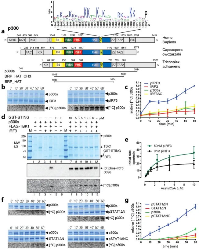
Figure 1. Transcription factor dimerization enables activation of p300. (a) Domain structure of p300. 516
Sequence conservation of the AIL is shown using WebLogo53. Constructs used are shown. (b) p300s was 517
incubated for the indicated times in the presence or absence of inactive, monomeric IRF3 or TBK1-518
phosphorylated, dimeric pIRF3. Samples were analyzed by SDS-PAGE followed by Coomassie staining 519
and autoradiography. (c) Quantification of autoacetylation of p300s. (d) p300 is activated by TBK1-520
mediated IRF3 phosphorylation. p300s was incubated with recombinant GST-STING, TBK1 and IRF3 in 521
the presence of ATP and [14C] acetyl-CoA. Top panel: Coomassie-stained SDS-PAGE gel. Middle panel:
522
Analysis of IRF3 phosphorylation on S396 using immunoblotting. Bottom panel: autoradiography. (e) 523
HAT scintillation proximity assay. 12.5 µM Histone H4 substrate peptide was incubated with 50 nM 524
p300s in the presence (green) or absence (black) of 50 nM pIRF3 and varying concentrations of [3H]
525
acetyl-CoA. The degree of Histone H4 substrate acetylation was quantified using scintillation counting. 526
(f) As in panel B but using inactive, monomeric STAT1ΔN or activated, dimeric pSTAT1ΔN. Activated, 527
dimeric pSTAT1ΔNC lacking the C-terminal TAD did not stimulate p300s autoacetylation. Samples were 528
analyzed as in panel (B). (g) Quantification of autoacetylation of p300s. Intensity values were normalized 529
by dividing by the maximum autoacetylation signal obtained after 60 minutes. Error bars shown in panels 530
(c), (e) and (g): Three independent experiments were performed and the mean value and error bars 531
representing the standard deviation are shown. Data analysis and plotting was done with Graphpad Prism 532
7.0. 533
535
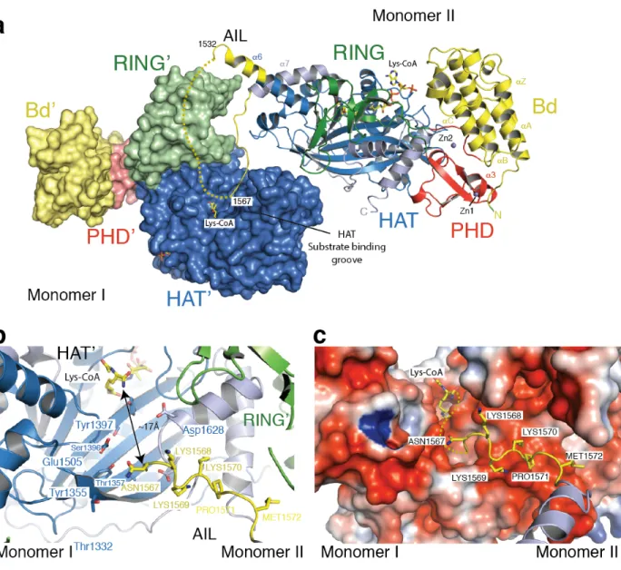
Figure 2: The structure of p300 adopts a AIL swap conformation. (a) Monomer I is surface rendered 536
and monomer II is shown as a cartoon. The AIL loop from monomer II is shown in yellow. The AIL lies 537
near the HAT substrate binding groove of monomer I. A disordered segment of the AIL is shown as a 538
dotted line. (b) Close up view of the residues of the AIL loop from monomer II and residues of monomer 539
I in the substrate binding pocket. (c) Binding of the positively charged AIL is mediated by interactions 540
with negatively charged residues in the HAT binding pocket. 541
542 543
544
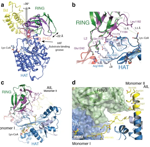
Figure 3: Structural rearrangement of the RING domain. (a) The RING domain (green) rotates ~39° 545
resulting in a 22 Å displacement away from the active site. The rotation axis is indicated as a grey rod. (b) 546
In the loop-swap conformation, residues in the RING-HAT interface are disrupted thus resulting in a 547
more open HAT active site. Leu1182 is positioned 15Å away from the Lys-CoA inhibitor in the loop-548
swap conformation (green) but within 5.5Å in the absence of the loop swap (magenta). (c) Repositioning 549
of the RING domain allows the AIL from monomer II to approach the HAT active site of monomer I. (d) 550
Details of the interaction surface of the AIL from monomer II with the RING domain of monomer I. 551
552 553
554 555
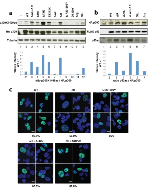
Figure 4: Regulation of HAT activity by flanking domains. (a) Indicated variants of p300 were 556
transiently co-transfected with p53 in COS cells and samples analyzed by western blotting using the 557
indicated antibodies. Bottom panel: quantification p300 K1499Ac signal. (b) Analysis of p53 acetylation. 558
Bottom panel: quantification p53 acetylation signal. (c) H1299 cells were transfected with the indicated 559
construct and analyzed by immunoflorescence using Anti-HA for p300 (green) and cell nuclei were 560
stained with Hoechst (blue). Bottom panels: Cells were treated with the A-485 HAT or the CBP30 561
Bromodomain inhibitor. Percentage of cells showing the indicated phenotype (n=200 cells) is indicated 562
below each panel. Scale bar, 10 µm. 563
564 565
566
Figure 5. Acetylation of the AIL regulates dynamic interaction with the substrate binding pocket of
568
p300. (a) Normalized distance between the AIL and residues in the inactive monomer. Inter-residue 569
distances are normalized by the distances expected if the AIL behaved as a self-avoiding random coil. 570
Electrostatic interaction mediated by conserved lysine residues between K1542 and K1560 of the AIL and 571
aspartic/glutamic acid residues around the active site of the HAT domain, as shown by the residues 572
highlighted (E1334, E1351, E1442, D1444, E1505, D1622, D1625, and D1628). The extensive contacts 573
between the AIL and the RING domain originates in part from the RING domain’s proximity to the AIL 574
in its inactive conformation. (b) Normalized distance between the AIL and all residues in the active 575
(acetylated) monomer. After acetylation, lysine-mediated electrostatic interactions are lost. (c) 576
Representative conformations with the AIL shown as an ensemble for the inactive deacetylated monomer 577
(left) and the active acetylated monomer (right). The Cα atoms of residues in the AIL are colored 578
according to charge: blue (positive), red (negative) and green (non-charged). The HAT substrate-binding 579
groove is more exposed in the active acetylated state, due to both the relative position of the RING 580
domain and the lack of preferential interactions by the AIL. (d) Inter-molecular interactions in the loop-581
swapped dimer between the AIL of one HAT and the adjacent subunit of the other. The adjacent subunit 582
is either in the active (top) and inactive (bottom) conformation. In the active state, the AIL is able to 583
directly engage with residues E1442 and E1444 from the adjacent HAT substrate binding groove, 584
suggesting the position of the RING domain has a steric impact on the accessibility of the AIL. (e) 585
Simulations of the AIL in context of the loop-swapped dimer. Left panel: Cartoon of the trajectory of the 586
AIL (dashed line). Right panel: Representative conformations with the AIL Cα backbone atoms are 587
colored according to charge as in panel (B). 588
590
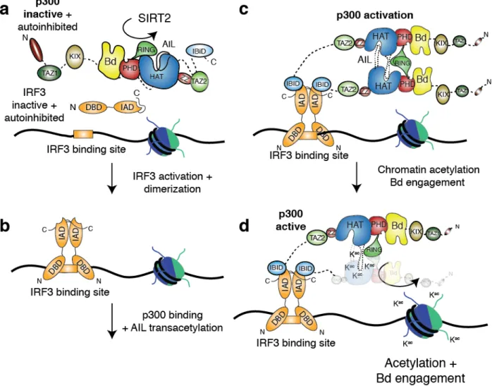
Figure 6: Molecular model for p300 activation and DNA targeting. (a) p300 is maintained in the 591
inactive state by deacetylases such as SIRT2. IRF3 is autoinhibited by a C-terminal segment in the IAD 592
domain. (b) TBK1 phosphorylation activates and dimerizes IRF3. The activated IRF3 dimer engages the 593
IBID domain of p300. (c) Recruitment of two copies of p300 results in trans-autoacetylation in the AIL 594
loop and HAT activation. (d) Activated p300 can acetylate chromatin and engage acetylated substrates 595
via the Bd. 596
598
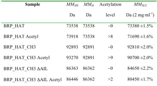
Table 1: Summary of SEC-MALLS and mass spectrometry experiments 599 Sample MMMS Da MMth Da Acetylation level MMSLS Da (2 mg·ml-1) BRP_HAT 73538 73538 ~0 73380 ±1.5% BRP_HAT Acetyl 73918 73538 >8 71690 ±1.6% BRP_HAT_CH3 92893 92891 ~0 92810 ±2.0% BRP_HAT_CH3 Acetyl 93270 92891 >9 90700 ±2.0% BRP_HAT_CH3 ΔΑΙL 86363 86362 ~0 84650 ±2.2% BRP_HAT_CH3 ΔΑΙL Acetyl 86446 86362 >2 80450 ±1.7%
Column labeling: Molar masses determined by Mass spectrometry (MMMS), MMth the theoretical molar 600
mass calculated from the appropriate primary sequences. Acetylation levels were estimated based on the 601
mass differences as compared to the non-acetylated sample. MMSLS (Molar masses determined by SEC-602
MALLS) at a concentration of 2 mg·ml-1. All p300 constructs contained the mutation Y1467F. The errors
603
reported for SEC-MALLS are the residual standard deviations of the observed data from the fitted values 604
calculated using Astra. 605
Methods 607
Constructs 608
For cell-free protein expression, cDNA of p300 (NCBI reference sequence: NM_001429.3) variants were 609
cloned into the pIVEX2.4d vector (Roche) with a N-terminal 6x His tag and a C-terminal FLAG tag. In 610
the ΔR constructs, the RING domain encompassing residues 1169–1241 was replaced by Glycine amino 611
acid residue linker. In the ΔAIL constructs, loop amino acid residues comprising residues 1520–1581 612
were replaced by the flexible linker sequence SGGSG. For E.coli expression, cDNA encoding residues 613
1048-1282, for the BRP or BPΔR were cloned into the vector pETM-33 (EMBL) with a TEV cleavable 614
N-terminal glutathione S-transferase (GST) tag. p300 BRP_HAT variants were cloned into pFASTBAC1 615
(Thermo Fisher) and expressed in insect cells as shown earlier29. p300s constructs, spanning amino acid 616
residues 324-2414, were cloned into pFASTBAC1 vector with an N-terminal FLAG tag. HA-tagged full-617
length p300 variants were cloned into pcDNA3.1 (Thermo Fisher). Point mutations were introduced by 618
QuikChange mutagenesis (Agilent). Point mutations and nucleotide deletions carried out in p300FL (1-619
2414) or p300s (324-2094) were done through transfer vectors as described previously29. STAT1ΔN 620
(136-748), STAT1ΔNC (136-713) and IRF3ΔC (1-382) with a C-terminal intein tag were cloned into the 621
pTXB1 vector (New England Biolabs) using the restriction enzymes NdeI (STAT1) or NcoI (IRF3) and 622
SpeI. IRF3 (1-427) with an N-terminal His-tag cleavable by TEV protease was cloned using the
623
restriction enzymes NcoI and XhoI into the vector pETM-11 (EMBL). All constructs were confirmed by 624
DNA sequencing. 625
626
Expression and Purification 627
Expression and purification of FLAG-tagged p300s constructs was done as described previously2. This
628
method allows purification of p300s variants that are already preacetylated. Expression and purification of 629
p300 BPR_HAT and SIRT2 were done as described in29. TBK1 was expressed in insect cells and purified
as described previously21. Cell-free protein synthesis was done in a 50 µL reaction volume. Briefly, 10 µg 631
mL-1 of His-p300 variants in pIVEX2.4d were added to a reaction mixture containing 1 mM amino acid
632
mix, 0.8 mM rNTPs (guanosine-, uracil-, and cytidine- 5’ triphosphate ribonucleotides), 1.2 mM 633
adenosine 5’-triphosphate, 55 mM HEPES, pH 7.5, 68 µM folinic acid, 0.64 mM cyclic adenosine 634
monophosphate, 3.4 mM dithiothreitol, 27.5 mM ammonium acetate, 2 mM spermidine, 5 µM ZnCl2, 80 635
mM creatine phosphate, 208 mM potassium glutamate, 16 mM magnesium acetate, 250 µg mL-1 creatine 636
kinase, 27 µg mL-1 T7 RNA polymerase, 0.175 µg mL-1 tRNA, and 67 µL mL-1 S30 E. coli bacterial
637
extract. Incubation was carried out at 22 °C with agitation for 16 h. Proteins were purified using Ni-NTA 638
chromatography (IMAC Sepharose 6 FF, GE healthcare) in buffer 1 (20 mM TRIS, pH 8.0, 300 mM 639
NaCl, 1 mM DTT, 5 µM ZnCl2) containing Complete Protease Inhibitors EDTA-Free (Roche). The resin
640
was washed with 20 CV of buffer 1 and the protein eluted with 5 CV buffer 1 containing 300 mM 641
Imidazole. The protein was concentrated in a prewashed Amicon Ultra 0.5 ml Ultracel 10K Centrifugal 642
filter (Molecular weight cut off = 10kDa; EMD Millipore). The protein was buffer exchanged into buffer 643
1 using 0.5 ml Zeba Spin desalting columns (Molecular weight cut off = 7kDa; Thermo Scientific), flash 644
frozen in liquid N2 and stored at -80 °C.
645
For expression of GST-BRP and GST- BPΔR fusion proteins in E. coli BL21 (DE3), LB medium 646
enriched with 100 µM ZnCl2 was used. Cell pellets were resuspended in buffer 1 containing Complete
647
Protease Inhibitors EDTA-Free (Roche) and lysed by using a Microfluidizer (Microfluidics Corp., MA, 648
USA). The lysate was clarified by centrifugation for 30 minutes at 39,000 g in a JA-25.5 rotor (Beckman) 649
and applied to a Glutathione Sepharose 4 Fast Flow resin according to instructions by the manufacturer 650
(GE Healthcare). The resin was washed with buffer 1 and incubated with His-tagged TEV protease (1:100 651
w/w) for 14-16 h at 4°C. Subtractive Ni-NTA chromatography (IMAC Sepharose 6 FF, GE Healthcare) 652
was then employed to remove the residual His-tag and TEV protease. The untagged protein was further 653
purified by gel filtration on a High Load 16/60 Superdex 75 column (GE Healthcare) equilibrated in 20 654
mM HEPES, pH 7.5, 300 mM NaCl, 0.5 mM TCEP and 5 µM ZnCl . The final protein was concentrated 655
to 15 mg/ml in a prewashed Amicon Ultra-15 Centrifugal filter (Molecular weight cut off = 10kDa; EMD 656
Millipore), flash frozen in liquid N2 and stored at -80 °C.
657
The expression and purification of non-phosphorylated STAT1 variants (STAT1ΔN, STAT1ΔNC) and 658
IRF3ΔC (1-382) was done in E.coli using the IMPACT expression system (New England Biolabs). For 659
the expression of Y701 phosphorylated variants (pSTAT1ΔN, pSTAT1ΔNC), proteins were co-expressed 660
with Elk receptor tyrosine kinase domain in E.coli BL21(DE3) TKB1 cells (Agilent). Cells were 661
harvested by centrifugation and resuspended in buffer 2 (20 mM HEPES pH 7.5, 500 mM NaCl). The 662
cells were lysed in a microfluidiser (Microfluidics Corp., MA, USA) and the soluble fraction was 663
obtained by centrifugation for 30 minutes at 39,000 g in a JA-25.5 rotor (Beckman). The supernatant was 664
first passed over chitin beads (New England Biolabs) and washed with buffer 2 for 10 column volumes. 665
The protein was cleaved at 4°C for 16h in buffer 2 containing 50 mM DTT, eluted and further purified by 666
gel filtration on a High Load 16/60 Superdex 200 column (GE Healthcare) equilibrated in buffer 2. 667
GST-STING, comprising the soluble cytoplasmic domain spanning amino acids 138-378, was expressed 668
in E.coli BL21(DE3) at 37°C for 3 h. The cells were harvested by centrifugation and resuspended in 669
buffer 3 (20 mM TRIS, pH 8.0, 300 mM NaCl, 1 mM DTT) containing Complete Protease Inhibitors 670
EDTA-Free (Roche). The cells were lysed in a microfluidiser (Microfluidics Corp., MA, USA) and the 671
soluble fraction was obtained by centrifugation as above. The supernatant was passed over equilibrated 672
Glutathione Sepharose 4 Fast Flow resin according to instructions by the manufacturer (GE Healthcare). 673
The resin was washed with buffer 3 and eluted with 10 mM reduced Glutathion in buffer 3. The protein 674
was further purified by gel filtration on a High Load 16/60 Superdex 200 column (GE Healthcare) 675
equilibrated in 20 mM HEPES, pH 7.5, 300 mM NaCl, 0.5 mM TCEP. The final protein was concentrated 676
to 16 mg/ml in a prewashed Amicon Ultra-15 Centrifugal filter (Molecular weight cut off = 30 kDa; EMD 677
Millipore), flash frozen in liquid N2 and stored at -80 °C.
IRF3 was expressed in E.coli BL21(DE3) at 18°C for 16 h. The cells were harvested by centrifugation 679
and resuspended in buffer 2 containing 10 mM imidazole. The cells were lysed in a microfluidiser 680
(Microfluidics Corp., MA, USA) and the soluble fraction was obtained by centrifugation as above. The 681
supernatant was passed over Ni2+–conjugated IMAC sepharose resin (GE Healthcare) and washed with 682
buffer 2 containing 20 mM imidazole. The protein was eluted in buffer 2 containing 500 mM imidazole 683
and was further purified by gel filtration on a High Load 16/60 Superdex 200 column in buffer 2 684
containing 0.5 mM TCEP. IRF3 was phosphorylated in vitro at a 1:10 molar ratio TBK1:IRF3 (1mg/ml) 685
in presence of 5 mM MgCl2 and 1 mM ATP. The reaction was incubated at 30°C for 1h and then for an 686
additional 10h at 21°C. Phosphorylated IRF3 was further purified by size exclusion chromatography on a 687
Superdex S200 16/60 column (GE Healthcare) in 20 mM HEPES, pH 7.5, 300 mM NaCl, 0.5 mM TCEP. 688
The production of recombinant histones was done following standard procedures54.
689 690
Crystallization and structure determination 691
The p300 BRP_HAT construct comprising the AIL and the mutation Y1467F was deacetylated as done 692
previously29. The protein at 4.5 mg ml-1 was incubated with a three-fold molar excess of the bi-substrate
693
inhibitor Lys-CoA34 prior to crystallization. Crystals in the P21 space group were grown by hanging-drop
694
vapor diffusion at 4 °C by mixing equal volumes of protein and crystallization solution containing 100 695
mM HEPES, pH 7.5, 18-22% polyethylene glycol 3350, 0.2 M NaCl. Crystals were cryoprotected in 20-696
25% ethylene glycol and drop frozen in liquid nitrogen. We collected native diffraction data to a 697
minimum Bragg spacing of 3.1 Å resolution at the ESRF on beamline ID29 under a nitrogen gas stream at 698
100 K, at a wavelength of 1.282 Å. We processed the data with XDS (Extended Data Table 1). The 699
structure of the p300 BRP_HAT was determined by molecular replacement using Phaser. There are four 700
copies in the asymmetric unit and the RING domains were initially not visible in the electron density map 701
and are partially disordered. Inspection of an anomalous difference map indicated peak density for the 702
zinc ions and allowed positioning of the RING domain in the outward rotated conformation. A final 703
model was produced by iterative rounds of manual model building in Coot and refinement using 704
PHENIX. The final model contains residues 1045 -1664 with a deletion of residues 1534-1567 and was 705
refined to a 3.1 Å resolution with an Rwork and an Rfree of 19% and 26%, respectively (Extended Data
706
Table 1). Analysis of the refined structure by MolProbity showed that there are no residues in disallowed 707
regions of the Ramachandran plot. The MolProbity all atom clash score was 1.91, placing the structure in 708
the 100th percentile among structures refined at 3.1 Å resolution (N=2108). 709
The BPΔR construct at 15 mg ml-1 was mixed with 2 mM of a 11-mer histone peptide H4 (10-20) 710
GLGKacGGAKacRHR (only the underlined amino acid sequence is visible in the electron density map) 711
containing two acetylated Lysine residues at positions K12 and K16 (H4K12K16). Crystals in the P212121 712
space group were grown by hanging-drop vapor diffusion at 21°C by mixing equal volumes of protein 713
and crystallization solution containing 1.6 M Ammonium Sulfate, 100 mM Bicine, pH 9.0. Crystals were 714
cryoprotected in 20% ethylene glycol and drop frozen in liquid nitrogen. We collected native diffraction 715
data to a minimum Bragg spacing of 2.5Å resolution at the ESRF on beamline ID29 under a nitrogen gas 716
stream at 100K, at a wavelength of 1.0Å (Extended Data Table 1). Data processing, molecular 717
replacement and refinement were done as indicated above. The final model contains two copies of the 718
BPΔR module corresponding to residues 1049 -1279 of p300 in the asymmetric unit. As expected, 719
replacement of the RING domain residues 1169-1241 by a single Glycine amino acid linker did not 720
adversely affect the remainder of the BP module. Analysis of the refined structure by MolProbity showed 721
that there are no residues in disallowed regions of the Ramachandran plot. The MolProbity all atom clash 722
score was 0.97 placing the structure in the 100th percentile (N=6960).
723 724
Monte Carlo simulations 725