Publisher’s version / Version de l'éditeur:
NeuroImage, 54, 1, pp. 10-15, 2010-07-17
READ THESE TERMS AND CONDITIONS CAREFULLY BEFORE USING THIS WEBSITE.
https://nrc-publications.canada.ca/eng/copyright
Vous avez des questions? Nous pouvons vous aider. Pour communiquer directement avec un auteur, consultez la
première page de la revue dans laquelle son article a été publié afin de trouver ses coordonnées. Si vous n’arrivez pas à les repérer, communiquez avec nous à [email protected].
Questions? Contact the NRC Publications Archive team at
[email protected]. If you wish to email the authors directly, please see the first page of the publication for their contact information.
NRC Publications Archive
Archives des publications du CNRC
This publication could be one of several versions: author’s original, accepted manuscript or the publisher’s version. / La version de cette publication peut être l’une des suivantes : la version prépublication de l’auteur, la version acceptée du manuscrit ou la version de l’éditeur.
For the publisher’s version, please access the DOI link below./ Pour consulter la version de l’éditeur, utilisez le lien DOI ci-dessous.
https://doi.org/10.1016/j.neuroimage.2010.07.028
Access and use of this website and the material on it are subject to the Terms and Conditions set forth at
Functional mapping in the corpus callosum: a 4 T fMRI study of white matter
Gawryluk, Jodie R.; D’Arcy, Ryan C. N.; Mazerolle, Erin L.; Brewer, Kimnerley D.; Beyea, Steven D.
https://publications-cnrc.canada.ca/fra/droits
L’accès à ce site Web et l’utilisation de son contenu sont assujettis aux conditions présentées dans le site LISEZ CES CONDITIONS ATTENTIVEMENT AVANT D’UTILISER CE SITE WEB.
NRC Publications Record / Notice d'Archives des publications de CNRC: https://nrc-publications.canada.ca/eng/view/object/?id=655153ee-c7d8-40ec-bd46-7ffbf90a2a1c https://publications-cnrc.canada.ca/fra/voir/objet/?id=655153ee-c7d8-40ec-bd46-7ffbf90a2a1c
Functional mapping in the corpus callosum: A 4 T fMRI study of white
matter
Jodie R. Gawryluka, b Ryan C.N. D'Arcya, b, c Erin L. Mazerollea, b Kimberly D. Brewera, d Steven D. Beyeaa, c, d a Institute for Biodiagnostics (Atlantic), National Research Council, Halifax, Nova
Scotia, Canada
b Department of Psychology/Neuroscience, Dalhousie University, Halifax, Nova
Scotia, Canada
c Department of Radiology, Dalhousie University, Halifax, Nova Scotia, Canada
d Department of Physics and Atmospheric Sciences, Dalhousie University,
Halifax, Nova Scotia, Canada
Received 16 April 2010. Revised 9 July 2010. Accepted 13 July 2010. Available Abstract
Introduction
The idea of fMRI activation in white matter (WM) is controversial. Our recent work has used two different approaches to investigate whether there is evidence for WM fMRI. The first approach used words and faces to elicit interhemispheric transfer activation in the posterior corpus callosum (Sperry task). The second approach used checkerboard stimuli to elicit similar activation in the anterior corpus callosum (Poffenberger task). Using these different tasks, it has been possible to detect WM activation in different regions. In the current study, we report the results of a critical experiment:
demonstrating that callosal activation can be experimentally manipulated within the same set of individuals.
Methods
All subjects completed both the Sperry and Poffenberger tasks. Functional MRI data were acquired at 4 T, using an asymmetric spin echo spiral sequence. Data were analyzed with FSL using a model-based approach. Analyses focused on group and individual activations in WM.
Results and discussion
Corpus callosum activation was elicited for both tasks, with activation varying according to task type. A statistical contrast of the two tasks revealed posterior callosal activation for the Sperry task and anterior callosal activation for the Poffenberger task. The Sperry task showed activation in the isthmus and middle body of the corpus callosum at the group level and in 100% of subjects. The Poffenberger task showed activation in the genu and middle body of the corpus callosum at the group level and in 94% of subjects. The WM activation replicated prior results, with the additional strength of functional mapping within the same group of individuals.
Research highlights
It is possible to map activation in white matter using fMRI.
Different functional pathways can be localized in the corpus callosum.
White matter fMRI activation can be observed at both the group and individual levels.
Keywords: Functional connectivity; White matter; High field fMRI; Spiral acquisition Introduction
White matter comprises approximately 50% of brain volume (Black, 2007), yet the
notion of using fMRI to examine these regions remains relatively unexamined and highly controversial. There are two main reasons for the controversy: 1) BOLD signals rely in part on relatively small fluctuations in cerebral blood volume and flow, which are then 3– 6 times lower in white matter ( [Preibisch & Haase, 2001], [Rostrup et al., 2000],
[Helenius et al., 2003], [van der Zande et al., 2005] and [Wise et al., 2004]); and 2) the primary source of fMRI signal is thought to arise from post-synaptic potentials (which occur mainly in gray matter) as opposed to action potentials (Logothetis et al., 2001). To put the situation in context, of the 254 287 fMRI studies that have been published to date (according to PubMed at the time this paper was written), there are only nine reporting activation in white matter to our knowledge ( [Tettamanti et al., 2002], [Omura et al., 2004], [Weber et al., 2005], [Mosier & Bereznaya, 2001], [D'Arcy et al., 2006], [Mazerolle et al., 2008], [Yarkoni et al., 2009], [Gawryluk et al., 2009] and [Mazerolle et al., 2010]). Approximately half of these studies detected white matter activation
incidentally.
Many of the fMRI studies that report white matter activation used tasks designed to elicit interhemispheric transfer (IHT; e.g., [Tettamanti et al., 2002], [Omura et al.,
2004] and [Weber et al., 2005]). The typical IHT task (Poffenberger, 1912) rapidly presents visual stimuli to each hemifield and requires a motor response from either the ipsilateral (no cross) or contralateral (motor cross) hand (e.g., Tettamanti et al., 2002). Another IHT task is modeled after so-called ―split-brain‖ patients (Gazzaniga et al., 1965), using visual field presentation of lateralized stimuli (words and faces) (e.g., D'Arcy et al., 2006).
Given the early evidence in support of functional activation in white matter, our group followed up with two prospective studies of white matter fMRI. In the first study, we used high field imaging (4 T) and a Sperry task to elicit WM activation in the corpus callosum (Mazerolle et al., 2008). The results revealed activation in the isthmus of the corpus callosum in 20% of the individual subjects and at the group level. In the second study, we replicated the demonstration of white matter activation using a Poffenberger task (Gawryluk et al., 2009). This time the results detected activation in the anterior corpus callosum, for 100% of the individual subjects and at the group level.
The increase in sensitivity was due largely to the use of a novel imaging technique called asymmetric spin echo (ASE) spiral (Brewer et al., 2009). ASE spiral acquires three images (per slice per volume) with increasing T2 weighting but equal
BOLD-contrast. Previous studies demonstrated that T2 weighting at high field is sensitive to
gray matter activation (Kim and Ugurbil, 1997). We have shown that increased T2
weighting combined with 4 T MRI is sensitive to white matter fMRI activation (Gawryluk et al., 2009). Given these results, ASE spiral acquisition at high field may lead to
increased detection of white matter fMRI activation when compared with other methods. Notably, the [Mazerolle et al., 2008] and [Gawryluk et al., 2009] studies detected
activation in different regions of the corpus callosum — both of which were functionally consistent with the tasks. The posterior callosal activation observed for the Sperry task was thought to connect parietal regions involved in integrating high level sensory information ( [Witelson, 1989] and [Zarei et al., 2006]). The anterior callosal activation observed for the Poffenberger task was thought to connect pre-motor regions
associated with the subject's response ( [Iacoboni & Zaidel, 2004], [Zarei et al., 2006], [Meyer et al., 2008] and [Stancak et al., 2000]). Previous work using the Poffenberger task has identified a more anterior cluster in the genu of the corpus callosum, which was also attributed to pre-motor interhemispheric transfer, although recent tractography studies suggest that the genu is structurally connected to pre-frontal regions (e.g., Zarei et al., 2006).
These results suggest that functional mapping in white matter may be possible. This observation needed to be confirmed using a within-subjects design. Accordingly, we sought to answer the following question: can two different tasks be used to map different callosal regions within the same subjects? To answer this question, we employed the Sperry and Poffenberger tasks. In order to continue optimizing our sensitivity to fMRI activation in white matter, we used the 4 T ASE spiral method weighting the combined data towards the third image (with the most T2 weighting). It
was hypothesized that: 1) both tasks would elicit white matter fMRI activation at the group and individual levels; 2) the Sperry task would elicit relatively more activation in the posterior corpus callosum and 3) the Poffenberger task would yield relatively more activation in the anterior corpus callosum.
Materials and methods Participants
Seventeen healthy, right handed subjects (8 females) participated in the study. The mean age of participants was 26.05 ± 4.79 years. The study was approved by the local ethics boards. Each participant provided written informed consent prior to their
participation.
Experimental design
Each participant completed the Poffenberger task followed by the Sperry task.1 The Sperry task utilized a block design (eight 22 s blocks with eight stimuli/block, alternated with 18 s rest blocks) to present words (left hemisphere stimuli) and faces (right
hemisphere stimuli) to the left and right visual fields. The stimuli were either real (i.e., typical faces or words) or pseudo (i.e., faces with rearranged features or non-words). Participants were asked to indicate if a given stimulus was a real or pseudo face or a real or pseudo word (four button forced response). Response hand was always crossed (i.e., left hand for words and right hand for faces). This combination resulted in two different IHT conditions: a motor cross and a visual-motor cross. Each block of stimuli contained only one condition; block order was randomized.
The Poffenberger task used a block design (ten 12 s blocks with eight stimuli/block, alternated with 18 s rest blocks) to present small checkerboard stimuli randomly to the left and right visual fields. Instructions were given prior to each block indicating whether the responses were to be made with the same or opposite hand as the side of the visual stimulus (two button forced response). Varying response hand in this way created two different IHT conditions: motor cross and no cross. Each block of stimuli contained only one condition; block order was randomized.
Participants were instructed to maintain central fixation throughout the experiment. All stimuli were presented laterally (> 2.3° from fixation to initially stimulate one
hemisphere, and rapidly (words/faces: 150 ms and checkerboards: 100 ms) to avoid saccades. The tasks were presented visually through back-projection to a screen mounted inside the bore (and viewed through a mirror mounted on the head coil) using E-prime (Psychology Software Tools, Inc). Each subject performed a short practice of each task with feedback to ensure compliance.
Functional MRI acquisition
Data were acquired from a 4 T Varian INOVA whole body MRI system. Gradients were provided by a body coil (Tesla Engineering Ltd.) operating at a maximum of 35.5 mT/m at 120 T/m/s, and driven by 950 V amplifiers (PCI). The RF coil employed was a TEM head coil (Bioengineering Inc.) driven by a 7 kW amplifier (Herley Inc.).
Functional MRI data were acquired using the ASE spiral sequence (Brewer et al.,
2009). The ASE spiral sequence collects three images (with increasing T2 weighting but
equal BOLD-contrast) per slice per volume. The number of slices was limited to 17 (4 mm axial slices, with no gap) in order to satisfy the time parameters required to collect the three ASE spiral images. Slices were prescribed to cover a slab extending from the ventral boundary of the corpus callosum to the cortex above. The parameters for functional imaging were as follows: 64 × 64 matrix (220 × 220 mm), 1 shot, TR = 2 s, TR/TE/TE* = 2000/68/27 ms (where TE is the spin echo center and TE* is the
asymmetric echo times).
Functional MRI data analyses
The three ASE images were combined using an inverted signal weighted averaging algorithm to increase T2 weighting (based on the findings of Gawryluk et al., 2009).
(Jenkinson et al., 2002), non-brain removal using BET (Smith, 2002), spatial smoothing using a Gaussian kernel of FWHM 6 mm, mean-based intensity normalization of all volumes by the same factor, and high-pass temporal filtering (100 s cutoff). Statistical analyses were performed using fMRI expert analysis tool (FEAT) version 5.3 in FMRIB Software Library (FSL). A model-based approach (General Linear Model) was taken using a gamma HRF and its temporal derivative, convolved with the block design of the two tasks. Time-series statistical analyses were carried out using FILM with local
autocorrelation correction (Woolrich et al., 2001). Statistical analyses were done with motion parameters as covariates.2 Except where otherwise stated, Z statistic images were first developed using a threshold for clusters determined by Z > 2.0 and a (corrected) cluster significance threshold of P = 0.05 (Worsley et al., 1992). Subsequently, activation maps were displayed in MRIcro (Z > 2.5). Images were registered to the high-resolution T1-weighted anatomical image (7 DOF) before being normalized to standard MNI space (12 DOF) using FLIRT ( [Jenkinson et al.,
2002] and [Jenkinson & Smith, 2001]). Registration was manually verified and optimized as needed.
Data were examined at both the group and individual levels. At the group level, the tasks were analyzed for activation versus rest. The main comparison of interest consisted of a statistical contrast of the Sperry and Poffenberger tasks at the group level, using a region of interest (ROI) approach focused on the corpus callosum to increase sensitivity (P < 0.005 uncorrected, displayed using Z > 2.5). For all group level analyses, local maxima (at least 5 mm apart) in callosal white matter were extracted for clusters with Z > 2.5 and extent > 2.
Results at the individual level were subsequently examined using activation versus rest contrasts. A region of interest (ROI) approach focused on the corpus callosum. The corpus callosum was masked within Featquery (FMRIB's Software Library) to determine the presence, size, and strength of activation in the ROI. Spatial coordinates (standard MNI space) were obtained for the maximally active voxel in callosal white matter in each task.
Results
Functional MRI results
Comparison of the Sperry and Poffenberger tasks at the group level
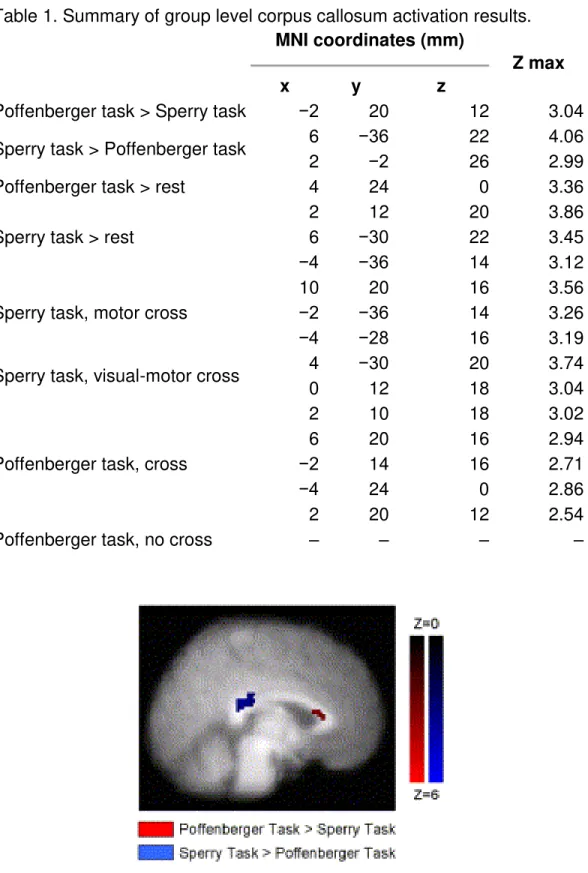
For all group level analyses, corpus callosum activation is summarized in Table 1. When the tasks were statistically compared to one another, activation was present in different regions of the corpus callosum. Specifically, the Sperry task showed greater activation in the posterior corpus callosum when contrasted with the Poffenberger task. The reverse pattern was also observed. The Poffenberger task showed greater
Table 1. Summary of group level corpus callosum activation results. MNI coordinates (mm)
Z max
x y z
Poffenberger task > Sperry task −2 20 12 3.04 Sperry task > Poffenberger task 6 −36 22 4.06
2 −2 26 2.99
Poffenberger task > rest 4 24 0 3.36 Sperry task > rest
2 12 20 3.86
6 −30 22 3.45
−4 −36 14 3.12
Sperry task, motor cross
10 20 16 3.56
−2 −36 14 3.26
−4 −28 16 3.19
Sperry task, visual-motor cross 4 −30 20 3.74
0 12 18 3.04
Poffenberger task, cross
2 10 18 3.02
6 20 16 2.94
−2 14 16 2.71
−4 24 0 2.86
2 20 12 2.54
Poffenberger task, no cross – – – –
Fig. 1. Group activation showing statistical difference between the Poffenberger and Sperry tasks. The Poffenberger task > Sperry task (displayed in red) shows activation in the anterior corpus callosum. The Sperry task > Poffenberger task (displayed in blue) shows activation in the posterior corpus callosum. Activation intensity is displayed in terms of Z-scores (N = 17).
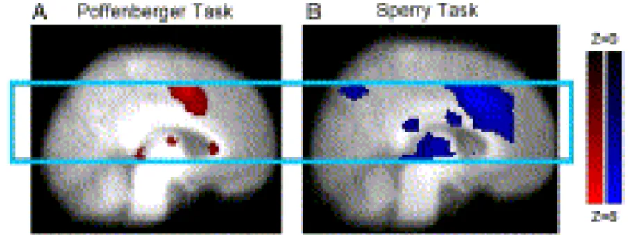
Examination of the Sperry and Poffenberger tasks at the group level
Separately, the tasks showed group level activation in different regions of the corpus callosum (Fig. 2). The Sperry task revealed activation clusters in the isthmus of the corpus callosum as well as in the middle body of the corpus callosum. Corresponding gray matter task-related activation was present in bilateral occipital (precuneus and lingual gyrus), bilateral parietal (precuneus and supramarginal gyrus), bilateral temporal (fusiform gyrus, inferior temporal gyrus, and insula), bilateral frontal (middle frontal gyrus and inferior frontal gyrus) regions as well as in the thalamus and anterior cingulate.
Fig. 2. Group activation for the Poffenberger task (A: left) and the Sperry task (B: right). Activation maps show both white matter activation in the corpus callosum and corresponding gray matter activation within the selected imaging slab (light blue box). All other details as per Fig. 1.
The Poffenberger task showed activation in the genu of the corpus callosum.
Corresponding gray matter activation was visible in bilateral occipital (lingual gyrus), bilateral parietal (precuneus and supramarginal gyrus), bilateral temporal (superior temporal gyrus, inferior temporal gyrus, and insula), bilateral frontal (middle frontal gyrus and inferior frontal gyrus) regions as well as in the thalamus and anterior and posterior cingulate.
Examination of the Sperry and Poffenberger conditions at the group level
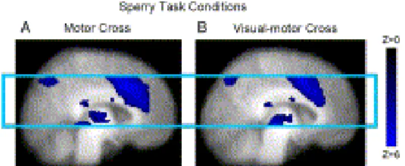
The results of group level conditional analyses were also examined. In the Sperry task, the motor cross and visual-motor cross conditions (compared to rest) showed activation in the isthmus and middle body of the corpus callosum (Fig. 3), as well as in the gray matter regions observed in the task versus rest analysis.
Fig. 3. Sperry task group activation showing similar clusters in both motor cross (A) and visual-motor cross (B) conditions. The posterior isthmus activation was specific to the Sperry task, whereas, the middle body activation was also present in the Poffenberger task. All other details as in Fig. 2.
In the Poffenberger task, the motor cross condition revealed activation in the genu and middle body of the corpus callosum (Fig. 4). The no cross condition did not show any activation in the corpus callosum. However, activation was present in the genu and middle body when analyzed at a lower threshold (Z = 2.0, P = 0.1).
Fig. 4. Poffenberger task group activation showing anterior and middle body clusters in the crossed condition (A). While there was no cluster in the uncrossed condition (B), similar activation was detected at lower thresholds. The pattern of results is consistent with prior observations of no ‗true‘ uncrossed condition ( [Mazerolle et al., 2008] and [Gawryluk et al., 2009]). All other details as in Fig. 2.
Examination of the Sperry and Poffenberger tasks at the individual level At the individual level, the Sperry task elicited white matter activation in 17/17
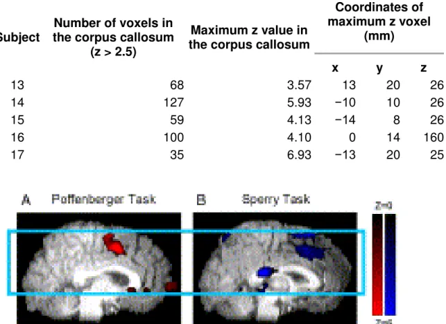
participants. The Poffenberger task yielded white matter activation in 16/17 participants. Table 2 shows the extent of activation, maximum Z score and coordinates of the
maximally active voxel in the corpus callosum for each participant during each task. Fig. 5 shows activation in a representative subject during each task. The average maximum Z score during the Sperry task was 5.23 (SD = 1.45) and the average maximum Z score during the Poffenberger task was 4.56 (SD = 1.53).
Table 2. Individual level results during the A. Sperry task and B. Poffenberger task: cluster extent, intensity and MNI space coordinates of the maximally active voxel in the corpus callosum.
Subject
Number of voxels in the corpus callosum
(z > 2.5)
Maximum z value in the corpus callosum
Coordinates of maximum z voxel (mm) x y z A. Sperry task 1 7 4.15 −8 17 24 2 208 6.31 6 10 20 3 105 3.86 0 −2 26 4 2 2.64 −8 −4 28 5 96 5.61 5 24 6 6 141 5.54 1 −30 18 7 83 7.33 13 20 27 8 30 4.61 −16 −46 10 9 27 3.38 −2 −38 12 10 78 5.70 12 22 20 11 35 4.17 6 4 26 12 50 5.90 4 −34 14 13 60 3.42 8 −38 16 14 48 5.46 −12 14 26 15 114 3.25 −2 −32 18 16 56 3.38 0 22 0 17 18 3.51 −8 0 28 B. Poffenberger task 1 0 0 2 98 5.04 17 24 9 3 2 2.85 −2 26 2 4 2 3.37 −10 24 16 5 48 3.65 2 −16 26 6 89 4.06 −6 22 14 7 9 3.21 22 −46 8 8 144 4.19 0 26 −2 9 80 3.78 10 32 2 10 31 3.06 −2 18 16 11 2 2.94 4 8 22 12 13 2.99 −8 28 4
Subject
Number of voxels in the corpus callosum
(z > 2.5)
Maximum z value in the corpus callosum
Coordinates of maximum z voxel (mm) x y z 13 68 3.57 13 20 26 14 127 5.93 −10 10 26 15 59 4.13 −14 8 26 16 100 4.10 0 14 160 17 35 6.93 −13 20 25
Fig. 5. Individual activation for the Poffenberger task (A: left) and the Sperry task (B: right). Images show white matter activation clusters clearly centered on the corpus callosum. All other details as in Fig. 2. Behavioral results
Analyses of behavioral data demonstrated that participants performed the Sperry task with a mean accuracy of 81.8%. The mean reaction time for the visual-motor cross condition (807.7 ms, SD = 93.9 ms) was shorter than the motor cross condition (819.4 ms, SD = 112.1 ms). However, this difference was not significant (p = 0.258). The Poffenberger task was completed with a mean accuracy of 97.5%.3 The mean reaction time of the motor cross condition (413.9 ms, SD = 73.8 ms) was longer than the mean reaction time in the uncrossed condition (351.7 ms, SD = 53.6 ms). The mean crossed–uncrossed difference was 62.2 ms, which was statistically significant
(p < 0.001). Discussion
The current study examined whether different interhemispheric transfer tasks could be used to functionally map patterns of activation in the corpus callosum. As predicted, the Sperry and Poffenberger tasks reliably detected white matter activation at both the group and individual level (Hypothesis 1). The Sperry task elicited activation in the posterior corpus callosum (Hypothesis 2). The Poffenberger task elicited activation in the anterior corpus callosum (Hypothesis 3). Both tasks elicited activation in the middle body of the corpus callosum.
Functional mapping of different callosal regions was demonstrated at the group level. Importantly, when the tasks were statistically compared, the Sperry task showed greater activation in the posterior corpus callosum, whereas the Poffenberger task showed greater activation in the anterior corpus callosum (Fig. 1). This finding is highly consistent with the differences observed between tasks across previous studies ( [Mazerolle et al., 2008] and [Gawryluk et al., 2009]). These results suggest that it is possible to functionally map white matter using a within-subjects design. This finding represents a critical experimental test of white matter activation.
The Sperry task elicited white matter activation in the isthmus of the corpus callosum (Fig. 2 and Fig. 3), replicating prior white matter (and gray matter) results (Mazerolle et al., 2008). This white matter activation is consistent with connections between parietal areas that serve to integrate high level sensory information ( [Witelson, 1989] and [Zarei et al., 2006]). Indeed, we used a subset of the same data to examine diffusion based tractography (Mazerolle et al., 2010).4 The results linked the white matter activation in the isthmus to gray matter activation in the parietal lobe (Mazerolle et al., 2010). In addition to the isthmus cluster, white matter activation was also detected in the
middle body of the corpus callosum for the Sperry task (Fig. 2 and Fig. 3). This region is thought to be associated with pre-motor cortical areas ( [Iacoboni & Zaidel, 2004], [Zarei et al., 2006], [Meyer et al., 2008] and [Stancak et al., 2000]). Given the motor
component of the Sperry task, this white matter activation is consistent with the task response requirements. Notably, the tractography results discussed above have also linked this cluster to gray matter activation in the pre-motor cortex (Mazerolle et al., 2010).
By comparison, the Poffenberger task yielded white matter activation in the genu (Fig. 3 and Fig. 4), which is highly consistent with previous studies ( [Tettamanti et al., 2002], [Omura et al., 2004] and [Weber et al., 2005]). The genu is thought to be associated with the pre-frontal cortex (Zarei et al., 2006). On-going tractography
analyses have linked the white matter activation in the genu to gray matter activation in the inferior frontal lobes (Mazerolle et al., 2009). This finding is not consistent with previous interpretations, which have linked genu activation to pre-motor
interhemispheric transfer ( [Tettamanti et al., 2002], [Omura et al., 2004] and [Weber et al., 2005]). Future studies are required to further examine the functional significance of genu activation.
Interestingly, conditional analyses of the Poffenberger task (Fig. 4; motor cross versus rest) also revealed a second corpus callosum cluster located in the middle body, replicating prior results (Gawryluk et al., 2009) and consistent with the middle body cluster in the Sperry task (above).5 The results indicated a degree of overlap in white matter activation, which is likely related to the motor responses common to both tasks. Similar to prior work using the ASE spiral sequence, improved sensitivity to white matter activation. At the individual level, callosal activation was elicited in 100% of participants for the Sperry task and 94% of participants for the Poffenberger task (Table 2, Fig. 5).
The proportion of subjects with white matter activation was similar to the prior
Poffenberger task results using the ASE spiral sequence (Gawryluk et al., 2009), and showed improved sensitivity for the Sperry task (compared to 20% in the previous study; Mazerolle et al., 2008). The increased sensitivity of ASE spiral may have also accounted for detecting more clusters in white matter.
Indeed, the use of ASE spiral likely represents a major factor accounting for the
detection of white matter activation. We previously found that the percent signal change and extent of activation in white matter increase with T2 weighting (the third ASE image
has the most sensitivity to white matter activation; Gawryluk et al., 2009). It is possible that the known extravascular diffusion effects related to T2 weighting ( [Yacoub et al.,
2003] and [Duong et al., 2003]) are different in gray and white matter, leading to the increased sensitivity to white matter activation in the third ASE spiral image. Another possibility is that the combination of three fMRI images leads to general increases in the signal-to-noise and contrast-to-noise ratios that augment the sensitivity of ASE spiral across tissue types. Along with ASE spiral, a number of other experimental factors may also play a role in enhancing sensitivity to fMRI activity in white matter (e.g., power, analysis parameters, etc.). On-going studies are focusing on establishing the source of sensitivity derived from ASE spiral and continuing to optimize the detection of white matter activation.
One caveat of the current study relates to the fact that the tasks were not
counterbalanced. It is possible that the lack of counterbalancing confounded task differences with other effects (e.g., fatigue or practice). While future work is needed to rule this out, the fact that the results replicated those of previous studies, in which these tasks were used in isolation, suggests that order did not influence the findings.
Conclusions
Functional MRI has been used to advance both basic and clinical science. However, the inability to study white matter function is a significant limitation. The current study
replicates prior work, and demonstrates that white matter can be functionally mapped in the corpus callosum using fMRI. Upcoming studies examine whether fMRI activation can be detected in other white matter structures and whether this technical advance can be utilized for diagnostic/assessment methods in white matter diseases.
Acknowledgments
The authors gratefully acknowledge the contributions C. Liu, K. Dillen, and J.
Quenneville, who assisted with data collection. This work was funded by the Natural Sciences and Engineering Research Council of Canada, the National Research Council, the Scottish Rite Charitable Foundation, the Nova Scotia Health Research Fund, the Killam Trusts, L'Oréal/UNESCO, and Dalhousie University.
1. S.E. Black Imaging white matter and the burden of small vessel disease Brain Cogn., 63 (2007), pp. 191–196
2. K.D. Brewer, J.A. Rioux, R.C. D'Arcy, C.V. Bowen, S.D. Beyea Asymmetric spin-echo (ASE) spiral improves BOLD fMRI in inhomogeneous regions NMR
Biomed., 22 (2009), pp. 654–662
3. R.C.N. D'Arcy, A. Hamilton, M. Jarmasz, S. Sullivan, G. Stroink Exploratory data analysis reveals visuo–visual interhemispheric transfer in fMRI Magn. Reson. Med., 55 (2006), pp. 952–958
4. T.Q. Duong, E. Yacoub, G. Adriany, X. Hu, K. Ugurbil, S.G. Kim Microvascular BOLD contribution at 4 and 7 T in the human brain: gradient-echo and spin-echo fMRI with suppression of blood effects Magn. Reson. Med., 49 (2003), pp. 1019– 1027
5. FMRIB's Software Library, www.fmrib.ox.ac.uk/fsl.
6. J.G. Gawryluk, K.D. Brewer, S.D. Beyea, R.C.N. D'Arcy Optimizing the detection of white matter fMRI using asymmetric spin echo spiral Neuroimage, 45 (2009), pp. 83–88
7. M.S. Gazzaniga, J.E. Bogen, R.W. Sperry Observations on visual perception after disconnexion of the cerebral hemispheres in man Brain, 88 (1965), pp. 221– 236
8. J. Helenius, J. Perkio, L. Soinne, L. Ostergaard, R.A. Carano, O. Salonen, S. Savolainen, M. Kaste, H.J. Aronen, T. Tatlisumak Cerebral hemodynamics in a healthy population measured by dynamic susceptibility contrast MR imaging Acta Radiol., 44 (2003), pp. 538–546
9. M. Iacoboni, E. Zaidel Interhemispheric visuo-motor integration in humans: the role of the superior parietal cortex Neuropsychologia, 42 (2004), pp. 419–425 10. M. Jenkinson, S.M. Smith A global optimisation method for robust affine
registration of brain images Med. Image Anal., 5 (2001), pp. 143–156
11. M. Jenkinson, P. Bannister, M. Brady, S. Smith Improved optimisation for the robust and accurate linear registration and motion correction of brain images Neuroimage, 17 (2002), pp. 825–841
12. S.G. Kim, K. Ugurbil Functional magnetic resonance imaging of the human brain J. Neurosci. Methods, 74 (1997), pp. 229–243
13. N.K. Logothetis, J. Pauls, M. Augath, T. Trinath, A. Oeltermann
Neurophysiological investigation of the basis of the fMRI signal Nature, 412 (2001), pp. 150–157
14. E.L. Mazerolle, R.C.N. D'Arcy, X. Song, S.D. Beyea Detecting fMRI activation in white matter: interhemispheric transfer across the corpus callosum BMC
Neurosci., 9 (2008), p. 84
15. E.L. Mazerolle, J.R. Gawryluk, K.D. Brewer, R.C.N. D'Arcy, C.V. Bowen, S.D. Beyea Co-localization of white matter fMRI and tractography in the corpus callosum Proceeding of the 15th Annual Meeting of the Organization for Human Brain Mapping, San Francisco, USA, # 234 (2009)
16. E.L. Mazerolle, S.D. Beyea, J.R. Gawryluk, K.D. Brewer, C.V. Bowen, R.C.N. D'Arcy Confirming white matter fMRI activation in the corpus callosum: co-localization with DTI tractography Neuroimage, 50 (2010), pp. 616–621
17. B.U. Meyer, S. Roricht, H. Grafin von Einsiedel, F. Kruggel, A. Weindi Inhibitory and excitatory interhemispheric transfers between motor cortical areas in normal humans and patients with abnormalities of the corpus callosum J. Neurosci., 28 (2008), pp. 1535–1536
18. K. Mosier, I. Bereznaya Parallel cortical networks for volitional control of swallowing in humans Exp. Brain Res., 140 (2001), pp. 280–289
19. K. Omura, T. Tsukamoto, Y. Kotani, Y. Ohgami, M. Minami, Y. Inoue Different mechanisms involved in interhemispheric transfer of visuomotor information NeuroReport, 15 (2004), pp. 2707–2711
20. A.T. Poffenberger Reaction time to retinal stimulation with special reference to the time lost in conduction through nervous centers Arch. Psychol., 23 (1912), pp. 1–73
21. C. Preibisch, A. Haase Perfusion imaging using spin-labeling methods: contrast-to-noise comparison in functional MRI applications Magn. Reson. Med., 46 (2001), pp. 172–182
22. E. Rostrup, I. Law, M. Blinkenburg, H.B.W. Larsson, A.P. Born, S. Holm, O.B. Paulson Regional differences in the CBF and BOLD response to hypercapnia: a combined PET and fMRI study Neuroimage, 11 (2000), pp. 87–97
23. S. Smith Fast robust automated brain extraction Hum. Brain Mapp., 17 (2002), pp. 143–155
24. A. Stancak Jr., C.H. Lucking, R. Kristeva-Feige Lateralization of movement-related potentials and the size of corpus callosum NeuroReport, 11 (2000), pp. 329–332
25. M. Tettamanti, E. Paulesu, P. Scifo, A. Maravita, F. Fazio, D. Perani, C.A. Marzi Interhemispheric transfer of visuomotor information in humans: fMRI evidence J. Neurophysiol., 88 (2002), pp. 1051–1058
26. F.H.R. van der Zande, P.A.M. Hofman, W.H. Backes Mapping
hypercapniainduced cerebrovascular reactivity using BOLD fMRI Neuroradiology, 47 (2005), pp. 114–120
27. B. Weber, V. Treyer, N. Oberholzer, T. Jaermann, P. Boesiger, P. Brugger, M. Regard, A. Buck, S. Savazzi, C.A. Marzi Attention and interhemispheric transfer: a behavioural and fMRI study J. Cogn. Neurosci., 17 (2005), pp. 113–123
28. R.G. Wise, K. Ide, M.J. Poulin, I. Tracey Resting fluctuations in arterial carbon dioxide induce significant low frequency variations in BOLD signal Neuroimage, 21 (2004), pp. 1652–1664
29. S.F. Witelson Hand and sex differences in the isthmus and genu of the human corpus callosum. A postmortem morphological study Brain, 112 (1989), pp. 799– 835
30. M.W. Woolrich, B.D. Ripley, J.M. Brady, S.M. Smith Temporal autocorrelation in univariate linear modelling of fMRI data Neuroimage, 14 (2001), pp. 1370–1386 31. K.J. Worsley, A.C. Evans, S. Marrett, P. Neelin A three-dimensional statistical
analysis for CBF activation studies in human brain J. Cereb. Blood Flow Metab., 12 (1992), pp. 900–918
32. E. Yacoub, T. Duong, P.-F. Van De Moortele, M. Lindquist, G. Adriany, S.-G. Kim, K. Ugurbil, X. Hu Spin-echo fMRI in humans using high spatial resolutions and high magnetic fields Magn. Reson. Med., 49 (2003), pp. 655–664
33. T. Yarkoni, D.M. Barch, J.R. Gray, T.E. Conturo, T.S. Braver BOLD correlates of trial-by-trial reaction time variability in gray and white matter: a multi-study fMRI analysis PLoS ONE, 4 (2009), p. e4257
34. M. Zarei, H. Johansen-Berg, S. Smith, O. Ciccarelli, A.J. Thompson, P.M. Matthews Functional anatomy of interhemispheric cortical connections in the human brain J. Anat., 209 (2006), pp. 311–320