compartments for mammalian cell engineering
The MIT Faculty has made this article openly available.
Please share
how this access benefits you. Your story matters.
Citation
Sigmund, Felix et al. "Bacterial encapsulins as orthogonal
compartments for mammalian cell engineering." Nature
Communications 9 (May 2018): 1990 © 2018 The Author(s)
As Published
http://dx.doi.org/10.1038/S41467-018-04227-3
Publisher
Springer Science and Business Media LLC
Version
Final published version
Citable link
https://hdl.handle.net/1721.1/126054
Terms of Use
Creative Commons Attribution 4.0 International license
Bacterial encapsulins as orthogonal compartments
for mammalian cell engineering
Felix Sigmund
1,2,3
, Christoph Massner
1,2,3
, Philipp Erdmann
4
, Anja Stelzl
1,2
, Hannes Rolbieski
1,2
, Mitul Desai
5
,
Sarah Bricault
5
, Tobias P. Wörner
6
, Joost Snijder
6,7
, Arie Geerlof
8
, Helmut Fuchs
9
,
Martin Hrab
ĕ de Angelis
9
, Albert J.R. Heck
6
, Alan Jasanoff
5,10,11
, Vasilis Ntziachristos
1,12
, Jürgen Plitzko
4
&
Gil G. Westmeyer
1,2,3
We genetically controlled compartmentalization in eukaryotic cells by heterologous
expression of bacterial encapsulin shell and cargo proteins to engineer enclosed enzymatic
reactions and size-constrained metal biomineralization. The shell protein (EncA) from
Myx-ococcus xanthus auto-assembles into nanocompartments inside mammalian cells to which
sets of native (EncB,C,D) and engineered cargo proteins self-target enabling localized
bimolecular
fluorescence and enzyme complementation. Encapsulation of the enzyme
tyr-osinase leads to the con
finement of toxic melanin production for robust detection via
mul-tispectral optoacoustic tomography (MSOT). Co-expression of ferritin-like native cargo
(EncB,C) results in ef
ficient iron sequestration producing substantial contrast by magnetic
resonance imaging (MRI) and allowing for magnetic cell sorting. The monodisperse,
sphe-rical, and iron-loading nanoshells are also excellent genetically encoded reporters for electron
microscopy (EM). In general, eukaryotically expressed encapsulins enable cellular
engi-neering of spatially confined multicomponent processes with versatile applications in
mul-tiscale molecular imaging, as well as intriguing implications for metabolic engineering and
cellular therapy.
DOI: 10.1038/s41467-018-04227-3
OPEN
1Institute of Biological and Medical Imaging, Helmholtz Zentrum München, Ingolstädter Landstraße 1, Neuherberg 85764, Germany.2Institute of Developmental Genetics, Helmholtz Zentrum München, Ingolstädter Landstraße 1, Neuherberg 85764, Germany.3Department of Nuclear Medicine, Technical University of Munich, Ismaninger Straße 22, Munich 81675, Germany.4Department of Structural Biology, Max Planck Institute of Biochemistry, Am Klopferspitz 18, Martinsried 82152, Germany.5Department of Biological Engineering, Massachusetts Institute of Technology, 77 Massachusetts Avenue, Cambridge 02139 Massachusetts, USA.6Biomolecular Mass Spectrometry and Proteomics Group, Bijvoet Center for Biomolecular Research and Utrecht Institute for Pharmaceutical Sciences, Utrecht University, Padualaan 8, Utrecht 3584CH, The Netherlands.7Snijder Bioscience, Spijkerstraat 114-4, Arnhem 6828 DN, The Netherlands.8Institute of Structural Biology, Helmholtz Zentrum München, Ingolstädter Landstraße 1, Neuherberg 85764, Germany. 9Institute of Experimental Genetics, Helmholtz Zentrum München, Ingolstädter Landstraße 1, Neuherberg 85764, Germany.10Department of Brain & Cognitive Sciences, Massachusetts Institute of Technology, 77 Massachusetts Avenue, Cambridge 02139 Massachusetts, USA.11Department of Nuclear Science & Engineering, Massachusetts Institute of Technology, 77 Massachusetts Avenue, Cambridge 02139 Massachusetts, USA.12Chair for Biological Imaging, Technical University of Munich, Ismaninger Straße 22, Munich 81675, Germany. Correspondence and requests for materials should be addressed to G.G.W. (email:gil.westmeyer@tum.de)
123456789
C
ompartmentalization, the spatial separation of processes
into closed subspaces, is an important principle that has
evolved on several biological scales: multi-enzyme
com-plexes that channel substrates, nanocompartments built entirely
from proteins, as well as membrane-enclosed organelles, cells,
and organs. Compartments make it possible to generate and
maintain specific local conditions that can facilitate interactions
and reactions in confined environments
1, such that they can
isolate toxic reaction products, protect labile intermediate
pro-ducts from degradation, or separate anabolic from catabolic
processes
2. Whereas eukaryotes possess many
membrane-enclosed organelles, membranous compartments are not known
in bacteria with a notable exception of magnetosomes in
mag-netotactic bacteria, in which specific reaction conditions are
maintained which enable magnetic biomineralization
3,4.
How-ever, nanocompartment shells built entirely from protein
com-plexes can serve functions in prokaryotes that are analogous to
eukaryotic organelles
5.
Intense work has been invested in engineering compartments
in prokaryotic systems and yeast to realize features such as
sub-strate channeling for biotechnological production processes
1,6,7.
In contrast, no orthogonal compartments with self-targeting
cargo molecules exist to date for use in mammalian cells. Such a
system could, for instance, enable cellular engineering of reaction
chambers that would endow genetically modified mammalian
cells with new metabolic pathways that may include labile
intermediate products or spatially confined toxic compounds.
Engineered orthogonal compartments in eukaryotic cells may
also enable size-constrained synthesis of biomaterials via, e.g.,
metal biomineralization processes occurring under specific
loca-lized environmental conditions.
With regards to protein complexes as building blocks for
addressable nanocompartments, viruses and virus-like particles
have been expressed in bacterial hosts to encase
fluorescent
proteins
8–12,
enzymes
13–16,
and
even
multi-enzymatic
processes
17,18. Similarly, bacterial microcompartments (BMC)
such as Eut microcompartments and carboxysomes have been
genetically engineered to load foreign cargo proteins such as
fluorescent proteins
19–21. In mammalian systems, vault proteins
(vaults) have been explored, which are ribonucleoprotein
com-plexes enclosed by ~60 nm large envelope structures
22into which
foreign cargo proteins such as
fluorescent proteins or enzymes
can be packaged
23–25. However, vaults have openings on both
ends and are endogenously expressed by many eukaryotic cells
26.
With respect to protein shell structures that can incorporate iron,
the iron storage protein ferritin has been overexpressed to
gen-erate MRI contrast under certain conditions although its core
size is only ~6 nm containing only ~2000 iron atoms on average
per core, which can result in only low magnetization
27–29.
Viral capsids such as the ones from cowpea chlorotic mottle
virus (CCMV) have also been equipped with iron-binding sites
that lead to accumulation of iron. Expression, assembly, and iron
loading in mammalian cells, however, have not yet been
demonstrated
30.
In search of a versatile nanocompartment-cargo system for
heterologous expression in eukaryotic cells, we were intrigued by
the recently discovered class of prokaryotic proteinaceous shell
proteins called encapsulins because they possess a set of attractive
features: (1) A single shell protein—without the need for
pro-teolytic processing—is sufficient to form comparably large
shell-like architectures (~18 nm or ~32 nm) auto-assembled from 60,
or 180 identical subunits with a triangulation number (T) of one
(T
= 1) or three (T = 3), respectively
31. (2) The assembled shells
are pH resistant and temperature stable
32. (3) A versatile set of
native cargo molecules including enzymes exist that are packaged
into shell structures via specific encapsulation signals defined by a
short terminal peptide sequence
32. (4) The pore size of ~5 Å
allows channeling small molecular substrates through the shell
33.
(5) Myxococcus xanthus (M. xanthus) encapsulin was also shown
to posses cargo proteins B and C, both containing rubrerythrin/
ferritin-like domains as well as highly conserved iron-binding
ExxH motifs
31enabling import and sequestration of iron inside
the nanoshell. Ferritin-like cargo proteins adopt an
“open ferritin
structure” and possess ferroxidase activity
31,33. A model based on
structural data from Thermotoga maritima encapsulins (with T
=
1) assumes that the ferritin-like protein docks into the shell where
it obtains ferrous iron through the pores which it then oxidizes
for deposition of up to an estimated 30,000 iron atoms per shell
in the case of M. xanthus
31,33,34. This amount is an order of
magnitude more than can be contained inside a ferritin core
expressed in eukaryotic cells. The function of the cargo protein D,
on the other hand, has so far not been understood. (6) The
ter-mini of the shell protein extend to the inner and outer surface,
respectively, such that surface functionalizations are conveniently
possible. The outer surface can, for instance, be functionalized to
install specific targeting moieties
35–37. Encapsulin variants were
also purified when secreted from HEK293 cells to present
gly-cosylated epitopes for an innovative vaccination approach
38.
Recently, the inner surface of the shell from T. maritima was also
modified with silver-binding peptides to cause local silver
pre-cipitation in Escherichia coli, but the ferritin-like cargo was
deleted to achieve this feature
39. (7) Non-native cargo proteins
including enzymes can be addressed to the inside of the
nano-compartment via a short encapsulation signal
11,40.
This excellent set of studies showed the feasibility and utility of
biotechnological production of encapsulins as biomolecular
scaffolds and targetable vehicles and probes.
We here introduce engineered encapsulins modified from
M. xanthus in the context of genetic programming of orthogonal
and addressable cellular compartments in mammalian cells. We
demonstrate that eukaryotically expressed encapsulins not only
auto-assemble at high density and without toxic effects but that
self-targeting and encapsulation of cargo molecules still efficiently
occur in mammalian cells. We furthermore show localized
enzymatic reactions in the nanocompartment useful for optical
and optoacoustic imaging, as well as confined iron accumulation
within the nanocompartments that labels cells for detection by
MRI. Importantly, we also show that encapsulins can serve as
excellent gene reporters for electron microscopy due their
sphe-rical shape and their ability to load iron. These data demonstrate
the value of encapsulins as genetic markers across modalities.
In addition, the iron sequestration inside the nanoshells
affords magnetic manipulation of cells genetically labeled with
encapsulins.
Results
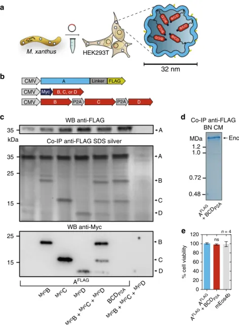
Encapsulin expression and self-assembly. Based on the favorable
set of features introduced above, we chose to heterologously
overexpress the encapsulin shell protein from M. xanthus in
HEK293T cells. We tagged the nanoshell with an outward facing
FLAG epitope (A
FLAG) and found it to express strongly without
and with the native cargo molecules from M. xanthus, denoted
encapsulins B, C, and D
31.
Co-expression of Myc-tagged B, C, or D alone, or a
combination of all three non-tagged proteins (via co-transfection
or a P2A construct, Fig.
1
b), co-immunoprecipitated with A
FLAGas visualized on silver-stained SDS-PAGE (Fig.
1
c, middle panel).
A corresponding western blot against the FLAG (Fig.
1
c, upper
panel) or Myc-epitope (Fig.
1
c, lower panel) confirmed the
identities of the protein bands (A
FLAG: 32.9 kDa,
MycB: 18.5 kDa,
MycC: 15.4 kDa,
MycD: 12.5 kDa).
Furthermore, a corresponding Blue Native PAGE (BN-PAGE)
of immunoprecipitated FLAG-tagged material from cells
expres-sing A
FLAGtogether with BCD
P2Arevealed a band with an
apparent molecular weight of above 1.2 MDa indicating
self-assembly of encapsulin protein complexes and self-targeting of all
native cargo proteins (Fig.
1
d).
The strong expression of A
FLAGwithout or with loaded cargo
did not result in a reduction of cell viability when compared to
cells overexpressing a
fluorescent protein as assessed by a viability
assay based on lactate dehydrogenase (LDH) release (Fig.
1
e).
We also generated a construct for a StrepTagII-labeled variant
of the shell that co-expresses the ferritin-like Myc-tagged C as
cargo protein via a scarless P2A site
41(
MycC-
IntP2A-A
STII, Fig.
2
a).
Material from HEK293T cells, conveniently purified via
Strep-Tactin affinity chromatography, showed assembled nanospheres
of 32.4 ± 1.7 nm as the major component in single particle
cryo-electron microscopy (cryo-EM) (Fig.
2
b, Supplementary Fig.
1
a,
b), corresponding to the single band >1.2 MDa in size on
BN-PAGE (Fig.
2
c, right panel). Again, no effect on cell viability was
detected for this construct tested by a luciferase-based viability
assay compared to A
FLAGwith and without cargo (BCD
P2A), as
well as controls without expression of encapsulins (EYFP and
untransfected HEK293T) (Fig.
2
d). Furthermore, N-terminal
addition of the human BM40 (osteonectin SPARC) secretory
signal peptide (SP) to the StrepTagII-modified encapsulin shell
protein resulted in entry into the secretory pathway and robust
a
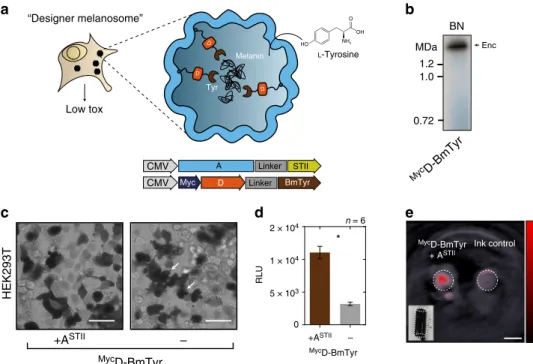
b
kDa A B C D 35 25 15Co-IP anti-FLAG SDS silver
A 35 WB anti-FLAG B C D AFLAG
MycB MycC MycD
MycB + MycC + MycD MycB + MycC + MycD BCD P2A 25 15 WB anti-Myc
c
d
AFLAG + BCD P2A n = 4 mEos4b 0 20 40 60 80 100 120 % cell viability AFLAG AFLAG + BCD P2A 0.48 1.2 1.0 0.72 MDa Enc Co-IP anti-FLAG BN CM A Linker FLAG CMV P2A Myc P2A B, C, or D CMV C B D CMV32 nm
HEK293T B/C/D B/C/D B/C/D B/C/D B/C/D B/C/D M. xanthuse
nsFig. 1 Assembly of encapsulins and targeting of cargo in HEK293T cells. a Schematic of the heterologous expression of surface-modified encapsulin variants loaded with endogenous cargo proteins.b Genetic constructs encoding the shell protein A (light blue) with a FLAG-tag as C-terminal surface modification as well as individual Myc-tagged cargo proteins (red) B, C, and D that can also be combined in a multi-gene expression construct (BCDP2A).c Co-immunoprecipitation of AFLAGand silver-stained SDS-PAGE from cells co-expressing just B, C, or D, or a combination of these three proteins expressed either via a mixture of individual DNA constructs (MycB+MycC+MycD), or by a multi-gene expression construct (BCD
P2A). The top panel shows a western blot (WB) against the exterior FLAG-tag in AFLAG. The bottom panel shows the corresponding WB against the Myc epitope.d Coomassie-stained Blue Native PAGE (BN CM) of purified material from HEK293T expressing AFLAGand BCDP2Ayielding a band above 1.2 MDa.e Cell viability after 48 h of overexpression of encapsulins (AFLAG) with or without cargo (BCD
P2A) assessed by an LDH release assay. A construct expressing thefluorescent protein mEos4b served as a control. The bars represent the mean ± SEM (p= 0.1965, Kruskal–Wallis, n = 4; no significant (ns) differences at α = 0.05 were found in Dunn’s multiple comparisons test between mEos4b and AFLAGexpressed without or with BCDP2A)
secretion of StrepTagII-modified encapsulins from HEK293T
cells as shown by Coomassie-stained BN-PAGE of material
present in the cell culture supernatant (Fig.
2
e).
In vivo expression of encapsulins. To achieve in vivo expression
of encapsulins, we generated a coexpression construct that
encoded both the nanoshell A
FLAGand the ferritin-like protein B
from a single plasmid that was small enough to be packaged into
an Adeno-associated virus (AAV) (Supplementary Fig.
1
c). After
transduction of murine brains via intracranial injections of this
viral vector co-expressing A
FLAGand B
M7by a P2A peptide, we
observed robust neuronal expression of the shell protein
(Sup-plementary Fig.
1
f, i). Silver-stained BN-PAGE and SDS-PAGE of
immunoprecipitated (anti-FLAG) proteins extracted from murine
brain showed that the nanocompartments assembled in vivo and
that the cargo B
M7was associated with the shell (Supplementary
Fig.
1
g, j). Similar in vivo results could be obtained by
co-expressing the nanoshell and ferritin-like B cargo via an IRES site
(Supplementary Fig.
1
h).
Encapsulation of engineered cargo. We next tested whether
non-natural cargo molecules could be efficiently targeted into the
nanocompartments. We thus C-terminally appended a minimal
encapsulation signal, which we found to only necessitate eight
amino acids (EncSig), to the photoactivatable
fluorescent protein
mEos4b
42, coexpressed it with A
FLAGand found by
co-immunoprecipitation and BN-PAGE analysis under an UV
imager that the cargo readily associated with the encapsulin shell
(Fig.
3
b).
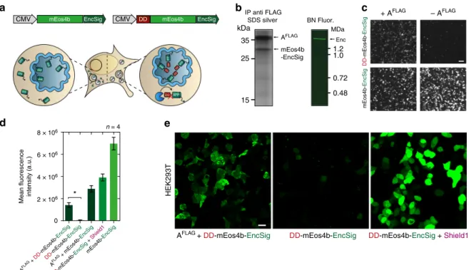
Selective degradation of non-encapsulated cargo proteins.
Importantly, we could also selectively enrich cargo proteins to the
encapsulin lumen by fusing an FKBP12-derived destabilizing
domain (DD) that labels the cargo for rapid degradation unless it
is shielded from proteasomal machinery
43. We show that
co-expressing A
FLAGand DD-mEos4b-EncSig in HEK293T yielded
significantly higher mean fluorescence values than
DD-mEos4b-EncSig alone, indicating that cargos inside the encapsulin are
protected from proteolytic degradation (Fig.
3
c, d). Confocal
microscopy revealed that coexpression of DD-mEos4b-EncSig
with A
FLAGshows green
fluorescence throughout the cytosol but
not in the nucleus, whereas the absence of the encapsulin shell
ablated the
fluorescence signal. In a positive control in which
DD-mEos4b-EncSig was stabilized by adding a small molecule instead
of encapsulating it,
fluorescence was observed throughout the cell
including the nucleus (Fig.
3
e).
We then purified encapsulins co-expressed with and without
DD-mEos4b-EncSig as cargo to determine their native mass and
found that in the absence of cargo, also smaller nanospheres
assembled consistent with the known configuration as 60-mers
with T
= 1 symmetry (Supplementary Fig.
2
). We estimated that
on average ~60
fluorescent proteins per nanoshell were enclosed
as confirmed by gel densitometry (Supplementary Fig.
3
a, b). We
furthermore found that the FLAG-tagged encapsulin shell is
phosphorylated (Supplementary Fig.
3c-e
).
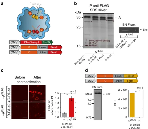
Simultaneous encapsulation of sets of engineered cargo. We
next wanted to assess whether multiple engineered cargo
mole-cules could be encapsulated together. We thus fused the two
halves of split PAmCherry1 (PA-s1, PA-s2) to either B or C
(B-PA-s2: 27.0 kDa, C-PA-s1: 33.1 kDa) and tested for
bimole-cular
fluorescence complementation (BiFC) within the
nano-compartment
44(Fig.
4
a, b). Either of these components could
be co-immunoprecipitated with A
FLAGas shown by silver-stained
SDS-PAGE (Fig.
4
b, Supplementary Fig.
4
a). The photoactivation
of the complemented split PAmCherry1 inside the encapsulins
could also be detected via
fluorescence imaging of the
corre-sponding BN-PAGE (Fig.
4
b, right panel, Supplementary Fig.
4
a).
Co-expression of both split halves together with A
FLAGlead to a
strong increase of photoactivatable
fluorescent signal throughout
the cytosol of HEK293T cells as quantified by confocal
micro-scopy compared to cells that did not express A
FLAG(Fig.
4
c).
a
b
1.2 1.0 0.72 0.48 MDa Enc Co-IP anti-STII BN CM Myc C-IntP2A-ASTII cryoEM BM40 A STII P2A Myc CMV C DnaE A Splicing STII STII A Linker BM40 CMV Secreted 1.2 1.0 MDa Enc BN CM 0 5 × 10⁴ 1 × 10⁵ 2 × 10⁵ RLU AFLAG + B CDP2A Myc C-IntP 2A -A STII Myc C-IntP2A -A STI I AFLAG EYF P ControlH 2O 2 n = 6 A 25 15 35 C kDa Co-IP anti-STII SDS silverd
c
e
nsFig. 2 Combined encapsulin:cargo construct and secreted encapsulin variant.a Scheme of a P2A bicistronic expression construct encoding StrepTagII-tagged (STII) nanocompartments containing Myc-tagged C as cargo protein (MycC-IntP2A-ASTII) as well as a variant with an N-terminal BM40 secretion peptide and StrepTagII (STII).b Cryo-electron microscopy image of material from HEK293T cells expressingMyc
C-IntP2A-ASTIIpurified via Strep-tag II/Strep-Tactin XT affinity chromatography showed the assembled nanospheres of ~32 nm diameter. Scale bar is 100 nm.c The corresponding BN-PAGE analysis of the identical material revealed a single band larger than 1.2 MDa. The accompanying silver-stained SDS-PAGE showed the coprecipitation of the cargoMycC with the StrepTagII-modified nanoshell.d Luciferase-based cell viability assay after 48 h of
overexpression ofMycC-IntP2A-ASTIIand AFLAGwith or without cargo BCDP2A. Cells overexpressing thefluorescent protein EYFP as well as untransfected HEK293T cells served as negative controls. To induce toxicity as positive control, untransfected HEK293T cells were treated with 1 mM H2O224 h prior to the assay. The bars represent the mean ± SEM (p = 0.442 excluding the positive control, Kruskal–Wallis, n = 6; no significant (ns) differences atα = 0.05 were found in Dunn’s multiple comparisons test between any of the encapsulin:cargo conditions and either EYFP or Control).e BN CM loaded with cell culture supernatant of HEK293T cells expressing ASTIIwith an N-terminal BM40 secretion signal showed a single band >1.2 MDa
Compartmentalized enzymatic reactions. To showcase the use
of the eukaryotically expressed encapsulins as bioengineered
reaction chambers with pores that can constrain passage of
reactants and reaction products, we targeted several enzymes to
the nanocapsules. In the presence of A
FLAG, the split luciferase
45parts LgBit and SmBit fused to C and B (C-LgBit: 32.7 kDa,
B-SmBit: 19.6 kDa) were complemented to functional enzymes as
demonstrated by bioluminescence detection from BN-PAGE
(Fig.
4
d, left) and from total lysate (Fig.
4
d, bar graph on the
right). Importantly, only very low luminescence signals were
detected when the encapsulin shell was not present indicating
that using split protein approaches can also ensure confined
enzyme activity inside the capsules, in addition to the strategy for
selective enrichment of cargo inside the nanocompartment as
shown in Fig.
3
.
Bioengineered melanosomes as gene reporters for MSOT. We
subsequently sought to utilize selective passage of small substrates
through the nanoshell to load the compartments with tyrosinase
as cargo which is the sole enzyme generating the photoabsorbing
polymer melanin from the amino acid tyrosine. Because of these
attractive features, tyrosinase has been used as a gene reporter for
optoacoustic tomography
46,47, an imaging modality that maps the
distribution of photoabsorbing molecules in tissue by locating the
ultrasonic waves that they emit in response to local heating upon
laser absorption
48,49. However, melanin production is toxic to
cells if not confined in melanosomes, which are membranous
compartments of specialized cells
50,51. We thus chose a soluble
tyrosinase from Bacillus megaterium
52that we thought could still
be functional as a fusion protein to the native cargo D (
MycD-BmTyr: 47.7 kDa) serving as targeting moiety (Fig.
5
a). Indeed we
could observe generation of melanin on the BN-PAGE band
corresponding to the assembled nanocompartment (Fig.
5
b). In
cells expressing the encapsulin-targeted tyrosinase and the
shell A
STII, we observed robust melanin formation by bright-field
microscopy without the strong toxicity apparent in the
mor-phology of control cells expressing just the tyrosinase (Fig.
5
c,
white arrows). Encapsulation of the tyrosinase also led to a
sig-nificant increase in cell viability as assessed by a luciferase-based
viability assay (Fig.
5
d). Cells expressing melanin-producing
encapsulins were dark in color (Fig.
5
e, inset) and thus
gen-erated intense photoacoustic signal even when referenced against
strongly absorbing synthetic ink with an optical density of 0.2
(Fig.
5
e).
Similarly, we showed that the engineered peroxidase APEX2
53can polymerize Diaminobenzidine (DAB) when targeted to the
nanocompartment (APEX2-EncSig; 31.0 kDa) as indicated by
the generation of photoabsorbing DAB polymers associated with
the BN-PAGE band corresponding to the assembled nanosphere
(Supplementary Fig.
4
b).
mEos4b EncSig CMV kDa 35 25 15 AFLAG mEos4b -EncSig IP anti FLAG SDS silver Enc BN Fluor. 1.2 1.0 MDa 0.72 0.48 Mean fluorescence intensity (a.u.) AFLAG + DD -mEos4b-EncSig AFLAG + mEos4b-EncSig mEos4b-EncSig DD -mEos4b-EncSig DD -mEos4b-EncSig + Shield1 2 × 10⁶ 0 n = 4 4 × 10⁶ 6 × 10⁶ 8 × 10⁶ *c
a
b
DD -mEos4b-EncSig mEos4b-EncSig + AFLAG – AFLAGd
e
mEos4b DD EncSig CMVAFLAG + DD-mEos4b-EncSig
HEK293T
DD-mEos4b-EncSig DD-mEos4b-EncSig + Shield1
XFP XFP XFP XFP XFP XFP XFP XFP XFP XFP XFP XFP XFP XFP XFP DDXFP XFP DD XFP DD XFP DD XFP DD XFP DD XFP DD XFP DD XFP DD
Fig. 3 Selective degradation of non-encapsulated cargo. a Schematic of genetic construct showing a minimal C-terminal encapsulation signal (EncSig) fused to the photoactivatablefluorescent protein mEos4b (mEos4b-EncSig) to associate it to the inner surface of the nanocompartment. When mEos4b-EncSig is N-terminally fused to an FKBP12-derived destabilizing domain (DD), it is degraded by the proteasome unless it is sequestered into the encapsulin shell. This strategy thus selectively enriches cargo inside the lumen of the nanocompartment.b Cargo loading of mEos4b-EncSig into the nanocompartment composed of AFLAGwas demonstrated by co-immunoprecipitation (Co-IP) against the FLAG epitope followed by silver-stained SDS-PAGE (left panel). Corresponding analysis of whole cell lysate by BN-PAGE on a UV imager showsfluorescence of the native encapsulin band indicating the presence of the mEos4b-EncSig cargo.c Representative 2 × 2 table of epifluorescence microscopy images from HEK293T cells co-expressing DD-mEos4b-EncSig with or without AFLAG(upper row) compared to coexpression of the cargo without destabilizing domain (mEos4b-EncSig, lower row). Scale bar represents 50µm. d Corresponding quantification of the fluorescence intensities as exemplified in c. Co-expression of AFLAGsignificantly protects degradation of DD-mEos4b-EncSig (p= 0.0286, Mann–Whitney test, n = 4 biological replicates, error bars represent mean ± SEM). DD-mEos4b-EncSig can also be stabilized by adding the small molecule Shield1 (0.5 mM, magenta label) to the cell culture medium.e Confocal microscopy images of HEK293T expressing DD-mEos4b-EncSig with or without AFLAG. As a reference DD-mEos4b-EncSig was stabilized via the addition of 0.5 mM Shield1. Please note that the contrast of all images was linearly adjusted to the same extent optimizing for the condition shown on the left, which resulted in partial oversaturation of the condition shown on the right. Scale bar represents 20µm
Since the electrophoretic mobility of protein complexes on
BN-PAGE also depends on their hydrodynamic size and shape
54,
cargo-loading could be confirmed by observing an identical
migration behavior of loaded as compared to unloaded capsules
(Supplementary Fig.
4
c).
Moreover, we targeted the putative cystathionine
γ-lyase
(SmCSE) to the nanospheres via an EncSig (smCSE-EncSig:
43.9 kDa) as shown by Co-IP with A
FLAG(Supplementary
Fig.
4
d). In the presence of
L-cysteine, this enzyme was reported
to catalyze a conversion of cadmium acetate in aqueous
solution into cadmium sulfide (CdS) nanocrystals such that
they would generate a photoluminescence signal under UV
illumination characteristic for crystal formation at quantum
confined sizes
55. Indeed, we could detect a photoluminescence
signal from the BN-PAGE band corresponding to encapsulin
loaded with SmCSE-EncSig after on-gel incubation with
cad-mium acetate and
L-cysteine indicating that the smCSE-EncSig
cargo was enzymatically active when bound into the shell
(Supplementary Fig.
4
d).
Size-constrained iron biomineralization. Another reason to
choose encapsulins from M. xanthus was that it was previously
reported to deposit iron via the ferritin-like cargo B and C
into relatively large compartments (~32 nm, T
= 3)
31. We thus
investigated whether this functionality could also be realized
in eukaryotic cells to enable spatially confined iron deposition
sequestered
away
from
the
complex
signaling
network
controlling mammalian iron homeostasis. We thus generated a
stable cell line co-expressing the nanoshell (A
FLAG) with all
native cargo proteins (B,C,D) via a dual-promoter construct
(Fig.
6
a). In this cell line, we observed long-term and robust
expression of all components shown by co-immunoprecipitation
with A
FLAGand by immunocytochemistry against the external
FLAG-epitope (Fig.
6
b, left panel, Supplementary Fig.
5
a).
Transient
co-expression
of
the
ferrous
iron
transporter
MmZip14
FLAG(Zip14) in the stable cell line resulted in a
robust dose-dependent iron loading (with ferrous ammonium
sulfate (FAS) at concentrations between 0.25–1.25 mM) already
after 48 h of supplementation as detected on BN-PAGE via
SmBit B B C C LgBit EncSig PAmCherry1 CMV Linker PA-s2 CMV B Linker PA-s1 CMV C A kDa 35 25 15 AFLAG PAmCherry1-EncSig C-PA-s1 B-PA-s2 IP anti FLAG SDS silverd
Enc w/o cargo B-PA-s2 C-PA-s1 B-P A-s2 + C-P A-s1 C-PA-s1 + B-P A-s2 PAmCher ry1– EncSig BN Fluor. AFLAG Before After B-P A-s2 +C-P A-s1 +A FLA G –A FLA G photoactivation + C-PA-s1 B-PA-s2 +AFLAG – +AFLAG – 1.0 1.1 1.2 1.3 1.4 1.5 Fluor . int. after / bef ore P A n = 3 * Linker SmBit CMV B Linker LgBit CMV C 1.2 1.0 MDa 0.72 BN Lum. Enc B-SmBit 0 2 × 10⁶ 4 × 10⁶ 6 × 10⁶ RLU n = 3c
a
b
S1 S2 B C * * ** *** ** *** * + C-LgBitFig. 4 Multi-component processes and enzymatic reactions can be targeted to encapsulins in mammalian cells. a Overview schematic of sets of cargo molecules for bimolecularfluorescence and enzyme complementation inside the nanocompartment. Targeting of foreign cargo proteins can be achieved either via a minimal C-terminal encapsulation signal (EncSig) or via C‑terminal fusions to the native cargo proteins B, C, or D. b Silver-stained SDS-PAGE from a co-immunoprecipitation (Co-IP) of AFLAGco-expressed with photoactivatable mCherry1 with EncSig (PAmCherry1-EncSig) or with either one of the halves of split PAmCherry1 fused to C or B, or a combination of both (C-PA-s1+ B-PA-s2). Fluorescence originating from complemented split PAmCherry1 inside the encapsulins was detected on BN-PAGE loaded with whole cell lysates of cells expressing AFLAGand C-PA-s1+ B-PA-s2 after 2 min of photoactivation (PA) on an UV imager.c Live cell confocal microscopy images (scale bar represents 20µm) of HEK293T cells expressing B-PAs1 and C-PAs2 with or without the shell-protein AFLAGbefore and after 60 s of photoactivation (PA) with 405 nm (upper panel) demonstrating efficient bimolecular fluorescence complementation inside encapsulin compartments. Fluorescence of photoactivated split PAmCherry1 was excited using a 561 nm laser. Fluorescence signals of the sample without and with AFLAGwere quantified by calculating the ratio of the mean signal after PA divided by the signal before PA. The bars in the lower panel represent the meanfluorescence intensity ratios averaged over independent transfection experiments ± SEM (p = 0.0123, unpaired t-test, n= 3). d Luminescence signal from BN-PAGE incubated with luciferase substrate and loaded with whole cell lysates of HEK293T co-expressing split luciferase fragments fused to either B or C (B-SmBit, C-LgBit) and AFLAG(left panel). The luminescent band corresponds to the complemented split luciferase inside the assembled nanocompartment. The bar graph (right panel) shows the corresponding total luminescence signals from the cell lysates expressing B-SmBit and C-LgBit with or without AFLAG, (mean ± SEM across three independent transfection experiments, in each experiment three technical replicates were averaged, p < 0.0001, unpaired t-test, n= 3)
DAB-enhanced Prussian Blue staining (DAB PB) (Fig.
6
b, right
panel).
Efficient iron loading could also be achieved by transient
expression of A
FLAG+ BCD
P2Atogether with Zip14. Under these
conditions, iron supplementation with ~0–3 mM FAS for 48 h led
to a substantial dose-dependent iron loading of the
nanocom-partment that saturated at ~1 mM FAS as shown by Coomassie
and DAB-enhanced Prussian Blue BN PAGE (Fig.
6
c, upper
panel, Supplementary Fig.
5
b). Interestingly, when we tested the
cargo molecules individually for their ability to load iron into the
nanosphere, we found that co-expression of only B or C
generated equally intense DAB PB bands as compared to
BCD
P2A, indicating that either B or C is sufficient for iron
deposition inside the nanocompartment.
In contrast, co-expression of D with A
FLAGor any of the cargo
molecules without the presence of A
FLAGdid not lead to
discernable DAB PB signals (Fig.
6
c, lower panel, Supplementary
Fig.
5
c). In a standard cell viability assay, we found no
impairment of the cells when Zip14 was co-expressed together
with A
FLAGand BCD
P2Aor just B. However, ~7% of cells showed
reduced viability when the cargos BCD
P2Awere expressed
without the nanocompartment (p
= 0.0238, Mann Whitney test,
n
= 3) or when only the fluorescent protein mEos4b-EncSig was
expressed (Supplementary Fig.
5
d) indicating that in the absence
of the nancompartment the imported iron was not sufficiently
sequestered by the endogenous iron homeostasis machinery.
We furthermore tested variants of A with N-terminal fusions
with peptide sequences from Magnetospirillum magneticum Mms
(6 and 7) proteins reported to aid in templating iron
mineraliza-tion
56but found no additional benefit of these modified inner
surfaces over A
FLAGusing our current readout (Supplementary
Fig.
5
e). In addition, we analyzed several variants of the cargo
proteins B and C, fused C-terminally to peptides from Mms
proteins (superscripts M6, M7, please see Supplementary Fig.
5
f).
These data confirmed that either B or C are sufficient to load the
nanocompartment with iron and showed that no obvious
additional iron loading resulted from the presence of the Mms
peptides.
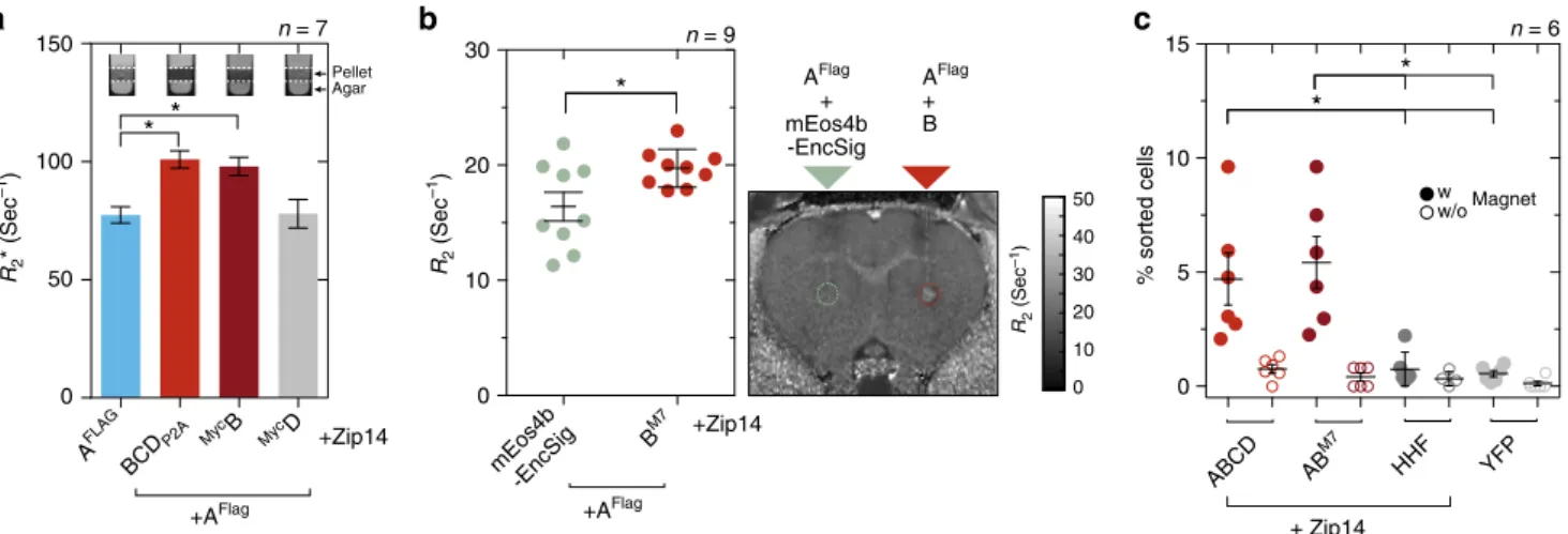
Encapsulins enable detection via MRI and magnetic sorting.
Next, we were interested in whether the strong iron accumulation
inside eukaryotically expressed encapsulin shells would yield
significant contrast by MRI. We thus expressed A
FLAGalone or
together with either all native cargos BCD
P2Aor just
MycB, or
MycD and Zip14 and subjected cell pellets to relaxometry
mea-surements by MRI. The nanocompartment A
FLAGco-expressed
with all native cargo proteins (BCD) lead to a significant increase
in R
2*-relaxation rates as compared to just A
FLAG. The same
effect was observed by co-expressing just the ferritin-like B
(Fig.
7
a, p
= 0.0047, Kruskal–Wallis with significant differences
at
α = 0.05 from Dunn’s multiple comparisons test vs. A
FLAG,
a
b
c
d
e
Low tox “Designer melanosome” Tyr Melanin L-Tyrosine D D D A Linker Linker STII BmTyr Myc D CMV CMV BN Enc MycD-BmTyr 1.2 1.0 MDa 0.72 n = 6 HEK293T MycD-BmTyr +ASTII – MycD-BmTyr 0 5 × 10³ 1 × 10⁴ 2 × 10⁴ RLU +ASTII – * MycD-BmTyr + ASTII Ink control O OH HO NH2Fig. 5 Bioengineering of a melanosome by targeting melanin-generating tyrosinase to the encapsulin compartment. a Schematic of the detoxifying effects of compartmentalized melanin production by encapsulated tyrosinase from Bacillus megaterium targeted to the nanocompartment via fusion to the native cargo D. The substrateL-tyrosine enters the compartment via the pores in the nanoshell.b BN-PAGE showing on-gel production of melanin via tyrosinase expressed in HEK293T cells fused to Myc-tagged encapsulin-cargo D (MycD-BmTyr) to encapsulate it in the assembled nanoshell. Dark colorization of the band was observed after incubation with 2 mML-tyrosine and 100µM CuCl2in PBS (pH 7.4) for 1 h at 37 °C.c Bright-field images of HEK293T cells expressingMycD-BmTyr with and without StrepTagII-modified shell (ASTII) after 48 h of expression. Twenty four hours post transfection, cells were supplemented with 1 mML-tyrosine and 10µM CuCl2. Cell protrusions (white arrows) were apparent indicating toxic effects of overexpression of non-encapsulated tyrosinase. Scale bar: 20µm. d Corresponding luciferase-based viability assay of HEK293T cells treated as in c overexpressingMycD-BmTyr with or without ASTIIafter 48 h. (The bars represent the mean ± SEM, n= 6, p < 0.0001, unpaired t-test.) e Images of two tubular phantoms (transversal slice) obtained by multispectral optoacoustic tomography (MSOT). The phantoms werefilled with ~107cells in 1.5% low melting agar expressingMyc D-BmTyr with ASTII(supplementation as inc and d) or containing highly concentrated ink (OD= 0.2) as control showing the intense contrast obtained between 690 nm and 900 nm from the melanin-producing encapsulins. The coefficients obtained from linear unmixing of the optoacoustic spectra with a melanin reference spectrum are displayed on the red colormap overlaid on the image obtained at 720 nm. The lower left inset shows a color photograph of the tubular phantom containing the cells. Scale bar: 3 mm
AFLAG CM + Zip14 CM DAB PB 0 0.25 0.75 1.0 2.0 3.0 mM FAS + Zip14 AFLAG + BCD P2A B/C/D B/C/D B/C/D B/C/D B/C/D B/C/D Zip14 CMV FLAG Zip14 A Linker FLAG CMV P2A C P2A B D CMV DAB PB 35 25 15 35 10 kDa A C D B WB
anti FLAG Enc
+ Zip14 1.25 0.25 mM FAS 48 h + Zip14 1.25 0.25 mM FAS 48 h 1.2 1.0 0.72 MDa CM DAB PB MycD MycC Myc B Myc B MycC MycD BCD P2A
a
b
c
Fig. 6 Efficient iron loading of eukaryotically expressed encapsulin nanospheres a Schematic of a dual-promoter construct used for generation of a stable cell line expressing AFLAGand all native cargos B, C, and D. Also depicted is a construct encoding the iron-transporter MmZip14FLAG(Zip14) used to transport additional amounts of iron into the cell.b Co-immunoprecipitation (Co-IP) against the FLAG epitope from a whole cell lysate of a stable HEK293T clone expressing AFLAGtogether with B, C, and D analyzed by silver-stained SDS PAGE and the corresponding WB against the FLAG epitope (left panel). The pair of Blue Native (BN) gels visualizes proteins from whole cell lysates via Coomassie staining (CM) (left panel) and iron content via treatment with DAB enhanced Prussian Blue (DAB PB) (right panel) from the same stable cell line. Robust iron loading of the assembled nanocompartments was achieved by transient co-expression of MmZip14FLAG-IRES-ZsGreen1 in which case 0.25 mM ferrous ammonium sulfate (FAS) for 48 h was sufficient to see strong iron loading.c BN gel stained with CM or DAB PB loaded with whole cell lysates of HEK293T cells transiently expressing AFLAG+ BCD
P2Aand Zip14FLAG supplemented with different concentrations of FAS (0–3 mM) for 48 h (upper panel). The strong bands, which correspond to the assembled nanoshell, indicate high expression levels of encapsulins and efficient, dose-dependent iron loading. The lower panel shows a CM and DAB PB-stained BN gel from whole cell lysates of HEK293T cells expressing Zip14FLAGand different combinations of native cargo molecules:MycB,MycC, andMycD alone, or all three (BCDP2A) with or without AFLAG. The robust DAB PB stains show that the ferritin-like cargo proteins B or C are sufficient for iron loading into encapsulins. FAS was supplemented at 2.5 mM for 48 h
+Zip14 +AFlag R2 * (Sec –1) 50 100 150 0
*
*
n = 7 Agar Pellet 0 5 10 15 % sorted cells*
Magnet w w/o + Zip14*
n = 6 R2 (Sec –1) +Zip14 +AFlag BM7 AFlag + B AFlag + mEos4b -EncSig 0 10 20 30 40 50 R2 (Sec –1 ) 0 10 20 30*
n = 9 MycD MycB AFLAG BCD P2A mEos4b -EncSig AB HHF YFP M7 ABCDa
b
c
Fig. 7 Iron-filled encapsulins enable detection by MRI and magnetic cell separation. a Relaxometry measurements by MRI conducted on cell pellets (~107 cells) from HEK293T cells transiently expressing AFLAG+ BCDP2A,MycB, orMycD, or AFLAGalone (1 mM FAS for 24 h and expression of Zip14FLAG). Expression of AFLAGwith BCDP2Aor withMycB showed a significantly enhanced R2*-relaxation rate as compared with AFLAGalone or loaded withMycD; ferritin-like cargo B was sufficient to generate an increase in R2*in the presence of the AFLAGnanocompartment (p= 0.0047, Kruskal–Wallis, n = 7 from four independent experiments, stars indicate significance at α = 0.05 from Dunn’s multiple comparisons test vs. AFLAG; the bars represent the mean ± SEM). The insets show MRI slices (13.5 ms echo time) through test tubes in which cells were pelleted on a layer of agar.b In vivo MRI detection of HEK293T cells transiently co-expressing AFLAGtogether with ferritin-like BM7that were xenografted into rat brains. As compared to cells co-expressing AFLAGtogether with thefluorescent protein mEos4b-EncSig as control cargo, we observed significantly increased transverse relaxation rates (p = 0.0078, Wilcoxon matched-pairs signed rank test, n= 9) measured at the injection site for AFLAG+ BM7expressing cells 24 h post injection. The horizontal lines represent the mean ± SEM. The image on the right shows a coronal R2map through a rat brain with the regions of interest (ROIs) defined over the injection sites by dashed circles.c HEK293T cells were co-expressing AFLAGand BCDP2A, AFLAGand BM7, or human H-chain ferritin (HHF) as a control together with Zip14 and were treated with 2.5 mM FAS for 48 h. Additional control cells were expressing only EYFP. Independent cell suspensions were subsequently sorted on commercial magnetic separation columns inside and outside the magneticfield to control for unspecific retention in the mesh of the column. The fraction of cells separated in the magneticfield for both encapsulin:cargo conditions was significantly higher than for any of the control conditions HHF + Zip14 or YFP (p= 0.0007, Kruskal–Wallis, n = 6 from three independent experiments, stars indicate significance at α = 0.05 from Dunn’s multiple comparisons test across all conditions with magneticfield; the horizontal lines represent the mean ± SEM)
n
= 7). This indicated again that co-expression of B was sufficient
to generate efficient iron deposition inside the nanoshell.
We subsequently sought to test whether cells genetically
labeled with encapsulins could be detected by MRI in vivo. As
an initial assessment, we thus xenografted cells co-expressing
A
FLAGtogether with B
M7into rat brains and obtained R
2-relaxation maps that showed elevated -relaxation rates (p
= 0.0078,
Wilcoxon matched-pairs signed rank test, n
= 9) at the injection
site as compared to xenografted cells in which the
fluorescent
protein mEos4b-EncSig was used as a control cargo (Fig.
7
b).
In addition to MRI contrast, the iron biomineralization inside
the encapsulins also allowed us to magnetically sort cells
co-expressing the shell A
FLAGwith BCD
P2Aor with B
M7at
significantly higher percentages than when human H-chain
ferritin (HHF) was expressed or just yellow
fluorescent protein
(EYFP) (p
= 0.0007, Kruskal–Wallis, with significant differences at
α = 0.05 from Dunn’s multiple comparisons test, n = 6; Fig.
7
c).
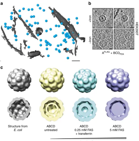
Encapsulins as markers for electron microscopy. Given that the
iron loading of the encapsulins was very efficient and observable
at the population level, we next assessed how well individual
nanocompartments could be detected by electron microscopy in
cells such that they could be used as genetically encoded markers.
We thus grew HEK293T cells stably expressing the shell protein
A
FLAGand BCD
P2Ausing a dual promoter vector on a
trans-mission electron microscopy (TEM) grid, vitrified them by
plunge-freezing and produced lamellae by cryo-focused ion beam
(cryo-FIB) milling for in situ cellular cryo-electron tomography
(cryo-ET). The heterologously expressed encapsulins were readily
detected as clearly discernible nanospheres (Fig.
8
a,
Supplemen-tary Fig.
6
a, b) that exhibited electron-dense cores when we
supplemented the growth media with ferrous iron (Fig.
8
b) and
were distributed as monodisperse spheres throughout the cytosol
(Supplementary Fig.
6
c, d). The electron density maps showed a
high similarity to the structure published from encapsulin shells
from M. xanthus expressed in E. coli (pdb 4PT2; EMDataBank
EMD-591728, Fig.
8
c). The clipped views from the encapsulins
(blue) furthermore show electron densities associated with
docked cargo proteins and most likely biomineralized iron as
compared with the inner surface of the shell from E. coli (gray)
that was mapped in the absence of any cargo (Fig.
8
c, lower row).
These data demonstrate that the spherical shape and high,
non-toxic expression levels make encapsulin very attractive as fully
genetically expressed markers for EM.
Discussion
In summary, we genetically controlled multifunctional orthogonal
compartments in mammalian cells via expressing N- or
Structure from E. coli ABCD untreated ABCD 0.25 mM FAS + transferrin ABCD 5 mM FAS Iron loading +Iron –Iron HEK293T AFLAG + BCDP2A
a
b
c
Fig. 8 Encapsulins as genetically encoded markers for cryo-electron tomography (Cryo-ET). a Cryo-ET data from HEK293T cells stably expressing encapsulins together with native ferritin-like cargo proteins (using the dual promoter construct AFLAG;BCDP2Ashown in Fig.6a). 3D rendering showing encapsulins in blue and membranes in gray colors. Scale bar:100 nm.b Example slices from tomograms show encapsulins with and without electron-dense cores from iron-accumulation (treatment with 5 mM FAS for 48 h prior to vitrification). Scale bars:20 nm. c In situ structures (displayed as 2× binned) derived from cryoelectron tomography of nanocompartments assembled in HEK293T cells without (beige), with 0.25 mM FAS and 1 mg/ml human transferrin (cyan) and 5 mM FAS (purple) as compared with the published structure shown in gray (pdb 4PT2; EMDataBank EMD-5917) that was obtained from M. xanthus EncA expressed in E. coli31. The cutaway views of the encapsulins show electron densities indicating the presence of cargo proteins (beige) and additional iron deposition (cyan and purple) as compared to published data from the EncA shell31that were obtained in the absence of cargo proteins
C-terminally modified encapsulins, which we found to
auto-assemble into abundant nanocompartments which readily
encapsulated sets of natural and engineered cargo proteins and
enabled size-constrained metal biomineralization.
The efficiency of self-targeting and auto-packaging of the
various cargo proteins in mammalian cells was remarkable given
that the number of possible protein interactions is even a few-fold
higher than in the original prokaryotic host organism
57,58. We
found that about 60 cargo proteins of a canonical
fluorescent
protein can be bound to the inner encapsulin surface via the
minimal encapsulation signal. Higher loading factors could be
achieved by providing cargo proteins with multidentate adapters
such that the entire encapsulin volume could be
filled. We
fur-thermore observed that without co-expression of endogenous or
engineered cargo, the abundance of the 60-mer encapsulin shell
with T
= 1 symmetry was increased, which is in line with a
previous observation made from M. xanthus encapsulin
expres-sed in E. coli
31and suggests that encapsulation of cargo leads to
the preferred assembly of the 180-mer in T
= 3 symmetry.
Also, iron storage inside the capsule via the ferritin-like
enzymes B or C targeted to the encapsulins was very efficient,
indicating that there was sufficient access to ferrous iron.
Whereas encapsulins heterologously expressed in E. coli were
shown to load iron
59, this could so far not been shown in
mammalian cells. We also found that just co-expression of B (or
C) with A is sufficient for robust iron storage such that a
single-piece reporter construct of just ~2.1 kb in size can be used.
In the context of optimizing T
2contrast in MRI, it would
certainly be valuable to explore modifications of the outer surface
that may control the agglomeration state and thus could
mod-ulate the apparent relaxivity of encapsulin ensembles
60. In this
context, it would also be desirable to explore capsid architectures
with more storage capacity such as ones with T
= 7
quasisym-metry known from bacteriophage HK97
31,61. Furthermore,
modifications of the inner surface of the shell may be engineered
and/or additional cargo could be designed that could facilitate the
nucleation process to support higher iron packing densities or
alter environmental parameters (e.g., pH and redox potential) to
potentially even generate superparamagnetic iron-oxides which
possess a substantially larger magnetization
62. Iterative
optimi-zation schemes such as directed evolution could also be employed
based on rescue assays from excess iron or magnetic microfluidic
sorting and could also be complemented by parallel screens in
prokaryotes if enough iron-influx can be achieved there.
Their dense monodisperse distribution, spherical shape, and
sufficient size, also render encapsulins excellent genetically
expressed EM markers in mammalian cells (Supplementary
Movie
1
) that are much more readily detectable than ferritins,
which have been visualized by EM in E. coli and yeast
63,64. In
addition, the iron-based contrast in encapsulins has the advantage
over semi-genetic methods such as metallothionein (MT),
min-iSOG, erHRP, or APEX/APEX2 that no
fixation and delivery of
artificial substrates and precipitation of electron-dense material is
necessary which may alter cellular structures
53,65–68. Instead, if
based EM contrast is desired, cells expressing the
iron-accumulating encapsulins can just be grown in regular growth
media containing sufficient iron for transferrin-mediated uptake
before direct plunge freezing and cryo-EM.
For future applications as EM gene reporters in, e.g.,
con-nectomics research, it would be desirable to generate further
encapsulin variants with surface-presented targeting moieties to
control their subcellular localization. In this regard, it is of note
that virtue of the self-assembling mechanism, the size of A is only
0.9 kb and that of B just 0.5 kb such that a combined construct is
small enough to be carried by viruses optimized for
trans-synaptic tracing
69. It should furthermore be feasible to perform
selective detection of encapsulins loaded with split
photo-activatable
fluorescent proteins via photoactivated localization
microscopy (PALM) and combine this with cryo-ET as was
demonstrated for photoactivatable GFP (cryo-PALM)
70.
Besides allowing the influx of metals for size-constrained
bio-mineralization for the type of applications discussed above, the
pore size of ~5
Å inside the encapsulin shell also affords selective
passage of small substrates, whereas reaction products may be
trapped inside the nanoshell. We have exploited this feature by
encapsulating tyrosinase for confined enzymatic production of
the toxic polymer melanin and utilized the engineered
“nano-melanosomes” as genetically encoded reporters for optoacoustic
imaging.
In future applications, encapsulins could thus be used as
ver-satile reaction chambers for, e.g., metabolic engineering of
orthogonal reactions in eukaryotic cells. The toolbox for
geneti-cally controlled compartmentalization in mammalian cells which
we introduce here could, for instance, enable multi-step
enzy-matic production involving labile or toxic intermediates but
yielding end-products that may have beneficial intracellular
effects or serve as molecular signals upon
“quantal” release from
the nanocompartment. The approach could for instance also
endow genetically modified mammalian cells used for cell
therapies with metabolic pathways that may augment their
therapeutic efficacy. Complementarily, endogenously produced
toxic products could be contained and detoxified in engineered
compartments for causal studies or potentially for cell or gene
therapies.
In addition to the encapsulins presented here, heterologous
expression of compartments with different sizes and shapes seem
possible, which could offer different sets of endogenous and
engineered cargo molecules with different subcellular targeting.
These alternative systems would ideally also be orthogonal to
each other such that multiplexing (maybe even nesting) of several
engineered compartments and multicomponent processes could
be achieved.
More generally, genetically controlled compartmentalization of
multi-component processes in eukaryotic cells—as demonstrated
for encapsulins here—is a fundamental biotechnological
cap-ability that has profound implications for mammalian cell
engi-neering and emerging cell therapies.
Methods
Genetic constructs. Mammalian codon-optimized MxEncA (UniProt: MXAN_3556) MxEncB, MxEncC, and MxEncD (UniProt: MXAN_3557, MXAN_4464, MXAN_2410) were custom synthesized by Integrated DNA Tech-nologies and cloned into pcDNA 3.1 (+ ) Zeocin (Invitrogen) using restriction cloning or Gibson assembly. The MxEncA surface tags (FLAG or StrepTagII) were C-terminally appended using Q5® Site-Directed Mutagenesis (New England Bio-labs). N-terminal Myc epitopes were added accordingly to the cargo proteins. Multigene expression of B, C, and D was achieved by generating a single reading frame containing all three genes separated by P2A peptides yielding BCDP2A. A “scarless” bicistronic construct encodingMycC and ASTIIwas custom synthesized by inserting a Ssp DnaE mini‐intein variant engineered for hyper‐N‐terminal autocleavage followed by a P2A peptide in between the genes as previously described41. For generating stable clones expressing MxEncABCD, MxEncAFLAG was cloned into the Cytomegalovirus promoter (CMV) driven expression cassette of pBudCE4.1 (Invitrogen) and BCDP2Awas cloned into the elongation factor 1 alpha promoter (EF1a) driven expression cassette of the vector via restriction cloning. To generate AAV enabling multigene expression of MxEncAFLAGand MxB-Mms7ct, two strategies were employed: MxEncAFLAGwas cloned upstream of an ECMV internal ribosome entry site (IRES) whereas MxB-Mms7ct was inserted downstream. The second approach employs MxB-Mms7ct followed by a P2A peptide and MxEncAFLAG. The two cassettes were subcloned into pAAV-CamKIIa (https://www.addgene.org/26969/) with BamHI and EcoRI. AAVs were custom prepared by the UNC Vector Core of the University of North Carolina at Chapel Hill. To test the bicistronic expression constructs used for the AAVs in HEK293T cells, the cassettes were also sub-cloned into the pcDNA 3.1 (+ ) Zeocin with EcoRI and NotI. To target PAmCherry1 and mEos4b as cargo to the encapsulin nanocompartments, thefluorescent proteins were C-terminally fused to 2 × GGGGS linkers followed by the minimal encapsulation signal LTVGSLRR
(EncSig). To generate the destabilized version of mEos4b, the L106P mutant of FKBP12 (DD-N)43was N-terminally appended to mEos4b-EncSig using Gibson-Assembly yielding DD-mEos4b-EncSig. For complementation of split PAm-Cherry1 inside the encapsulin nanoshell, amino acids 1–159 of PAmPAm-Cherry1 were fused to MxEncC via a 2 × GGGGS linker and amino acids 160–236 of PAm-Cherry1 were directly fused to the C-terminus of MxEncB. For complementation of a split luciferase, the split part LgBit (NanoBiT system, Promega) was fused C-terminally to MxEncC via a 2 × GGGGS linker. SmBit was directly fused to the C-terminus of MxEncB. SmCSE55(UniProt: Smal_0489) and APEX253were fused to 2 × GGGGS linker followed by the minimal encapsulation signal. Mammalian codon-optimized Bacillus megaterium tyrosinase (BmTyr) was C-terminally appended toMycD separated by 2 × GGGGS linker in custom gene synthesis. C-terminally FLAG-tagged Mus musculus Zip14 was inserted into pcDNA 3.1 (+ ) or pIRES2-ZsGreen1 via restriction cloning. To yield secreted encapsulins, MxEncASTII was N-terminally fused to a human BM40 secretion peptide. In order to generate encapsulin derivatives featuring C-terminal acidic peptides of magnetotactic bacteria Mms proteins that are implicated in mediation of magnetite formation either the C-terminal peptide of Mms6 (YAYMKSRDIESAQSDEEVELRDALA) or Mms7 (YVWARRRHGTPDLSDDALLAAAGEE) of Magnetospirillum magneticum were fused either to the inward-facing N-terminus of MxEncAFLAGor to the C-terminus of either the MxEncB or C using Q5® Site-Directed Mutagenesis. For a complete list of the genetic constructs featuring their composition refer to Supplementary Table1. Cell culture. Low passage number HEK293T (ECACC: 12022001, obtained via Sigma-Aldrich) and CHO (ECACC: 85050302, obtained via Sigma-Aldrich) cells were cultured in advanced DMEM with 10 % FBS and penicillin–streptomycin at 100 µg/ml at 37 °C and 5% CO2. Cells were transfected with X-tremeGENE HP (Roche) according to the protocol of the manufacturer. DNA amounts (ratio shell to cargos) were kept constant in all transient experiments to yield reproducible DNA-Lipoplex formation. To generate a stable HEK293T cell line expressing MxEncABCD, cells were transfected with pBudCE4.1 MxEncABCD and stable transfectants were selected with 300 µg/ml Zeocin (InvivoGen).
Protein expression and lysis. Cells were harvested between 24 and 48 h post transfection. Cells were lysed with M-PER Mammalian Protein Extraction Reagent (Pierce Biotechnology) containing a mammalian protease inhibitor cocktail (SIGMA P8340, Sigma-Aldrich) according to the protocol of the manufacturer in all experiments using FLAG-tagged encapsulins. For lysis of cells expressing StrepTagII-modified encapsulins, cells were resuspended in Buffer W (150 mM NaCl, 100 mM Tris-Cl, pH 8.0) and exposed to four freeze–thaw cycles in LN2. After spinning down cell debris at 10,000 × g for 15 min, cell lysates were kept at 4 ° C for downstream analyses. Protein concentrations of lysates were determined by measuring OD at 280 nm.
Co-immunoprecipitation of encapsulins. Cell lysates were incubated with Anti-FLAG® M2 Magnetic Beads or Anti-FLAG® M2 affinity gel (SIGMA M8823 and A2220, Sigma-Aldrich) according to the protocol of the manufacturer. After binding, the magnetic beads were washed four times on a magnetic separator rack (DYNAL separator, Invitrogen) with M-PER buffer. Bound FLAG-tagged encap-sulins were eluted using M-PER buffer containing 100 µg/ml FLAG-peptide (SIGMA F3290, Sigma-Aldrich). In the case of encapsulins with an external StrepTagII, MagStrep“type3” XT beads or Strep-Tactin®XT resin (IBA Life-sciences) was used according to the protocol of the manufacturer. Proteins were eluted using Buffer BXT (150 mM NaCl, 100 mM Tris-Cl, pH 8.0, 50 mM Biotin). To analyze the eluted proteins, samples were mixed with SDS-PAGE sample buffer and incubated at 95 °C for 5 min. Samples were loaded onto pre-cast 12% Bio-Rad Mini-PROTEAN® TGX™ (Bio-Rad Laboratories) gels and run for 45 min at 200 V. Accordingly, gels were either directly silver-stained using SilverQuest™ Silver Staining Kit (Novex) according to the protocol of the manufacturer or immuno-blotted onto PVDF membranes. After blotting, membranes were blocked in 5% non-fat milk in TBS for 1 h at room temperature. Subsequently, membranes were incubated in TBS containing 5% non-fat milk and 1 µg/ml Monoclonal ANTI-FLAG® M2 antibody (SIGMA F1804, Sigma-Aldrich) or 1 µg/ml Anti-Myc Tag Antibody clone 9E10 (05–419, EMD Millipore) for 2 h at room temperature. After five washing cycles with TBS, membranes were incubated with anti-mouse IgG HRP-conjugate (SIGMA A5278, Sigma-Aldrich) for 1 h at room temperature in 5% non-fat milk in TBS. Protein bands were detected using Amersham ECL Prime Western Blotting Detection Reagent (GE Healthcare Bio-Sciences AB) on a Fusion FX7/SL advance imaging system (Peqlab Biotechnologie GmbH). For depho-sphorylation of protein material from the Co-IP, 10 units of calf intestinal phos-phatase (New England Biolabs) were added to protein solutions in 1× CutSmart Buffer (New England Biolabs) and incubated for 1 h at 37 °C. For densitometric determination of SDS-PAGE bands, band intensity integrals were measured using ImageJ (NIH).
Blue Native gel electrophoresis and on-gel analyses. For detection of native encapsulin nanocompartments, the NativePAGE™ Novex® Bis-Tris Gel System (Life Technologies) was used. Either eluted material from the Co-IP/purification or whole cell lysates of cells expressing encapsulins in NativePAGE™ Novex® sample
buffer were loaded onto pre-cast NativePAGE™ Novex® 3–12% Bis-Tris gels. NativeMark™ Unstained Protein Standard (Life Technologies) covering a size range between 20 and 1200 kDa was used as a marker. The total protein amount of whole cell lysates loaded per well was adjusted to ~1–3 µg. Blue native (BN) gels were run for 90–180 min at 150 V according to the protocol of the manufacturer. Gels loaded with samples from Co-IP/purification were silver-stained using SilverQuest™ Silver Staining Kit (Novex) or Coomassie-stained using Bio-Safe™ Coomassie Stain (Bio-Rad Laboratories). For protein detection, gels loaded with whole cell lysate samples were Coomassie-stained accordingly. For detection of iron-containing proteins, gels loaded with samples containing iron loaded encapsulins were Prussian Blue (PB) stained. Briefly, gels were incubated in 2% potassium hexacyanoferrate(II) in 10% HCl for 45 min. For 3,3′-diaminobenzidine-enhancement (DAB PB), gels were washed three times with ddH2O and incubated in 0.1 M phosphate buffer (pH 7.4) containing 0.025% DAB and 0.005% H2O2until dark-brown bands appeared. To stop DAB polymerization, gels were washed three times with ddH2O. For detection offluorescent signals from native encapsulin bands (fluorescent cargos: mEos4b, PAmCherry1, split PAmCherry1 or mineralized CdS), unstained BN gels were imaged on a Fusion FX7/SL advance imaging system (Peqlab Biotechnologie GmbH) using the UVfluorescence mode. For on-gel detection of luminescence signal generated by encapsulated split NanoLuciferase, unstained BN gels were soaked in 1 ml of Nano-Glo® Luciferase substrate (Nano-Glo® Luciferase Assay, Promega) and imaged on a Fusion FX7/SL advance imaging system (Peqlab Bio-technologie GmbH) in chemiluminescence mode. For whole cell lysate lumines-cence detection, cell lysates were mixed with the substrate at a 1:1 ratio and luminescence readings were taken on a Centro LB 960 (Berthold Technologies) at 0.1 s acquisition time. For detection of APEX2 peroxidase activity inside encap-sulins, unstained BN gels were incubated in 0.1 M phosphate buffer (pH 7.4) containing 0.025% DAB and 0.005% H2O2for 15 min until black bands appeared on the gel. For microscopic detection of DAB polymerization in cells expressing APEX2-loaded encapsulins, cells werefixed in 4% PFA in PBS for 15 min. Sub-sequently, cells were incubated in 0.1 M phosphate buffer (pH 7.4) containing 0.025% DAB and 0.005% H2O2for 5 min. The reaction was stopped by washing three times with PBS. For the on-gel detection of melanin generation associated with encapsulins, gels loaded with whole cell lysates of HEK293T cells expressing encapsulins loaded with tyrosinase were incubated in PBS containing 2 mML -tyrosinase and 100 µM CuCl2for 1 h at 37 °C until a black encapsulin band became visible.
Size exclusion chromatography. Size exclusion chromatography (SEC) of purified AFLAGwith or without DD-mEos4b-EncSig was performed on an Äkta Purifier (GE Healthcare) equipped with an analytical size exclusion column (Superose 6 10/ 300 GL, GE Healthcare) at 4oC. For refractive index (RI) detection, a Viscotek TDA 305 triple array detector (Malvern Instruments) downstream of the column was used. In total, 100 µl samples were run at aflow rate of 0.4 ml/min in 50 mM Tris-HCl, 150 mM NaCl, 1 mM EDTA, pH 7.4, at a concentration of 0.3 mg/ml. Dynamic light scattering. Dynamic light scattering experiments were performed on a DynaPro NanoStar instrument and analyzed with DYNAMICS 7.1.9 software (Wyatt Technology). Measurements were performed at 22 °C using standard rec-tangular cuvettes containing 60 µl of protein sample in the concentration range between 0.15 and 0.5 mg/ml. For each measurement, 100 acquisitions with an acquisition time of 5 s were recorded.
Native mass spectrometry. Purified sample material from HEK293T cells expressing AFLAG, with and without co-expression of the photoactivatable fluor-escent protein DD-mEos4b-EncSig, was buffer exchanged to 150 mM aqueous ammonium acetate, pH 7.5 using Micro Bio-Spin Columns with Bio-Gel P6 (Biorad, USA) following the manufacturer’s protocol for buffer exchange. Samples were analyzed at a concentration of 0.1–0.45 g/l, corresponding to an estimated monomer concentration ranging from 3 to 14μM. Gold-coated nanoelectrospray needles were made in-house from borosilicate capillaries (Kwik-Fil, World Preci-sion Instruments, Sarasota, FL) on a P97 puller (Sutter Instruments, Novato, CA) and being coated by using an Edwards Scancoat six pirani 501 sputter coater (Edwards Laboratories, Milpitas, USA). Measurements were carried out in positive ion mode on a modified Q-ToF 2 (Waters, UK) instrument71,72, operated at
ele-vated pressure in the source region (~10 mbar), using Xenon as collision gas at 2*10−2mbar in the collision cell. Capillary and sample cone voltage was set to 1400 V and 150 V, respectively. The voltage before the collision cell was either set to 100 V or 250–300 V, optimizing for desolvation of the intact complex or the subsequent ejection of subunits, respectively. Spectra were calibrated using an aqueous solution of cesium iodide (25 mg/ml) and exported from MassLynx. All further data analysis was performed with in-house developed python scripts (Python 3.6). When applicable, charges were assigned to charge state resolved peak series by extracting the top position for consecutive charge states and minimizing the standard deviation (SD) of the average mass by trying different charge states. Centroids for empty and cargofilled encapsulins (T = 3) were calculated using all data points above 40 % of the base peaks intensity in the appropriate region (m/z 30,000–40,000 for empty and m/z 35,000–45,000 for cargo filled encapsulins). The average was taken over three technical replicates and the error represents the