HAL Id: hal-02177344
https://hal.archives-ouvertes.fr/hal-02177344
Submitted on 24 Nov 2020HAL is a multi-disciplinary open access
archive for the deposit and dissemination of sci-entific research documents, whether they are pub-lished or not. The documents may come from teaching and research institutions in France or abroad, or from public or private research centers.
L’archive ouverte pluridisciplinaire HAL, est destinée au dépôt et à la diffusion de documents scientifiques de niveau recherche, publiés ou non, émanant des établissements d’enseignement et de recherche français ou étrangers, des laboratoires publics ou privés.
David Daudé, Alizée Vergès, Emmanuelle Cambon, Stéphane Emond, Samuel Tranier, et al.. Neutral Genetic Drift-Based Engineering of a Sucrose-Utilizing Enzyme toward Glycodiversification. ACS Catalysis, American Chemical Society, 2019, 9 (2), pp.1241-1252. �10.1021/acscatal.8b03609�. �hal-02177344�
Neutral Genetic Drift-Based Engineering of a Sucrose-Utilizing
Enzyme toward Glycodiversi
fication
David Daudé,
†,§Alizée Vergès,
†,§Emmanuelle Cambon,
†Stéphane Emond,
†Samuel Tranier,
‡Isabelle André,
*
,†and Magali Remaud-Siméon
*
,††Laboratoire d’Ingénierie des Systèmes Biologiques et Procédés (LISBP), Université de Toulouse, CNRS, INRA, INSA, 135, avenue
de Rangueil, F-31077 cedex 04 Toulouse, France
‡Département Biophysique Structurale, Institut de Pharmacologie et de Biologie Structurale, Université de Toulouse, Université Paul
Sabatier, CNRS, F-31077 Toulouse, France
*
S Supporting InformationABSTRACT: Neutral drift (also called purifying selection) is an attractive approach to generate polymorphic variant libraries for enzyme engineering. Here, we have
applied this strategy to modify the substrate specificity of a transglucosylase. Our
model enzyme, the amylosucrase from Neisseria polysaccharea, is a glucosylation biocatalyst of prime interest because it uses the widespread substrate sucrose as a glucosyl donor and shows broad acceptor promiscuity. A library of 440 functional amylosucrase variants was generated after four rounds of neutral drift at a low mutation rate. The functional variations present in this library were investigated by assaying the ability of these variants to use an alternative glucosyl donor
(p-nitrophenyl-α-D-glucopyranoside, pNP-Glc) and to glucosylate a range of acceptors
(including methyl-α-L-rhamnopyranoside, which is not naturally recognized by the
parental enzyme). The impact of these mutations on the thermal stability of the variants was also assessed. Large variations of acceptor promiscuity were observed, ranging from the complete loss of detectable activity to a 2-fold increase relative to
the parental enzyme. Variants showing increased catalytic efficiency toward the alternative pNP-Glc donor were also identified.
Specifically, one variant combining four unprecedented amino acid changes was 25-fold more efficient at utilizing pNP-Glc than
the parental enzyme and acquired glucosylation activity toward methyl-α-L-rhamnopyranoside. Enzymes with improved thermal
stability were also identified. Overall, our work demonstrates that neutral drift is an effective and powerful strategy to engineer
transglycosylases with enhanced or even acquired substrate specificities from small-sized functional libraries compatible with
accurate low-throughput multi-parameter analyses.
KEYWORDS: enzyme engineering, neutral drift, transglucosylase engineering, glycodiversification, amylosucrase
■
INTRODUCTIONDirected evolution has been extensively applied in the laboratory to mimic the Darwinian process of evolution in
vitro through iterative cycles of gene diversification followed by
selective screening to retain variants with desired properties.1−3
The success of this strategy, however, relies heavily on the
development of efficient high-throughput screening (HTS)
procedures for large variant libraries due to the extremely rare
occurrence of beneficial mutations.4−7 In the past decade,
powerful HTS tools have been described.8 However, the
development of universal and miniaturized screening protocols allowing the simultaneous monitoring of diverse and non-related parameters remains critical for large library analysis. To circumvent such technical limitations, there is a growing interest in approaches allowing the generation of small-sized but high-quality enzyme libraries, compatible with
low-throughput and accurate functional assays.9−11 In particular,
neutral genetic drift (named after Kimura’s neutral theory of
evolution; also dubbed purifying selections) is considered to be
a powerful approach for building polymorphic libraries
comprising variants with enhanced or novel characteristics.12
It is now largely assumed that neutral mutations, i.e., sequence changes occurring under selection pressure for maintaining the protein’s original function, are essential to generate genetic polymorphism that may confer selective advantage under subsequent changing selection pressure. The groups of F.
Arnold and D. Tawfik first investigated the role of such
non-adaptive neutral mutations in laboratory-driven evolution of promiscuous enzymes, demonstrating that the accumulation of
amino acid changes that maintained enzymes’ functional
integrities largely modified their promiscuous activities.13−15
In subsequent works, the role of neutral mutations was further
considered by subjecting the model protein TEM-1
β-lactamase to purifying selection at high mutational loads.
Received: September 7, 2018
Revised: December 11, 2018
Published: December 28, 2018
Research Article
pubs.acs.org/acscatalysis
Cite This:ACS Catal. 2019, 9, 1241−1252
copying and redistribution of the article or any adaptations for non-commercial purposes.
Downloaded via 39.52.188.156 on October 21, 2019 at 13:24:44 (UTC).
These intense neutral drifts yielded variants with back-to-consensus/ancestor stabilizing mutations that in turn enabled
the fixation of otherwise deleterious mutations. Mutations
occurring under neutral drift can thus improve thermodynamic stability, yielding robust variants that may have selective
advantages under changing environmental conditions.13
More-over, neutral-drift-based investigations have provided exper-imental insights into the role of gradual genotype changes and
the rise of new phenotypes in the course of evolution.16,17The
capacity of neutral variants to be further used as evolutionary
starting points has also been investigated by shuffling neutrally
evolved variants, leading to the identification of improved
variants with increased promiscuous activities.18,19 Neutral
genetic drift thus appears to be a powerful approach to identify superior starting points for protein engineering toward the selection of improved variants from small polymorphic libraries
(i.e., containing on average≤500 variants).20,21
In the work described in the present Article, we investigated the potential of neutral drift to evolve and engineer a transglycosylase for the glycosylation of new acceptor
Figure 1.Application of neutral drift to expand transglucosylation activities of amylosucrase. (A) Reactions catalyzed by NpAS amylosucrase on sucrose in the presence or the absence of exogenous acceptors. (B) Overall engineering strategy: (a) generation of libraries of functional variants by neutral drift (i.e., repeated rounds of random mutagenesis followed by a pH-based colony-screening assay to select active NpAS variants33); (b) secondary activity and stability screening; and (c) structure−function studies of the most improved transglucosylation variants.
substrates. Our model enzyme is the amylosucrase from Neisseria polysaccharea (NpAS), a member of glycoside-hydrolase family 13 (GH13) according to the
carbohydrate-active enzyme classification.22,23 This α-transglycosylase uses
sucrose (α-D-glucopyranosyl-1,2-β-D-fructofuranoside) as a
glucosyl-donor substrate and catalyzes the formation of an amylose-like polymer with the concomitant release of
fructose.24,25 NpAS can also glucosylate exogenous acceptors
to synthesize various glycoconjugates (Figure 1A).
Interest-ingly, while NpAS is strongly specific for sucrose (i.e., its
natural donor substrate), its glucosylation activity toward
acceptor substrates is highly promiscuous.26 This acceptor
ambiguity is furthermore a highly evolvable feature of this enzyme, as previously demonstrated when engineering NpAS
for the synthesis of antigenic oligosaccharides,27−29 original
disaccharides,30maltooligosaccharides,31or glucoconjugates.32
Here, we evaluated the potential of neutral drift to engineer a multipurpose transglucosylase. We generated a library of 440 functional NpAS variants after four rounds of neutral drift under low mutational load and examined the promiscuity
profile of each variant using an alternative donor and various
acceptors. Additionally, we investigated the effect on enzyme
stability of the substitutions accumulated over the neutral drift experiment.
■
RESULTS AND DISCUSSIONNeutral Genetic Drift-Based Library. Our aim was to generate a library of functional variants from which enzymes
with improved performances in terms of glycodiversification
could be derived. As previously mentioned, NpAS is a transglucosylase showing naturally an interesting acceptor promiscuity due to the panel of acceptors that can be
glucosylated by the wild-type (WT) enzyme (Figure 1A).
We therefore subjected NpAS to four successive rounds of
neutral drift (Figure 1B). Briefly, we randomly mutated the
gene encoding NpAS by error-prone PCR at an average of 1.2 amino acids exchange per gene and, after cloning, screened the resulting library of around 5000 clones for activity toward sucrose. A previously reported pH-based colony screening assay was used to purge deleterious mutations and identify
variants retaining activity for sucrose cleavage.33 This screen
exploits the ability of the colonies expressing an active amylosucrase variant to cleave sucrose and release fructose,
which subsequently leads to medium acidification that is
detectable with a chromogenic pH indicator. As such, it does not discriminate between glucoside-hydrolase and trans-glucosylase activities. Among the NpAS variants deemed active at this step, we selected between 200 and 500 clones at random
and subjected them to a further round of gene diversification
and screening. This process was iterated four times, yielding
260, 458, 492, and 440 functional NpAS variants after thefirst,
second, third, and fourth rounds, respectively. After the fourth round, the enzymatic activities of crude cell extracts of the 440 selected variants were determined using a reducing sugar assay to measure sucrose cleavage ability and HPLC analysis to evaluate hydrolysis and transglucosylation levels. This analysis
confirmed the retention of transglucosylation activity among all
the variants selected in the fourth round (as reflected by the
low proportion of released glucose and the presence of
maltooligosaccharides on the HPLC profiles; data not shown).
The sucrose cleavage activity among the fourth round variants ranged between 10% and 350% of that of the parental
NpAS (standard deviation (SD)∼15% of its average activity; n
= 56). To address the importance of the strong specificity for
sucrose in the context of the neutral drift experiment, we chose
to differentiate two subsets of variants: (i) neutral variants with
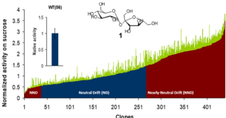
parent-like activity toward sucrose and (ii) nearly neutral variants with altered (i.e., enhanced or decreased) activity toward sucrose. Those variants that displayed sucrose cleavage activity within three standard deviations of the average activity of parental NpAS were considered as neutral, while those with activity levels outside the range of three standard deviations
were defined as nearly neutral (Figure 2). This segregation was
established from the overall phenotypes of the variants as measured from their crude cell extracts, which does not take into account possible variation in expression levels. This
arbitrary classification enabled the discrimination of two
populations of variants with equivalent size (230 neutral variants and 210 nearly neutral variants), which are compared in this work.
To further characterize the mutational changes that occurred over the neutral drift experiment, we sequenced 100 neutral
and 81 nearly neutral variants (representing ∼41% of the
fourth round library). Overall, the sequenced variants were
mutated at an average rate of 2.9± 2.0 nucleotide substitutions
per variant (corresponding to 1.4± 1.3 amino acids exchanged
per variant; Table 1). Nearly neutral variants harbored a
slightly higher number of substitutions per variant than the neutral ones (1.6 versus 1.2 amino acid exchanges for the nearly neutral and the neutral variants, respectively). Fifty-three sequences (29.3% of the sequenced variants) were found
Figure 2.Relative activity of variants selected after the fourth round of neutral drift. Activities toward cleavage of sucrose (1) were determined in triplicate by measuring the release of reducing sugars in crude cell extracts over time and were normalized to the parental NpAS activity level (WT). Standard deviations (SD) for each variant are shown in green. For the parental enzyme, the calculated SD represents 15% of the average activity level (n = 56). Variants within 3SD of parental mean activity were arbitrarily defined as neutral (blue), and variants outside 3SD were defined as nearly neutral (red).
Table 1. Sequence Analysis of Variants Selected after the Fourth Round of Neutral Drift
no. of variants no. of sequenced variants average nucleotide mutations per gene
(±SD)
average amino acid mutations per
gene (±SD) library 440 181 2.86 (±1.99) 1.35 (±1.33) neutral variants 230 100 2.76 (±1.83) 1.18 (±1.34) nearly neutral variants 210 81 2.99 (±2.17) 1.57 (±1.28) ACS Catalysis
to be identical to the parental enzyme at the protein level and 16 (8.9%) had no mutations at all (i.e., the other 37 sequences displayed silent mutations). Overall, these sequencing data are in accordance with the fact that the neutral drift experiment was performed under low mutational load.
Effect on Stability of Neutral and Nearly Neutral
Amino Acid Exchanges. Differential scanning fluorimetry
(DSF) was used to measure the melting temperature (Tm) of
215 purified variants randomly chosen from the fourth round
library (Figure 3A). The measured Tmvalues fell within a range
of 7 °C (from 46 to 53 °C) around the Tmof the parental
enzyme (∼48.4 °C) among both neutral and nearly neutral
variants, demonstrating that the thermostability of the parent
was marginally affected under the low mutational load of our
neutral drift experiment. Interestingly, nearly neutral variants increased in both thermostability and activity toward sucrose
cleavage were also identified (Figure 3B). Notably, a higher
number of variants with increased stability were found in the upper part of the near-neutral library (among the more active crude extract). As previously mentioned, our arbitrary
classification did not consider the possible variations of
expression levels. Consequently, the more stable variants may be better expressed, as expression improvement is often correlated with enhanced stability.
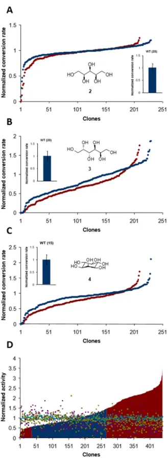
Modulation of Native Enzyme Promiscuity. Toward Acceptor. NpAS has previously been shown to process a large range of exogenous molecules as acceptor substrates. Among them, alditols (e.g., xylitol, dulcitol, and myo-inositol) are
glucosylated to various extents by the parental enzyme.22Both
neutral and nearly neutral variants selected from the fourth round of neutral drift (produced as crude cell extracts) were
assayed toward these substrates, and thefinal conversion for
each of the three acceptors was tested independently for each variant. Results for both populations were normalized to the conversion obtained with the parental enzyme (corresponding to 75%, 35%, and 10%, for xylitol, myo-inositol, and dulcitol,
respectively) (Figure 4). Interestingly, both populations shared
similar behavior in terms of maximum and range of acceptor conversion, indicating that acceptor glucosylation efficiency is not related to sucrose cleavage activity determined in the absence of acceptor. Up to 2-fold increases in conversion rates
were observed for myo-inositol and dulcitol. A relatively flat
profile was obtained for xylitol conversion, and no significant
improvement was observed, possibly because this substrate was
already very well recognized by the parental enzyme (75% conversion). Conversely, variants with impaired glucosylation capabilities toward the three substrates were also identified,
indicating that neutral drift may also lead to the identification
of variants with restricted promiscuity. Interestingly, glucosy-lation levels for the three tested acceptor molecules were
altered differently among both neutral and nearly neutral
variants (Figure 4D). Taken together, these observations
demonstrate how neutral drift at a low mutational rate may lead to the generation of active variants with various functionalities, including many variants of interest with increased or restricted ability to convert polyols, some of
them being highly specific for one polyol type.
Toward Donor. NpAS is highly specific for its natural donor
substrate sucrose but possesses promiscuous activity toward a
few alternative non-natural donors, includingα-fluoroglucoside
and p-nitrophenyl-α-D-glucopyranoside (pNP-Glc).26,34 In
particular, we have previously reported that the kinetic parameters of NpAS were much lower for pNP-Glc than for
sucrose with up to 490-fold decrease in kcat/KMfor pNP-Glc in
comparison to sucrose in the same concentration range.26To
investigate whether neutral drift could yield variants with increased hydrolytic activity toward pNP-Glc, we screened our fourth neutral drift round library of functional variants against
that substrate. The variants were first purified using a
miniaturized purification procedure35 to avoid background
noise associated with the presence ofα-glycosidase activity of
the host cell. Upon addition of pNP-Glc, the donor consumption rate for each variant was determined by monitoring the absorbance at 405 nm of released p-nitrophenoxide. The resulting measured activities ranged from the limit of detection up to 9-fold improvement relative
to the parental enzyme (Figure 5). Furthermore, the activities
of neutral and nearly neutral variants showed similar
distribution profiles as observed for acceptor glucosylation.
Although no clear correlation could be established between the activities toward sucrose and pNP-Glc under our experimental conditions, it is noteworthy that the two most improved variants against pNP-Glc belonged to the neutral library, namely 7946C7 (V102M/S238T) and 7945E5 (D37H/ V331D/D526N/V609I). Moreover, the two best improved variants from the nearly neutral library, namely 7942D2 (N76S/S430L) and 7942F3 (A289T/K469R), were also increased by 2.5-fold in their activity for sucrose and suggest
Figure 3.Thermostability of neutral and nearly neutral variants. (A) Changes in thermostability of neutral and nearly neutral variants. A total of 215 variants were randomly chosen from the fourth round of neutrally drifted variants, expressed in E. coli, and purified, and their melting temperatures (Tm) were measured in triplicate using differential scanning fluorimetry. (B) Tmvalues of neutral and nearly neutral variants plotted
that these two activities are intrinsically associated. Among the improved variants against pNP-Glc, the melting temperature values were found similar to that of the parental enzyme except for 7945E5 that displayed the lowest thermodynamic stability out of the 440 variants.
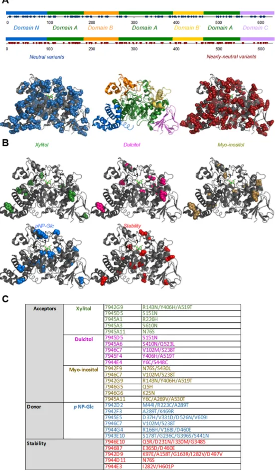
Profiling of Drifted Variants. The amino acid
sub-stitutions are distributed throughout the 3D structure of NpAS (Figure 6). The enzyme’s domains (e.g., A, B, and C domains
common to the GH13 family and N and B′
amylosucrase-specific domains) all seem to be relatively tolerant to mutations
(Figure 6A). Surprisingly, the B′ domain, which is involved in
sucrose specificity,36 was more affected by neutral mutations
than nearly neutral ones. Some substitutions are located on the enzyme surface and others are buried, indicating that neutral mutations are not necessarily distant from the active site. Moreover, highly conserved residues previously shown to be
involved in sucrose (Y147F in subsite −1) or acceptor
(A289T, I330M, V331D in the first shell of subsite +1)
recognition were also affected by substitutions. Analysis of
sequence structure−function relationships was further
consid-ered for the most beneficial mutations (Figure 6B and Table
S1). The combination of substitutions Y406H and A519T, as
well as the S138T substitution, appeared to improve conversion rates toward the glucosylation of all three assayed acceptors.
Variants exhibiting a higher reactivity with pNP-Glc contained more substitutions (from 2 to 4) on average.
Substitution A289T, found in two distinct and highly efficient
variants, and V331D are located in subsite +1 onflexible loops
surrounding the NpAS active site. These substitutions, also deemed critical for non-natural acceptor glucosylation, are likely responsible for enhanced activity toward pNP-Glc, possibly by helping with accommodation of the pNP leaving
group or promoting its release.27
With respect to stability improvement, a wide variety of mutations in terms of positions and number of substitutions (from 1 to 5) were observed, suggesting that combined mutations may act synergistically to improve the enzyme
Figure 4.Acceptor promiscuity of NpAS active variants after four rounds of neutral drift. Thefinal conversion rates for alditols, namely (A) xylitol (2), (B) dulcitol (3), and (C) myo-inositol (4), using sucrose as donor were measured and normalized relative to the conversion rate of the parental enzyme. Replicate conversion rate measurements using parental NpAS were performed for each acceptor, with the number of repeats indicated in brackets. Conversion rates are represented in increasing order and further compared with respect to their corresponding sucrose activity, colored
Figure4. continued
in cyan, purple, and olive green for 2, 3, and 4, respectively (D). Neutral variants and nearly neutral variants are colored in blue and red, respectively.
Figure 5. Donor promiscuity of NpAS active variants after four rounds of neutral drift. Purified variants were assayed toward the promiscuous substrate pNP-Glc (5). The consumption rates were determined relative to the parental enzyme. Neutral and nearly neutral variants are indicated in blue and red, respectively. Variant activities are ranged in increasing order.
Figure 6. Sequence structure−function analysis of variants. (A) Localization of neutral (blue spheres) and nearly neutral (red spheres) substitutions on the NpAS structural domains (PDB code: 1JGI). (B) Representation of all substitutions harbored by improved variants toward xylitol (green), dulcitol (magenta), myo-inositol (yellow), pNP-Glc (blue), and stability (red). (C) Amino acid substitution in improved variants.
thermostability. The highest Tm value was observed for a
variant harboring four amino-acid substitutions (7946E10:
Q5R/D231N/I330M/G348S) with a 4 °C increase in Tm
compared to the parent enzyme.
Search for Novel Specificity. The results described thus
far emphasize how neutral and nearly neutral mutations can
significantly affect NpAS substrate promiscuity. Next, we
investigated whether neutral drift could yield variants with improved glucosylation activity on a non-natural acceptor of
interest, methyl-α-L-rhamnopyranoside (α-L-Rhap-OMe), one
of the sole reported monosaccharide non-naturally used as an acceptor by the WT enzyme. Glucosylation products of this monosaccharide are key intermediates toward the synthesis of Shigella f lexneri antigens. We previously developed a computer-aided enzyme engineering approach to generate NpAS variants
able to glucosylate this monosaccharide.27−29This work led to
the identification of a single-substitution variant (I228Y) which
was able to glucosylateα-L-Rhap-OMe intoα-D-Glcp-(1
→3)-Figure 7.Neutral drift variant 7945E5 displays new acceptor activity towardα-L-Rhap-OMe. (A) LC/MS analyses were performed to determine
the molar mass of the glucosylation products obtained with variant 7945E5. (B) The structure of the monoglucosylated product was deduced from comparisons with chemically and enzymatically synthesized standards, namely α-D-glucopyranosyl-(1→3)-α-L-rhamnopyranoside and α-D
-glucopyranosyl-(1→4)-α-L-rhamnopyranoside.
α-L-Rhap-OMe.33 Here, we evaluated the capacity of the
drifted variants to catalyze reaction ofα-L-Rhap-OMe with no
prior structural design.
Surprisingly, we detected one variant in the fourth-round neutral library (7945E5: D37H/V331D/D526N/V609I)
which had acquired the ability to glucosylateα-L-Rhap-OMe.
HPLC/MS analyses further enabled the detection of three
different products corresponding to mono-, di-, and
triglucosylated products (Figure 7A). Interestingly, the
retention time of the monoglucosylated form was clearly
different from those of α-D-Glcp-(1→3)-α-L-Rhap-OMe and
α-D-Glcp-(1→4)-α-L-Rhap-OMe (Figure 7B), indicating that
variant 7945E5 catalyzed the synthesis of an unprecedented glucosylation product never previously obtained using NpAS variants. Unfortunately, the precise structure of this product could not be determined within the scope of the present work due to poor reaction yields and hence insufficient isolable
material for NMR characterization. Nevertheless, given that
α-L-Rhap-OMe harbors only three hydroxyl groups (at positions
C-2, C-3, and C-4), we may reasonably assume it to beα-D
-glucopyranosyl-(1→2)-α-L-rhamnopyranoside, a molecule not
currently recorded as enzymatically produced in nature. It is noteworthy that this motif is part of the repeating unit of the
Escherichia coli O13 O-antigen.37 Overall, this preliminary
result demonstrates the utility/potential of neutral drift to open access to novel enzyme activities by yielding superior alternative starting points for further protein engineering.
Characterization of 7945E5 Variant Showing a Novel
Specificity. Structural Analysis. To obtain further insight on
how acceptor specificity was changed, the X-ray structure of
the variant 7945E5 was determined at a resolution of 1.60 Å
resolution (Table S2, PDB code: 5N6V). With a rmsd
calculated on 628Cα atoms of 0.25 Å, the structure is highly
similar to that of the WT NpAS (PDB code: 1G5A).36As often
observed in NpAS crystal structures, a Tris molecule was found
in the active site28,30,36despite the fact that the crystals were
soaked in 100 mM α-L-Rhap-OMe and sucrose.
Crystallo-graphic data also revealed the presence of other ligands in the
structure (Figure S1): two sucrose molecules bound in SB2
and SB3 binding sites previously described,38,39 and two
fructose molecules found in a binding site that we named SB5,
corresponding to the previously identified oligosaccharide
binding site OB3. Both SB3 and SB5 sites are located at the enzyme surface. SB5 is also positioned at the interface between two-symmetry-related molecules. A glucose molecule was also
found bound in OB2 site. Regarding the role of the introduced mutations, three positions (i.e., 37, 526, and 609) are remote from the active site and not in direct contact with the product (Figure S2). Position 331 is located in the +1 subsite and was
investigated by saturation mutagenesis in previous work.27
However, none of the corresponding single variants were able
to glucosylate α-L-Rhap-OMe.33 This observation clearly
indicates that the additional substitutions at positions 37, 526, and 609 of variant 7945E5 are key in the acquisition of
the new acceptor specificity. Such a variant would not have
been designed by rational or even semi-rational engineering
approaches as the combinatorial effects of amino acid changes
is difficult to predict, especially when involving residues far
from the active site. These results further demonstrate that neutral genetic drift is a powerful strategy for generating enzyme variants with original combinations of amino acid substitutions and unprecedented catalytic capabilities.
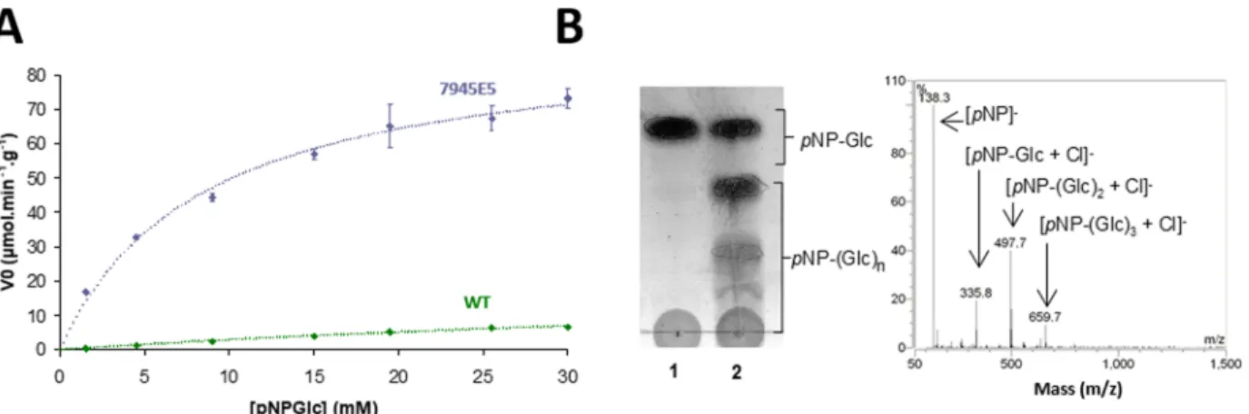
Increased Activity toward pNP-Glc. Interestingly, variant 7945E5 also displayed increased activity on pNP-Glc (7-fold increase over parental NpAS under screening conditions). The kinetic parameters of this variant toward pNP-Glc revealed a
25-fold increase in catalytic efficiency (kcat/KM) compared to
the parental enzyme (Figure 8A, Table 2). Thin-layer
chromatography (TLC) and mass spectrometry (MS) revealed that pNP-Glc acts as both donor and acceptor substrate during
the transglucosylation reaction (Figure 8B). It is noteworthy
that variant 7945E5 had the lowest Tmvalue among the 215
variants evaluated by DSF (Tmdecreased by 3°C compared to
parental NpAS), which was further confirmed by circular
Figure 8.Activity of variant 7945E5 toward pNP-Glc. (A) Michaelis−Menten plot for parental NpAS and variant 7945E5. (B) MS and TLC analyses of transglucosylation capacity of variant 7945E5 when pNP-Glc was used as sole substrate.
Table 2. Kinetic Parameters towardpNP-Glc and TmValues
of 7945E5 andNpAS Enzymes
NpAS variant 7945E5 Kinetic Parameters
KM(mM) 66.9a 8.5
kcat(min−1) 1. 6 6.5
VM(μmol product released·min−1·g−1) 22.3a 91
kcat/KM(mM−1·min−1) 0.03a 0.76
Thermostability
Tm,DSF(°C) 48.4 45.7
Tm,CD(°C) 48.1 45.7
dichroism (CD) (Table 2). Analysis of the average B-factors
suggested that loop 8 (residues 495−502) is more flexible in
variant 7945E5 in comparison to parental NpAS (Figure S3).
This observation is in agreement with previous conclusions
stating that substitutions affecting functionality (e.g., substrate
specificity) are often detrimental to protein stability.28,40,41
■
CONCLUSIONSIn the present Article, we describe a low mutational rate neutral-drift engineering approach applied to a sucrose-utilizing transglycosylase, amylosucrase from Neisseria poly-saccharea. Four repeated rounds of random mutagenesis and purifying selection were performed, yielding a library of 440 functional enzyme variants with parent-level activity on
sucrose. The introduced neutral variations (1.4± 1.3 amino
acid exchange per variant) did not affect overall protein
stability, as only marginal variations in thermostability (±3.5
°C around the Tm of the parent) were recorded among the
variants. Extensive screening revealed large variations in the neutrally drifted population for both donor and acceptor
reactions. Analysis of thefinal conversion rates for three polyol
acceptors revealed variations spanning from activity loss (below detectable levels) to a 2-fold increase relative to the parent enzyme. Additionally, activity toward the promiscuous
donor pNP-Glc alsofluctuated significantly among the variants,
with a 25-fold improvement in catalytic efficiency (kcat/Km)
observed for a neutral variant harboring four amino acid substitutions (namely 7945E5: D37H/V331D/D526N/ V609I). Moreover, this variant also showed transglucosylation
activity toward a new acceptor, methyl-α-L-rhamnopyranoside,
which is a precursor of prime interest for the synthesis of antigenic oligosaccharides from Shigella f lexneri and Escherichia coli. Interestingly, this nearly neutral variant was also the least
stable variant (Tm≈ 45.7 °C) recorded among the neutrally
drifted population. Altogether, these results further demon-strate that neutral drift may help to enlarge the subdemon-strate scope of enzymes while maintaining their natural functional integrity.
Our data also demonstrate that this approach is very effective
for identifying biocatalysts with enhanced or even new catalytic properties by evolving small-sized libraries compatible with
low-throughput but accurate analyses. The combined effects
on catalytic activity, substrate conversion rate, and thermo-stability further suggest that neutral drift can be a powerful enzyme engineering strategy for synthetic biology. The use of such neutral and polymorphic libraries can lead to the
generation of finely tuned variants with modified properties
that have proven essential for balancing new enzymatic pathways.
■
EXPERIMENTAL AND THEORETICAL PROCEDURESBacteria, Plasmid, Strain, and Chemicals. Plasmid pGST-AS derived from the pGEX-6P-3 vector (GE Healthcare Biosciences) and containing the gene encoding amylosucrase from Neisseria polysaccharea amylosucrase (NpAS) was used as the template for the construction of saturation mutagenesis libraries. Escherichia coli Top 10 was used as the host strain for plasmid transformation and gene expression. Sucrose was purchased from Merck KGaA (Darmstadt, Germany). Xylitol,
myo-inositol, dulcitol, p-nitrophenyl-α-D-glucopyranoside,
glu-tathione reduced, dithiothreitol, bromothymol blue (BTB), and lysozyme were purchased from Sigma-Aldrich (St. Louis,
MO, USA). Methyl-α-L-rhamnopyranoside, methyl-α-D
-gluco-pyranosyl-(1→3)-α-L-rhamnopyranoside, and α-D
-glucopyra-nosyl-(1→4)-α-L-rhamnopyranoside were chemically
synthe-sized and characterized at the Unité de Chimie des
Biomolécules, Institut Pasteur, France.27,42
Ampicillin,
iso-propyl β-D-thiogalactopyranoside (IPTG), and
deoxyribonu-clease I were purchased from Euromedex (Souffelweyersheim,
France). DpnI, EcoRI HF, and NotI HF restriction enzymes, T4 DNA ligase, and Antarctic Phosphatase were purchased from New England Biolabs (Beverly, MA, USA). Oligonucleo-tides were synthesized by Eurogentec (Liege, Belgium). DNA
extraction (GenElute HP plasmid Miniprep) and purification
(GenElute Gel Extraction) kits were purchased from Sigma-Aldrich.
Construction, Selection, and Expression of Variant Library. Mutations were introduced randomly by error-prone PCR using the GeneMorph II Random Mutagenesis kit
(Agilent Technology) according to the manufacturer’s
instructions, with pGST-AS (350 ng) as vector template for
thefirst round of mutagenesis. PCR amplifications were carried
out with Mutazyme II DNA polymerase (2.5 U). The cycling
included a denaturation step at 95°C (2 min) and then 30
cycles composed of (i) a denaturation step at 95°C (30 s), (ii)
a hybridization step at 55°C (30 s), and (iii) an elongation
step at 72 °C (4 min). A final elongation step at 72 °C (10
min) was performed. DNA was digested with DpnI and further
purified. The pool of genes was then double-digested by EcoRI
HF and NotI HF restriction enzymes and gel purified. In
parallel, a cloning vector was prepared by double-digesting the pGST-AS with EcoRI HF and NotI HF restriction enzymes, followed by dephosphorylation with Antarctic Phosphatase and gel purification following the manufacturer’s recommenda-tions. Insert (50 ng) and vector (50 ng) were ligated using T4
DNA ligase (10 min; 25 °C). The ligation product was
precipitated with 5 volumes of absolute ethanol and 1/10
volume of sodium acetate (3 M; pH 5.3) at−20 °C overnight.
DNA was centrifuged (30 min, 18000g, 4°C), and the pellet
was rinsed two times with 70% ethanol and dried under vacuum using an Eppendorf concentrator 5301. The pellet was
resuspended in water, and 2μL was used to transform 50 μL of
E. coli Top 10 electro-competent cells before plating on LB
agar plates supplemented with 1% glycerol and 100μg·mL−1
ampicillin on which a hydrophilic membrane (Durapore
membrane filter, 0.22 μm, Millipore) had been overlaid.
After 24 h at 37 °C, the membrane was transferred onto
another LB agar plate supplemented with ampicillin (100μg·
mL−1), sucrose (146 mM), IPTG (1 mM), Tris buffer (50
mM, pH 7.5), and BTB indicator (0.1 g·L−1) and incubated for
24 h at 30 °C as previously described.33A library of around
5000 recombinant clones was obtained. Cells expressing amylosucrase variants still active on sucrose were colored yellow and thus easily differentiated, and 260 clones were picked at random and transferred into 96-well microplates,
containing in each well 200 μL of 2XYT medium
supplemented with 100 μg·mL−1 ampicillin. After 24 h of
incubation at 30 °C, the variants were plated on LB agar
supplemented with 100μg·mL−1ampicillin. The clones were
grown overnight at 37°C, and the colonies were scraped from
the plate for plasmid extraction, constituting the first-round
library. This plasmid pool was used as the template for the second round of mutagenesis following the procedure outlined above. Altogether, four rounds of mutagenesis were performed. Finally, 260, 458, 492, and 440 variants were selected from
concentration of 1 mM) to induce glutathione S-transferase-amylosucrase variant fusion protein expression. After 6 to 8 h,
plates were centrifuged (20 min, 3000g, 4 °C), and the
supernatant was removed. The cell pellet was resuspended in
200 μL of phosphate-buffered saline (PBS) 2X (pH 7.3,
containing lysozyme 0.5 mg·mL−1and 1.5 U of
deoxyribonu-clease I) followed by freezing at −80 °C for 8−12 h. After
thawing at room temperature, the plates were centrifuged (20
min, 3000g, 4 °C), and 150 μL portions of each the four
supernatants were transferred to a single GST MultiTrap 4B
(GE Healthcare Biosciences) 96-well plate (600 μL of
supernatant per well). Unbound proteins were removed by
centrifugation (4 min, 100g, 4°C). PBS (600 μL) was added
to each well to wash bound samples, and this operation was
repeated three times. The final elution was performed by
adding 200μL of Tris buffer (50 mM, pH 7.0) containing 1
mM dithiothreitol and 15 mM reduced glutathione. The purity of the proteins was then monitored by sodium dodecyl sulfate-PAGE.
Screening of Variants toward Acceptor Substrates. Storage microplates containing the neutral-drift library were thawed and replicated to inoculate starter culture in 96-well
microplates containing in each well 200μL of 2XYT medium
supplemented with 100μg·mL−1 ampicillin. After growth for
12 h at 30°C (700 rpm), plates were duplicated four times
into 96-Deep Well plates containing in each well 1 mL of
ZYM-5052 auto-inducible medium supplemented with 100μg·
mL−1ampicillin and then grown for 24 h at 30°C (700 rpm).
The plates were then centrifuged (20 min, 3000g, 4°C), and
the supernatants were removed. The cell pellets were
resuspended in 200 μL of PBS 2X (pH 7.3, containing
lysozyme 0.5 mg/mL and 1.5 U of deoxyribonuclease I),
followed by freezing at−80 °C for 8−12 h. Next, 100 μL of
enzymatic extract was transferred into a new microplate, and
50 μL of sucrose was added as well as 50 μL of acceptors.
Sucrose:acceptor ratios were 1:1 for dulcitol (146:146 mM)
and methyl-α-L-rhamnopyranoside (73:73 mM) and 1:2 for
xylitol (146:292 mM) and myo-inositol (146:292 mM).
Reactions were carried out for 24 h at 30 °C, inactivated by
heating at 95 °C, and centrifuged (3000 g, 20 min). The
acceptor reaction media were analyzed by direct injection mass
spectrometry (MSQ Plus, Thermo Scientific) for methyl-α-L
-rhamnopyranoside and by high-pressure liquid chromatog-raphy (HPLC) analysis using a Dionex P 680 series pump, a Shodex RI 101 series refractometer, and an HTC PAL autosampler for other acceptors. HPLC analyses were
performed on an HPX-87C column (300 mm × 7.8 mm,
Biorad) maintained at 65°C using ultrapure water as eluent at
aflow of 0.6 mL/min for dulcitol and xylitol and with an
HPX-87K column (300 mm× 7.8 mm, Biorad) maintained at 65 °C
using ProtParam tool on ExPASy portal (web.expasy.org/
protparam/).
Activity Assays. Activities toward sucrose were determined
by transferring 50μL of the crude enzymatic extracts into a
microplate with 50 μL of sucrose (final concentration 146
mM) and incubating for 2 h at 30 °C. The reaction was
stopped by adding 100μL of 3,5-dinitrosalicylic acid followed
by heating at 95 °C for 10 min, and the concentrations of
released reducing sugars were determined by measuring the absorbance at 540 nm with a Sunrise spectrophotometer. Reactions were performed in triplicate to estimate the variability of the experiment. Relative activities were calculated with respect to the optical density of the culture (at 600 nm) and were normalized to the parental value calculated from 56 full repeats (i.e., 56 cultures of 1 mL in 96-Deep Well plates).
Activities toward p-nitrophenyl-α-D-glucopyranoside were
determined by transferring 50μL of the purified enzymes into
a microplate with 50μL of p-nitrophenyl-α-D-glucopyranoside
(final concentration 15 mM) after incubating for 15 h at 30 °C.
The conversion of p-nitrophenyl-α-D-glucopyranoside was
determined by measuring the absorbance of p-nitrophenoxide released at 405 nm with a Sunrise spectrophotometer. Relative activities were calculated with respect to the optical density of the cultures and were normalized to the parental value calculated from 20 full repeats.
High-throughput screening experiments were performed using equipment from the ICEO facility (Laboratoire
d’Ingénierie des Systèmes Biologiques et des Procédés
(LISBP), Toulouse, France).
Thermostability of Enzymes. The melting points (Tm) of
GST-fused amylosucrase variants were recorded in triplicate experiments by DSF. 215 randomly chosen variants were
purified using the microplate purification protocol described
previously.35A mix of 19 μL of enzyme and 1 μL of
Sypro-orange (5X final) (Invitrogen, Paisley, UK) was incubated
using a temperature gradient ranging from 25 to 80°C with a
0.3°C increment. The thermal transition was monitored with a
RTQ-PCR CFX96 Real-Time System (Biorad, Marnes-la-Coquette, France). The melting temperatures were determined
as the maximums of thefirst derivative for each temperature−
fluorescence curve and averaged, and the standard deviation
for each Tmwas determined.
Circular dichroism spectra of recombinant NpAS and 7945E5 purified enzymes were recorded on a JASCO J815 spectropolarimeter equipped with a Peltier cell temperature
controller. Tm values were obtained by heating the samples
(enzyme 2μM in 50 mM Tris, pH 7.0, 150 mM NaCl, 1 mM
DTT, 1 mM EDTA) at 1 °C·min−1 and recording the
30 s. The melting temperatures were determined by fitting analyses using SigmaPlot 10.0.
Structure Determination. Crystallization Conditions. Variant 7945E5 was crystallized using conditions described
previously.39Experiments were carried out in 24-well plates at
12 °C using the hanging drop vapor diffusion method. One
volume of protein at 10 mg·mL−1(in Tris 50 mM, NaCl 150
mM, EDTA 1 mM, DTT 1 mM, pH 7.3) was mixed with one volume of precipitant solution (PEG 6000 at 25, 30, 35%, or 40% (w/v), 0.1 M HEPES at pH 6.8, 7.0, 7.3, 7.6, 7.9, or 8.2). Platelet crystals appeared after 1 week. The best crystallization conditions were obtained with PEG 6000 at 30% (w/v) and 0.1 M HEPES pH 7.9.
Data Collection and Structure Determination. X-ray
experiments were carried out at 100 K. Prior toflash cooling,
crystals of variant 7945E5 were soaked for 3 min in the
reservoir solution supplemented with 100 mM methyl-α-L
-rhamnopyranoside and 100 mM sucrose. Diffraction data sets
were collected to a maximum resolution of 1.60 Å on beamline ID29 at the European Synchrotron Radiation facility (ESRF,
Grenoble, France). Diffracted intensities were integrated and
scaled using XDS,46 and 5% of the scaled amplitudes were
randomly selected and excluded from the refinement
procedure. Crystals of variant 7945E5 belong to orthorhombic
space group P22121 with one molecule per asymmetric unit
giving a Matthews coefficient of 2.4 Å3/Da. Data collection
statistics are given inTable S2.
Model Building and Refinement. Structure refinement was
performed using the structure of parental NpAS (PDB code:
1G5A) as an initial model and refmac5 from CCP4 GUI.47
The model was built manually in SigmaA weighted electron
density maps using COOT.48Water molecules were
automati-cally assigned, and ligand molecules werefitted manually into
residual maps. Thefinal model was refined to final R and Rfree
of 14.8% and 18.0%, respectively. Refinement statistics are
given in Table 2. Coordinates of the structure of variant
7945E5 have been deposited at the Protein Data Bank (PDB code: 5N6V).
■
ASSOCIATED CONTENT*
S Supporting InformationThe Supporting Information is available free of charge on the
ACS Publications websiteat DOI:10.1021/acscatal.8b03609.
Supporting tables andfigures showing details of variant
analysis, crystallographic data collection, and structural
analysis of variant 7945E5 (PDF)
■
AUTHOR INFORMATION Corresponding Authors *E-mail:[email protected]. *E-mail:[email protected]. ORCID Isabelle André:0000-0001-6280-4109 Magali Remaud-Siméon: 0000-0002-2658-672X Author Contributions§D.D. and A.V. contributed equally to this work.
Notes
The authors declare no competingfinancial interest.
■
ACKNOWLEDGMENTSThis work was supported by the French National Research Agency (ANR Project GLUCODESIGN, ANR-08-PCVI-002-02). D.D. and A.V. were supported by Ph.D. grants from the French Ministry of Research. The authors are grateful to S. Bozonnet and S. Pizzut-Serin for assistance when using the enzyme screening equipment of the ICEO facility (located in the LISBP and part of the Integrated Screening Platform of Toulouse PICT, IBiSA). They also thank members of the biophysical facility from IPBS (PICT, IBiSA) for providing access to DSF and crystallography equipment. The authors thank the European Synchrotron Radiation Facility (ESRF) at
Grenoble (France), particularly the beamline ID29 staff. The
authors would like to thank A.-M. Besançon, S. Cantos, and N.
Monties for technical assistance. We are also grateful to L. A. Mulard for the precious gift of the disaccharides used as standards. The authors are grateful to Sean Devenish for careful and meticulous reading of the manuscript.
■
REFERENCES(1) Peisajovich, S. G.; Tawfik, D. S. Protein Engineers Turned Evolutionists. Nat. Methods 2007, 4, 991−994.
(2) Stemmer, W. P. DNA Shuffling by Random Fragmentation and Reassembly: In Vitro Recombination for Molecular Evolution. Proc. Natl. Acad. Sci. U. S. A. 1994, 91, 10747−10751.
(3) Zhao, H.; Giver, L.; Shao, Z.; Affholter, J. A.; Arnold, F. H. Molecular Evolution by Staggered Extension Process (StEP) in Vitro Recombination. Nat. Biotechnol. 1998, 16, 258−261.
(4) Bornscheuer, U. T.; Huisman, G. W.; Kazlauskas, R. J.; Lutz, S.; Moore, J. C.; Robins, K. Engineering the Third Wave of Biocatalysis. Nature 2012, 485, 185−194.
(5) Dalby, P. A. Strategy and Success for the Directed Evolution of Enzymes. Curr. Opin. Struct. Biol. 2011, 21, 473−480.
(6) Davids, T.; Schmidt, M.; Böttcher, D.; Bornscheuer, U. T. Strategies for the Discovery and Engineering of Enzymes for Biocatalysis. Curr. Opin. Chem. Biol. 2013, 17, 215−220.
(7) Turner, N. J. Directed Evolution of Enzymes for Applied Biocatalysis. Trends Biotechnol. 2003, 21, 474−478.
(8) Longwell, C. K.; Labanieh, L.; Cochran, J. R. High-Throughput Screening Technologies for Enzyme Engineering. Curr. Opin. Biotechnol. 2017, 48, 196−202.
(9) Goldsmith, M.; Tawfik, D. S. Directed Enzyme Evolution: Beyond the Low-Hanging Fruit. Curr. Opin. Struct. Biol. 2012, 22, 406−412.
(10) Reetz, M. T.; Bocola, M.; Carballeira, J. D.; Zha, D.; Vogel, A. Expanding the Range of Substrate Acceptance of Enzymes: Combinatorial Active-Site Saturation Test. Angew. Chem. 2005, 117, 4264−4268.
(11) Jochens, H.; Bornscheuer, U. T. Natural Diversity to Guide Focused Directed Evolution. ChemBioChem 2010, 11, 1861−1866.
(12) Kimura, M. The Neutral Theory of Molecular Evolution; Cambridge University Press: Cambridge, 1983.
(13) Bershtein, S.; Goldin, K.; Tawfik, D. S. Intense Neutral Drifts Yield Robust and Evolvable Consensus Proteins. J. Mol. Biol. 2008, 379, 1029−1044.
(14) Bloom, J. D.; Romero, P. A.; Lu, Z.; Arnold, F. H. Neutral Genetic Drift Can Alter Promiscuous Protein Functions, Potentially Aiding Functional Evolution. Biol. Direct 2007, 2, 17.
(15) Aharoni, A.; Gaidukov, L.; Khersonsky, O.; Gould, S. M.; Roodveldt, C.; Tawfik, D. S. The “evolvability” of Promiscuous Protein Functions. Nat. Genet. 2005, 37, 73−76.
(16) Amitai, G.; Gupta, R. D.; Tawfik, D. S. Latent Evolutionary Potentials under the Neutral Mutational Drift of an Enzyme. HFSP J. 2007, 1, 67−78.
(17) Wroe, R.; Chan, H. S.; Bornberg-Bauer, E. A Structural Model of Latent Evolutionary Potentials Underlying Neutral Networks in Proteins. HFSP J. 2007, 1, 79−87.
1053-3_5.
(22) Coutinho, P.; Henrissat, B. Carbohydrate-Active Enzymes: An Integrated Database Approach Server; 1999.
(23) Moulis, C.; André, I.; Remaud-Simeon, M. GH13 Amylosu-crases and GH70 Branching SuAmylosu-crases, Atypical Enzymes in Their Respective Families. Cell. Mol. Life Sci. 2016, 73, 2661−2679.
(24) André, I.; Potocki-Véronèse, G.; Morel, S.; Monsan, P.; Remaud-Siméon, M. Sucrose-Utilizing Transglucosidases for Bio-catalysis. In Carbohydrates in Sustainable Development I; Rauter, A. P., Vogel, P., Queneau, Y., Eds.; Topics in Current Chemistry; Springer: Berlin/Heidelberg, 2010; pp 25−48. https://doi.org/10.1007/128_ 2010_52.
(25) Monsan, P.; Remaud-Siméon, M.; André, I. Transglucosidases as Efficient Tools for Oligosaccharide and Glucoconjugate Synthesis. Curr. Opin. Microbiol. 2010, 13, 293−300.
(26) Daudé, D.; Champion, E.; Morel, S.; Guieysse, D.; Remaud-Siméon, M.; André, I. Probing Substrate Promiscuity of Amylosucrase from Neisseria Polysaccharea. ChemCatChem 2013, 5, 2288−2295.
(27) Champion, E.; André, I.; Moulis, C.; Boutet, J.; Descroix, K.; Morel, S.; Monsan, P.; Mulard, L. A.; Remaud-Siméon, M. Design of α-Transglucosidases of Controlled Specificity for Programmed Chemoenzymatic Synthesis of Antigenic Oligosaccharides. J. Am. Chem. Soc. 2009, 131, 7379−7389.
(28) Champion, E.; Guérin, F.; Moulis, C.; Barbe, S.; Tran, T. H.; Morel, S.; Descroix, K.; Monsan, P.; Mourey, L.; Mulard, L. A.; Tranier, S.; Remaud-Siméon, M.; André, I. Applying Pairwise Combinations of Amino Acid Mutations for Sorting Out Highly Efficient Glucosylation Tools for Chemo-Enzymatic Synthesis of Bacterial Oligosaccharides. J. Am. Chem. Soc. 2012, 134, 18677− 18688.
(29) Verges, A.; Cambon, E.; Barbe, S.; Salamone, S.; Le Guen, Y.; Moulis, C.; Mulard, L. A.; Remaud-Siméon, M.; André, I. Computer-Aided Engineering of a Transglycosylase for the Glucosylation of an Unnatural Disaccharide of Relevance for Bacterial Antigen Synthesis. ACS Catal. 2015, 5, 1186−1198.
(30) Vergès, A.; Cambon, E.; Barbe, S.; Moulis, C.; Remaud-Siméon, M.; André, I. Novel Product Specificity toward Erlose and Panose Exhibited by Multisite Engineered Mutants of Amylosucrase. Protein Sci. 2017, 26, 566−577.
(31) Vergès, A.; Barbe, S.; Cambon, E.; Moulis, C.; Tranier, S.; Remaud-Siméon, M.; André, I. Engineering of Anp Efficient Mutant of Neisseria Polysaccharea Amylosucrase for the Synthesis of Controlled Size Maltooligosaccharides. Carbohydr. Polym. 2017, 173, 403−411.
(32) Malbert, Y.; Pizzut-Serin, S.; Massou, S.; Cambon, E.; Laguerre, S.; Monsan, P.; Lefoulon, F.; Morel, S.; André, I.; Remaud-Simeon, M. Extending the Structural Diversity ofα-Flavonoid Glycosides with Engineered Glucansucrases. ChemCatChem 2014, 6, 2282−2291.
(33) Champion, E.; Moulis, C.; Morel, S.; Mulard, L. A.; Monsan, P.; Remaud-Siméon, M.; André, I. A PH-Based High-Throughput Screening of Sucrose-Utilizing Transglucosidases for the Develop-ment of Enzymatic Glucosylation Tools. ChemCatChem 2010, 2, 969−975.
Antigens of Shigella Flexneri. Carbohydr. Res. 2010, 345, 1594−1599. (38) Albenne, C.; Potocki de Montalk, G.; Monsan, P.; Skov, L. K.; Mirza, O.; Gajhede, M.; Remaud-Simeon, M. Site-Directed Muta-genesis of Key Amino Acids in the Active Site of Amylosucrase from Neisseria Polysaccharea. Biol., Bratisl. 2002, 57 (Suppl 11), 119−128. (39) Skov, L. K.; Mirza, O.; Sprogøe, D.; van der Veen, B. A.; Remaud-Simeon, M.; Albenne, C.; Monsan, P.; Gajhede, M. Crystal Structure of the Glu328Gln Mutant of Neisseria Polysaccharea Amylosucrase in Complex with Sucrose and Maltoheptaose. Biocatal. Biotransform. 2006, 24, 99−105.
(40) Tokuriki, N.; Tawfik, D. S. Stability Effects of Mutations and Protein Evolvability. Curr. Opin. Struct. Biol. 2009, 19, 596−604.
(41) Tokuriki, N.; Stricher, F.; Serrano, L.; Tawfik, D. S. How Protein Stability and New Functions Trade Off. PLoS Comput. Biol. 2008, 4, No. e1000002.
(42) Champion, E.; André, I.; Mulard, L. A.; Monsan, P.; Remaud-Siméon, M.; Morel, S. Synthesis of L-Rhamnose and N -Acetyl-D-Glucosamine Derivatives Entering in the Composition of Bacterial Polysaccharides by Use of Glucansucrases. J. Carbohydr. Chem. 2009, 28, 142−160.
(43) Emond, S.; Mondeil, S.; Jaziri, K.; André, I.; Monsan, P.; Remaud-Siméon, M.; Potocki-Véronèse, G. Cloning, Purification and Characterization of a Thermostable Amylosucrase from Deinococcus Geothermalis. FEMS Microbiol. Lett. 2008, 285, 25−32.
(44) De Montalk, G. P.; Remaud-Simeon, M.; Willemot, R. M.; Planchot, V.; Monsan, P. Sequence Analysis of the Gene Encoding Amylosucrase from Neisseria Polysaccharea and Characterization of the Recombinant Enzyme. J. Bacteriol. 1999, 181, 375−381.
(45) Pizzut-Serin, S.; Potocki-Véronèse, G.; van der Veen, B. A.; Albenne, C.; Monsan, P.; Remaud-Simeon, M. Characterisation of a Novel Amylosucrase from Deinococcus Radiodurans. FEBS Lett. 2005, 579, 1405−1410.
(46) Kabsch, W. Automatic Processing of Rotation Diffraction Data from Crystals of Initially Unknown Symmetry and Cell Constants. J. Appl. Crystallogr. 1993, 26, 795−800.
(47) Murshudov, G. N.; Vagin, A. A.; Dodson, E. J. Refinement of Macromolecular Structures by the Maximum-Likelihood Method. Acta Crystallogr., Sect. D: Biol. Crystallogr. 1997, 53, 240−255.
(48) Emsley, P.; Cowtan, K. Coot: Model-Building Tools for Molecular Graphics. Acta Crystallogr., Sect. D: Biol. Crystallogr. 2004, 60, 2126−2132.