HAL Id: inserm-02232741
https://www.hal.inserm.fr/inserm-02232741
Submitted on 1 Aug 2019
HAL is a multi-disciplinary open access
archive for the deposit and dissemination of
sci-entific research documents, whether they are
pub-lished or not. The documents may come from
teaching and research institutions in France or
abroad, or from public or private research centers.
L’archive ouverte pluridisciplinaire HAL, est
destinée au dépôt et à la diffusion de documents
scientifiques de niveau recherche, publiés ou non,
émanant des établissements d’enseignement et de
recherche français ou étrangers, des laboratoires
publics ou privés.
Distributed under a Creative Commons Attribution| 4.0 International License
controls mucosal biofilms
Jean-Paul Motta, Alexandre Denadai-Souza, David Sagnat, Laura Guiraud,
Anissa Edir, Chrystelle Bonnart, Mireille Sebbag, Perrine Rousset, Ariane
Lapeyre, Carine Seguy, et al.
To cite this version:
Jean-Paul Motta, Alexandre Denadai-Souza, David Sagnat, Laura Guiraud, Anissa Edir, et al.. Active
thrombin produced by the intestinal epithelium controls mucosal biofilms. Nature Communications,
Nature Publishing Group, 2019, 10 (1), pp.3224. �10.1038/s41467-019-11140-w�. �inserm-02232741�
Active thrombin produced by the intestinal
epithelium controls mucosal bio
films
Jean-Paul Motta
1,6
, Alexandre Denadai-Souza
1,6
, David Sagnat
1
, Laura Guiraud
1
, Anissa Edir
1
,
Chrystelle Bonnart
1
, Mireille Sebbag
1
, Perrine Rousset
1
, Ariane Lapeyre
1
, Carine Seguy
1
, Noa Mathurine-Thomas
1
,
Heather J. Galipeau
2
, Delphine Bonnet
3
, Laurent Alric
3
, Andre G. Buret
4
, John L. Wallace
5
, Antoine Dufour
5
,
Elena F. Verdu
2
, Morley D. Hollenberg
5
, Eric Oswald
1
, Matteo Serino
1
, Celine Deraison
1
&
Nathalie Vergnolle
1,5
Proteolytic homeostasis is important at mucosal surfaces, but its actors and their precise role
in physiology are poorly understood. Here we report that healthy human and mouse colon
epithelia are a major source of active thrombin. We show that mucosal thrombin is directly
regulated by the presence of commensal microbiota. Speci
fic inhibition of luminal thrombin
activity causes macroscopic and microscopic damage as well as transcriptomic alterations of
genes involved in host-microbiota interactions. Further, luminal thrombin inhibition impairs
the spatial segregation of microbiota bio
films, allowing bacteria to invade the mucus layer
and to translocate across the epithelium. Thrombin cleaves the bio
film matrix of
recon-stituted mucosa-associated human microbiota. Our results indicate that thrombin constrains
biofilms at the intestinal mucosa. Further work is needed to test whether thrombin plays
similar roles in other mucosal surfaces, given that lung, bladder and skin epithelia also
express thrombin.
https://doi.org/10.1038/s41467-019-11140-w
OPEN
1IRSD, Université de Toulouse, INSERM, INRA, ENVT, UPS, U1220, CHU Purpan, CS60039, 31024 Toulouse, France.2Farncombe Family Digestive Health
Research Institute, McMaster University, Health Science Center, Rm 3N4, 1280 Main Street West, Hamilton, ON L8S 4K1, Canada.3Department of Internal
Medicine and Digestive Diseases, 1, avenue Jean Poulhes-TSA 50032, 31059 Toulouse, France.4Department of Biological Sciences, University of Calgary,
2500 University Drive NW, Calgary, Ab T2N 4N1, Canada.5Departments of Physiology & Pharmacology, and Medicine, University of Calgary Cumming
School of Medicine, 3330 Hospital Drive NW, Calgary, Ab T2N 4N1, Canada.6These authors contributed equally: Jean-Paul Motta, Alexandre Denadai-Souza.
Correspondence and requests for materials should be addressed to N.V. (email:[email protected])
123456789
E
pithelia can serve as a local source of proteases at the
surface of mucosal organs such as in the skin
1,2, the lung
3and the digestive tract
4,5. Previous work indicates that
bacterial infection
6, stress
5, ischemia
7, low-grade
8or high-grade
inflammation
9,10can trigger mucosal and epithelial release of
active proteases by the host. Functional proteomic profiling has
recently identified active proteases released by human intestinal
mucosa in health and in inflammatory bowel diseases
11. Among
the identified active proteases, thrombin was found to be present
both in healthy and inflamed human mucosa, although the
cel-lular source of thrombin was not identified. Thrombin is a serine
protease known to be synthesized in the liver. It plays a central
role in hemostasis by converting plasma
fibrinogen into fibrin
and by promoting platelet aggregation via proteinase-activated
receptor (PAR) activation. In addition to its coagulation pathway
role, thrombin influences a number of pathophysiological
pro-cesses, including inflammation, tissue repair, angiogenesis, and
tumor invasion
4,12–14. While the reported increased presence of
thrombin in inflamed mucosa from inflammatory bowel disease
patients could easily be explained by the bleeding associated with
tissue damage
11, it was more surprising to detect active thrombin
in tissues from healthy individuals. One of the objectives of the
present study was therefore to investigate the cellular source of
thrombin in the gut mucosa.
Intestinal microbiota naturally grows as a mucus-coated
polymicrobial community, embedded as a biofilm organization,
separated from the epithelial surface by sterile mucus layer
15–19.
Nevertheless, biofilm encroachment at the surface of mucosal
tissue is one of the most relevant drivers of persistent bacterial
infections, thus constituting a major challenge for human and
animal health
20. Not only have such biofilms been associated
with disease, they contribute directly to the foundation of
inflammatory-based mucosal injuries
21–24. Given the importance
of spatial segregation between commensal bacteria and the
epi-thelia, we postulated that the epithelium uses specific mechanisms
to prevent the formation of a deleterious microbiota biofilm
blanket in contact with host tissues.
We identify here the presence of thrombin in intestinal
epi-thelium and its constitutive release on the apical side in
physio-logical conditions. We show that epithelial thrombin is regulated
by the presence of microbiota, and further that thrombin exerts a
protective role at mucosal surface. Thrombin maintains mucosal
homeostasis via its ability to cleave microbiota biofilm-derived
proteins, thereby preventing biofilm contact with tissues, and
limiting bacterial invasion.
Results
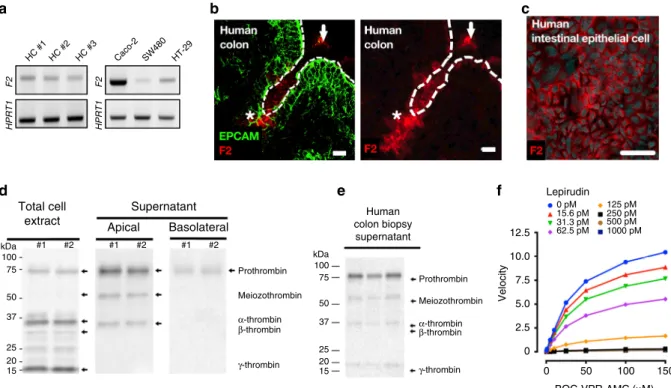
Intestinal epithelium releases constitutive active thrombin.
Reverse transcription analysis showed the presence of thrombin
mRNA in intestinal epithelium from healthy human colon crypts
(HC) as well as in three different human intestinal epithelial cell
lines (Caco-2, HT-29, SW480, Fig.
1
a, Supplementary Table 1).
HC #1 HPRT1 F2 HPRT1 F2 HC #2 HC #3 Caco-2 SW480 HT-29 Total cell extract kDa #1 #2 #1 #2 #1 #2 100 75 50 37 25 20 15 kDa Prothrombin Meiozothrombin α-thrombin β-thrombin γ-thrombin Prothrombin 12.5 Lepirudin Human colon biopsy supernatant 0 pM 15.6 pM 31.3 pM 62.5 pM 125 pM 250 pM 500 pM 1000 pM 10.0 7.5 5.0 2.5 0 0 50 BOC-VPR-AMC (μM) Velocity 100 150 Meiozothrombin α-thrombin β-thrombin γ-thrombin 100 75 50 37 25 20 15 Supernatant Apical Basolateral
a
b
c
d
e
f
Fig. 1 Intestinal epithelium releases active thrombin in gut lumen. a F2 mRNA transcripts (Factor 2, thrombin) were detected in colonic crypt epithelium from three different healthy human (left), and three different human cancer cell lines (Caco-2, SW480 and HT-29, right). Representative blots from 3
independent experiments.b Immunostaining of Epithelial cell adhesion molecule (EPCAM, green, epithelial cell marker) and thrombin (red) in human colon
biopsy displays thrombin-expressing epithelial cells (stars) and secreted thrombin in the lumen (arrows). Images are representative of three human
donors. Scale bar corresponds to 20µm. c Immunostaining of thrombin (purple) and nuclei (cyan blue, 4′,6-diamidino-2-phenylindole or DAPI) confirmed
the specific staining of thrombin in Caco-2 cells. Images are representative of at least 3 independent fields. Scale bar corresponds to 50 µm. d Western
blots revealed the presence of prothrombin (72 kDa) and its active isoforms (50, 32, 28 and 15 kDa) in Caco-2 cell protein extract (left blot). Released
thrombin quantity was higher in apical (middle blot) compared to basolateral media (right blot). Representative blots from 3 independent experiments.e
Western-blot confirmed the presence of human prothrombin and its active isoforms in colonic biopsy supernatant from three healthy donors.
Representative blot from 3 independent experimentsf Caco-2 cultured for 24 h in serum-free medium releases a trypsin-like activity (boc-VPR-AMC
fluorescent substrate) that is dose-dependently inhibited by the thrombin inhibitor (lepirudin). Activity assay was reproduced in 3 independent experiments
Immunofluorescence detection of thrombin in healthy human
colonic biopsies illustrated the expression of thrombin protein in
the intestinal epithelium (stars), and in the lumen (arrows,
Fig.
1
b). Thrombin protein expression was confirmed in the
human epithelial cell line (Caco-2) by immunohistochemistry
(Fig.
1
c). Western-blot analysis of cell extracts and supernatants
from Caco-2 cells grown on transwells; confirmed the presence of
released thrombin, both in its inactive proform (72 kDa band)
and in its active forms (meizothrombin at 50 kDa, thrombin
alpha at 32 kDa, thrombin beta 28 kDa, thrombin gamma 15 kDa,
Fig.
1
d). Most of the thrombin protein was released on the apical
side of Caco-2 monolayers grown in transwells, only discrete
bands were detected in Caco-2 basolateral supernatants (Fig.
1
d).
Western-blot analysis also confirmed the presence of thrombin in
supernatants from incubated human colon biopsies harvested
from healthy donors (Fig.
1
e). Both the proform and active forms
of thrombin were found in human colon biopsy supernatants,
mucus scraped at the surface of colon mucosa, fecal samples
from healthy human volunteer and naïve mice (Supplementary
Figure 1). Proteolytic activity released by unstimulated Caco-2
cells (24 h) was concentration-dependently inhibited by lepirudin,
a specific inhibitor of thrombin, further demonstrating that
epithelial cells release active thrombolytic activity in the range of
50 mU·ml
−1(Fig.
1
f). Thrombin detection in human colonic
tissues did not co-localize with Thioflavin T staining, indicating
that in physiological conditions, mucosal thrombin is not
asso-ciated with amyloid
fibrin protein aggregates (Supplementary
Figure 2). Overall, these data demonstrate that intestinal epithelial
cells are a local source of active thrombin, released and active
mostly on the luminal side of intestinal mucosa.
Thrombin regulation in intestinal epithelium. Prothrombin
activation is known to be tightly regulated by the prothrombinase
complex, composed of heterodimers of coagulation factors 10a
and 5a. Because active thrombin was detected in supernatants
from intestinal epithelial cell line (Caco-2, Fig.
1
f), we
investi-gated the possible presence of transcripts from the
pro-thrombinase complex F5 and F10 genes in two human intestinal
epithelial cell lines (Caco-2 and HT-29) as well as in isolated
human colonic crypts. In all cases, transcripts from F5 and F10
genes were detected (Fig.
2
a), showing that intestinal epithelial
cells possess all the machinery required for the production of
active thrombin. Further, we confirmed the presence of the F10
and F5 proteins in cell lysates of Caco-2 (Fig.
2
b). Finally, in the
presence of the prothrombinase F10-specific inhibitor Apixaban
(0.01, 0.1 and 1 µM), thrombin activity produced by intestinal
epithelial cells was significantly inhibited, proving that
pro-thrombinase complex is present in intestinal epithelial cell
cul-tures and is involved in active thrombin generation (Fig.
2
c).
Because most of the thrombin protein produced by
unstimu-lated intestinal epithelial cells was released at the apical side, we
postulated that thrombin expression might be regulated by
luminal factors, and potentially the presence of microbiota at the
epithelial surface. We therefore investigated the mucosal
expres-sion of thrombin in the colons of germ-free mice. Average
threshold cycle was 16 for the liver and 28 for colon mucosa. We
Prothrombinase complex Caco-2 HT-29 Colon crypts F5 kDa Caco2 proFX proFV FVa (heavy) FVa (light) FXa kDa Caco2 F10 HPRT1
a
b
c
100 50 0 Vehicle 0.01 0.1 Apixaban [μM] % Thrombin-lik e activity mRNA f old change compared to WT 1 4 2 1 0.5 0.25 0.125 0.0625 0.03125 conv Colon mucosa Liver conv GF GF GF >SPF GF >SPFd
Fig. 2 Thrombin is activated by epithelial-prothrombinase complex and is regulated by commensal microbiota. a mRNA transcripts for the two genes of the prothrombinase complex (gene F5 and gene F10) were detected in two different human cancer cell lines (Caco-2 and HT-29) and isolated human colonic
crypts. The same samples used in Fig.1a to detect F2 transcripts were used to detect F5 and F10 transcripts in Caco-2 and HT-29. Representative blots
from 3 independent experiments.b Western blots revealed the presence of Factor 10 (inactive, 74 kDa and active form 54 kDa, left blot) as well as Factor 5
(inactive, 300 kDa; active heavy chain 94 kDa and active light chain 74 kDa, right blot) in Caco-2 cell protein extract (left blot). Representative blots from 2
independent experiments.c Epithelial thrombin activity produced by Caco-2 was dose-dependently inhibited by the presence of the F10 inhibitor Apixaban
during 24 h (0.01, 0.1 and 1µM). Activity assay was reproduced in 3 independent experiments. Data are presented as mean ± SEM. One-way ANOVA with
Fisher’s LSD test ***P < 0.001 vs. vehicule group (d). Germ-free mice (GF, n = 5), germ-free recolonized with specific-pathogen-free (SPF) cecal content
(GF > SPF, n= 5) and conventionally-bred mice (CONV, n = 8) were sacrificed at 10–14 weeks old. Relative expression of F2 mRNA transcript (thrombin)
was unchanged in germ-free mouse liver compared to conventionally-bred mouse liver and GF > SPF. In germ-free mouse, F2 mRNA was significantly
reduced in colon mucosa compared to conventionally-bred mouse colon mucosa. GF recolonized with SPF microbiota had identical transcription of
epithelial thrombin compared to conventional mice. One-way ANOVA with Fisher’s LSD test **P < 0.01 vs. GF group. Data are represented as scatter plot
observed that thrombin mRNA was significantly reduced (by
70%) in germ-free mice, compared to the levels detected in
conventionally-bred mice (Fig.
2
d). Liver expression of thrombin
was not different in germ-free compared to conventional mice
(Fig.
2
d). Interestingly, mucosal transcription of thrombin
mRNA was completely restored in germ-free mice 3 weeks after
recolonization with pathogen-free standard microbiota (cecal
content) (Fig.
2
d). These results confirmed the constitutive
production of active thrombin in intestinal epithelium, and its
control by the presence of microbiota.
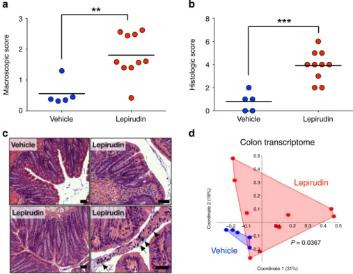
Constitutive thrombin activity preserves mucosal homeostasis.
To determine the physiological role of thrombin at mucosal
surfaces, we inhibited its activity, by administering the irreversible
thrombin-specific inhibitor, lepirudin, intracolonically to mice
25.
After ten daily administrations (10 µg per day per mouse), we
observed a significant increase of macroscopic damage compared
to animals receiving intracolonic administration of vehicle (NaCl
0.9%) (Fig.
3
a). Histological analysis also revealed significant
damage in the distal colon of animals treated with lepirudin
(Fig.
3
b). The histological damage consisted of crypt elongation,
area of goblet cell depletion, as well as neutrophil transmigration
into the lumen (Fig.
3
c). We performed a transcriptomic qPCR
analyses of genes involved in host-microbiota related functions
(Defb4, Tff3, Reg3g Reg4b, Camp, Muc2), tight junctions
(Zo1, Ocln, Cldn1, Cldn2, Cldn5) as well as inflammatory
markers (Cox2, Nos2, TNFa, IFNg, IL17A, Adgre1, Cxcl1, IL1b) in
the distal colon of vehicle- and lepirudin-treated animals
(Supplementary Figure 3). We used a principal coordinate
ana-lysis (PCoA) with Bray-Curtis dissimilarity matrix to show
overall distance in each mouse transcriptome (Fig.
3
d). As
illu-strated by PcoA, but also by hierarchical clustering dendrogram
(Supplementary Figure 3A), there was a significant shift between
the transcriptome of vehicle- and lepirudin-treated groups
(Permanova P value
= 0.0367). The strongest discriminants in
this shift were group of genes involved in host-microbiota
interactions (Camp, Muc2 and Tff3; Supplementary Figs. 3 and 4)
as well as the inducible nitric oxide synthase (Nos2). This
separation caused by lepirudin failed to reach significance for
inflammatory genes (Permanova P value = 0.0949,
Supplemen-tary Figure 3C) or tight junction genes (Permanova P value
=
0.553, Supplementary Figure 3D). Together, these results
demonstrate that inhibition of epithelial thrombin activity in the
lumen is sufficient to cause intestinal injuries and to cause
alterations in the host-microbiota related transcriptome.
Impact of constitutive thrombin activity on gut microbiota. To
determine whether constitutive thrombin activity would change
the composition and relative abundance of gut microbiota, we
sequenced the 16 S rDNA V3–V4 regions from fecal samples
of mice at day 0 and after 10 days treatment with lepirudin
(Supplementary Figure 5A). Principal component analysis
revealed that the shift induced by the intracolonic administration
of the thrombin inhibitor lepirudin was modest as group’s convex
hulls were largely superimposed (Supplementary Figure 5B).
However, the cladogram obtained by LEfSe (Linear discriminant
a
3
Macroscopic score
Vehicle Lepirudin Vehicle
Colon transcriptome Lepirudin Vehicle P = 0.0367 Coordinate 2 (18%) Coordinate 1 (31%) –0.2 –0.2 –0.1 0.1 0.1 0.2 0.2 0.3 0.3 0.4 0.4 0.5 0.5 –0.1 Lepirudin Histologic score 2 1 0 8 6 4 2 0
b
c
d
Fig. 3 Intestinal homeostasis is compromised by inhibition of thrombin activity in colon lumen. C57Bl/6 mice were treated daily with either vehicle (n= 5)
or lepirudin (n= 10, 10 µg/day) via intracolonic route, euthanized after 10 days and used for further analysis (a–d). a Macroscopic (Wallace scoring
endpoint58) andb. Microscopic damage scores49in lepirudin-treated group were significantly greater than the scores measured in vehicle controls. a, b
Data are represented as scatter plot with the mean bar. Unpaired Mann–Whitney two-tailed test ***P < 0.001 c Hematoxylin-eosin images revealed a
normal histology in vehicle-treated group, while in lepirudin-treated group, we observed crypt elongation (lower left panel), areas with goblet cell depletion
(upper right panel), as well as neutrophils transmigration in the lumen (lower right panel, arrows). Scale bars represent 50µm. Representative images of
vehicle (n= 5) and lepirudin-treated (n = 10) group are reported. d Transcriptome analysis of Muc2, Camp, Tff3, Defb4, Reg3g, Reg3b, Cox2, Nos2, Tnf, Ifng,
Cxcl1, IL17a, Adgre1, IL1b, Cldn1, Cldn2, Cldn5, Tjp1 Zo1 and Ocln genes was performed by qPCR. Principal coordinate analysis with Bray-Curtis dissimilarity
matrix illustrates distances between each mouse transcriptome (each dot represents an individual mouse transcriptome), and demonstrate a significant
analysis effect size) analysis showed a lepirudin-specific microbial
signature characterized by a higher abundance of the genus
Barnesiella (belonging to the phylum Bacteroidetes and family
Porphyromonadaceae), compared to the vehicle-treated group
(Supplementary Figure 5C).
As the physiology of complex microbial communities is
strongly dependent on the immediate surroundings of each
microbe, we then asked if thrombin would alter the microbial
microenvironment. Fluorescent in situ hybridization was used to
visualize spatial organization of microbiota biofilm. In animals
treated with vehicle, no mucosa-adherent bacteria were observed,
and no bacteria were detected deep in the crypts, within the
epithelial cells or in the submucosa. Microbiota in the distal
colonic mucosa was clearly separated from the epithelial surface
by a dense mucus layer, free of bacteria, although high density
microbial biofilms were obvious in areas where mucus contacted
fecal material (Fig.
4
a). Intestinal biofilms in animals treated with
lepirudin had a strikingly different spatial organization from that
of vehicle-treated mice (Fig.
4
b), resulting in an overall higher
biofilm damage score (Fig.
4
c, Table
1
). Short and long rod
bacteria could be seen forming microcolonies or partially
segregated from each other at the outer edge of the mucus layer
(when present) and feces. These microcolonies were largely
encased in a loose polysaccharide-rich matrix (Fig.
4
b). Lepirudin
allowed isolated planktonic bacteria to colonize the submucosal
tissues (yellow star, Fig.
4
b). As this result indicated a possible
breach of the mucosal barrier, we investigated whether
commen-sals were able to translocate across the mucosa to distant organs
such as mesenteric lymph nodes. The results show that the
numbers of both aerobic and anaerobic bacteria were significantly
increased in the mesenteric lymph nodes of mice treated with
intracolonic administration of the thrombin inhibitor, lepirudin,
compared to animals treated with the vehicle alone (Fig.
4
d).
Overall, the
findings indicate that while ablation of mucosal
thrombin activity modestly alters microbiota community
com-position, it does create opportunities for microbial invasive
behavior. These results suggest that constitutive thrombin activity
at the surface of intestinal mucosa exerts a constraining role on
mucosal biofilms, that prevent its contact with the epithelial
surface.
Thrombin alters human gut microbiota bio
film structure. To
further explore if and how thrombin could contribute further to
Bacterial translocation into MLNs
Vehicle Vehicle 106 10 8 6 4 2 0 CFU/g of tissue
Biofilm damage score
105 104 103 102 Lepirudin Lepirudin Vehicle Lepirudin Aerobic (24 h) Anaerobic (48 h) Vehicle Lepirudin
a
b
c
d
Fig. 4 Suppression of basal thrombin activity modifies spatial organization of microbiota biofilm. C57Bl/6 mice were treated daily with either vehicle (n =
5) or lepirudin (n= 10, 10 µg per day) via intracolonic route, euthanized after 10 days. a, b Distal colon sections were Carnoy’s fixed. All bacterial cells were
labeled with the universal probe Eub338 forfluorescent in situ hybridization (red color). Wheat germ agglutinin was used to stain the polysaccharides-rich
mucus layer on these samples (green color). All images were counterstained with the nuclear stain, 4’,6-diamidino-2-phenylindole DAPI (blue color).
a In vehicle-treated colon, double-arrows highlights the presence of sterile mucus layer separating the colon epithelium (dashed line) from the dense
microbiota biofilm. b In contrast, lepirudin-treated animals were characterized by a complete disorganization of microbial biofilms and mucus layer in distal
colon. Adherent and isolated microcolonies were evident (arrows) and rods were visible in submucosal tissue (asterisks). Images are representative of n=
5 mice. Scale bars represent 20μm. c Abnormal alterations to gut microbiota biofilm organization were blindly recorded and demonstrated an increased
damage score in animals treated with lepirudin compared to vehicle control groups. Data are represented as scatter plot with the mean bar. Unpaired
Mann–Whitney two-tailed t test ***P < 0.001. d Bacterial translocation, of aerobes and anaerobes, into mesenteric lymph nodes (MLN) was significantly
greater in animals treated with lepirudin compared to vehicle-treated animals. Data are represented as scatter plot with the mean bar. Unpaired
the spatial segregation of commensal biofilms, we first cultured
mucosa-associated microbiota, from 4 healthy human colon
biopsies, in the presence of active human thrombin in liquid
media. After 15 h exposure under these specific conditions of
incubations, no direct bactericidal or bacteriostatic property was
detected upon thrombin exposure (Supplementary Figure 6). We
then reconstituted mucosa-associated human microbiota under
its natural biofilm phenotype in anaerobic conditions. Mature
human biofilms were generated for 48 h and were then exposed
for an additional 24 h to active or inactivated human thrombin.
Biofilm biomass (cells and total matrix-associated content) was
significantly reduced concentration-dependently, starting from
10 mU ml
−1of active thrombin (Fig.
5
a). Boiled thrombin
(500 mU ml
−1) did not cause such an effect (Fig.
5
a), nor did
Table 1 Bio
film damage score of the distal colon microbiota
Biofilm damage score
0 1 2 3
Inner mucus layer invasion Rare Few Numerous Numerous and dense colonies
Biofilm distance to epithelia >20µm >10µm Numerous contact Dense biofilm in contact
Biofilm density High Mild Scattered Mostly planktonic
Bacterial translocation into lamina propria None Few Numerous Dense colonies
Immune cell transmigration into the biofilm None Scattered Dense Dense and extracellular DNA
Bacteria morphology More than 5 morphotypes <4 morphotypes <3 morphotypes Predominance of one morphotype
donor #1 120 100 80 60 40 20 0 120 100 80 60 40 20 0 0 1 10 100 1000 1000 0 10 Active hF2 (mU/ml) T
otal biofilm biomass
(% of v ehicle) Boiled hF2 (mU/ml) 50 100 500 500 donor #2 donor #3 donor #4 donor #5 Average Average donor #1 donor #3 donor #2 Active hF2 (mU/ml) Boiled hF2 (mU/ml) Matr ix proteins content (% of v ehicle) Vehicle +F2 Vehicle +F2 Bacteria Bacteria Matrix proteins Matrix proteins Vehicle +F2
a
b
c
d
Fig. 5 Thrombin alters multispecies human biopsy microbiota anaerobic biofilms. Multispecies anaerobic biofilms from 5 different human healthy donors
were generated ex-vivo. Mature biofilms were then exposed to various concentrations of human active thrombin or inactive boiled thrombin for 24 h.
a Active thrombin, but not boiled thrombin, dose-dependently reduced total biofilm biomass (crystal violet assay), (b) as well as reduced the total content
of matrix-associated proteins (FilmTracer Ruby matrix biofilm stain). A & B Data are presented as mean ± SEM. One-way ANOVA with Fisher’s LSD test vs.
control group without thrombin *P < 0.05, **P < 0.01, ***P < 0.001. Each concentration with > 12 biofilms per donor, N = 3 independent experiments.
c Representative confocal 3D-surfaces reconstructions from human biofilms confirmed a strong effect of thrombin (100 mU ml−1, equivalent to 1 nM, 24 h)
on matrix-associated proteins (green color), while total adherent bacteria staining (propidium iodide, in red) was marginally affected. Scale bars represent
20µm. Representative images from at least 3 independent experiments. d Scanning electron microscopy was performed on the same human biofilm
treated or not with thrombin (100 mU ml−1, equivalent to 1 nM, 24 h). Biofilms from vehicle-treated group were fully covered with a thick matrix slime with
unnoticeable bacteria (upper panel). Biofilm treated with thrombin had an altered matrix structure, bacteria cells becomes clearly visible (lower panel).
thrombin in the presence of its specific inhibitor lepirudin
(Supplementary Figure 7). At concentrations lower than 100 mU
ml
−1, thrombin had no effect on bacterial viability within
bio-films (Supplementary Figure 8A). Starting at concentrations of
50 mU ml
−1, active human thrombin increased the dispersion of
live biofilm-derived bacteria (Supplementary Figure 8B). To
understand further the origin of biofilm biomass reduction, we
stained matrix-associated proteins (SYPRO biofilm matrix) and
polysaccharides (wheat germ agglutinin). We then extracted the
biofilm matrices by disruption of electrostatic interactions using
1.5 M NaCl buffer (pH 7.4), and measured the amount of
fluor-escently labeled matrix-associated proteins and polysaccharides.
We found that thrombin, starting at 1 mU ml
−1, significantly
reduced total content of matrix-associated proteins (Fig.
5
c). At
higher concentrations (>100 mU ml
−1), thrombin reduced the
total polysaccharide content in biofilms (Supplementary
Fig-ure 8C and 8D). Scanning electron microscopy revealed bacterial
cells hidden beneath the dense and fully covering matrix slime in
untreated human biofilms (Fig.
5
d, upper panel). When a biofilm
from the same patient was treated with human thrombin
(100 mU ml
−1for 24 h), individual bacteria became visible as the
matrix was severely damaged (Fig.
5
d, lower panel). Further, we
have investigated the molecular mechanisms by which active
thrombin (100 mU ml
−1for 24 h) cleaves matrix-associated
proteins by using N-terminomics/TAILS (Terminal Amine
Iso-topic Labeling of Substrates, identification of endoproteolysis
site)
26,27and shotgun proteomics analysis (Supplementary
Fig-ure 9). The list of biofilm-peptides processed by human thrombin
including the cleavage site positions are shown in Supplementary
Table 2, together with the putative protein corresponding to the
cleaved peptides and the microbial species known to express such
peptides. Overall, these data provide evidence that human
thrombin can damage mature human multispecies biofilms,
through enzymatic processing of selective protein constituent of
the biofilm matrix backbone.
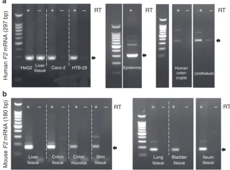
Thrombin is expressed in skin, lung, bladder and ileon. As
thrombin (F2 gene) appeared to be important for intestinal
mucosa homeostasis by constraining microbial biofilms, we
hypothesized that epithelia from all major host-microbiota
sur-faces can also produce thrombin for the same purpose. We
detected F2 mRNA in epithelial cell lines derived from intestine,
skin and lung, in crypt epithelium and bladder urothelium
har-vested from healthy human tissues (Fig.
6
a) as well as from
C57Bl/6 mice (Fig.
6
b). We sequenced the specific F2 amplicon
(297 bp for human, 180 bp for mouse), and performed a blast
alignment revealing a > 99% homology with the respective human
and mouse F2 genes, confirming an active transcription of the
thrombin gene. These data further expand our knowledge on the
presence of active thrombin in epithelial organs, which at least in
the intestine is under the direct regulation of microbiota. Our
data thus point to a previously unknown role for thrombin
epithelia-biofilms interactions.
Discussion
The present
findings demonstrate that thrombin, a serine
pro-tease classically involved in the coagulation cascade and known to
be produced in the liver, can originate from intestinal epithelium,
where it can in principle, contribute to mucosal protection by
Human F2 mRNA (297 bp) Mouse F2 mRNA (180 bp) Liver
tissue tissue mucosa tissue Colon Colon Skin
tissue Ileum tissue tissue Bladder Lung + – + – + – + – + – + – + – + – + – + – + RT + – + – RT RT RT RT HeG2 Liver
tissue Caco-2 HTB-29 Epidermis
Urothelium Human colon crypts
a
b
Fig. 6 The F2 gene encoding thrombin is expressed in all major epithelia under healthy conditions. F2 mRNA transcript (thrombin) was detected by reverse
transcription PCR in (a) human and (b) C57Bl/6 mouse epithelia. a F2 mRNA is present in lane RT+ corresponding to human hepatocyte cell line (Hep
G2), liver tissue, intestinal epithelial cell line (Caco-2), lung epithelial cell line (HTB-29), healthy skin epidermis, human isolated epithelial cells from colon
crypts and primary culture of urothelium. Lanes RT- are negative control for amplification of genomic contamination. Small gaps represent the same gel
from which irrelevant lanes were cut out. Larger gaps correspond to three different gels, PCR conditions were 40 cycles at 60 °C for HepG2, Caco-2,
HTB-39 and 35 cycles at 62 °C for epidermis, colon crypts and urothelium. Amplicon at 297 bp (arrow) was sequenced and confirmed to be human F2 mRNA.
b F2 mRNA is present in lane+ RT corresponding to liver tissue, colon tissue, colon mucosa, skin tissue, lung tissue, bladder tissue, and ileum tissue from
mouse. Lanes -RT are negative control for amplification of genomic contamination. Small gaps represent the same gel from which irrelevant lanes were cut
out. Larger gaps correspond to two different gels, PCR conditions were 40 cycles at 60 °C for liver, colon tissue, colon mucosa, skin and 35 cycles at 62 °C
for lung, bladder and ileum tissue. Amplicon at 180 bp (arrow) was sequenced and confirmed to be mouse F2 mRNA. RT-PCR experiments have been
maintaining healthy spatial segregation between the host and the
microbiota.
Adding to our current report, ectopic expression of thrombin
(both mRNA and protein) has been suspected in the epithelium
from benign and malignant prostate tumors
28, in brain
endo-thelial cells
29, as well as in epithelial-like cells that form ovarian
follicle granulosa cells
30. In our study, we detected unequivocal
expression of thrombin in non-tumoral intestinal, lung, skin and
bladder epithelium. Furthermore, we demonstrated that the
epi-thelial production of thrombin mRNA is directly controlled by
microbiota colonization in the intestine. Interestingly, thrombin
mRNA production in liver remained unchanged in germ-free
mice, suggesting that the intestinal epithelium possesses specific
regulatory mechanisms for the local production of thrombin in
the digestive tract.
Although anticoagulants and direct thrombin inhibitors
administered orally are increasingly used for treatment of chronic
cardiovascular pathologies
31, their safety remains a concern.
Patients commonly suffer from gastrointestinal adverse effects
that may be severe and even fatal in the case of gastrointestinal
bleeding
25,32. Our results demonstrated a protective role of
con-stitutively produced epithelial thrombin. It thus can be
hypo-thesized that chronic inhibition of epithelial thrombin activity by
oral anticoagulant treatments could possibly lead to uncontrolled
and invasive biofilm phenotypes, initiating tissue damage.
Indeed, in Supplementary Figure 10, mice were orally treated with
dabigatran etexilate, a direct thrombin inhibitor used in humans
for cardiovascular pathologies, or with warfarin, a vitamin K
antagonist. We observed in these mice gastrointestinal bleeding
(presence of blood in feces), tissue damage in stomach (petechia,
purpura, edema), and bacterial translocation to mesenteric
lymph nodes.
In addition to its transcriptional regulation, inactive
pro-thrombin has to be cleaved into active pro-thrombin. This activation
can be achieved not only by autoproteolysis, but also by the
activity of many other proteases of the coagulation cascade.
Among the many different possible ways to activate thrombin, we
investigated the concurrent presence along with thrombin, of the
prothrombinase complex composed of Factors 5a and 10a. Both
factors are present (both mRNA and protein) and biologically
active in the intestinal epithelium to transform inactive to active
thrombin. Further, we found that thrombin activity is under the
control of the prothrombinase complex, as Apixaban, an inhibitor
of this complex, was able to inhibit the release of thrombin
activity from intestinal epithelial cells. Pro-thrombin activation
also requires the vitamin K-dependent carboxylase (specifically
the VKORC1 subunit)
33known to be produced by commensal
microbiota. Germ-free mice reach an euthanasia endpoint when
fed an irradiated AIN-76A diet, if not supplemented with vitamin
K
34. Of note, vitamin K deficiency in humans resulting from
broad-spectrum parenteral antibiotic treatment, is associated with
severe gastrointestinal damage, such as bleeding and perforated
gastric ulcers
35. Here we propose that thrombin inhibition in the
lumen of distal colon might directly predispose to mucosal
injuries, at least in part due to microbiota biofilm adhering to
mucosal surfaces. In addition to this previously unknown role for
thrombin in microbiota organization, other pathophysiological
roles for epithelial thrombin are plausible, but yet unexplored.
More research is needed to determine whether constitutive
expression of epithelial thrombin might also regulate epithelial
biology, potentially by protease-activated receptors activation.
On the basis of our results, a role for thrombin activity in the
modulation of Barnesiella abundance is suspected, although
thrombin’s effect on shaping overall microbiota composition is
likely to be minor. Under inflammatory conditions, human
neutrophil elastase is able to cleave out small C-terminal highly
cationic fragments from thrombin, which in turn exert
bacter-icidal effects on isolated pathogens
36,37. Adding to these reports,
our data suggest that full-length active thrombin, exerts a specific
biofilm-disrupting activity that depends on its proteolytic
func-tion. Together, thrombin seems to have a dual action on
microbes: to segregate host mucosa from bacterial biofilms for the
full-length active thrombin, and bactericidal effects on planktonic
bacteria for truncated, proteolytically inactive thrombin.
Inter-estingly, western-blot analysis demonstrated that different forms
of thrombin (pro-form, active form, truncated forms) are present
at the epithelial surface. Each form might exert diverse effects on
bacteria in vivo, adding to the impact of proteolytically active
thrombin on bacterial biofilm biomass.
Biofilm bacteria are embedded in a protective matrix having a
complex
composition
(e.g.,
RNA/DNA,
proteins,
poly-saccharides). The development of this biofilm organization
con-stitutes a major process directly relevant to human and animal
health
20. Therefore, the prevention of biofilm overgrowth and
disruption of already established deleterious biofilms is crucially
important. Destroying the biofilm matrix backbone, for example
via enzymatic lysis, seems to be an interesting approach for
biofilm eradication. Several microbially-derived enzymes have
indeed been reported to degrade components of bacterial biofilm
matrix, although these reports relied exclusively on monospecies
biofilms
38–42. Our study adds a previously unsuspected
compo-nent of biofilm matrix regulation: active thrombin produced by
the host epithelial mucosa. Epithelial-derived thrombin can thus
be added to the list of matrix-degrading antibiofilm agents, with a
strong effect on biofilms originating from complex multispecies
human microbiota. Our study suggests that epithelial-derived
proteolytic factors are produced as a physiological response
towards microbiota biofilms growing at the mucosal surface of
the digestive tract. Our demonstration that thrombin is also
expressed by epithelia from lung, bladder and skin, where the
same role for thrombin could be expected, further suggests that
this novel mechanism may be a target for new therapies to
modify pathogenic biofilm encroachment at these host-microbial
surfaces.
We determined that the concentration of thrombin released by
monolayers of intestinal epithelial cells was in the range of
50 mU ml
−1. We used a similar thrombin concentration for
in vitro experiments on biofilms. Of note, this thrombin
con-centration range is very low compared to the systemic blood
thrombin activity required to induce coagulation (in the range of
10 U ml
−1)
43. Although we report a protective function for basal
thrombin activity, where low concentrations of epithelial
thrombin could be beneficial to control mucosal biofilms and
avoid their direct contacts with tissue, increased thrombin activity
is also associated with chronic inflammation of the gut in cases of
inflammatory bowel disease patients
11. In that case, one can
hypothesize that these relatively high concentrations of thrombin
at mucosal surfaces might be detrimental, fragmenting and
dispersing biofilms to cause tissue damage. Indeed, an inflamed
area of the colon is associated with an overall destruction
of normal biofilm organization, with heterogeneous morphology,
ranging from isolated cells to clusters of epithelia-adherent
biofilms
15,18,22,23,44. Further studies related to the role of
epithelial-derived thrombin in pathologies, and to the control of
thrombin expression and activity, are thus warranted.
The biofilm phenotype of intestinal microbiota has been well
established in the healthy digestive tract and is conserved
throughout the animal kingdom
15,17,19,21,45. The ability to
maintain such biofilms that distance the microbiota from host
tissue is likely of evolutionary significance for digestive health as
well as for host survival. We propose that epithelial thrombin
helps maintain a spatial segregation between the microbiota
biofilm and the host. The present findings can have a direct
impact on human health in view of the link between biofilms
abnormal overgrowth on mucosal surface, in the gut, and beyond
(e.g., inflammatory bowel diseases, colorectal cancer, cystic
fibrosis, urinary tract infections and chronic wound ulcers)
20,46.
Methods
Human tissue collection. Colon cancer resections from human donors were provided by the Centre Hospitalier de Toulouse (France). The Ethics Committee (Comités de Protection des personnes de la Région Occitanie) approved the human research protocol (ClinicalTrials.gov Identifier: NCT01990716) and is compliant with all relevant European ethical regulations. Written and verbal informed con-sent was obtained before enrollment in the study. Biopsies used in the study were collected from macroscopically healthy area at distance from cancer tissue and when necessary transferred in sterile tubes for anaerobic transport (BBL, BD Bioscience). Tissue was manipulated under aseptic conditions and maintained throughout on ice and on PBS sterile buffer. Human crypts were isolated, cultured,
and colon organoids were generated as described in47, the absence of inflammatory
cells in organoid cultures was confirmed (lack of CD45-positive staining on flow
cytometry).
Animals. All animal procedures were approved by the Animal Care and Ethics Committee of US006/CREFE (CEEA-122, APAFIS#7762-20161125092278235) and are compliant with all relevant ethical regulations. C57BL/6 mice were kept in ventilated cages and acclimatized to the study conditions for 2 weeks before entering experiments. Mice used for experiments were 8 weeks old. Germ-free C57BL/6 mice (10–12 weeks old) generated by 2-stage embryo transfer and housed
inflexifilm gnotobiotic isolators were obtained from McMaster University’s Axenic
Gnotobiotic Facility. All protocols were approved by the McMaster University Animal Care Committee and McMaster Animal Research Ethics Board (AREB) in an amendment to the Animal Utilization Protocol (#170836). Tissue samples from these mice were shipped to INSERM-IRSD.
In conventionally bred mice, thrombin inhibition was induced by daily
intracolonic instillation of Lepirudin (10 µg, Bachem, Ki= 0.2 pM) under 3%
isoflurane anesthesia. Control animals were treated with an equal volume of vehicle
(0.9 % NaCl). After instillation, the animals were kept upside-down for 2 min. Animals were euthanized by cervical dislocation after 10 days of experimentation. The cumulative macroscopic damage was obtained by measuring colon thickness
edema (in mm) and blind Wallace scoring endpoints48. Histological damage
scoring was performed on formalin-fixed, paraffin-embedded sections stained with
hematoxylin-eosin, and according to a previously published scoring system49, on 5
fields per tissue section. Macroscopic and histologic scoring was blindly performed by skilled experimenters (one experimenter for macroscopic and two
experimenters for histological scoring). Mesenteric lymph nodes were collected aseptically, weighed, homogenized and plated on Columbia blood agar (BD Biosciences) for 24 h for aerobes and 48 h for anaerobes (anaerobic jar) at 37 °C.
To investigate gastrointestinal damage associated with oral anticoagulants, one
group of mice (n= 12) was treated orally with the direct thrombin inhibitor
Dabigatran etexilate (50 mg/kg/day in mice corresponding to a human dose of∼4
mg/kg/day, SelleckChem, Euromedex, France). Another group (n= 7) was given
warfarin in drinking water ad libitum (10 mg L−1, Bristol-Myers Squibb, France).
The dose based on water consumption intake was 2 mg/kg/day, which is equivalent to a human dose of 10 mg daily. Fecal score was recorded daily, based on i) fecal consistency (score 0 for normal feces, 1 for soft feces, 2 for diarrhea) and ii) the presence of blood in the feces (Hemoccult tests, score 0 for negative, score 1 for positive, score 2 for gross bleeding). We measured potential macroscopic lesions in the stomach according to the following criteria: no macroscopic damage (0), presence of purpura or petechia (1), tissue edema (1). Bacterial translocation to the mesenteric lymph nodes (presence of aerobic and anaerobic bacteria) was also assessed as described above.
Cell culture. Human intestinal epithelial cell lines (Caco-2 (ATCC HTB-37), HT-29 (ATCC HT-38), SW480 (ATCC-CCL-228)), human hepatocyte (Hep G2 from ATCC HB-8065), human lung epithelial cells (A549, ATCC CCL-185) were grown in DMEM high glucose GlutaMAX Supplemented with 1x non-essential
aminoa-cids, 1x penicillin/streptomycin and 10% FBS (Gibco). Briefly, 3 × 10⁵IECs were
plated onflat-bottom 6-well plates and grown for 7 days at 37 °C 5% CO2with
culture medium replacement three times a week. Then, 1 × 10⁵Caco-2 cells were
plated on polycarbonate 12-transwells and grown for 21 days, as described above.
For cellular supernatants harvesting, cells were washed twice with PBS Ca2+/Mg2+
-free and kept for 24 h in the cell culture medium described above, but without FBS.
Gut microbiota biofilms. For mucosa-associated biofilm reconstitution, human
colonic biopsies were transferred in sterile tubes for anaerobic transport (BBL, BD Bioscience) just after collection in the endoscopy room. Colon biopsies were
homogenized in a microtube pestle and mucosa-associated microbiota wasfirst
cultured overnight in rich anaerobe media (Wilkins-Chalgren broth, ThermoFisher
Scientific) supplemented with L-cysteine (5%, Sigma-Aldrich). Biofilms were
gen-erated using the Calgary Biofilm Device (Innovotech, Edmonton, Canada)15,21,50,51.
All steps described herein were performed in anaerobic conditions in jar (Anae-ropack, ThermoFisher Scientific). Mature biofilms (48 h) were transferred onto a new plate of minimal M63 media supplemented with glucose (2%), L-cysteine (0.5%) and containing various concentration of human active thrombin (thrombin
from human plasma, maximal concentration of 1000mU ml−1was equivalent to
10 nM, Sigma T6884), thrombin inhibitor lepirudin (Bachem) or vehicle (phosphate buffered saline, pH 7.4 with 0.1 % bovine serum albumin) for 24 h. The biofilm biomass density and bacterial viability were determined respectively by crystal violet (1% in water, Reactifs RAL 317980) and rezasurin (2% in water, Sigma-Aldrich
R7017)15,21. Matrix-associated proteins and matrix-associated polysaccharides
(N-acetlyglycosamines and sialic acid) were stained respectively with FilmTracer Ruby matrix biofilm stain (ThermoFisher Scientific F10318) and wheat germ agglutinin (1/1000 dilution, Sigma L4895) for 2 h. Matrix was then extracted after incubation for 30 min in 1.5 M NaCl buffer, based on previously described
method52, and specific fluorescence was measured on fluorescent
spectro-photometer (Tecan). Biofilm rate of dispersal was assessed by measuring the optical density (600 nm) and assessing colony-forming unit (CFU) of biofilm-dispersed planktonic bacteria recovered in the challenge plate. All results were expressed as percentages change of that of the means in the vehicle-treated group set as 100 %.
Imaging of biofilms. Biofilms were stained for 1 h, without fixation, with
Film-Tracer Ruby matrix biofilm stain (specific matrix-associated protein stain), fluorescein-labeled wheat germ agglutinin (matrix-associated polysaccharides stain) and propidium iodide (DNA-RNA stain, 1/1000 dilution, Invitrogen P1304MP) and visualized on a confocal microscope (Zeiss LSM 710). Biofilm
damage score was blindly evaluated in 13fields for vehicle group (N = 5 animals)
and 22fields in lepirudin (N = 5 animals), according to the criteria described in
Table1. Three-dimension surface rendering of stained biofilm was performed on
Imaris Bitplane (v8, Concord, MA, USA). Alternatively, pegs containing treated
biofilms were broken with needle nose pliers, and fixed in 2 % glutaraldehyde
(Sigma-Aldrich) in 0.1 M Sorensen phosphate buffer (pH 7.4). Biofilms were dehydrated, dried by critical point drying (Leica EM CPD 300), and coated with 6 nm Platinium on a Leica EM Med 020 before being examined on a FEI Quanta 250 FEG scanning electron microscope, at accelerating voltages of 5 and 10 kV. FIJI
freeware was used forfinal image mounting (v.1.51).
Bacterial growth rate. Human mucosa-associated microbiota were grown for 24 h in rich anaerobe media (Wilkins-Chalgren). Saturated cultures were then diluted in
96-well microplate to optical density of 0.1 (OD600 nm) in minimal M63 media
supplemented with glucose (2%), L-cysteine (0.5%) and containing various
con-centration of human active thrombin (0 to 1250 mU ml−1). The growth curves of
each inoculum were generated from continuous OD600nmreading every 20 min for
15 h. Each value was expressed using means of duplicate experiment for each microbiota.
Tissue imaging. Forfluorescent in situ hybridization (FISH), Carnoy’s-fixed mice
colon tissues were paraffin-embedded. Slides were hybridized with 1 µM of a
universal bacterial 16 Sfluorescent rRNA probe (EUB338-Cy3,
5′-GCTGCCTCCCGTAGGAGT-3′ Cy5, Eurofins) and counterstained for DNA by
4′,6-diamidino-2-phenylindole (DAPI, 1/1000 dilution Sigma-Aldrich) and
poly-saccharides content (wheat germ agglutinin labeled withfluorescein, 1/1000,
Sigma-Aldrich L4895). Human colonic biopsies were cryopreserved in Optimal Cutting Temperature (OCT, Dako) and sectioned at 6 µm of thickness. Slides were thawed at room temperature for 20 min and blocked for 1 h (1% BSA, 0.3% Triton X-100, PBS 1×). Tissues were immuno-stained with goat polyclonal anti-thrombin (1/250 dilution, Santa Cruz, sc-16972) overnight, and double labeled with sec-ondary antibody (anti-goat alexa-555, 1/1000 dilution Life Technologies A211432). Slides were counterstained with 4′,6-diamidino-2-phenylindole (DAPI, Invitrogen,
France), Thioflavin T (0.001% dilution, Invitrogen, T3516) and/or
fluorescein-labeled wheat germ agglutinin (1/1000 dilution, Sigma-Aldrich L4895). Epithelial cells were highlighted with anti-Epcam staining (1/200 dilution; Abcam, ab-32392). To reveal unspecific staining of thrombin antibody, isotype control (Santa Cruz, sc 2028) was used and incubated at the same concentration and under the same experimental conditions (Supplementary Figure 11). Representative images were
obtained from blind acquisition of 4 differentfields per animals. We acquired all
images on Leica LSM 710 confocal microscope, and FIJI freeware was used forfinal
image mounting (v.1.51).
16S rDNA Sequencing. Total DNA was extracted from feces at both baseline and after the treatment with lepirudin according to manufacturer’s protocols (QIAamp
DNA stool mini kit, Qiagen 51604) with slight modifications as described in ref.53.
The 16 S bacterial rDNA V3–V4 regions were targeted by the 357wf-785R primers and analyzed by MiSeq at RTLGenomics (Texas, USA). An average of 11,000 (between 6896 and 15901) sequences was generated per sample. A complete
description of the applied bioinformaticfilters is available atwww.rtlgenomics.com.
(huttenhower.sph.harvard.edu/galaxy/) via the LEfSe (Linear Discriminant
Ana-lysis Effect Size) algorithm54.
Transcription assays. mRNA from intestinal epithelial cells, crypts and organoids were extracted by using the Nucleospin RNA/Protein kit (Macherey-Nagel). mRNAs from other human and animal tissues were extracted using the Qiagen RNeasy kit according to the manufacturer’s instructions (Qiagen) and reversely transcribed into cDNA (iScript cDNA synthesis kit, Biorad). The PCR was per-formed on 384-well plates and on LightCycler 480 (Roche). The sequences used in the study are detailed in Supplementary Table 1. The expression levels of genes were normalized to both Glyceraldehyde 3-phosphate dehydrogenase (GAPDH) and hypoxanthine-guanine phosphoribosyltransferase (HPRT) as reference genes.
Fold changes in the mRNA levels were calculated with the comparative 2−ΔΔCt
method. For RT-PCR blot analysis, 1 µg mRNAs were reverse transcripted using Maxima First Strand cDNA kit (Thermo Fisher). Subsequent PCR was performed on 50 ng of cDNA at 60 °C for 40 cycles or 62 °C for 35 cycles. The PCR products were separated on a 2% agarose gel and stained with ethidium bromide. Gel images were captured using Quantum ST4 1000/26MX (Fisher Scientific). Specific band were sequenced and were blasted on National Center for biotechnology informa-tion (blastn NCBI), and aligned using Clustal Omega program. All the sequences correspond to thrombin (99% homology). Representative gels were selected from at least three independent experiments.
Measurement of thrombin activity. Proteolytic activity was measured in Caco-2 cell supernatant samples with BOC-Val-Pro-Arg-amino-4-methylcoumarin hydro-chloride (0.5–150 µM, Sigma-Alrich B9395) as substrate in 50 mM Tris, 10 mM
CaCl2, 150 mM NaCl, pH= 8.3. Thrombin activity was identified from overall
Arg-cleaving enzymes by pre-incubating supernatants with increasing concentrations of the specific thrombin inhibitor lepirudin for 30 min at 37 °C (15.6–1000 pM; Bachem, GmbH), or specific F10 inhibitor Apixaban (0.01 µM, 0.1 µM, 1 µM in 0.1% DMSO vehicle, Selleckchem S1593) for 24-h in serum-free culture medium.
Velocity (reaction rate per min) was calculated by the change influorescence
(excitation: 355 nm, emission: 460 nm), measured over 15 min at 37 °C on a Var-ioskan Flash microplate reader (Thermo Fisher Scientific). No thrombin activity was detected using this assay in pure FBS nor in heat-inactivated FBS.
Western blots. Total protein extract of Caco-2 cells was prepared by using the Nucleospin RNA/Protein kit (Macherey-Nagel, GmbH). Proteins from Caco-2 cell supernatant were precipitated in 15% trichloroacetic acid at 4 °C during 90 min.
The pellet was washed twice in cold acetone (−20 °C) and solubilized in 20 µL of
protein solving buffer with tris-(2-carboxyethyl)-phosphine hydrochloride (PSB-TCEP; Macherey-Nagel). Samples were then heated at 95 °C for 5 min, clarified by centrifugation at 12,000 × g for 5 min and the solubilized sample was loaded into
4–20% Mini-Protean TGX precast gels (Bio-Rad, GmbH). Human feces from 3
donors (1 g) were suspended in 1 mL PBS buffer, homogenized, centrifuged and
supernatants were stored in−20 °C. The mucus was collected in buffer (Tris 40
mM, NaCl 150 mM, EDTA 20 mM, pH 8 and protease inhibitor cocktail, Sigma) by scrapping the colon mucosa of human resection. Lysed samples from human and mouse tissues and feces were diluted in Laemmli buffer 4 × (Biorad), supplemented with 2-mercaptoethanol and heated at 95 °C. Samples were run with at least 20 µg
of total protein on Precast Gel 4–15% (Biorad) and transferred to nitrocellulose
membrane (Biorad). The membrane was blocked 1 h (PBS, 5% milk and 1% bovine serum albumine) and incubated overnight with anti-thrombin antibody (Santa Cruz sc-16972, 1/200 dilution), anti-Factor 10 (Abcam, Ab79929, 1/200 dilution), or anti-Factor 5 (Abcam, Ab108614, 1/200 dilution), in blocking buffer. Detection was achieved using secondary antibody coupled to horseradish peroxidase (donkey anti-goat IgG, 1/3 000 dilution, Promega V805A), and anti-rabbit (1/3 000, Pro-mega, W401B) during 1 h and a chemiluminescent substrate (ECL from Amer-sham, Chemidoc XRS, Biorad). Pro-thrombin and active forms of thrombin were used as control (70 and 60 ng of proteins respectively, Sigma-Aldrich). Repre-sentative blots were selected from at least three independent experiments.
N-terminomics/TAILS workflow. Human microbiota biofilms from 3 different
healthy donors have been generated on the Calgary Biofilm Device as describe above. Biofilms were treated with either vehicle (PBS) or recombinant human
thrombin (100 mU ml−1, equivalent to 1 nM) for 24 h at 37 °C. Biofilm-associated
proteins were extracted in 6 M Urea, 4% SDS buffer, precipitated in trichloroacetic
acid (15%final) and were further processed to N-terminomics/TAILS (and shotgun
proteomics analysis (Supplementary Figure 10). Briefly, samples were alkylated with iodoacetamide, peptide were then labeled with isotopically heavy [40 mM
13CD2O+ 20 mM NaBH3CN (sodium cyanoborohydride)] or light labels [40 mM
light formaldehyde (CH2O)+ 20 mM NaBH3CN]. Samples were then processed
for N-terminal enrichment27,55and processed to high performance liquid
chro-matography (HPLC) and mass spectrometry (MS) at the Southern Alberta Mass Spectrometry (SAMS) core facility at the University of Calgary, Canada. Spectral data were matched to peptide sequences in the human UniProt protein database of twelve common bacterial species using the Andromeda algorithm as implemented in the MaxQuant software package v.1.6.0.1, at a peptide-spectrum match false discovery rate (FDR) of < 0.05. The cleavage site specificity was set to semi-ArgC
(free N-terminus), with up to two missed cleavages allowed. Significant outlier cut-off values were determined after log(2) transformation by boxplot-and-whiskers
analysis using the BoxPlotR tool56(Supplementary Figure 10).
Statistical analyses. Graphic representation and statistical analyses were per-formed using GraphPad Prism (v6, La Jolla, USA). Mann–Whitney’s non-parametric t tests were used accordingly after D’Agostino-Pearson normality test.
For multiple variables, we used two-way ANOVA followed with Fisher’s LSD test.
An associated P value less than 5% was considered significant. All central values are means for dot-plots and histograms. Error bars represent standard error of the mean. To represent visual distances in multivariate data set (metagenomic and transcriptomic), we used principal co-ordinate analysis (PCoA) using Past 3
soft-ware57. Permutational multivariate analysis of variance (PERMANOVA
Bonfer-roni corrected) with Bray-Curtis dissimilarity was used for comparing
transcriptomic data set (Past 3). Images for western-blots, RT-PCR and microscopy were obtained from at least 3 independent experiments and/or at least 3 inde-pendent human donors.
Reporting Summary. Further information on research design is available in the Nature Research Reporting Summary linked to this article.
Data availability
The sequencing data presented in the manuscript are found in the NCBI database under BioProjectPRJNA549826. This project contains the raw 16 S sequencing data deposited in the Sequence Read Archive (SRA) with accession codeSRP201956. The raw proteomic data have been deposited in the ProteomeXchange Consortium via the PRIDE partner repository with the data set identifierPXD014315. A reporting summary for this article is available as a Supplementary Informationfile. The source data used for Fig. 1–6 and Supplementary Figures 1–10 are provided as a Source Data file. Other data supporting thefindings of this manuscript are available from the corresponding author upon request.
Received: 22 November 2018 Accepted: 25 June 2019
References
1. Bonnart, C., Deraison, C., Lacroix, M. & Hovnanian, A. [Elastase 2, a key
player in the integrity of the epidermal barrier and in Netherton syndrome]. Med. Sci. 26, 681–685 (2010).
2. Descargues, P. et al. Spink5-deficient mice mimic Netherton syndrome
through degradation of desmoglein 1 by epidermal protease hyperactivity. Nat. Genet. 37, 56–65 (2005).
3. Meyer, M. & Jaspers, I. Respiratory protease/antiprotease balance
determines susceptibility to viral infection and can be modified by nutritional antioxidants. Am. J. Physiol. Lung Cell Mol. Physiol. 308, L1189–L1201 (2015).
4. Vergnolle, N. Protease inhibition as new therapeutic strategy for GI diseases.
Gut 65, 1215–1224 (2016).
5. Rolland-Fourcade, C. et al. Epithelial expression and function of trypsin-3 in
irritable bowel syndrome. Gut 66, 1767–1778 (2017).
6. Hansen, K. K. et al. A major role for proteolytic activity and
proteinase-activated receptor-2 in the pathogenesis of infectious colitis. Proc. Natl Acad. Sci. USA 102, 8363–8368 (2005).
7. Gobbetti, T. et al. Serine protease inhibition reduces post-ischemic granulocyte
recruitment in mouse intestine. Am. J. Pathol. 180, 141–152 (2012).
8. Cenac, N. et al. Role for protease activity in visceral pain in irritable bowel
syndrome. J. Clin. Invest. 117, 636–647 (2007).
9. Motta, J. P. et al. Food-grade bacteria expressing elafin protect against
inflammation and restore colon homeostasis. Sci. Transl. Med. 4, 158ra144 (2012).
10. Motta, J. P. et al. Modifying the protease, antiprotease pattern by elafin
overexpression protects mice from colitis. Gastroenterology 140, 1272–1282
(2011).
11. Denadai-Souza, A. et al. Functional Proteomic Profiling of Secreted Serine Proteases in Health and Inflammatory Bowel Disease. Sci. Rep. 8, 7834 (2018).
12. Cirino, G. & Vergnolle, N. Proteinase-activated receptors (PARs): crossroads between innate immunity and coagulation. Curr. Opin. Pharm. 6, 428–434 (2006).
13. Siller-Matula, J. M., Schwameis, M., Blann, A., Mannhalter, C. & Jilma, B. Thrombin as a multi-functional enzyme. Focus on in vitro and in vivo effects. Thromb. Haemost. 106, 1020–1033 (2011).
14. Huntington, J. A. Thrombin plasticity. Biochim Biophys. Acta 1824, 246–252