MITLibraries
Document Services Room 14-0551 77 Massachusetts Avenue Cambridge, MA 02139 Ph: 617.253.5668 Fax: 617.253.1690 Email: docs@mit.edu http://libraries.mit. edu/docsDISCLAIMER OF QUALITY
Due to the condition of the original material, there are unavoidable
flaws in this reproduction. We have made every effort possible to
provide you with the best copy available. If you are dissatisfied with
this product and find it unusable, please contact Document Services as
soon as possible.
Thank you.
Some pages in the original document contain color
Cloning, Sequencing, Expression, and Characterization of the Adenosylcobalamin-Dependent Ribonucleotide Reductase from
Lactobacillus leichmannii
by
Squire J. Booker
B.A. Chemistry, Austin College (1987)
Submitted to the Department of Chemistry in Partial Fulfillment of the Requirements
for the Degree of DOCTOR OF PHILOSOPHY
at the
Massachusetts Institute of Technology September 1994
© Massachusetts Institute of Technology, 1994 All rights reserved
Signature of Author___
.. . -
Department of Chemistry
September 7, 1994
Certified by
Professor JoAnne Stubbe
Thesis Supervisor
Accepted by
Dietmar Seyferth
Chairman, Departmental Committee on Graduate Students
MASSACHi n'INSTTU JEThis doctoral thesis has been examined by a Committee of the
Department of Chemistry as follows:
Professor William H. Orme-Johnson _,- - .. .- - T *
-k4j-irperson
Professor JoAnne Stubbe :W -Is-- ( I., %II, -~ - -S T hs p.eris
Thesis Supervisor
To my grandmother, Cleona Price,
Cloning, Sequencing, Expression, and Characterization of the Adenosylcobalamin-Dependent Ribonucleotide Reductase from
Lactobacillus leichmannii by
Squire J. Booker
Submitted to the Department of Chemistry on September 7, 1994 in Partial Fulfillment of the Requirements for the Degree of
Doctor of Philosophy
ABSTRACT
The ribonucleotide reductases (RNRs) catalyze the rate limiting step in DNA biosynthesis, the conversion of ribonucleotides to 2'-deoxyribonucleotides. Four classes of RNRs are currently acknowledged, with each class differing in cofactor requirement. The two best-characterized reductases to date are the differic iron center-tyrosyl radical-dependent reductase isolated from Escherichia coli (RDPR),
and the adenosylcobalamin-dependent reductase isolated from Lactobacillus
leichmannii (RTPR). Although these two enzymes differ in the cofactor used to
effect nucleotide reduction, numerous studies in our laboratory have suggested
that their mechanisms of catalysis are astonishingly similar. In the work
reported herein, the RNR from L. leichmannii was cloned, and its DNA and protein sequences determined. In addition, the protein was hyperexpressed in
E. coli under the control of the tac promoter. The enzyme has been purified to
>95% homogeneity, yielding --90 mg of protein from 2.5 g of the recombinant
E. coli. Although the amino acid sequence of RTPR displays no statistical
homology with that of RDPR, a thorough review of the sequence by eye resulted in the discovery of 4 cysteines which lay in sequence motifs similar to those of
cysteines previously found to be important in catalysis in RDPR. An additional cysteine was implicated from early biochemical studies as being important in catalysis. These five cysteines, as well as a control cysteine, were mutated to serines, and the effect of the resulting mutant proteins on nucleotide reduction was analyzed. Two of the mutant proteins were unable to catalyze nucleotide
reduction, but produced similar products to the wild-type enzyme when
incubated with the mechanism-based inhibitor 2'-chloro-2'-deoxyuridine
5'-triphosphate. One mutant protein produced no product at all. The mutated cysteines from these proteins were assigned to the active site of RTPR. Two additional cysteines were inactive in the presence of the in vivo reductant, the protein thioredoxin, but catalyzed nucleotide reduction at the same rate as wild-type RTPR when DTT, a small organic dithiol, was used as the reductant. These cysteines were thus assigned the role of a redox shuttle. Their function in the cell is to deliver reducing equivalents from thioredoxin into the active site disulfide formed concomitant with nucleotide reduction.
Thesis Supervisor: Professor JoAnne Stubbe
Acknowledgments
My MIT graduate school experience has finally come to an end, and I need to thank all of the people who contributed to making it a net positive one. First, I
want to thank Professor JoAnne Stubbe for her support (both financial and
scientific) and guidance. I am indeed awed by her unwavering committment to science. In my (x-l) years in her lab, I have never found her unwilling to discuss
science, or too busy to answer questions about biochemistry. Her
almost-childlike fascination with biochemistry is both refreshing and inspiring. As
appropriately stated by Ernie Mueller, "Her example is one I will never forget." Secondly, I want to thank all of the past and present members of the Stubbe lab - especially the early group members who experienced many of the "classic moments." I can never read a paper on prostaglandin synthase and not think of Ernie. In my mind he is indelibly linked to that enzyme. I have the utmost respect for Ernie and his "strength in the face of adversity." Johannes Rudoph had two "classic moments," both of which involved the postal service. I wish him the best, and I hope he's able to channel is way to success. Heidi, Regis, and Erik were also fortunate enough to experience and contribute to the "classic moments." Heidi's "busy social schedule" classic ranks with the best there is. I want to thank Rgis for helping to arrange my stay in Paris (merci beaucoup Rgissimo), and Erik for the long and exhausting bike rides. I wish them all the best. Pam England has experienced "classic moments" in her own lab, which she
has shared with various members of our laboratory. I've enjoyed talking and
networking with Pam, and I especially appreciate her in-depth analyses of many aspects of being a scientist. I'll definitely remember her for her famous quote, "You haven't really been to hell if you're willing to go back." Dr. Shishan Mao
was a classic in and of himself, and an exceptional scientist. His studies on the
E. coli enzyme paved the way for the mutagenesis studies on the L. leichmannii
enzyme.
Other members whom I'd like to thank are Jennifer Banzon, who made coming to lab a pleasant experience, despite the fact that I was in 3 2P/Southern hybridization hell at the time. Wing Hang Tong has always been a special friend, and was also 1/3 of the Rainbow Coalition at Marty's wedding. I want to especially thank her for the many scrumptious meals that she cooked. In addition, I certainly have to thank Wei Wu (the consumer/mover/shaker) for getting us all set up on our $40,000 mailbox/CD player, and for giving me a place to stay for the last two months. I thank Dr. Joan Broderick and Stuart Licht for their many contributions to this thesis. I especially want to thank Stuart for his intellectual contributions to our project, and for doing more than his share of the grunt work. I also want to thank Diana Lewicki, our secretary, for looking out for me and taking good care of me.
The members of the Stubbe lab that I want to thank most are Mike Absalon and Marty Bollinger. We three have certainly experienced the "classic moments." In fact, I wonder if Marty has gotten that review article out yet. The times that I spent with these guys were my best in Boston. I learned a huge amount from the large and detailed discussions that we had. Mike and Marty are both exceptional scientists, and I know they'll do really well.
I thank the people in the graduate office, especially Margo Tyler and Ike Colbert, for their support and encouragement. The graduate office is like Cheers - it's where everyone knows your name. I appreciate their endeavors to make MIT a more "colorful" place.
I definitely have to thank my roomy of 5 years, Delton Cox. Delton's unique outlook on life kept me laughing continuously. Living at 505 Washington
Ave in Chelsea is an experience that I'll never forget. I wish him a speedy recovery, and continued success in finishing his studies here at MIT.
My most sincere thanks goes to my family. All of them. I never would have gotten through the front door of MIT if it weren't for their steadfast support. I was extremely fortunate to grow up around three super uncles (Uncle 0, Uncle A.J., and Uncle Richard), who got me interested in learning, and who provided me with more than I needed to be successful in my endeavors. I have to thank my mother for passing on her sense of humor to me. It's truly a survival tool. Her phone calls kept me in stitches even during some of my most trying times. I thank my father too for his support and help. I have to thank Dr. Joseph Francisco who got me interested in doing doctoral work at MIT. I also wish him continued success in his scientific endeavors. I have to thank my cousin DeLisa for always being supportive. (I know she'll probably be perturbed that I didn't include a photo alongside her name.) Finally, I want to thank my grandmother and my aunt Anita Price (she's gonna love seeing her name in print) for taking good care of me, and providing me with a home filled with love.
Table of Contents
Page Abstract Acknowledgments Table of Contents Figures Schemes Tables AbbreviationsChapter 1: The Ribonucleotide Reductases: Radical Enzymes Employing Radically Different Cofactors
Characterization of Ribonucleotide Reductase Activity Classes of Ribonucleotide Reductase
E. coli Ribonucleotide Reductase
Lactobacillus leichmannii Ribonucleotide Reductase
Ribonucleotide Reductasefrom Anaerobically-grown Escherichia coli
Brevibacterium ammoniagenes Ribonucleotide Reductase Mechanism of Ribonucleotide Reduction
Modelsfor the Mechanism of Ribonucleotide Reduction The Nature of Xe
Interaction of Ribonucleotide Reductase with
2'-Chloro-2'-deoxyuridine 5'-di(tri)phosphate
Attempts to Locate the Redox-active Cysteines of RNR
4 5 8 15 20 22 25 27 32 34 34 37 43 44 46 46 51 55 61
Investigation of the Mechanism of Ribonucleotide Reduction with
Protein
Analogs
. .References . . . .
Chapter 2: Isolation and Sequencing of the Gene for Ribonucleoside Triphosphate Reductase from Lactobacillus leichmannii
Materials and Methods
Materials
...
Peptide Mapping .
Synthesis and Purification of Oligomers
Isolation of High Molecular Weight Genomic DNA Southern Transfers . .
Radiolabeling of Oligomers .
Screening of L. leichmannii Genomic DNA
Isolation of 6.6 kb HindIII Fragments and Construction of a Subgenomic Library . .
The Polymerase Chain Reaction (PCR)
Cloning and Sequencing of the Fragment Isolated by PCR
Results . . .
Peptide Mapping .. . .
Design and Construction of Hybridization Probes and PCR Primers for the Polymerase Chain Reaction (PCR) . .
Initial Attempts to Clone the L. leichmannii Ribonucleotide
Reductase .
Isolation of High Molecular Weight Genomic DNA from
L. leichmannii . . .
Screening of L. leichmannii Genomic DNA .
63 70 80 89 90 91 92 94 95 95 96 97 98 101 101 105 112 118 119
Attempts to Create a Subgenomic Library .
Cloning of the L. eichmannii Genefor RTPR by PCR
Partial Cloning of the Fragment Obtainedfrom the PCR Reaction Determination of the Gene Sequence of RTPR
Discussion .
References . . 134 . 143 · 147 . 150 · 159 · 175Chapter 3: Expression, Purification, and Characterization of Ribonucleoside Triphosphate Reductase from Lactobacillus leichmannii . 180
Materials and Methods . . . . 192
Materials . . . . . 192
Expression-Cassette Amplification of the N-terminal Half of the
RTPR Gene . . . . . . 193
Expression-Cassette Amplification of the C-terminal Half of the
RTPR Gene . . . . . . . . 195
Cloning of the Full-length RTPR Gene into Plasmid pKK223-3 . 195
Expression of RTPR in E. coli HB101 . . 196
Purification of Recombinant RTPR . . . 198
RTPR Activity Determination . . . 199
Kinetic Characterization of Recombinant RTPR . . 199
Analysis of the Ability of dGTP to Stimulate the Reduction of ATP
using Recombinant RTPR . . 200
Transformation of pSQUIRE into E. coli JM105 and Expression of
RTPR under the Control of an Inducible System 200
Expression of RTPR in D20/Minimal Media and Purification of the
Results
Expression-Cassette Cloning of the Entire RTPR Gene into Plasmid
pKK223-3
Expression of RTPR in E. coli HB101
Purification of Recombinant RTPR Characterization of Recombinant RTPR
Inducible Expression of RTPR in E. coli JM105
Expression of RTPR in Minimal Media Containing D20 as the
Solvent Discussion References
er 4: Evidence for the Participation of Five Cysteine Residues in Ribonucleotide Reduction
Materials and Methods
Materials
Preparation of[2-14C]CTP
Preparation of Site-Directed Mutants Growth and Expression of Mutants
Enzyme Assays Using [2-14C]CTP and NaOAc
Enzyme Assays Using [2-14C]CTP and Allosteric Effector . Single-Turnover Experiments with Mutants C731S, C736S, and
C731&736S
Characterization of Oxidized RTPR
Determination of Product Production with Mutants C1 19S and
C419S.
Analysis of the Ability of C408S to Catalyze Nucleotide Reduction
203 203 208 214 217 225 226 229 235 . 237 . 241 241 242 243 247 247 248 . 249 . 249 250 250 Chapt
Circular Dichroism Spectra . . . 251
Characterization of C305S RTPR . .. 251
Results . . . . . .. 252
Preparation of Site-Directed Mutants . . . 252
RTPR Assays . . . .. 253
Characterization of C731S, C736S, and the Double Mutant
C731&736S . .. 254
Assays with C119S and C419S RTPR 259
Characterization of C408S RTPR 263
Characterization of C305S RTPR . 264
Discussion .
.
264
References . .. 278
Chapter 5: A Reinvestigation of the Adenosylcobalamin-dependent Exchange Reaction Catalyzed by the Ribonucleoside
Triphosphate Reductase from Lactobacillus leichmannii . 284
Materials and Methods . .. 294
Materials . . . . . . .294
Purification of [5'-3H]AdoCbl 297
Assayfor Tritium Exchangefrom [5'-3H]AdoCbl 298 Determination of a Kmfor AdoCbl in the Exchange Reaction with
wt RTPR
.
.
299
Determination of a Kmfor dGTP in the RTPR-Catalyzed Exchange
Reaction . . 300
Analysis of the Ability of NaOAc to Replace dGTP in the Exchange
Analysisfor Consumption of Reductant during the Exchange
Reaction . . . . . .301
Analysis of the Ability of Mutants C119S and C419S to Catalyze the
Exchange Reaction . . . . ..
301
Analysis of the Ability of Mutant C408S to Catalyze the Exchange
Reaction . . . . . . .302
Analysis of the Ability of Pre-reduced RTPR to Catalyze the
Exchange Reaction . .. 302
Analysis of the Ability of Pre-oxidized RTPR to Catalyze the
Exchange Reaction . . 304
Isolation of Mutant C32S TR . . 304
Results . . . 305
Assayfor Exchange: Isolation and Quantitation of3H
2O . 305
Deleterious Effects of DTT on the Exchange Reaction . 307
Dependence of the Exchange Reaction on the Choice of Buffer . 311 Determination of an Apparent Kmfor dGTP in the Exchange
Reaction . . .
313
Determination of a Kmfor AdoCbl in the Exchange Reaction . 316
Analysis of the Ability of NaOAc to Substitutefor dGTP in the
Exchange Reaction . . 318
Characterization of the Reductant Dependency on the Exchange
Reaction . 320
Characterization of the Ability of Mutant C731&736S RTPR to
Catalyze the Exchange Reaction 325
Characterization of the Ability of the Active-Site C--S Mutants to
Characterization of the Ability of Oxidized RTPR to Catalyze the
Exchange Reaction . 332
Discussion . . . 337
Figures
Pathway for the de novo biosynthesis of DNA in L. leichmannii. Structure of coenzyme B12.
Postulated role of 5 cysteines involved in nucleotide reduction in the RDPR from E. coli.
Generation of cDNA from isolated messenger RNA.
Isolation of a gene by hybridization to a complementary probe. Peptide map of RTPR generated by trypsin digestion.
Amplification of a target sequence by PCR.
Restriction analysis and gel electrophoresis of zoos 21, 22, and 23. Phosphorimage of Southern transfer and hybridization with primer
I-1.
UV-visible spectrum of genomic DNA isolated from L. eichmannii. Restriction digest of L. leichmannii genomic DNA.
Phosphorimage of a Southern blot probed with primer I-2. Phosphorimage of a Southern blot probed with primer I-2. Phosphorimage of a Southern blot probed with primer I-2. Southern blot probed with primer I-2.
1.1: 1.2: 1.3: 2.1: 2.2: 2.3: 2.4: 2.5: 2.6: 2.7: 2.8: 2.9: 2.10: 2.11: 2.12:
Southern blot of L. leichmannii genomic DNA digested with Eco R1
and probed with primer I-1.
Southern blot of L. leichmannii genomic DNA digested with Eco R1 and probed with primer I-1.
Strategy for creating a subgenomic DNA library.
Amplification of a 2.2 kb fragment from L. leichmannii genomic DNA by PCR.
Agarose gel analysis of PCR fragment after digestion with Eco R1 and Bam H1.
Cloning of the fragment amplified by PCR. Initial sequence read from the insert of pTK-3.
Nucleotide and deduced amino acid sequence of RTPR from Lactobacillus leichmannii.
Amino acid sequence of RDPR from E. coli.
Deduced amino acid sequence of the ribonucleotide reductase from
E. coli grown under anaerobic conditions.
Sequence alignment of the cobalamin binding domain of methionine synthase and RTPR.
Sequence alignment of a conserved motif from amino acid
sequences of methylmalonyl-CoA mutases, methionine synthase, and RTPR.
Alignment of a conserved region of several of the non-heme iron-dependent reductases with RTPR of L. leichmannii.
2.13: 2.14: 2.15: 2.16: 2.17: 2.18: 2.19: 2.20: 2.21: 2.22: 2.23: 2.24: 2.25:
A simple schematic showing the process by which proteins are synthesized from a DNA message.
The lac operon.
Strategy for the cloning of the RTPR gene.
Construction of the N-terminal PCR primer for expression-cassette PCR.
Expression of RTPR. Expression of RTPR.
SDS/PAGE analysis of the purification of RTPR. Lineweaver-Burk plot of initial rate data vs [ATP]. Lineweaver-Burk plot of initial rate data vs [AdoCbl]. Initial rate of dATP production as a function of [dGTP]. Initial rate of ATP reduction as a function of [ATP].
Inducible expression of RTPR in E. coli JM105.
Expression of RTPR in minimal media with D20 as the solvent. Circular map of plasmid pSQUIRE showing unique restriction sites as well as those which may be important for subcloning.
Time course for the production of dCTP under single-turnover conditions using wt RTPR.
Analysis of the production of cytosine and the mystery peak with pre-oxidized wt RTPR. 3.1 3.2: 3.3: 3.4: 3.5: 3.6: 3.7: 3.8: 3.9: 3.10: 3.11: 3.12: 3.13: 3.14: 4.1: 4.2:
Circular dichroism spectra of mutant C408S RTPR and wt RTPR. Postulated model for the role of five cysteines in nucleotide reduction.
Analysis of cob(II)alamin binding to C408S RTPR. Structure of coenzyme B12.
Working hypothesis for the RTPR-catalyzed exchange reaction. Time course for the exchange of tritium from [5'-3H]AdoCbl. The background rate of exchange at varying concentrations of [5'-3H]AdoCbl
Effect of buffer on the exchange reaction. Initial rate of exchange as a function of [dGTP].
Determination of an apparent Km for dGTP in the presence of the TR/TRR/NADPH reducing system.
Determination of a Km for AdoCbl using the TR/TRR/NADPH reducing system.
Exchange of tritium from [5'-3H]AdoCbl with pre-reduced wt RTPR.
Re-characterization of the ability of TR to inhibit the exchange reaction.
Exchange studies carried out with pre-reduced mutant C731&736S RTPR. 4.3: 4.4: 4.5: 5.1: 5.2: 5.3: 5.4: 5.5: 5.6: 5.7: 5.8: 5.9: 5.10: 5.11:
Characterization of exchange using pre-oxidized wt RTPR. 5.13: Exchange studies with pre-oxidized mutant C731&736S RTPR.
Schemes
The reaction catalyzed by ribonucleotide reductase.
X-ray structure of the diferric iron center-tyrosyl radical cofactor of the R2 subunit of RDPR.
Proposed mechanism for the reaction of Fenton's reagent with 1,2-ethanediol.
Working hypothesis for the mechanism of ribonucleotide reduction.
Proposed mechanism for the enzyme dioldehydrase.
Postulated intermediate leading to inactivation of RDPR when incubated with 2'-chloro-2'-dUTP.
Proposed mechanism for the inactivation of RDPR by 2'-chloro-2'-dUDP.
Model study for the formation of the species giving rise to the chromophore having a Xmax at -320 nm.
Strategy for locating the cysteines which are oxidized concomitant with substrate reduction.
Isolated peptides from active-site labeling studies.
Calculation showing the minimum length of an oligonucleotide necessary for its sequence to occur only one time in a mammalian genome.
Design of reverse PCR primer.
1.1: 1.2: 1.3: 1.4: 1.5: 1.6: 1.7: 1.8: 1.9: 1.10: 2.1: 2.2:
4.1: Interaction of ribonucleoside di(tri)phosphate reductase with 2'-chloro-2'-deoxyuridine 5'-di(tri)phosphate.
4.2: Interaction of mutant C225SR1 of the E. coli reductase with cytidine
diphosphate.
4.3: Model for the generation of a protein radical upon binding AdoCbl. 5.1: Model proposed by Ong et al. for the exchange reaction.
5.2: A model for exchange which suggests that it might be mediated through the reductant.
5.3: Hypothesis for the inactivation of RTPR during the exchange reaction in the presence of NaOAc.
Tables
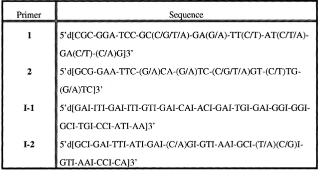
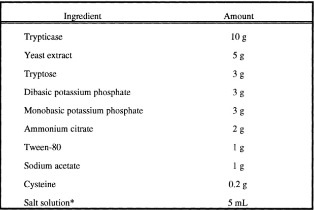
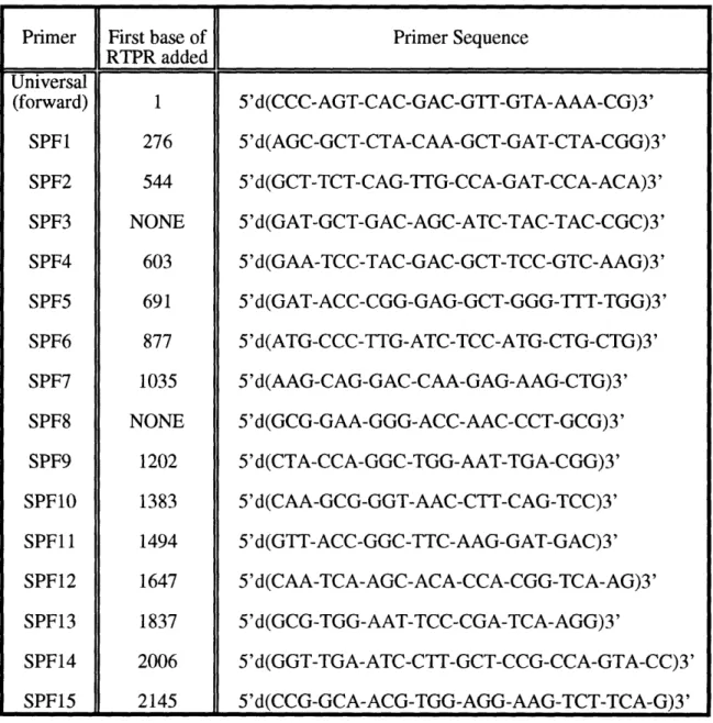
Synthesized oligonucleotide primers used in cloning. Recipe for 1 L of Lactobacillus carrying medium.
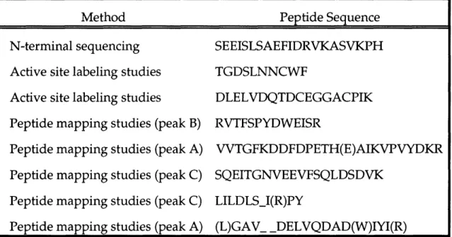
Forward oligonucleotide primers used in sequencing RTPR. Reverse oligonucleotide primers used in sequencing RTPR. Peptides of L. eichmannii RTPR.
Codon usage for L. leichmannii RTPR. Primers used for expression-cassette PCR.
Purification of RTPR from E. coli HB101/pSQUIRE. Amino acid composition of recombinant RTPR.
Kinetic constants for recombinant and non-recombinant RTPRs. Primers used for mutagenesis.
Phenotypes of the C-terminal RTPR mutants.
Equivalents of dC, cytosine, and the mystery peak as a function of time.
Phenotypes of the active site C-*S mutants: Equivalents of product per equivalent of RTPR.
dNTP/dNDP stimulation of the tritium exchange reaction between [5'-3H]AdoCbl and solvent.
2.1: 2.2: 2.3: 2.4: 2.5: 2.6: 3.1: 3.2: 3.3: 3.4: 4.1: 4.2: 4.3: 4.4: 5.1:
Profile of 3H20 elution from Sep-pak C
18 cartridge.
5.3: DTT-dependent background rate of exchange as a function of [5'-3H]AdoCbl concentration.
5.4: Time course for exchange in a reaction in which 1 M NaOAc is substituted for dGTP.
5.5: Effort to show enzyme dependence on an exchange reaction in which 1 M NaOAc is substituted for dGTP.
5.6: Effect of various thiols on the transfer of tritium from [5'-3H]AdoCbl to H20.
5.7: Characterization of the ability of pre-reduced RTPR to catalyze the exchange reaction under several conditions.
5.8: Re-characterization of the ability of TR to inhibit the exchange reaction in the absence of NADPH.
5.9: Characterization of mutant C731&736S RTPR with respect to the exchange reaction under pre-reduced conditions.
5.10: Characterization of the ability of mutant C408S to catalyze the exchange reaction.
5.11: Characterization of the ability of the active site RTPR mutants to catalyze the exchange reaction.
5.12: Primary data for the characterization of mutants C119S and C419S RTPRs.
5.13: Characterization of the ability of oxidized wt RTPR and pre-oxidized mutant C731&736S RTPR to catalyze the exchange reaction.
Similarity of the nucleotide requirement for exchange and the rapid production of cob(II)alamin.
Abbreviations
aa amino acid AdoCbl 5'-deoxyadenosylcobalamin Bl2r cob(II)alamin bp base pair Bq becquerelBSA bovine serum albumin
cDNA complementary DNA
Ci curie
CIP alkaline phosphatase from calf intestine
dA 5'-deoxyadenosine
Da Daltons
dC deoxycytidine
DE-52 diethylaminoethyl cellulose from Whatman
ds double-stranded
E. coli Escherichia coli
EDTA ethylenediaminetetraacetic acid
EPR electron paramagnetic resonance
HEPES N-(2-hydroxyethyl)piperazine-N'-(2-ethanesulfonic acid)
HPLC high pressure liquid chromatography
IPTG isopropyl -D-thiogalactoside
kb kilo base pairs
Km Michaelis constant
Lac lactose
LB Luria-Bertani broth
mRNA messenger RNA
MW molecular weight
NADH nicotinamide adenine dinucleotide (reduced form)
NADPH nicotinamide adenine dinucleotide phosphate (reduced form)
NMR nuclear magnetic resonance
OAc acetate
OD optical density
PAGE polyacrylamide gel electrophoresis
PCR polymerase chain reaction
PEG polyethylene glycol
PMSF phenylmethanesulfonyl fluoride
PNK T4 polynucleotide kinase
PVDF poly[(vinylidene difluoride)]
RNase H ribonuclease H
RDPR ribonucleoside diphosphate reductase from E. coli
RTPR ribonucleoside triphosphate reductase from L. eichmannii
SDS sodium dodecylsulfate
TAE tris-acetate/EDTA
TBE tris-borate/EDTA
TE tris-EDTA, pH 8
TEA triethylamine
TEAB triethylammonium bicarbonate
TLC thin layer chromatography
Tris tris(hydroxymethyl)aminomethane
Vmax maximal velocity
Wt wild-type
Chapter 1:
The Ribonucleotide Reductases: Radical Enzymes Employing Radically Differenct Cofactors.
The de novo biosynthesis of DNA is a complex process involving several metabolic pathways and many different enzymes (Figure 1.1). One well known enzyme in this overall process is DNA polymerase. Using the parent strands as templates, this enzyme constructs the nascent DNA polymer from four monomer
units, dATP, dTTP, dCTP, and dGTP, with the concomitant release of
pyrophosphate (PPi) upon the addition of each monomer. The action of this enzyme is understandably dependent upon the presence of the monomer units,
or deoxyribonucleoside triphosphates (dNTPs); however, the fidelity of this
polymerization reaction, and hence the unflawed transmittance of genetic
information during cell division, is dependent not only upon the presence of the four dNTPs, but upon a well-regulated balance of each (Reichard, 1988). Several enzymes participate in the fine tuning of the cellular pools of dNTPs; however, in all self sufficient organisms, as well as some viruses and phages, nature has invented one and only one mechanism for the formation and regulation of the
dNTPs necessary for DNA biosynthesis. The enzyme that governs these
processes is ribonucleotide reductase.
The ribonucleotide reductases (RNRs) constitute a unique class of metalloenzymes which catalyze the reduction, at a single active site, of all four
ribonucleotides to the corresponding 2'-deoxyribonucleotides (Thelander &
Reichard, 1979; Lammers & Follmann, 1983; Stubbe, 1990b). This transformation is the first committed step in DNA biosynthesis, and as a consequence of being at such a critical juncture in metabolism, RNRs are under very rigid regulation.
Indeed, several studies have shown that RNRs can be controlled at the
transcriptional level (Tuggle & Fuchs, 1986), the translational level (Noronha et al., 1972; Standart et al., 1985), at the level of cofactor synthesis and destruction (Barlow et al., 1983), and also by an astonishing network of allosteric interactions
CD 3 0
F
n} ¢D O .su ¢D A~~~1
~
l --. 0 tr C. o o U) %z
-z 1.~0 >:D :r¢ m CC~ U). (a. o F ' 0 I-Iz CD m 0 a 0 O >z -
> 0IVX- --
--
_q
IV 0~~~0 -.
0 Co
-0.~~~~~~~r 3 -o ,m E z~ , om~0
D 10--/ C I a~ Q -Z 0. 3 0 CD . ~ a. ,0
- I i
0
>
0r 0 ms 0 0 0 C. C' 0 -Zo
Cc
O Q. (D CD 0,_ IC - -I(Singh et al., 1977; Eriksson & Sj6berg, 1989; Reichard, 1993b). Because of their key role in producing the dNTPs for DNA biosynthesis, as well as regulating the only pathway that the cell has for making these dNTPs, it might be presumed that the reductases are extremely conserved. Surprisingly however, in contrast to most other enzymes which govern key metabolic processes, the RNRs are not conserved at all. This lack of homology applies to the level of the quarternary structure of the enzymes, the cofactors employed in effecting substrate turnover, and as will be shown in Chapter 2, in primary sequence (Stubbe, 1990a; Booker & Stubbe, 1993; Reichard, 1993b).
The study of ribonucleotide reductases has developed into quite an
expansive area of biological science. Many scientists are interested in the enzyme from an evolutionary standpoint, as it is presumed to be the major player in the transformation from an RNA-based world to a DNA-based world (Benner et al., 1989). There are those who are interested in the enzyme from a therapeutic standpoint, as a target for antineoplastic and antiviral agents (Stubbe, 1990b). Still, some researchers are interested in RNR as a model for allosteric regulation while others want to understand its role in the regulation of the cell cycle. Lastly, there are those scientists who are driven by its mode of catalysis. Although the net transformation is ostensibly simple - the replacement of a hydroxyl group with a hydrogen - the mechanism by which this is carried out is deceivingly complicated (Ashley & Stubbe, 1985; Stubbe, 1990a). This introduction seeks to provide an overview of the ribonucleotide reductase class of enzymes. However, because of the broad scope of the subject, it will focus primarily on the structural and mechanistic aspects of the enzymes, describing the initial characterization of
ribonucleotide reductase activity, the classes of RNR, and the mechanistic
Characterization of Ribonucleotide Reductase Activity
Studies in the early 1950s by Hammarsten, Reichard, and Saluste,
suggested that ribonucleotides were the precursors to deoxyribonucleotides
(Hammarsten et al., 1950). Using whole rats in combination with 15N labeled cytidine and uridine, these researchers showed that the isolated DNA contained
significant amounts of labeled cytosine and uridine. Earlier experiments by
Bendich, Getler, and Brown, revealed that the free base cytosine could not be used for the synthesis of DNA (Bendich et al., 1949), and studies by Irwin Rose
showed that cytidine labeled in both the base and sugar did not change in
specific activity upon its incorporation into DNA (Rose & Schweigert, 1953). The
results of these investigations in combination with experiments that
demonstrated that RNA is not the direct precursor to DNA (Abrams, 1950), led to the advancement of the hypothesis that deoxyribonucleotides were synthesized
directly from ribonucleotide precursors, and not from a free base and a
deoxyribose sugar. Relatively soon after, several laboratories were able to show evidence in cell free extracts from several sources that deoxycytidine phosphates could be formed from cytidine 5'-phosphate (Abrams et al., 1960; Moore & Hurlbert, 1960; Reichard, 1961). The most extensive characterization of this enzymatic activity came from studies in Sweden in the laboratory of Professor Peter Reichard. Working in E. coli, he isolated two protein fractions which were able to convert cytidine monophosphate (CMP) to deoxycytidine diphosphate (dCDP) (Reichard, 1962). Fraction A was ideintified to be CMP kinase, a
previously characterized enzyme which phosphorylates CMP to cytidine
diphosphate (CDP). Fraction B, which could be fractionated further into two proteins, B1 and B2, required the presence of fraction A for activity when CMP
was used as substrate, but turnover could be achieved in the absence of fraction A when CDP was used as the substrate.
Studies of nucleotide metabolism in the bacterium Lactobacillus leichmannii
suggested that a cofactor is required for deoxynucleotide production. Acting on results from E.E. Snell's laboratory at the University of Wisconsin, which showed
that L. eichmannii needed vitamin B
12for growth in the absence of
deoxyribonucleosides (Kitay et al., 1950), R.L. Blakley and H.A. Barker (1964) demonstrated that cell-free extracts of this bacterium synthesized deoxyribosides from the corresponding riboside only in the presence of vitamin B12. Subsequent studies by Blakley (1965) as well as Beck and Hardy (1965) led to the identificaion of 5'-deoxyadenosylcobalamin, a derivative of vitamin B12, as the active cofactor in the L. leichmannii reductase. Meanwhile, experiments in the laboratory of P. Reichard demonstrated rather convincingly that the reductase isolated from
E. coli was not cobamide dependent (Moore & Reichard, 1963). In fact, the small
subunit, B2, was shown to contain two atoms of non-heme iron per mol of
protein, the removal of which resulted in loss of enzyme activity (Brown et al., 1969). No labile sulfide was detected upon removal of the irons, suggesting that the iron was not present as an Fe/S cluster. The UV-vis spectrum of protein B2 displayed a very broad feature at 360 nm, a very steep shoulder at 325 nm, and a very sharp peak at 410 nm. The removal of iron from protein B2 resulted in the loss of these features; the reconstituion of protein B2 with Fe2 +resulted in the reappearance of these features (Brown et al., 1969). Subsequent electron spin resonance (EPR) studies of protein B2 suggested that it also contained an organic free radical, the presence of which was dependent upon the formation of the diiron center (Ehrenberg & Reichard, 1972). In seminal experiments, EPR spectroscopy was used in combination with protein B2 that had been expressed in the presence of specifically deuterated amino acids to show that this organic
radical resided on a tyrosine residue (Sjdberg et al., 1977). This was the first example of a stable protein radical that played an essential role in an enzymatic reaction.
As uncommon as it is for enzymes that play key roles in metabolism to employ more than one cofactor, nature has devised at least three, and perhaps four different classes of RNRs, each employing a unique cofactor for catalysis. As will be discussed subsequently, a ribonucleotide reductase has been isolated from E. coli grown under anaerobic conditions, that is distinct from the E. coli reductase discussed above (Fontecave et al., 1989; Eliasson et al., 1990; Eliasson et al., 1992). In addition, a RNR isolated from Brevibacterium ammoniagenes appears
to require manganese and not iron for catalytic activity (Willing et al., 1988b; Willing et al., 1988a).
Classes of Ribonucleotide Reductase
E. coli Ribonucleotide Reductase
The RNR isolated from E. coli (EC 1.17.4.1) is by far the best characterized of all the reductases - especially with respect to its structure and modes of regulation. Although this is partly due to the novelty of this enzyme harboring a stable organic radical, the fact that it is the prototype for reductases isolated from viruses as well as mammalian systems has also intensified its study for medicinal reasons (Stubbe, 1990b). This enzyme acts on nucleoside substrates that are diphosphorylated, giving rise to the name ribonucleoside diphosphate reductase, or simply RDPR. The newly-generated dNDPs are then phosphorylated to the
corresponding dNTP by nucleoside diphosphate kinase before being
incorporated into DNA via an appropriate polymerase.
As mentioned
previously, RDPR is composed of two subunits, B1 and B2, which are now universally known as R1 and R2. In the holoenzyme both of these subunits are
dimeric, yielding a putative overall tetrameric 22 quarternary structure
(Thelander & Reichard, 1979; Eriksson & Sjbberg, 1989; Stubbe, 1990b). The R1 subunit is the larger of the two, having a monomeric molecular weight of 86 kDa. It contains the binding site for NDP substrates, as well as binding sites for NTP and dNTP allosteric effectors. Also, the R1 subunit contains catalytically important cysteine residues which become oxidized concomitant with substrate reduction (Thelander, 1974). In order to achieve multiple turnovers, the resulting disulfide bond must be rereduced. This can be achieved in vitro with small
dithiols such as dithiothreitol (DTT) or dihydrolipoic acid (DHL), albeit at
concentrations around 25-30 mM. In vivo however, these cysteines are reduced by a low molecular weight (12 kDa) protein, thioredoxin (TR), which derives its
reducing equivalents from thioredoxin reductase (TRR). TRR is a
flavin-containing protein which in turn obtains its reducing equivalents from the
oxidation of NADPH. This process is summarized in Scheme 1.1. The
Scheme 1.1: The reaction catalyzed by ribonucleotide reductase
3i
(P)PP
P
(P)PP
/ 'HO HSH
s
SH
Is
TR/TRRINADP TR/TRR/NADPHviability of E. coli mutants deficient in thioredoxin activity led to the discovery of glutaredoxin as a hydrogen donor for RDPR (Holmgren, 1985). Glutaredoxin derives its reducing equivalents ultimately from NADPH as well, via a system coupled to glutathione reductase and glutathione.
The R1 subunit contains two different binding sites which regulate the activity and substrate specificity of the enzymes (Eriksson & Sjdberg, 1989). The high affinity site - named for its strong affinity for the allosteric effectors as compared to the low affinity site - interacts with various NTPs and dNTPs to determine which particular NDP will be turned over. The method of actual allosteric regulation is complex, however a simple scenario is described below. When ATP (or dATP at low concentrations) is bound the enzyme reduces CDP and UDP. When dGTP is bound, the enzyme reduces ADP; and when dTTP is bound, GDP is reduced. The low affinity site binds either ATP or dATP. When ATP is bound to this site, the activity of the enzyme is enhanced. When dATP is bound to this site, the enzyme is turned off. These interactions act in concert to maintain a balanced level of the deoxynucleotides needed for DNA biosynthesis.
Although the R1 subunit of the E. coli reductase appears to be the business end of the enzyme, catalysis cannot occur without the second subunit, R2. The R2 subunit of the E. coli reductase is indeed a novel protein. Early spectroscopic studies suggested that the protein contains two high-spin Fe(III) atoms that are antiferromagnetically-coupled through a t-oxo bridge (Bunker et al., 1987; Scarrow et al., 1987; Sj6berg et al., 1987; Backes et al., 1989). This fully-assembled iron center is a necessary requirement for the maintenance of the tyrosyl radical, which in turn is required for catalysis. This tyrosyl radical has a half-life that is on the order of hours at room temperature, and years at -80°C. Based on an
reductases, as well as the use of the mutant protein Y122F-R2, the tyrosyl radical was assigned to tyrosine 122 of the E. coli RDPR (Sj6berg et al., 1977). Very recent work by Nordlund and Eklund has resulted in the solving of the 3-dimensional
structure of the R2 protein, and the diiron cofactor (Scheme 1.2) (Nordlund et al., 1990; Nordlund & Eklund, 1993). The two proteins of R2 fit together in an
arrangement which makes the dimeric protein appear heart-shaped. Each
monomeric subunit, contains 1 iron center and potentially 1 tyrosyl radical,
which are both situated deep within each protein. The tyrosyl radical is 5.3 A from the nearest Fe(III) atom and 10 A away from the nearest surface of the R2 protein. Each Fe(III) atom is attached to the protein via histidine and carboxylate ligands, and in addition to the t-oxo bridge, the carboxylate from glutamate 115 also bridges the two irons which are 3.3 A apart (Scheme 1.2).
Lactobacillus leichmannii Ribonucleotide Reductase
Although the enzyme isolated from E. coli is the best characterized
reductase to date, a resurgence in interest of the enzyme isolated from
L. eichmannii is taking place. This is no doubt due in large part to the fact that
the L. eichmannii reductase (EC 1.17.4.2) is structurally the simplest of all of the known reductases, as well as the desire among scientists to explore the function
and catalytic capabilities of nature's only proven organometallic cofactor,
5'-deoxyadenosylcobalamin (AdoCbl). This cofactor is composed of a cobalt-containing corrin ring, a dimethylbenzimidazole group, and a 5'-deoxyadenosine moiety coordinated to the cobalt via a unique Co(III)-carbon bond (Figure 1.2). The cobalt is held in place by four coordinating nitrogen atoms provided by the corrin macrocycle. The nitrogen of the dimethylbenzimidazole group provides the proximal ligand to the octahedral Co(III) atom, while the distal ligand is provided by the 5' carbon of 5'-deoxyadenosine.
Scheme 1.2: X-ray structure of the diferric iron center-tyrosyl radical cofactor of
the R2 subunit of RDPR. Adapted from Nordlund et al. (1990).
Ty- 1
O2
Tyr 122
Asp
8 4
H
2
0
V
_ Fe
"'%%%
N
0
7His8His1 1 8
AHo Glu
2 3 8H,0
.0
O0x
tClGIull
5
H1is
2 41
G1u
2 0 4
Figure 1.2: Structure of coenzyme B1 2. The 5'-methylene carbon of 5'-deoxyadenosine is surrounded by a rectangle, as it is the carbon which forms the unique organometallic bond in the cofactor. The four pyrroline rings are labeled A, B, C, and D, and the chiral centers of the corrin macrocycle are denoted with asterisks. Figure adapted from Vitamin B12 (Zagalak & Friedrich, 1979).
0
The reductase
from L. eichmannii catalyzes the reduction of
ribonucleotides that are triphosphorylated, giving rise to the name
ribonucleoside triphosphate reductase (RTPR). This enzyme is a single
polypeptide of Mr = 82,000 (Panagou et al., 1972; Booker & Stubbe, 1993). Analogously to the E. coli reductase, RTPR is also allosterically regulated (Beck, 1967). Moreover, the pattern of allosteric regulation is similar to that of the E. coli reductase; CTP reduction is stimulated by dATP, UTP reduction by dCTP, ATP reduction by dGTP, and GTP reduction by dTTP (Beck, 1967). This elaborate array of allosteric regulation can be abrogated in the presence of various cations and anions (Jacobsen & Huennekens, 1969). When acetate is present as the anion, the activating effect of cations is in the order Na+<K+<Rb+<Cs+<NH4<<Li+. In the case of NaOAc, the maximum activating effect is obtained with a concentration of 1 M. Also analogously to the E. coli reductase, RTPR containscysteines which become oxidized concomitant with substrate reduction.
Artificial reductants such as DTT and DHL can be used to rereduce the active site disulfide so that multiple turnovers can occur. In vivo, however, L. eichmannii contains a TR/TRR/NADPH reducing system which can re-reduce the active site disulfide (Orr & Vitols, 1966). The E. coli TR is fully capable of supplying reducing equivalents to RTPR, and displays a Km for substrate turnover of 4 ,M (Blakley, 1978). The TR from L. eichmanni displays a Km that is only slightly lower (Blakley, 1978). Because the E. coli TR and TRR have been cloned and overexpressed, they are routinely used as the reductant for RTPR (Lunn et al., 1984; Russel & Model, 1985).
RTPR catalyzes two other reactions in addition to deoxynucleotide
production. The first reaction is the equilibration with solvent of the 5'
methylene hydrogens of the cofactor (Abeles & Beck, 1967; Hogenkamp et al., 1968). In a reaction requiring NTPs or dNTPs, and reductant, RTPR will catalyze
the washout of tritium from [5'-3H]AdoCbl to solvent. Likewise, in a reaction performed under similar conditions but in 3H20, RTPR will catalyze the washin of tritium into unlabeled cofactor. When this reaction was allowed to proceed to equilibrium, a maximum of 1.4 atoms of tritium per molecule of AdoCbl was found to be incorporated (Hogenkamp et al., 1968). In the reverse direction, [5'-3H]AdoCbl labeled by chemical means was found to transfer all of its tritium
to solvent. These results provide strong evidence that this exchange reaction
proceeds through an intermediate in which the two 5' methylene hydrogens are
equivalent.
This intermediate is suspected to be cob(II)alamin and
5'-deoxyadenosine. The exchange reaction is the focus of Chapter 5, and a detailed history and analysis of it will be presented therein.
Evidence
which
is
highly
suggestive
of
the
cob(II)alamin/5'-deoxyadenosine intermediate comes from the second reaction that is catalyzed by RTPR. This reaction is the slow degradation of the cofactor to cob(II)alamin and 5'-deoxyadenosine (Yamada et al., 1971). As in the exchange reaction, this slow decomposition of the cofactor is also dependent upon the presence of reductant and an NTP or dNTP. The products of this reaction bind tightly to the enzyme in a mutally cooperative fashion, and display Kds of 37 iM [cob(II)alamin] and 14 ptM (5'-deoxyadenosine) (Yamada et al., 1971).
Although the decomposition of the cofactor is too slow to be on the
catalytic pathway, stopped flow spectrophotometric studies by Tamao and
Blakley (1973) revealed that cob(II)alamin is also produced in a very rapid reaction with a first order rate constant of 38-46s-1. This rapid reaction is also contingent upon the presence of a dithiol and an NTP or dNTP, but unlike the slow reaction, this rapid reaction is fully reversible, and upon cooling to 5°C the UV-vis spectrum indicative of cob(II)alamin disappears. Confirmation that this species is indeed cob(II)alamin was established by Orme-Johnson et al. (1974)
using rapid freeze-quench EPR. The paramagnetic species formed was produced
with a rate constant similar to that observed in the spectrophotometric
experiments of Tamao and Blakley. The EPR spectrum differed from that of free
cob(II)alamin, and later studies suggested that it was consistent with a
cob(II)alamin species interacting with an organic radical (Hamilton et al., 1972).
All of the AdoCbl-dependent reductases are not monomeric enzymes.
Tsai and Hogenkamp (1980) have isolated an AdoCbl-dependent reductase from
Corynebacterium nephridii which is dimeric, and which uses nucleoside
diphosphorylated substrates. This enzyme is also allosterically regulated;
however, the the pattern of regulation is very complex. In general, as in the enzyme from E. coli, it appears that dNTPs act as effectors (Tsai & Hogenkamp, 1980). The AdoCbl-dependent reductases appear to be restricted to prokaryotes, with the exception of the enzyme isolated from Euglenophyta (Gleason & Frick,
1980).
Ribonucleotide Reductasefrom Anaerobically-grown Escherichia coli
The ability of E. coli to grow in the absence of oxygen necessitates that they be able to anaerobically synthesize the dNTPs necessary for DNA biosynthesis. Genetic experiments suggested that E. coli possessed a separate reductase for anaerobic growth (Jamison & Adler, 1987; Hantke, 1988). This reductase, (which constitutes the third class of RNRs) was subsequently isolated in the laboratory of P. Reichard, and found to be a ribonucleoside triphosphate reductase. It contains what appears to be a [4Fe-4S] cluster (Mulliez et al., 1993), and requires
S-adenosylmethionine (AdoMet) and other low molecular weight factors
(including K+ and HCO2-) for catalytic activity (Eliasson et al., 1990; Eliasson et al., 1992). The gene for this reductase has been cloned, and its primary sequence
determined. The protein appears to be monomeric, and from the its primary sequence, the molecular weight was determined to be 80.1 kDa (Sun et al., 1993). The pattern of allosteric regulation of this enzyme is simlar to those discussed above. Adenosine triphosphate promotes the reduction of CTP and UTP, dTTP promotes the reduction of GTP, and dGTP promotes the reduction of ATP. As in the E. coli RDPR, dATP is a general inhibitor of the enzyme (Reichard, 1993a).
Although very little sequence homology exists between this reductase and either of the subunits of the E. coli RDPR, the C terminus of the anaerobic reductase contains a sequence (RVCGY) which is very similar to a sequence (RVSGY) from the enzyme pyruvate formate-lyase (pfl). In pfl, a radical on glycine-734 in the conserved sequence shown above has been shown to be generated in the presence of AdoMet and 5-deazariboflavin (Wagner et al., 1992). In the presence of oxygen, the radical is destroyed, and the protein is cleaved into two fragments. The role of the glycyl residue in catalysis has not been clearly defined in either protein, however oxygen similarly inactivates the anaerobic ribonucleotide reductase.
Brevibacterium ammoniagenes Ribonucleotide Reductase
The enzyme isolated from B. ammoniagenes
remains still relatively
uncharacterized. This has oftentimes resulted in its exclusion as a class of RNRs (Reichard, 1993b). Early studies of B. ammoniagenes and Micrococcus luteus by several labs showed that manganese deficiency in these organisms resulted in
unbalanced growth, filamentous morphology, and an arrest of DNA synthesis,
by not RNA synthesis nor protein synthesis. The addition of Mn(II) to these cells restored DNA synthesis as well as the normal pattern of growth exhibited by
these organisms. Further studies showed that the abrogation of DNA synthesis
caused by a deficiency in Mn was due to an inhibition of ribonucleotide
reduction and not DNA replication (Schimpff-Weiland et al., 1981). A
Mn-dependent reductase from Brevibacterium ammoniagenes was subsequently
isolated (Willing et al., 1988b). This enzyme contains two subunits, a large monomeric subunit of Mr = 80,000, and a smaller dimeric subunit of Mr = 50,000 per monomer, giving rise to an overall c 2 quarternary structure (Willing et al., 1988b). The growth of B. ammoniagenes in the presence of 5 4MnC1
2 and the
subsequent isolation of the reductase, in combination with
non-denaturing/denaturing gel electrophoresis, showed that 54Mn migrates with the smaller subunit of the reductase (Willing et al., 1988b).As in the L. leichmannii and aerobic E. coli reductases, a small protein, thioredoxin, supplies the reducing equivalents necessary for multiple turnover in the B. ammoniagenes reductase. The E. coli TR will also serve in the same capacity (Willing et al., 1988a). The Mn-dependent enzyme is a ribonucleoside diphosphate reductase, and a study of its nucletide specificity revealed a similar pattern of allosteric regulation to that of the E. coli RDPR and the L. eichmanni enzyme. The reduction of CDP (and to a lesser extent UDP) is stimulated by dATP, ADP by dGTP, and GDP by dTTP (Willing et al., 1988a).
Although evidence for an organic radical is not presently available in this reductase, several lines of reasoning might suggest that this enzyme is similar to the E. coli RDPR. Firstly, the enzyme is inhibited by hydroxyurea, a compound known to inhibit the E. coli RDPR presumably by scavenging the tyrosyl radical (Willing et al., 1988b). Secondly, the R2 subunit of the E. coli RDPR has been
reconstituted with Mn(II), and the resulting structure characterized by
spectroscopic and crystallographic studies (Atta et al., 1992). Although this
ions occupy the iron-binding sites of the protein, and are bridged by E115 and E238, with no oxygen bridge present. Thirdly, the UV-vis spectrum of the
B. ammoniagenes RDPR is similar to model compounds that contain two Mn(III)
atoms coupled through a pt-oxo bridge (Sheats et al., 1987). Whether this enzyme contains a tyrosyl radical or perhaps some other organic radical awaits its further characterization.
The Mechanism of Ribonucleotide Reduction
Models for the Mechanism of Ribonucleotide Reduction
Despite the difference in quarternary structure as well as cofactor
requirement, studies on the L. eichmannii and E. coli reductases have suggested that these two ostensibly different enzymes may function by similar mechanisms of catalysis (Stubbe, 1990b). This hypothesis stems from the fact that (1) in both cases the hydrogen that replaces the 2' hydroxyl group is derived from solvent with retention of configuration (2) both enzymes couple substrate reduction to the oxidation of two cysteine residues on the protein to a disulfide (3) in both enzymes the disulfide bond formed concomitant with substrate reduction can be
rereduced by a TR/TRR/NADPH reducing network, with the E. coli TR being
able to function with the L. leichmannii enzyme.
Although the presence of a tyrosyl radical in the E. coli RDPR suggested to
some that radicals might be involved in catalysis, at what stage they were
involved and in what capacity was a mystery. Studies by Walling and Johnson (Walling & Johnson, 1975), as well as Gilbert et a. (Gilbert et al., 1972) on the mechanism of Fenton's reagent [Fe(II)/H202] provided an initial clue as to how this reaction might proceed (Scheme 1.3). When Fenton's reagent is reacted with
Scheme 1.3: Proposed mechanism for the reaction of Fenton's reagent with
1,2-ethanediol.
Fe(II)
+ H202HO + HO-CH2-CH2-OH
O- Fe(III) + HOo +
HO-HO-CH2-CH-OH + H20 + 0 H+ H20-CH2-CH-OH CH+ C2H-+ .H CH2-CH-OH - CH2-CH=OH Fe(II) Fe(III) CH3-CH:O
products are generated due to side reactions. The reaction requires acid catalysis, and is initiated by the reduction of H202 by Fe(II) to yield HO,HO-, and Fe(III). The hydroxyl radical abstracts a hydrogen atom from substrate, which upon protonation of the 0-hydroxyl, yields H20 and a radical-cation species which can be drawn in the shown resonance forms. A subsequent e- reduction yields an
enolate which subsequently affords the aldehyde upon tautomerization. This
resulting product is 2e- oxidized from the expected product of ribonucleotide reduction. Since both the L. teichmannii and E. coli reductases were known to couple substrate turnover to the oxidation of two protein cysteines to a disulfide bond, the above model proved to be an attractive working hypothesis for the reaction catalyzed by ribonucleotide reductase. Based on this model, X (some
oxidizing species) plays the role of the hydroxyl radical created by Fenton's
reagent. The first step in the reaction is the abstraction of a hydrogen atom from the 3' position of the substrate. The 2' hydroxyl group is protonated - ultimately by one of the redox-active cysteines on the protein - and the loss of a molecule of H20 affords a radical-cation intermediate, as proposed in the mechanism of
Fenton's reagent with 1,2-ethanethiol. This radical-cation intermediate is
proposed to undergo two stepwise le- reductions by the redox-active cysteines, the first of which affords a disulfide radical anion and a 3'-keto deoxynucleotide upon the addition of a proton. The second le- reduction is followed by the return of the initial hydrogen atom (Ha) abstracted back to the 3' position of the deoxynucleotide product (Scheme 1.4).
The postulated mechanism for ribonucleotide reduction (Scheme 1.4)
makes several predictions that have been experimentally tested. The first
prediction is that the 3' C-H bond of the substrate is cleaved. This was tested
using substrates which were specifically tritiated at the 3' position of the
nucleotide. When [3'-3H]UD(T)P or [3'-3H]AD(T)P is used as substrate with RDPR (RTPR), small V/K isotope effects are observed on the reduction of these radiolabeled nucleotides (Stubbe et al., 1981; Stubbe et al., 1983; Ashley et al., 1986). In the case of RTPR, these isotope effects are essentially invarient with pH (1.6-1.8, pH 6.1-8.3, for UTP; 1.9-2.1 pH 8.3-5.5, for ATP); however, in the case of RDPR the isotope effects vary from 1.4 to 1.9 (pH 8.6-6.6) for the reduction of
Scheme 1.4: Working hypothesis for the mechanism of ribonucleotide reduction. I X. B SH SH I I I XHa XHa PPF B SH SH I I I I XHa PPP B SH S-I I I I XHa PPP B SH -I I I XHa XHa PPP BH S:."! S I I I I XHa HO H B S -S , I I I
ADP, and 2.8 to 4.7 (pH 8.4-6.6) for the reduction of UDP. In addition very small but reproducible amounts (up to 1%) of 3H20 are produced in a time dependent
I XHa N PPP 'H OB H .I . . I PPI W.
fashion upon the incubation of these radiolabeled substrates with both of the
above RNRs. This result provides additional evidence that the above isotope effects are indeed primary effects rather than secondary effects, unambiguously establishing that the cleavage of the 3' C-H bond is required for nucleotide reduction.
The second prediction that this model makes, is that at the end of
nucleotide reduction the hydrogen atom originally abstracted from the 3'
position of the substrate is returned to the 3' position of the product. This prediction was investigated using nuclear magnetic resonance (NMR) techniques in combination with substrates that were specifically deuterated at the 3' postion. When substrate reduction is carried out with [3'-2H]UD(T)P, the NMR spectrum of the isolated product shows that it is completely deuterated (Ashley & Stubbe,
1985).
Although none of the intermediates shown in Scheme 1.4 have been seen during normal turnover, each step has precedent in the chemical literature [For a recent review see Stubbe (1990)]. As discussed above, all steps leading to the radical cation intermediate are supported by the chemistry of Fenton's reagent with 1,2-ethanediol. Evidence supporting the 3'-keto intermediate comes from work with the substrate analog 2'-chloro-2'-deoxyuridine 5'-di(tri)phosphate, as well as studies with several RDPR protein analogs, and will be presented subsequently. Furthermore, there is precendent for the reduction by e- transfer of a formylmethyl radical by DTT at high pH (Stubbe, 1990b). This reduction would afford a disulfide-radical anion and the 3'-keto intermediate as shown in Scheme 1.2.
The Nature of Xo
The mechanism in Scheme 1.4 presents a reasonable working hypothesis
for the reaction catalyzed by the ribonucleotide reductases. The burning
question thus becomes the nature of X°. From the standpoint of the E. coli enzyme it appears that X * might be the tyrosyl radical on the R2 subunit. Recent crystallographic studies by Nordlund and Eklund (1993) (1990) strongly suggest that this is indeed not the case. As discussed above, the tyrosyl radical is buried deep within the R2 subunit approximately 10
A
from the surface of the protein, and is therefore not capable of mediating 3' hydrogen atom abstraction unless gross conformational changes occur. This corroborates the long-held hypothesis by Stubbe and coworkers that the function of the R2 subunit is to generate aradical on the R1 subunit (the subunit which binds substrates and allosteric
effectors, and which contains the redox-active cysteines) by long-range e-/H+ transfer (Stubbe, 1990b). It is then the radical on the R1 subunit which initiates catalysis by abstracting the 3' hydrogen of the substrate. Evidence that suggests that the transient radical formed on protein R1 is a thiyl radical will be provided below.
That radical intermediates might be involved in the AdoCbl-dependent
reductase has huge precedent in the chemical literature [For a review see
Dolphin (1982)]. AdoCbl participates in a class of enzymes that carry out a rearrangement between a hydrogen atom on one carbon, and some functional group that is located on an adjacent carbon. The enzyme dioldehydrase is a prototype for this class, and its reaction is shown in Scheme 1.5. The tremendous amount of evidence in support of this scheme has been reviewed by Babior and Krower (1979). The reaction is initiated upon the homolytic cleavage of the cofactor and the abstraction of a hydrogen atom from substrate. The resulting substrate radical rearranges (by a mechanism that is unclear to date) to give the
product radical, which re-abstracts a hydrogen atom from XHa to give a geminal diol. The geminal diol stereospecifically loses a molecule of H20 in a reaction which is catalyzed by the enzyme to afford the product aldehyde. Seminal
studies carried out in the Abeles laboratory on dioldehydrase showed that
AdoCbl functions as an intermediate hydrogen atom carrier. In the event that the substrate [1,1-3H]DL-propanediol is incubated with the enzyme, tritium is found to be located in the cofactor. If [5'-3H]AdoCbl is incubated with enzyme and substrate, tritium is found to be located in the product (Abeles & Zagalak, 1966; Frey et al., 1967). These studies show that AdoCbl can act as a hydrogen carrier in this reaction; however, the degree to which it functions in this capacity is uncertain. Indeed, studies have shown that kH/kT = 125 for the transfer of tritium from cofactor to product (Essenberg et al., 1971). Cleland has proposed
that this anomalously high isotope effect could be explained by a pool of
hydrogen atoms on the enzyme with which the migrating hydrogen can
equilibrate, or by an alternate pathway (perhaps a protein radical?) with which the hydrogen can be transferred (Cleland, 1982). In the event that the transfer
occurs through a protein residue 90% of the time, and through
5'-deoxyadenosine 10% of the time, the true isotope effect (12.5) would be in the realm of what is normally observed for tritium.
These studies with dioldehydrase present a starting point to ask whether AdoCbl functions in the same manner in the reaction catalyzed by RTPR. That radical intermediates might be involved in the reaction catalyzed by RTPR was evidenced by early biophysical studies on the enzyme. As previously mentioned stopped flow UV-vis studies by Tamao and Blakley (1973) as well as rapid freeze-quench EPR studies by Orme-Johnson, Beinert, and Blakley (1974) showed that the L. leichmannii RNR catalyzes the homolytic cleavage of the Co-carbon bond of AdoCbl with a first order rate constant of 38-46 s-1in the presence of dGTP and