HAL Id: hal-01365426
https://hal.archives-ouvertes.fr/hal-01365426
Submitted on 13 Sep 2016
HAL is a multi-disciplinary open access
archive for the deposit and dissemination of
sci-entific research documents, whether they are
pub-lished or not. The documents may come from
teaching and research institutions in France or
abroad, or from public or private research centers.
L’archive ouverte pluridisciplinaire HAL, est
destinée au dépôt et à la diffusion de documents
scientifiques de niveau recherche, publiés ou non,
émanant des établissements d’enseignement et de
recherche français ou étrangers, des laboratoires
publics ou privés.
Copyright
Determination of Energy-Transfer Distributions in
Ionizing Ion-Molecule Collisions
S. Maclot, R. Delaunay, D. g. Piekarski, A. Domaracka, B. a. Huber, L.
Adoui, F. Martín, M. Alcamí, L. Avaldi, P. Bolognesi, et al.
To cite this version:
S. Maclot, R. Delaunay, D. g. Piekarski, A. Domaracka, B. a. Huber, et al.. Determination of
Energy-Transfer Distributions in Ionizing Ion-Molecule Collisions. Physical Review Letters, American
Physical Society, 2016, 117 (7), pp.073201. �10.1103/PhysRevLett.117.073201�. �hal-01365426�
S. Maclot,1, ∗ R. Delaunay,1 D.G. Piekarski,2 A. Domaracka,1 B.A. Huber,1 L. Adoui,1
F. Mart´ın,2, 3, 4 M. Alcam´ı,2, 3 L. Avaldi,5 P. Bolognesi,5 S. D´ıaz-Tendero,2, 4 and P. Rousseau1, †
1
Normandie Universit´e - CIMAP - UMR 6252 CEA/CNRS/ENSICAEN/UNICAEN
- Boulevard Henri Becquerel BP 5133 - 14070 Caen cedex 5 - France
2
Departamento de Qu´ımica - M´odulo 13 - Universidad Aut´onoma de Madrid -28049 Madrid - Spain
3
Instituto Madrile˜no de Estudios Avanzados en Nanociencias (IMDEANanociencia) -Cantoblanco 28049 Madrid - Spain
4Condensed Matter Physics Center (IFIMAC), Universidad Aut´onoma de Madrid -28049 Madrid - Spain
5CNR - Istituto di Struttura della Materia, Area della Ricerca di Roma 1, Monterotondo Scalo, Italy
(Dated: June 22, 2016)
The ionization and fragmentation of the nucleoside thymidine in the gas phase has been inves-tigated by combining ion-collision with state-selected photoionization experiments and quantum chemistry calculations. The comparison between the mass spectra measured in both types of ex-periments allows us to accurately determine the distribution of the energy deposited in the ionized molecule as a result of the collision. The relation of two experimental techniques and theory shows a strong correlation between the excited states of the ionized molecule with the computed dissociation pathways, as well as with charge localization or delocalization.
The understanding of the electronic and nuclear dy-namics in molecular systems induced by sudden ion-ization/excitation, which drives chemical reactions, of-fers new opportunities for controlled ultrafast chem-istry [1]. For example, it has been recently observed the charge migration on the fs timescale after hole forma-tion, which triggers atomic motion and molecular frag-mentation [2] or the localization of multiple charges in specific molecular groups and the subsequent Coulomb explosion [3, 4]. Ultrafast nuclear rearrangement have been also observed in pump-probe experiments [5], such processes compete with the expected charge separation in multiply charged molecules [6–8]. Therefore, a detailed knowledge of the response of complex molecular sys-tems to ionization/excitation and its influence on chem-ical reactivity is still today a relevant topic [9, 10]. In this context, recent combined experiment/theory works have been very valuable in providing pictures of the ion-induced ionization/fragmentation of complex molecular systems [7, 8, 11, 12]. However, a meaningful compari-son between experimental and theoretical results requires the knowledge of the energy transferred in the collision, which is in fact represented by a wide energy distribu-tion due to interacdistribu-tions at different impact parameters. Pioneering experimental work reported in [13, 14] has al-ready been performed in order to determine the actual energy-deposit distributions in ion-molecule collisions, as well as to study its relationship with the observed frag-mentation patterns. However, these methods require the knowledge of the initial and final projectile states which is only straightforward in the case of double-electron cap-ture by singly charged ions (e.g. H+ → H−), which is
more the exception than the rule.
Here we report on the ionization and fragmentation of a DNA building block, the nucleoside thymidine com-bining i) ion collisions, ii) VUV photoionization along with iii) ab initio calculations. Combining such
state-of-the-art techniques, we provide a complete picture of the charge localization and the excitation energy distribution in complex molecular systems after interaction with ion-izing radiation. More importantly, it becomes possible to determine the energy deposited in the target as a result of an ionizing collision with ions, which is the primary pro-cess associated with radiation damage. With the devel-opment of cancer therapies based on ionizing particles, as hadrontherapy [15], a better understanding of the radia-tion damage via a multi-scale and -disciplinary approach has become unavoidable [16]. At the molecular scale, this relies on the investigation of ionization/fragmentation of molecules of biological interest in the gas phase at differ-ent energy ranges [11, 17–19].
The experiments have been performed at ARIBE, the low-energy ions facility of GANIL (Caen, France) and at the GASPHASE beamline of the synchrotron radiation facility ELETTRA (Trieste, Italy). Both experiments are based on crossed-beam set-ups using coincidence time-of-flight mass spectrometry. The photoionization ex-periments are based on state-selected mass spectrome-try using photoelectron-photoion coincidence measure-ments (PEPICO). The effusive beam of neutral thymi-dine molecule (2’-deoxy-thymithymi-dine abbreviated dThy, see structural formula in Fig.1(a)) was produced by heat-ing a powder in a oven at a temperature low enough to avoid thermal decomposition [20]. Both experimental set-ups have been described in detail elsewhere [21, 22] and a brief description is given in the Supplemental Ma-terial [23].
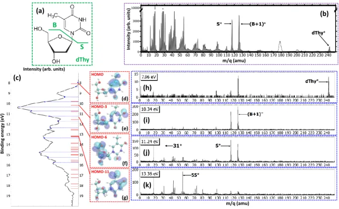
In order to have a picture of the stability of the charged thymidine in the gas phase, the mass spectrum of the charged products detected after the production of singly-charged thymidine in the interaction with 48 keV O6+ ions is shown in Fig.1(b). The peak located at
m/q = 242 amu corresponds to the intact singly charged dThy+and shows that a fraction of the parent population
2 S B (c) (d) (e) (f) (g) (h) (i) (j) (k) (a) (b) 31+ HOMO HOMO-11 HOMO-6 HOMO-3 dThy Inten sit y (a rb . u nit s)
Intensity (arb. units)
m/q (amu) m/q (amu) Bi ndi ng energ y (e V)
FIG. 1. (a) Structural formula of thymidine. Considering the glycosidic bond cleavage, the fragments produced are noted B
and S for the base and sugar parts, respectively. (b) Mass spectrum of thymidine after the ionization by 48 keV O6+ ions.
White peaks around m/q = 180 and 200 amu are due to pollutions. (c) Photoelectron spectrum (PES) of thymidine obtained at 50 eV (black curve). The blue dashed lines show the energy values chosen for PEPICO measurements. Red bars correspond to orbital energy values computed with OVGF method. Panels (d-g) show the electron density of different molecular orbitals. Panels (h-k) show the PEPICO mass spectra recorded for different binding energies of the electron corresponding to closest energies to the orbitals presented.
can be stable, at least in the µs timescale, after single ion-ization. The main peaks among the heaviest fragments are observed at m/q = 117 amu and m/q = 126 amu. The first one corresponds to the sugar part S+ whereas
the second one is assigned to the fragment (B+1)+
corre-sponding to an intramolecular rearrangement associated with a hydrogen transfer to the base part B [24]. Both fragments are the result of the glycosidic bond cleav-age, an important mechanism in the radiation damage of DNA [24–26], and contribute to 8% of the spectrum. A very small amount of fragments heavier than the base or sugar parts, i.e. loss of neutral fragments keeping in-tact the glycosidic bond, are also observed. This is due to the large distribution of impact parameters in the case of ion collisions [27] which leads to energy transfers, span-ning from few meV to few tens of eV and involves a dis-tribution of vibrational energy transfer and electron cap-tures in various electronic states. Thus, the knowledge of the distribution of the energy transferred to the molecule plays a key role to unravel its fragmentation dynamics. It is difficult to assess experimentally this energy distri-bution even if translational spectroscopy can provide it in the case of multiple electron capture [13, 14].
A method that can provide direct insight on the
frag-mentation following a selected energy deposition is the PEPICO technique, where the kinetic energy of the pho-toelectrons allows to pin point the energy left in the tar-get [28]. The photoelectron spectrum of thymidine mea-sured at 50 eV is shown in Fig.1(c). From this spectrum, thirteen photoelectron binding energy values Eb, covering
the main features, have been chosen to study the evolu-tion of the fragmentaevolu-tion. A simulaevolu-tion of the photoelec-tron binding energy spectra was carried out by comput-ing the ionization energies for the 31st highest molec-ular orbitals using the outer-valence Green’s function (OVGF) method [29] in combination with a 6-311G(d,p) basis set of the Gaussian09 package [30]. The results are plotted in the panel (c). This method incorporates the effects of electron correlation in the computation of molecular ionization potentials as one-particle theory for the description of ultrafast electron charge density dy-namics after ionization of an outer-valence electron. The uncertainty of the calculated energies by this method is about ±0.3 eV [31]. The Fig.1(d-g) shows the com-puted electron densities of four orbitals corresponding to the HOMO, HOMO−3, HOMO−6 and HOMO−11 with binding energies of 8.09, 10.19, 11.36 and 13.18 eV, re-spectively, which are the closest in energy to the four
I n te n s it y ( a rb . u n it s )
Excitation energy (eV)
0 1 2 3 4 5 6 7 8
Binding energy (eV)
8 9 10 11 12 13 14 15 16
FIG. 2. Determined distribution of the excitation energy in
the ion collision (see text). The R2 coefficient of
determina-tion for this fit is 0.86.
selected PEPICO mass spectra shown in the Fig.1(h-k). They illustrate that the charge localization after ioniza-tion strongly depends on the orbital and may lead to different fragmentation channels. In the first mass spec-trum recorded at Eb= 7.96 eV (Fig.1(h)) we observe the
peak due to the parent ion, i.e. the singly charged thymi-dine molecule. Due to the experimental energy resolution in the PEPICO measurements, not only photoelectrons from the ground ionic state but also from deeper orbitals can be detected [23]. This may lead to molecular dissoci-ation and indeed some fragments are also observed. The main fragment corresponds to (B+1)+ indicating that the charge is mainly located on the base part as suggested by the electronic density of the HOMO (panel (d)). In the next mass spectrum, measured at Eb = 10.34 eV,
two mains peaks are observed with similar intensities. They are assigned to the fragment (B+1)+ and S+ as
observed in the case of the fragmentation induced by mul-tiply charged ions. The similar intensities show that the charge has almost the same probability to be located on each one of the two moieties of the molecule as shown by the non preferential charge localization in the orbital HOMO−3, in panel (e), and neighboring orbitals [23]. The panel (j) presents the mass spectrum recorded at Eb = 11.24 eV. The same main peaks are present, but
the fragment S+ is now prominent. This is consistent
with the preferential charge localization of the associated orbital HOMO−6 (panel (f)), although the neighboring orbitals can contribute to S+and (B+1)+peaks [23]. At larger excitation energy (Fig.1k), the mass spectrum is characterized by a strong fragmentation showing a redis-tribution of the transferred energy leading to the cleavage of several bonds in the molecule.
Using the energy selected PEPICO mass spectra we can evaluate the excitation energy distribution in an ion collision. This is achieved by fitting the results of the
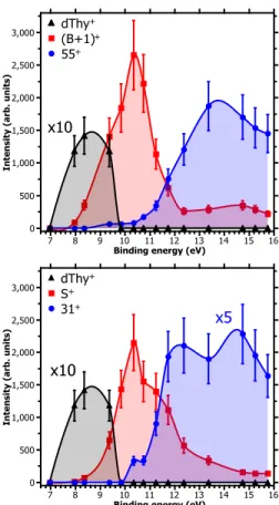
FIG. 3. Partial ion yields of some products as a function
of the binding energy obtained from PEPICO. (a) Ionized
thymidine (dThy+) and base fragments ((B+1)+ and 55+).
(b) Ionized thymidine and sugar fragments (S+ and 31+).
Each point of the curves represents the areas of a gaussian fit to the mass spectra peaks. Error bars are estimated to 20% of the value due to the fitting method.
PEPICO spectra via a constrained linear least-square regression to the the ion-induced mass spectrum con-sidering eleven most relevant features. The fit param-eters represent the contribution of each PEPICO mass spectrum, i.e. the contribution of the fragmentation of a bunch of excited states of the singly charged ion, to the ion spectrum (See method in Supplemental Mate-rial [23]). The result is displayed in Fig.2 as a function of the excitation energy defined as the difference between the energy left in the target and the ionization potential. The energy distribution increases smoothly up to a max-imum around 2 − 3 eV and then it extends up to 8 eV and likely also above this energy, in a region not investi-gated in the present PEPICO experiments. Collisions at closer impact parameters can explain the extended tail towards larger deposited energy [27, 32, 33]. Penetrat-ing trajectories are associated with large deposit energy of several tens of eV [33]. However, in the present in-teraction of 48 keV O6+ with thymidine, peripheral
col-lisions leading to small energy transfer are dominating. This is due to the fact that the electron capture radius
4 dThy 0.0 1st VIP 8.00 7.66 dThy+ 8.89 S+ 12.35 (125+) (117+) 10.92 (55+) 13.66 (55+) 13.60 (31+) 7.66 8.93 (B+1)+ 9.36 (S-1)+ (126+) (116+) 13.14 (55+) 7.66 (31+) HOCH2+ 10.77 12.30 (55+) C3H3O+ C3H3O+ (e) (a) (b) (c) (d) C3H3O+ C3H3O+ HOCH2+ B+ Ex ci ta ti on En er gy (e V)
Intensity (arb. units)
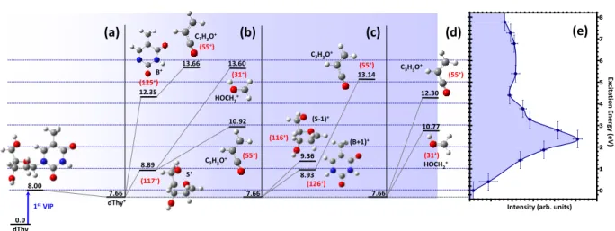
FIG. 4. (a) First vertical ionization potential and the most stable conformer of the singly charged thymidine (dThy+). (b),
(c) and (d) Energy levels of some fragmentation pathways obtained after the exploration of the potential energy surface of the most stable conformer of the singly charged thymidine. The calculated barriers of all pathways are not shown here for sake of clarity. (e) Determined distribution of excitation energy.
is large (∼ 20 a.u. considering the classical-over-barrier model [34]) compared to the molecular size (”diameter” of ∼ 4.5 a.u.). The form of the distribution shown in Fig. 2 is qualitatively similar to those obtained by fitting theoretical fragmentation probabilities to experimental measured branching ratios in small carbon clusters [27] and fullerenes [33, 35], in which the energy distribution was the fitting parameter, thus showing that the present results are also compatible with previous empirical es-timations. Notice that, although the set of accessible target states can in principle be different in photoioniza-tion and collision processes, due to the different conser-vation rules that can apply in each case, this is not a problem in the present work because the absence of any symmetry in the molecular target does not restrict the number of accessible states in either process. Moreover, the single-electron capture, which is the dominant pro-cess at impact energies considered in this work, is not accompanied by excitation of target and projectile elec-trons [33, 35]. Therefore, one can safely assume that the mass spectra resulting from the collision involves the same target states as the PEPICO spectra.
According to the PEPICO results, the maximum in the distribution of excitation energy corresponds to the region of the HOMO−3 state. The charge distribution (Fig.1e) leads to the cleavage of the glycosidic bond and the production of both (B+1)+ and S+ fragments as observed in ion-collision. Larger excitation energy will cause further fragmentation. The partial ion yields of the parent ion and the leading fragments in the PEPICO mass spectra are plotted as a function of the binding en-ergy Eb in Fig.3. The parent ion has a maximum yield
centered around 8.5 eV and then vanishes above 10 eV, while the partial yields of the main fragments (B+1)+
and S+are observed over a wide E
brange starting around
8.5 eV and a display maximum around 10 eV (Fig.3). Secondary dissociation of these fragments is observed for higher Ebwhich corresponds to the tail towards larger
ex-citation energies in Fig.2. Fragments at m/q = 55 amu have been previously assigned to C3H3O+ arising from
the base part [24]. Several pathways leading to this frag-ment have been calculated, as shown in Fig.4. The quan-tum chemistry calculations for the secondary fragmenta-tion rely on an explorafragmenta-tion of the potential energy sur-face in the ground state, i.e. assuming fast redistribution of the excitation energy over the vibrational degrees of freedom. The structure of the neutral molecule in the gas phase, its ionic form and the fragments produced in the relevant exit channels, together with the associated dis-sociation energy, have been computed with the density functional theory at the B3LYP/6-31G(d,p) level of the-ory, using Gaussian09 [30]. The simulations show that fragment C3H3O+ can be produced from the base part
B+ (panel (b)) and from (B+1)+ (panel (c)), but also from the sugar part S+ (panel (b)). More surprisingly it is also possible to form this fragment directly from dThy+ without glycosidic bond breaking (panel (d)). The sec-ond fragment at m/q = 31 is assigned to HOCH+2. This fragment arises from the sugar part [25] and certainly from the outside part of the furanose ring [32]. Computed formation mechanisms show that it is possible to obtain this fragment from the sugar part while keeping intact the glycosidic bond (Fig.4(b) and (d), respectively). Thus, combining the partial ion yields measured in the PEPICO experiments, the calculation of the binding energies of the different ionic states with the OVGF method and the dis-sociation pathways one can evaluate the contribution of the different fragmentation channels to the distribution of the energy transfer in the ion collision.
a method to accurately determine the excitation energy distribution of complex molecular ions produced in colli-sions with fast ions without knowledge of the initial and final states of the projectile nor the ionization potential of the target. The method relies on the combination of photon and ion experiments. The additional support of quantum chemistry calculations allows us to rationalize the measured energy distributions in terms of the elec-tronic states of the singly charged ion and fragmentation channels. Thus the combination of ion and electron ve-locity resolved spectroscopies with in situ photoioniza-tion experiments appears as a promising tool to obtain a complete picture of the molecular dynamics that follows a collision with fast ions.
We thank F. Noury, S. Guillous, R. Richter, M. Coreno and K. Prince for support as well as L.J. Avaldi for ad-vices in the statistical analysis of the data. We acknowl-edge the allocation of computer time at the Centro de Computaci´on Cient´ıfica at the Universidad Aut´onoma de Madrid (CCC-UAM). This work was partially sup-ported by the projects FIS2013-42002-R and CTQ2013-43698-P (MINECO), NANOFRONTMAG (CAM), PHC Galil´ee 28137PE and an Advanced Grant of the Eu-ropean Research Council XCHEM No. 290853. Re-search was conducted in the framework of the Interna-tional Associated Laboratory (LIA) ”Fragmentation DY-NAmics of complex MOlecular systems - DYNAMO” and in the COST actions XLIC (CM1204) and Nano-IBCT (MP1002). D.G.P. acknowledges the FPI doctorate grant of the Universidad Aut´onoma de Madrid. S. D.-T. grate-fully acknowledges the ”Ram´on y Cajal” program of the Spanish Ministerio de Educaci´on y Ciencia.
∗
sylvain.maclot@fysik.lth.se
†
patrick.rousseau@ganil.fr
[1] R. de Nalda and L. Ba˜nares, eds., Ultrafast Phenomena in Molecular Sciences, Springer Series in Chemical Physics, Vol. 107 (Springer International Publishing, Heidelberg New York Dordrecht London, 2014).
[2] F. Calegari, D. Ayuso, A. Trabattoni, L. Belshaw, S. De Camillis, S. Anumula, F. Frassetto, L. Poletto, A. Palacios, P. Decleva, J. B. Greenwood, F. Mart´ın, and M. Nisoli, Science 346, 336 (2014).
[3] B. Erk, D. Rolles, L. Foucar, B. Rudek, S. W.
Epp, M. Cryle, C. Bostedt, S. Schorb, J. Bozek, A. Rouzee, A. Hundertmark, T. Marchenko, M. Si-mon, F. Filsinger, L. Christensen, S. De, S. Trippel,
J. K¨upper, H. Stapelfeldt, S. Wada, K. Ueda, M.
Swig-gers, M. Messerschmidt, C. D. Schr¨oter, R. Moshammer,
I. Schlichting, J. Ullrich, and A. Rudenko, Phys. Rev. Lett. 110, 053003 (2013).
[4] A. N. Markevitch, D. A. Romanov, S. M. Smith, and
R. J. Levis, Phys. Rev. Lett. 92, 063001 (2004).
[5] Y. Jiang, A. Rudenko, O. Herrwerth, L. Foucar,
M. Kurka, K. K¨uhnel, M. Lezius, M. Kling, J. van
Tilborg, A. Belkacem, K. Ueda, S. D¨usterer, R. Treusch,
C. Schr¨oter, R. Moshammer, and J. Ullrich, Phys. Rev.
Lett. 105, 263002 (2010).
[6] S. De, J. Rajput, A. Roy, P. Ghosh, and C. Safvan, Phys. Rev. Lett. 97, 213201 (2006).
[7] S. Maclot, D. G. Piekarski, A. Domaracka, A. M´ery,
V. Vizcaino, L. Adoui, F. Mart´ın, M. Alcam´ı, B. A. Hu-ber, P. Rousseau, and S. D´ıaz-Tendero, J. Phys. Chem. Lett. 4, 3903 (2013).
[8] D. G. Piekarski, R. Delaunay, S. Maclot, L. Adoui, F. Mart´ın, M. Alcam´ı, B. A. Huber, P. Rousseau, A.
Do-maracka, and S. D´ıaz-Tendero, Phys. Chem. Chem.
Phys. 17, 16767 (2015).
[9] L. Levin, W. Skomorowski, L. Rybak, R. Kosloff, C. P.
Koch, and Z. Amitay, Phys. Rev. Lett. 114, 233003
(2015).
[10] Z. Li, O. Vendrell, and R. Santra, Phys. Rev. Lett. 115, 143002 (2015).
[11] P. L´opez-Tarifa, M.-A. Herv´e du Penhoat, R.
Vuilleu-mier, M.-P. Gaigeot, I. Tavernelli, A. Le Padellec, J.-P. Champeaux, M. Alcam´ı, P. Moretto-Capelle, F. Mart´ın, and M.-F. Politis, Phys. Rev. Lett. 107, 023202 (2011). [12] M. Capron, S. D´ıaz-Tendero, S. Maclot, A. Domaracka,
E. Lattouf, A. Lawicki, R. Maisonny, J.-Y. Chesnel, A. M´ery, J.-C. Poully, J. Rangama, L. Adoui, F. Mart´ın, M. Alcam´ı, P. Rousseau, and B. A. Huber, Chem.-Eur. J. 18, 9321 (2012).
[13] L. Chen, S. Martin, J. Bernard, and R. Br´edy, Phys.
Rev. Lett. 98, 193401 (2007).
[14] R. Br´edy, J. Bernard, L. Chen, G. Montagne, B. Li, and
S. Martin, J. Chem. Phys. 130, 114305 (2009).
[15] M. Durante and J. S. Loeffler, Nat. Rev. Clin. Oncol. 7, 37 (2010).
[16] A. V. Solov’yov, E. Surdutovich, E. Scifoni, I. Mishustin, and W. Greiner, Phys. Rev. E 79, 011909 (2009). [17] B. Liu, S. Nielsen, P. Hvelplund, H. Zettergren, H.
Ced-erquist, B. Manil, and B. A. Huber, Phys. Rev. Lett. 97, 133401 (2006).
[18] S. Ptasi´nska, S. Denifl, V. Grill, T. M¨ark, E. Illenberger, and P. Scheier, Phys. Rev. Lett. 95, 093201 (2005). [19] E. C. Montenegro, M. B. Shah, H. Luna, S. W. J. Scully,
A. L. F. de Barros, J. A. Wyer, and J. Lecointre, Phys. Rev. Lett. 99, 213201 (2007).
[20] H. Levola, K. Kooser, E. Rachlew, E. N˜ommiste, and
E. Kukk, Int. J. Mass Spectrom. 353, 7 (2013).
[21] T. Bergen, X. Biquard, A. Brenac, F. Chandezon, B. A. Huber, D. Jalabert, H. Lebius, M. Maurel, E. Monnand,
J. Opitz, A. Pesnelle, B. Pras, C. Ristori, and J. C.
Rocco, Rev. Sci. Instrum. 70, 3244 (1999).
[22] O. Plekan, M. Coreno, V. Feyer, A. Moise, R. Richter, M. de Simone, R. Sankari, and K. C. Prince, Phys. Scr. 78, 058105 (2008).
[23] See Supplemental Material at [URL will be inserted by publisher] for experimental details, fitting method and list of electron densities of orbitals.
[24] E. It¨al¨a, M. A. Huels, E. Rachlew, K. Kooser, T. H¨agerth, and E. Kukk, J. Phys. B: At. Mol. Opt. 46, 215102 (2013).
[25] Z. Deng, I. Bald, E. Illenberger, and M. Huels, Phys.
Rev. Lett. 95, 153201 (2005).
[26] S. Ptasi´nska, P. Candori, S. Denifl, S. Yoon, V. Grill,
P. Scheier, and T. M¨ark, Chem. Phys. Lett. 409, 270
(2005).
[27] G. Martinet, S. D´ıaz-Tendero, M. Chabot, K. Wohrer,
6
A. L. Padellec, D. Gard´es, L. Lavergne, G. Lalu,
X. Grave, J. F. Clavelin, P.-A. Hervieux, M. Alcam´ı, and F. Mart´ın, Phys. Rev. Lett. 93, 063401 (2004).
[28] P. Bolognesi, J. A. Kettunen, A. Cartoni, R. Richter,
S. Tosic, S. Maclot, P. Rousseau, R. Delaunay, and
L. Avaldi, Phys. Chem. Chem. Phys. 17, 24063 (2015). [29] L. S. Cederbaum, J. Phys. B: At. Mol. Phys. 8, 290
(1975).
[30] M. J. Frisch et al., “Gaussian 09 Revision C.01,” Gaus-sian Inc. Wallingford CT 2009.
[31] D. L. Yeager, Applied Many-Body Methods in Spec-troscopy and Electronic Structure, D. Mukherjee ed. (Springer US, New York, 1992) pp. 133–161.
[32] M. A. Herv´e Du Penhoat, P. L´opez-Tarifa, K. K. Ghose, Y. Jeanvoine, M. P. Gaigeot, R. Vuilleumier, M. F.
Poli-tis, and M. C. Bacchus-Montabonel, J. Mol. Model. 20, 2221 (2014).
[33] A. Rentenier, L. F. Ruiz, S. D´ıaz-Tendero, B. Zarour,
P. Moretto-Capelle, D. Bordenave-Montesquieu,
A. Bordenave-Montesquieu, P. A. Hervieux, M. Al-cam´ı, M. F. Politis, J. Hanssen, and F. Mart´ın, Phys. Rev. Lett. 100, 183401 (2008).
[34] A. B´ar´any, G. Astner, H. Cederquist, H. Danared,
S. Huldt, P. Hvelplund, A. Johnson, H. Knudsen, L. Lil-jeby, and K.-G. Rensfelt, Nucl. Instrum. Meth. B 9, 397 (1985).
[35] S. D´ıaz-Tendero, L. F. Ruiz, B. Zarour, J. Hanssen, M. Alcam´ı, M. F. Politis, P.-A. Hervieux, and F. Mart´ın, J. Phys.: Conf. Ser. 194, 012047 (2009).