HAL Id: tel-02971170
https://tel.archives-ouvertes.fr/tel-02971170
Submitted on 19 Oct 2020HAL is a multi-disciplinary open access archive for the deposit and dissemination of sci-entific research documents, whether they are pub-lished or not. The documents may come from teaching and research institutions in France or abroad, or from public or private research centers.
L’archive ouverte pluridisciplinaire HAL, est destinée au dépôt et à la diffusion de documents scientifiques de niveau recherche, publiés ou non, émanant des établissements d’enseignement et de recherche français ou étrangers, des laboratoires publics ou privés.
The regulatory role of cytokines on neurodevelopment
and behaviour during the early postnatal period : An
investigation on the impact of TNF on mouse behaviour
in the early postnatal period and a novel approach of
data analysis applied to the immune activation mouse
model of autism
Cristina Paraschivescu
To cite this version:
Cristina Paraschivescu. The regulatory role of cytokines on neurodevelopment and behaviour during the early postnatal period : An investigation on the impact of TNF on mouse behaviour in the early postnatal period and a novel approach of data analysis applied to the immune activation mouse model of autism. Molecular biology. COMUE Université Côte d’Azur (2015 - 2019), 2019. English. �NNT : 2019AZUR6027�. �tel-02971170�
Le rôle régulateur des cytokines dans le neurodéveloppement
et le comportement au début de la période postnatale
Etude de l'impact du TNF sur le comportement de la souris au début de la période postnataleet une nouvelle approche d'analyse de données appliquée au modèle murin de l'autisme basée sur l'activation de l’immunité maternelle
Cristina Paraschivescu
Université Côte d’Azur, CNRS, Institut de Pharmacologie Moléculaire et
Cellulaire, Valbonne, France
LABEX Signalife
Présentée en vue de l’obtention du grade de docteur en Immunologie et Microbiologie
de l’Université Côte d’Azur
Dirigée par : Laetitia Davidovic / Nicolas Glaichenhaus
Soutenue le : 19 décembre 2019
Devant le jury, composé de :
Jacques Barik, Président, UCA, IPMC, Nice
Ulrike Weber-Stadlbauer, Rapporteur, Université de Zurich, Zurich
Sophie Layé, Rapporteur, Université Bordeaux 2, NutriNeuro, INRA, Bordeaux
Jérôme Becker, Examinateur, Université Rabelais de Tours, PRC, INRA, Tours
Laetitia Davidovic, Co-directrice de thèse, UCA, IPMC, Nice
Nicolas Glaichenhaus, Co-directeur de thèse, UCA, IPMC, Nice
Le rôle régulateur des cytokines dans le neurodéveloppement
et le comportement au début de la période postnatale
Etude de l'impact du TNF sur le comportement de la souris au début de la période postnataleet une nouvelle approche d'analyse de données appliquée au modèle murin de l'autisme basée sur l'activation de l’immunité maternelle
Jury :
Président du jury*
Jacques Barik, Maître de Conférence, Université Côte d'Azur Rapporteurs:
Ulrike Weber-Stadlbauer, Assistant Professor, Team Leader, Université de Zurich Sophie Layé, Directrice de Recherche INRA, Université Bordeaux 2
Examinateur:
Jérôme Becker, Chargée de Recherche Inserm, Université Rabelais de Tours Directeurs de Thèse:
Laetitia Davidovic, Chargée de Recherche CNRS, Université Côte d'Azur Nicolas Glaichenhaus, Professeur, Université Côte d'Azur
Titre (en français) : Le rôle régulateur des cytokines dans le neurodéveloppement et le comportement au début de la période postnatale
Résumé (de 1700 à 4000 caractères espaces compris)
Plusieurs études ont montré que l’activation du système immunitaire maternel (MIA) pendant la grossesse augmentait le risque de troubles neurologiques et d’anomalies du comportement dans la descendance. Afin d’étudier les mécanismes impliqués, plusieurs auteurs ont comparé le comportement de souris nées de mères injectées pendant la grossesse avec du poly(I:C), une molécule mimant une infection par le virus de la grippe, et celui de souris nées de mères injectées avec une solution saline. Bien que ces études aient permis de confirmer que l’activation du système immunitaire maternel pouvait induire des troubles du comportement, la majorité d’entre elles se sont fondées sur des tests comportementaux effectués chez la souris adulte. Ainsi, il reste à déterminer si la modification des niveaux d’autres cytokines pendant la période périnatale peut avoir une incidence sur le neurodéveloppement précoce et sur le comportement de la jeune souris. Pour répondre à cette question, nous avons caractérisé la descendance de plusieurs cohortes de mères injectées avec du poly(I:C) ou avec une solution saline, pour leur comportement entre 5 et 15 jours après la naissance et pour la concentration de plusieurs cytokines dans le sérum. Parce que le neurodéveloppement et la production de cytokines sont affectés par plusieurs variables, nous avons utilisé une analyse multivariée pour identifier les variables environnementales et biologiques associées au fait d’être le descendant d’une mère injectée avec du poly(I :C) (par opposition au fait d’être le descendant d’une mère injectée avec une solution saline). Nous avons constaté que la diminution du poids et de la température corporelle de la mère après injection de poly(I:C), la taille de la portée, le poids de la souris à 15 jours, le nombre de vocalisations ultrasonores (USV) émises par la souris à 6 jours, la distance parcourue par le souris et le temps passé immobile à 13 jours, ainsi que les concentrations sériques de TNF, IL-5, IL-15 et CXCL10 à 15 jours étaient associés au fait d’être le descendant d’une mère injectée avec du poly(I :C). Pour continuer à explorer le rôle régulateur du TNF, nous avons injecté quotidiennement du TNF recombinant à des souris nouveau-nées entre le jour 1 et le jour 5 après leur naissance, et nous avons étudié leur développement et leur comportement entre le jour 8 et le jour 15. Contrairement à nos attentes, l’injection de TNF à des souris nouveau-nées n’a pas d’impact négatif sur le développement, mais favorise plutôt l’acquisition de réflexes sensorimoteurs et le comportement exploratoire. Pris dans leur l’ensemble, nos résultats confirment que les cytokines jouent un rôle crucial dans le neurodéveloppement et que des variations dans l’abondance de certaines d’entre elles, et notamment du TNF, ont un impact sur l’acquisition de certains réflexes et comportement pendant les premiers jours de la vie. Bien que nos études ne nous aient pas permis d’explorer les mécanismes par lesquels cytokines influent sur le neurodéveloppement, les protocoles que nous avons élaborés et les résultats que
nous avons obtenus fournissent un cadre pour d’autres études visant à mieux comprendre ces mécanismes.
Mots-clés : neurodéveloppement, cytokines, immunité maternelle, inflammation, autisme, protection neuronale, comportement, analyse statistique multivariée
Title (in English): The regulatory role of cytokines on neurodevelopment and behaviour during the early postnatal period
Abstract (from 1700 to 4000 prints including spaces)
Both preclinical and clinical studies have shown that immune activation and inflammation during the early stages of neurodevelopment increase the risk of neurodevelopment disorders and behaviour abnormalities in adults. While the underlying mechanisms have only been partially elucidated, experiments in the maternal immune activation mouse model (MIA) – in which pregnant dams are injected with the viral mimic poly(I:C) – have demonstrated the critical role of two cytokines: interleukin (IL)-6 and IL-17A. However, the vast majority of the studies performed to date have used behavioural tests in adult mice as a read out to study the impact of cytokines on neurodevelopment. Therefore, it is not clear whether altered levels of other cytokines during the perinatal period could impact neurodevelopment and behaviour in infant mice. To address this issue, we have analysed the progeny of several cohorts of poly(I:C)- and saline-injected mothers for behaviour between postnatal day 5 (P5) and P15 and serum cytokine levels at P15. Because both perinatal neurodevelopment and cytokine production are known or believed to be impacted by many environmental variables, we analysed our data using a multivariable statistical model to identify features associated with being born to a poly(I:C)-injected mother (as opposed to being born to a saline-injected mother). We found that the drop of body weight and temperature of the mother after poly(I:C) injection, the litter size, the pup weight at P15, the number of ultrasonic vocalizations (USV) emitted by the pup at P6, the distance travelled by the pup and the time it spent mobile at P13, as well as serum levels of Tumour Necrosis Factor (TNF), IL-5, IL-15 and C-X-C motif chemokine (CXCL)10 were all associated with altered odds of being born to a poly(I:C)-injected mother. To further explore the role of TNF during the early postnatal period, we injected mouse pups daily from P1 to P5 and assessed these animals for both developmental milestones and behaviour from P8 to P15. Unexpectedly, injection of recombinant TNF did not have a detrimental impact on neurodevelopment but rather promoted sensorimotor reflexes acquisition and exploratory behaviour. Altogether, our results confirm that cytokines play a critical role during neurodevelopment and that altered levels of specific cytokines, and in particular TNF, could regulate the acquisition of developmental milestones and behaviour in infant mice. While
we have only obtained preliminary insights into underlying mechanisms, the protocols that we have developed provide a framework for further studies.
Keywords: neurodevelopment, cytokines, maternal immunity, inflammation, autism, neuronal protection, behaviour, multivariable statistical analysis
Acknowledgements
I would particularly like to thank my supervisors Laetitia and Nicolas for their invaluable guidance and help throughout my PhD. They chose to give me the opportunity to carry out my PhD in their team and believe in me during the past 3 years, for which I will be forever grateful. I learned a lot from you and by working with you, thank you very much for that! Additionally, I would like to thank the entire NG team, especially:
- Susana, for your amazing help with statistics and intriguing conversations; - Patricia, for the nice occasional team work and for being a good friend; - Olfa, for teaching me molecular techniques;
- Emanuela, for bringing an extra ray of sun into our sometimes gloomy lab routine; - Last, but not least, Aidan, for your constant support and for making these past years
more fun than they would’ve been otherwise.
I want to thank Pierre Gressens and Juliette Van Steenwinckel, our collaborators from Paris, who taught me how to properly perform injections into mouse neonates, as well as how to carefully control the mouse experimental design.
I would also like to give massive thanks to the animal facility, particularly to: - Thomas Lorivel, for your constant help with our projects, in terms of the
experimental planning, technical support, discussions about the analysis of our data and the speed at which you solve Animex problems
- Lucien Relmy, for taking care of my exorbitant amount of mice at any given time Special thanks to Gauthier Alavoine, for building many of our experimental setups for behavioural tests, oftentimes on short notice!
I would also like to thank the entire institute, for providing an excellent environment for growth and scientific development.
I want to thank my family, especially my mother Emilia Nica, who always supported and encouraged me to find my path in life, even in the most difficult situations! I wouldn’t be here without her! (I hope you got the pun)
Merci mult mum! Îți mulțumesc că m-ai ajutat întotdeauna, în special că m-ai înțeles și îndrumat în decizia grea de a înceta un doctorat, fără de care nu aș fi fost aici și nu aș fi învățat toat ce am învățat.
Last, but not least, I want to thank my English and French language professors from
Romania, Mrs. Galaseanu and Mrs. Dinescu, without whom I couldn’t have carried out a PhD in a foreign country and a foreign language! Vă mulțumesc din tot sufletul!
I dedicate this work to Greta, my beloved sister, who left us in 2016. You will never be forgotten.
“La steaua care-a răsărit “So far it is athwart the blue
E-o cale-atât de lungă, To where yon star appears,
Că mii de ani i-au trebuit That for its light to reach our view
Luminii să ne-ajungă. Has needed thousand years.
Poate de mult s-a stins în drum Maybe those ages gone it shed
În depărtări albastre,
Its glow, then languished in the skies, Iar raza ei abia acum
Yet only now its rays have sped Luci vederii noastre,
Their journey to our eyes.
Icoana stelei ce-a murit
The icon of the star that died Încet pe cer se suie:
Slowly the vault ascended;
Era pe când nu s-a zărit, Time was ere it could first be spied,
Azi o vedem, şi nu e. We see now what is ended.
Tot astfel când al nostru dor So is it when our love's aspire
Pieri în noapte-adâncă,
Is hid beneath night's bowl, Lumina stinsului amor
The gleam of its extinguished fire Ne urmăreşte încă”
Enkindles yet our soul.”
Table of contents
List of abbreviations ... 10
Introduction ... 13
1. Interactions between the immune system and the brain during immune activation ... 13
1.1. Overview... 13
1.2. Cytokines and cytokine receptors ... 13
1.3. Pathogen recognition ... 14
1.4. Pathogen Associated Molecular Patterns ... 16
1.5. The peripheral immune system and the brain ... 17
1.6. Cytokines and behaviour ... 19
2. How cytokines shape neurodevelopment ... 20
2.1. The essential role of cytokines during embryonic development ... 20
2.2. Microglia: neuroimmune interactions in shaping neuronal circuitry ... 20
2.3. Neonatal immune system vs. adult immune system ... 21
2.4. The essential role of cytokines during postnatal development ... 22
2.5. Specific role of cytokines in neurodevelopment: the example of TNF ... 22
2.6. Human studies supporting a role of cytokines in neurodevelopment ... 23
3. Cytokines can interfere with neurodevelopment and contribute to neurodevelopmental disorders, including autism spectrum disorders ... 30
3.1. Neurodevelopmental disorders - overview ... 30
3.2. Autism spectrum disorders ... 31
3.3. Human studies: the case of ASD ... 33
3.4. Mouse models of neurodevelopmental defects triggered by immune activation ... 36
4. Scientific hypotheses and objectives ... 41
Manuscript #1 ... 43
1. Introduction ... 44
2. Materials and Methods ... 46
3. Results ... 50
4. Discussion ... 52
5. Conclusions & future directions ... 55
References ... 56
Figures ... 60
Manuscript #2 ... 67
1. Introduction ... 68
2. Materials and Methods ... 70
4. Discussion ... 76 5. Conclusions ... 80 References ... 81 Figures ... 85 Discussion ... 91 Overview... 91
1. How does TNF impact neurodevelopment? ... 95
2. What is the physiological relevance of our experimental model in which pups are injected with recombinant TNF? ... 97
3. Vulnerability vs. resilience to psychological stress ... 99
4. What else could we do to further investigate the impact of TNF on neurodevelopment? ... 102
5. What is the impact of poly(I:C) injection on pregnant dams? ... 103
6. Why do pups born to poly(I:C)-injected mothers gain weight more rapidly than control pups? ... 104
7. Why do pups born to poly(I:C)-injected mothers exhibit communication impairment? ... 105
8. Why do pups born to poly(I:C)-injected mothers exhibit decreased locomotor activity? ... 107
9. Why did we analyse our data using a multivariable statistical approach, and which conclusions could we draw? ... 108
10. What is known about the role of TNF, CXCL10, IL-5 and IL-15 in neurodevelopment and brain function? ... 110
Take home message ... 113
List of abbreviations
5'—C—phosphate—G—3' CpG
Activator protein 1 AP1
Alzheimer’s disease AD
Analysis of variance ANOVA
Applied behaviour analysis ABA
Asperger's syndrome AS
Attention Deficit Hyperactivity Disorder ADHD
Autism Spectrum Disorder ASD
Blood-brain-barrier BBB
Bone morphogenetic proteins BMP
Caesarean-section C-section
Cardiotrophin-1 CT-1
C-C motif ligand CCL
Central Nervous System CNS
Ciliary neurotrophic factor CNTF
cochlear root neurons CRN
Colony stimulating factors CSF
Confidence Interval CI
C-X-C chemokine receptor CXCR
C-X-C motif ligand CXCL
Cyclooxygenase type 2 COX2
degrees Celsius °C
Diagnostic and Statistical Manual of Mental Disorders, version 5 DSM-V
Embryonic day E
Fragile X syndrome (gene) FMR1
G protein-coupled receptor GPCR
Gamma-aminobutyric acid receptor subunit beta-3 GABRB3
Gastrointestinal GI
Generalized Estimating Equations GEE
Hypothalamic-Pituitary-Adrenal axis HPA axis
hour h
Intellectual quotient IQ
Interferon IFN
Interferon Regulatory Factor IRF
Interleukin IL
Interleukin-6 receptor IL-6R
kiloDalton kDa
kilogram Kg
kiloHertz kHz
Knock-out KO
Leukemia inhibitory factor LIF
Lower Limit of Detection LLOD
Major Histocompatibility Complex I MHC-I
Maternal immune activation MIA
microgram μg
microliter μL
milliliter ml
millisecond ms
m-opioid receptor knock-out mice Oprm
-/-Natural Killer NK cell
neurexin NRXN
Neurodevelomental disorder NDD
neuroligins NLGN
Neuronal stem cells NSC
N-methyl-D-aspartate NMDA
Nuclear factor kappa-light-chain-enhancer of activated B cells NF-kB
nucleus reticularis pontis caudalis PnC
number n
Odd Ratios OR
Oxytocin receptor OXTR
Positive And Negative Symptoms Scale PANSS
Pathogen-associated-molecular patterns PAMP
Pattern recognition receptors PRR
Peripheral blood mononuclear cell PBMC
Pervasive developmental disorder-not otherwise specified PDD-NOS
Polyinosinic:polycytidylic acid Poly(I:C)
Postnatal day P
Prepulse inhibition PPI
Principal component analysis PCA
Prostaglandin E2 PGE2
p-value p
radial glial cells RGS
Rett syndrome (gene) MECP2
Ribonucleic acid RNA
Room temperature RT
second s
Segmented filamentous bacteria SFB
SH3 and multiple ankyrin repeat domains 1 knock-out mice Shank1 -/-SH3 and multiple ankyrin repeat domains 2/3 (gene) Shank2/3
soluble Tumour Necrosis Factor sTNF
Strengths and Difficulties Questionnaire SDQ
T helper 17 cell Th17 cell
T helper cell 1 Th1 cell
T helper cell 2 Th2 cell
Toll-like receptor TLR
Transforming growth factors TGF
transmembrane form Tumour Necrosis Factor mTNF
Tumour Growth Factor β TGFβ
Tumour Necrosis Factor TNF
Tumour Necrosis Factor receptor 1 TNFR1
Tumour Necrosis Factor receptor 1-knock-out Tnfr1-KO
Tumour Necrosis Factor receptor 2 TNFR2
Ultrasonic vocalisations USV
United States US
Introduction
1. Interactions between the immune system and the brain during immune activation
1.1. Overview
The immune system represents the totality of factors and entities in a given organism, which act in coordination as a defence machinery against invading pathogens (for review see (Chaplin 2010)). It is broadly divided into two main branches, the innate and the adaptive immune systems. The innate immune system includes germline-encoded mechanisms and represents the first step of defence, acting within hours of a given infection. Its main components are physical barriers, such as the epithelial cell and mucus layers, soluble secreted proteins, among which complement proteins, cytokines and lipid mediators, cells, such as mononuclear phagocytes (monocytes, macrophages, dendritic cells) and granulocytes (neutrophils, basophils and eosinophils) and membrane bound receptors which detect microbial presence. Unlike the innate immune system, the adaptive immune system is able to adapt its detection mechanisms with high specificity towards a particular antigen, by using antigen specific receptors found on the cell surfaces of T and B lymphocytes. The adaptive response is slower to react and follows the innate immune response.
1.2. Cytokines and cytokine receptors
The key molecules which regulate and connect the innate and adaptive immune responses are cytokines and the receptors through which they signal (Illustration 1). Cytokines represent a large group of signalling proteins which play key roles in immune protection against invading pathogens, homeostatic maintenance of the organism shaping the nervous system during early neurodevelopment and adulthood. Cytokines are produced by both immune and non-immune cells and have cell type – and location – dependent functions, capable of acting as autocrine and paracrine signalling. Cytokines are grouped into several main classes: interleukins, interferons, chemokines, tumour necrosis factors, transforming growth factors (TGFs) and colony stimulating factors (CSFs). The main producers of peripheral cytokines are macrophages and T cells, and to a lesser extent, monocytes, fibroblasts and endothelial cells. In the central nervous system (CNS), cytokines are mainly
produced by microglia, which are the brain-resident innate immune cells, but also by non-immune cell types, such as neurons and astrocytes (Turner et al. 2014). Cytokines signal through a variety of receptors which differ both structurally and functionally. Proinflammatory cytokines, such as IL-1, IL-6 and TNF signal through type I transmembrane receptors with distinct extracellular and intracellular domains used for signal transduction, while chemokines, such as CXCL12, signal through specific chemokine receptors, belonging to the seven-transmembrane-G protein-coupled receptor (GPCR) family (Turner et al. 2014).
Illustration 1. Example of cytokine classification by immune response and family. Adapted from Turner et al. 2014.
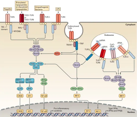
1.3. Pathogen recognition
Cytokines and other immune mediators are produced upon immune cell recognition of invading pathogens. Toll-like receptors (TLRs) operate the cellular mechanisms of pathogen recognition. TLRs are germline-encoded pattern recognition receptors (PRRs), capable of recognizing a wide range of pathogens based on their shared molecular structures, termed pathogen-associated-molecular patterns (PAMPs). Upon PAMP recognition, PRRs trigger an intracellular signalling cascade which ends in the nuclear translocation and activation of transcription factors, such as NF-kB, AP1, IRF3 or IRF7, which subsequently mediate the induction of type I interferon (IFN) and pro-inflammatory cytokines. Specific TLRs respond to specific types of pathogens, an effect driven by TLR localization. TLRs are localized both at the plasma membrane and intracellularly, on endosomes, phagosomes and the endoplasmatic reticulum (Illustration 2). Therefore, TLRs, such as TLR-1, TLR-2, TLR-4, TLR-5 and TLR-6 mostly recognise components of microbial membranes, such as lipopolysaccharide (LPS) and flagellin, and are therefore involved in fungal and bacterial
immune responses, while TLR-3, TLR-7, TLR-8 and TLR-9 mostly recognise nucleic acids, such as CpG DNA motifs as well as double- and single-stranded RNA, and are involved in anti-viral responses (O’Neill, Golenbock, and Bowie 2013; Kawai and Akira 2011).
TLRs have also been found to be expressed in the brain in neurons and glial cells. Microglia, the main immune cells in the brain, express the largest repertoire of TLRs. Moreover, TLR expression has been analysed in mouse models during multiple stage of brain development and it was shown that distinct TLR, such as TLR-7 and TLR-9, change their pattern of expression during the different stages of embryonic brain development (Kaul et al. 2012). TLR-3 plays a particularly important role during the early stages of development, as it was found to be the most highly expressed. It acts as an inhibitor of neural progenitor cell proliferation and axonal growth and therefore plays a key role in modulating neurogenesis (Lathia et al. 2008).
Illustration 2. Representation of the TLR signalling pathways. The different types of TLRs present extra- and intra- cellularly are presented in association with their individual ligands.
Upon ligand binding, several mechanisms assist in the activation of transcription factors, which leads to cytokine production. Adapted from O’Neill, Golenbock, and Bowie 2013.
1.4. Pathogen Associated Molecular Patterns
Molecules which mimic viral and bacterial infections, whether synthetic or naturally occurring, can also bind to TLRs and induce a similar immune response to actual infections. Some of the most commonly used substitutes for inducing infection of viral and bacterial origin are polyinosinic:polycytidylic acid (Poly(I:C)) and LPS, respectively. Poly(I:C) is a synthetic double-stranded RNA analogue used to mimic aspects of a viral infection. It acts by binding to TLR3, which initiates a signalling cascade which, in turn, elicits an acute-phase response in the host, including fever and induction of pro-inflammatory cytokines (IL-1, IL-6, TNF), chemokines, type I IFN and complement proteins (Boksa 2010). LPS is a natural component of the cell wall of Gram-negative bacteria and is used to mimic a bacterial infection. It binds to TLR4 and, similarly to Poly(I:C), leads to upregulation of pro-inflammatory cytokines. Although similar in their mode of action, Poly(I:C) and LPS immune responses can differ in the magnitude of the induced cytokine responses and the type of activated cells. The advantage of using these immunogens to induce immune response is the possibility to experimentally control the time-course and dose of antigen exposure. However, it should be noted that neither Poly(I:C), nor LPS completely mimic the entire time course of a propagating viral or bacterial infection (Harvey and Boksa 2012).
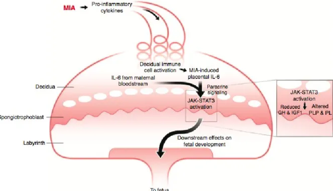
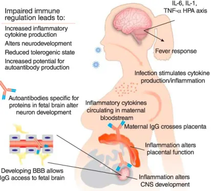
For instance, Poly(I:C) is widely used to mimic maternal viral infection in mice during pregnancy. Epidemiologic evidence suggests that maternal infection is a risk factor for autism spectrum disorders (ASD) and schizophrenia (Hornig et al. 2018; Atladóttir et al. 2010; H. yin Jiang et al. 2016) and it has been modelled in several rodent models. Poly(I:C) injection in pregnant dams during the middle stages of pregnancy, activates the maternal immune system, leading to increased serum levels of the proinflammatory cytokines TNF, IFN-β, IL-1β, IL-6 and IL-17A (Careaga et al. 2018; Mueller et al. 2019; Choi et al. 2016; Kim et al. 2017; E. Y. Hsiao and Patterson 2011). This maternal cytokine dysregulation alters the placental immune environment, in which IL-6 is specifically involved (W. L. Wu et al. 2017), and results in the activation of decidual immune cells, such as macrophages, granulocytes
and uterine NK cells. This increases the placental levels of IL-6, which has downstream effects on the foetal brain, subsequently causing deleterious behaviour in the offspring (Hsiao 2011; Patterson 2011) (Illustration 3).
Illustration 3. Representation of the mechanisms involved in the dysregulaton of the placental immune environment, as a result of Poly(I:C) injection in pregnant dams. This immune activation has deleterious effects on the faetal brain. Adapted from Patterson 2011.
1.5. The peripheral immune system and the brain
Immune activation can spread to areas which are considered immune privileged in homeostatic conditions, such as the placental environment and the CNS. Moreover, during an initial immune activation, endothelial barriers across the body, such as the placental, intestinal and blood-brain-barriers (BBB) can become more permeabilized, commonly referred to as “leaky”, and allow passage of immune cells and larger immune mediators, such as cytokines, into these environments with particular immunological functions. In the brain, this immunological peripheral influx, often caused by infections, can trigger neuroinflammation, which is associated with sickness behaviour (Konsman, Parnet, and Dantzer 2002), as well as other behavioural alterations, described in the next sections. In addition, cytokines can be directly synthesized in the brain by the glia, as well as neurons, a
process which has been seen after brain insults in the absence of infection, through “sterile” damage (Mayer 2013).
The brain parenchyma part of the CNS is an immune privileged site. However, other parts constituting the CNS, such as the meninges, choroid plexus, circumventricular organs, and ventricles undergo immune response similar to those present in the periphery (Hagberg, Gressens, and Mallard 2012). In homeostatic conditions, the BBB restricts entry of potentially harmful molecules and cells to the parenchyma. During peripheral infections, as well as traumatic sterile injury in the brain, pro-inflammatory cytokines, such as IL-1β, IL-6 or TNF, access the brain through saturable transport systems (W. A. Banks, Kastin, and Broadwell 1995; Gutierrez, Banks, and Kastin 1993). Moreover, peripheral leukocytes, such as macrophages and neutrophils, can cross the BBB through a process mediated by changes in adhesion molecules, and contribute to activate glial cells mediating neuroinflammatory processes (Soares et al. 1995; Kubes and Ward 2006; R. S. b. Clark et al. 1994). Microglia, which are the primary brain-resident immune cells, play an important role in sensing CNS damage, by continuously sampling their immediate environment for pathogens or tissue injury. They are mostly responsible for phagocytosing and eliminating microbes, dead cells and protein aggregates (Colonna and Butovsky 2017). Following injury, they rapidly become activated and start releasing pro-inflammatory mediators, such as cytokines, chemokines and reactive oxygen species. Prolonged microglial activation can induce excitotoxic neuronal death and contribute to progressive CNS disorders (C. Mayer 2013; Ye et al. 2013). Another important class of glial cells in the brain is formed by astrocytes which are also the most abundant cell type in the brain, working mainly to support neuronal and synaptic functions. Following tissue injury, activated astrocytes deposit a proteoglycan matrix which forms glial scars that can lead to inhibition of axonal regeneration and function under chronic injury (Yiu and He 2006). Both types of glial cells secrete cytokines and can harm CNS functioning under specific conditions (Illustration 4).
Illustration 4. Summary of the brain-immune interaction in mediating behavioural abnormalities. Immune cells and their mediators act directly on neurons and glia and alter important developmental and functional processes. Adapted from Meltzer and Van De Water 2017.
1.6. Cytokines and behaviour
An important mechanism by which glial cells contribute to lead to pathological processes is through the over-expression of pro-inflammatory cytokines, mostly TNF and IL-1β. Numerous studies have shown that cytokines can control brain function and behaviour, with mostly detrimental effects. Cytokines can notably induce anxiety (Simen et al. 2006; Spadaro and Dunn 1990), sickness behaviour (Konsman, Parnet, and Dantzer 2002; Anisman and Merali 1999), depression (Maes et al. 1993; Dowlati et al. 2010; Réus et al. 2017) and impair cognitive processes (Heyser et al. 1997; Menza et al. 2010). This is further supported by association studies in psychiatric cohorts and in the general population, which have suggested that some pro-inflammatory cytokines are associated with mental health disorders (O’Shea et al. 2014; Kuban et al. 2016; N. M. Jiang et al. 2014), including neurodevelopmental disorders such as ASD (Goines et al. 2011; Spann et al. 2018).
2. How cytokines shape neurodevelopment
Although the immune system is a key player of the host’s defence, it also plays a critical role in the maintenance of tissue integrity and general homeostasis. The immune system has also evolved to regulate physiological processes, such as development, reproduction, metabolism and several aspects of CNS development (Sattler 2017).
2.1. The essential role of cytokines during embryonic development
Cytokines can have pro- or anti-inflammatory functions and be neuroprotective or destructive, depending on their timing of expression (age-related), level of expression (acute vs. chronic) and concentrations (Morganti-Kossman 1997). During neurogenesis, radial glial cells (RGCs) derived from neuroepithelial cells (Hatakeyama et al. 2004) act as precursors for all neurons, astrocytes, oligodendrocytes and adult neural stem cells and guide the migration of immature neurons to their final location (Pinto and Götz 2007). The cytokines of particular importance during this process are the gp130/IL-6 family cytokines and the bone morphogenetic proteins (BMPs), part of the TGFβ superfamily. Members of the gp130 family cytokines, such as IL-6, leukemia inhibitory factor (LIF), ciliary neurotrophic factor (CNTF) and cardiotrophin-1 (CT-1) regulate RGCs self-renewal (Gregg and Weiss 2005; Hatta et al. 2002; Yoshimatsu et al. 2006), while inhibition of the neural induction repressors BMPs contribute to neural induction (Gaulden and Reiter 2008). Moreover, chemokines, such as the CXCL12 through their receptor CXCR4, promote migration and proliferation of newly generated neurons and glia (Klein et al. 2001; Zhu et al. 2002; Lu, Grove, and Miller 2002), and play an important role in axonal pathfinding (Chalasani et al. 2003). Another important role of cytokines during neurodevelopment is that of regulators of synaptogenesis and synaptic pruning (Sedel et al. 2004; Barker et al. 2001), such as in the case of microglia-derived TNF.
2.2. Microglia: neuroimmune interactions in shaping neuronal circuitry
Apart from cytokines, immune cells, and in particular, microglia, also have specific roles in neurodevelopment during the early stages of neurogenesis, as well as during the postnatal period and adulthood. They are the first glial cells to migrate into the CNS during embryonic brain development. This is an important period of neuronal migration, during which
microglia guide neurons and their axons to form prenatal circuits (Colonna and Butovsky 2017), as well as influence neural precursor cell differentiation (Aarum et al. 2003). Moreover, in vitro coculture of microglial and neuronal stem cells (NSCs) show that microglial-secreted factors are necessary for NSC self-renewal (Walton et al. 2006). During postnatal development, microglia modulate synaptic pruning. This activity is achieved by the phagocytosis of dendritic spines that did not receive synaptic inputs (Colonna and Butovsky 2017). Also, microglia phagocyte the debris of surnumerary neurons which had to be eliminated as they were unable to form functional circuits. All these effects contribute to microglial shaping of the neuronal networks during early development.
2.3. Neonatal immune system vs. adult immune system
There is increasing knowledge about the involvement of immune cells and their mediators in early brain development, as well as the immunological differences between the perinatal and adult brain (Garay and McAllister 2010). In comparison to the adult immune system, the neonatal immune system is polarized towards Th2 responses (Maródi 2002; Levy 2007; Wynn and Levy 2010). Moreover, stimulated neonatal serum monocytes secrete less TNFα, a Th1-polarising cytokine, and more IL-6, a Th2-polarising cytokine, than adult monocytes (Angelone et al. 2006). There are also clear age-related differences in immune responses in the brain. In the adult CNS and in particular in the brain parenchyma, the response to inflammatory stimuli, such as pro-inflammatory cytokines or LPS, is characterized by the ability to restrict peripheral leukocyte migration (Andersson, Perry and Gordon, 1992a; Andersson, Perry and Gordon, 1992b). In contrast, during the early stages of mouse CNS development, neutrophil and monocyte are recruited to the brain parenchyma upon endotoxin injection, but the characteristics of this response are age-dependent (Lawson and Perry 1995): immediately after birth, at post-natal day (P) 0, the brain inflammatory response is relatively weak, showing reduced microglial response upon intracerebral LPS administration, as well as slow and reduced neutrophil and monocyte recruitment from the periphery. By P7, the microglial response following LPS injection becomes fast and efficient and there is increased neutrophil recruitment, as compared to P0 (Lawson and Perry 1995).
2.4. The essential role of cytokines during postnatal development
During postnatal development, cytokines in homeostatic conditions have been shown to display a dynamic pattern of expression, both in blood and brain tissue. This pattern is age- and region-specific, which is suggestive of the need for a timely and restricted expression of specific cytokines during neurodevelopment (Garay et al. 2013; Deverman and Patterson 2009; Dziegielewska et al. 2000; Bauer, Kerr, and Patterson 2007). The expression of IL-6, a cytokine involved in neurogenesis, as well as its receptor, IL-6R, have been demonstrated to be tissue-specific in the rat brain, depending on the postnatal developmental stage: Il6 and
Il6r mRNAs levels are highest in adult hippocampus, whereas the levels of Il6 mRNA are
highest in all other brain regions during early brain development (Gadient and Otten 1994).
2.5. Specific role of cytokines in neurodevelopment: the example of TNF
Tumour Necrosis Factor (TNF) is a proinflammatory cytokine historically known as a chief orchestrator of the innate immune response (Holbrook et al. 2019). TNF is normally present in minute amounts, however, following an immune challenge, TNF is massively induced in activated macrophages in peripheral tissues. TNF is expressed as a 27 kDa transmembrane form (mTNF) which acts by cell-to-cell contacts, and as a soluble 17 kDa form (sTNF) produced by regulated cleavage of mTNF that is released in tissues and blood (Kriegler et al. 1988). TNF signals through two membrane receptors, TNFR1 and TNFR2. While both sTNF and mTNF activate TNFR1 signalling transduction pathway, only mTNF triggers TNFR2 signaling (Probert 2015). TNF and its receptors also expressed outside the immune compartment, and notably in the CNS.
Evidence of the role of TNF in early neurodevelopment comes from studies in young mice. Slight increases in TNF levels are observed in the hippocampus and in the cortex during the first 2 postnatal weeks of life, a time of active neurogenesis and synaptogenesis (Garay et al. 2013). Moreover, low doses of TNF promoted the survival, proliferation, and neuronal differentiation mouse neonatal neural precursor cells cultures, while higher doses were apoptotic (Bernardino et al. 2008). Furthermore, young Tnf-knockout (KO) mice exhibit an accelerated maturation of the dentate gyrus hippocampal region, but with pyramidal neurons harbouring a smaller dendritic arborisation in CA1 and CA3 regions (Golan et al. 2004). Finally,
both in vitro and in vivo studies have shown that developing pyramidal neurons from the cortex of Tnf-KO mice are deficient in synaptic scaling, a form of homeostatic plasticity that enables adjustment of synaptic strength at the neuron-scale in response to sustained activity, which is critical for the activity-dependent refinement of neural circuitry during early development (Stellwagen and Malenka 2006; Kaneko et al. 2008; Ranson et al. 2012) . This suggests a critical role for TNF in shaping the nervous system during early developmental stages.
TNF is also required for CNS functioning during adulthood. In physiological conditions, it is constitutively secreted in minute amounts by neurons and glia (Probert 2015). In these conditions, TNF is required for brain cell maintenance and homeostasis. Notably, TNF promotes proliferation of oligodendrocyte progenitors and remyelination (Arnett et al. 2001). Moreover, TNF is known to enhance excitatory synaptic scaling (Beattie et al. 2002). In this context, TNF secreted by astrocytes controls the exposure of AMPA receptors at the synapse, thereby directly regulating synaptic neurotransmission in the hippocampus, cortex and striatum (Beattie et al. 2002; Lewitus et al. 2014; Santello, Bezzi, and Volterra 2011). In the cerebellum, TNF increases the intrinsic excitability of cerebellar Purkinje cells by controling the release of glial glutamate (Shim et al. 2018). In vivo, both Tnf-knockout (KO) and Tnfr1-KO mice have elevated hippocampal adult neurogenesis, while lack of TNFR2 decreases baseline neurogenesis (Chen and Palmer 2013; Iosif et al. 2006).
2.6. Human studies supporting a role of cytokines in neurodevelopment
Although many studies link disrupted patterns of cytokines with neurodevelopmental conditions (see next section 3.), some have identified both deleterious and beneficial links between the levels of specific gestational cytokines and neurocognitive behaviour in the general population.
Gestational cytokine levels and neurocognitive behaviour: To assess the influence of maternal cytokine levels on offspring neurocognitive development, one study studied the
association between maternal serum cytokine levels (measured longitudinally during the 2nd
age. The authors first estimated the cumulative exposure to each cytokine and then studied the associations with neurocognitive outcomes at 7 years. Among the cytokines assessed, they found two pro-inflammatory cytokines - TNF and IL-8 - to be associated with negative or positive neurocognitive outcomes, respectively. TNF was associated with problems in visual-motor functioning and lower cognitive scores, whereas IL-8 was associated with better cognitive performance and motor functioning (Ghassabian et al. 2018). While association does not necessarily imply causation, this study draws attention to the possible involvement maternal cytokines in neurodevelopmental processes at early life stages.
Another study investigated whether the socioeconomic environment can influence maternal immune activity during gestation and whether this was associated with adverse behavioural outcome in the offspring during the first year of life (Gilman et al. 2017). Several
proinflammatory cytokines were measured in the maternal serum during the 3rd trimester of
pregnancy. They found that gestational levels of IL-8 were lower in the most disadvantaged pregnancies experiencing more social-economic distress. Furthermore, maternal socio-economic disadvantage was associated with higher risk of structural and sensorimotor-related neurological abnormalities in the offspring. Finally, they found that decreased maternal IL-8 levels in disadvantaged pregnancies were positively associated with increased risk of neurological abnormalities. Together with previous studies reviewed in Hantsoo et
al., 2018, this study suggests the involvement of maternal stress response to adversity,
which can translate into maternal immune dysregulation and contribute to increase the offspring’s vulnerability to neuropsychiatric disorders.
The following figure (Illustration 5) provides more insight into the link between maternal stressors during pregnancy, which include immune dysfunction, and offspring neuropsychiatric development.
Illustration 5. The impact of stressful events on the maternal immune system during gestation, involving negative outcomes for offspring neurodevelopment and behaviour. Adapted from Hantsoo et al. 2018.
Cytokine levels at birth and neurocognitive behaviour: Another study that should be mentioned when describing the impact of cytokines on child neurocognitive development is the one conducted by Von Ehrenstein et al., 2012 on the link between cytokine levels at birth, measured in cord blood, and child’s intellectual development measured by the intellectual quotient (IQ). The study found that increased levels of IFN-γ and IL-12p70 at birth were associated with reduced odds of low IQ (IQ<70) at 5 years of age. Also, increased cord blood levels of TNF was associated with reduced odds of low IQ (IQ<70) in preterm children. This suggests important links between proinflammatory cytokines and early brain development and that dysregulation of cytokine patterns could contribute to later abnormal child behaviour.
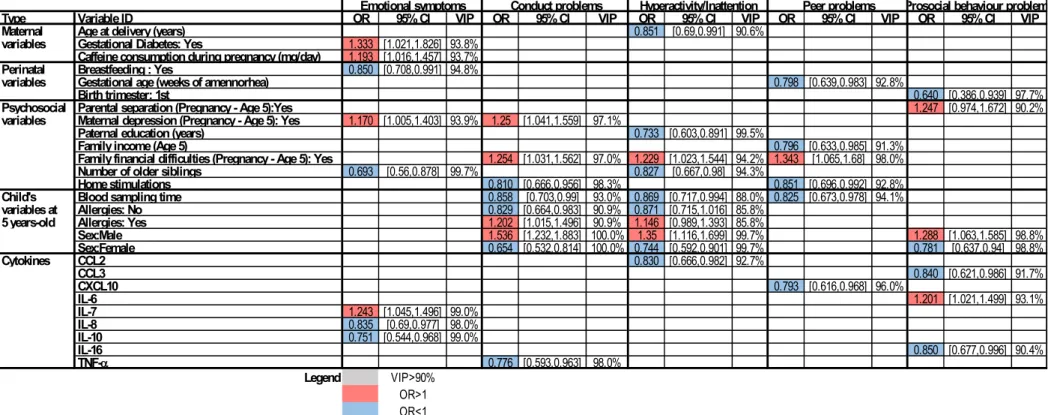
Our team has also recently investigated the association of cytokines at birth with child’s behavioural outcome in a cohort of healthy children of 5 years of age. We used data and biological samples from 786 child pairs participating to the French national
mother-child cohort EDEN. Maternal serum was collected in the 2d trimester of pregnancy. At the age of 5, children were assessed for behavioural difficulties using the Strengths and Difficulties Questionnaire (SDQ). Serum samples were analysed for levels of well-characterized effector or regulatory cytokines. We then used the Elastic net model, a penalized logistic regression method, to investigate associations between serum levels of cytokines and each of the five SDQ-assessed behavioural dimensions after adjustment for relevant covariates and confounders, including parental data for various socio-economic parameters, such as the age of delivery, breastfeeding, C-section, parental education, etc. We found five cytokines to be associated with increased odds of developing problems in one or more behavioural dimensions: CXCL10, IL-10 and IL-12p40 with emotions, CCL11 with both conduct problems and peer problems. In contrast, five cytokines were associated with decreased odds of problems in one or more behavioural dimensions: IL-7, IL-15 and TNF-β with emotions, IL-15 and CCL26 with peer problems, IL-15, CCL26 and TNF with prosocial behaviour. Table 1 summarizes these results. This supports the notion that cytokines at birth could contribute to shape the developing CNS and impact the behavioural outcome of the child later in life.
Table 1. Adjusted associations between cytokines measured in cord blood serum and high-risk of behavioural problems at 5 years of age. Weighted mean Odd Ratios (OR), weighted 95% Confidence Interval (CI) and Variable Inclusion Probability (VIP) for each of the variables selected by the Elastic Net are shown. The VIP was used as a measure of the stability of an association as it can be interpreted as the posterior probability of including a given variable in the model. Only variables with VIPs above 90% are presented.
Variables description ORs 95% CI VIP ORs 95% CI VIP ORs 95% CI VIP ORs 95% CI VIP ORs 95% CI VIP Age at delivery (years) 0.954 [0.911,0.995] 97.4%
Pre-pregnancy BMI (kg.m-2) 1.031 [1.002,1.078] 96.2% 0.97 [0.926,1.001] 92.3%
Smoking during pregnancy (cigarettes/day) 1.086 [1.01,1.151] 98.8% 1.058 [0.993,1.135] 90.4%
Alcohol drinking during pregnancy (mean glasses/week) 1.089 [0.982,1.228 91.8%
Perinatal variables Birth weight (kg) 1.001 [1,1.001] 97.7%
Sex: Male 1.331 [1.158,1.671] 100.0% 1.33 [1.128,1.71] 100.0% 1.222 [1.056,1.461] 100.0%
Sex: Female 0.753 [0.604,0.866] 100.0% 0.755 [0.589,0.888] 100.0% 0.825 [0.697,0.955] 100.0%
Maternal prenatal anxiety (STAI scale) 1.003 [0.98,1.013] 97.3%
Maternal depression during pregnancy (no) 0.726 [0.578,0.915] 97.3% 0.849 [0.658,1.005] 90.6%
Maternal depression during pregnancy (yes) 1.374 [1.095,1.736] 97.3% 1.179 [0.998,1.519] 90.6%
Maternal education (years) 0.918 [0.831,0.988] 99.9% 0.948 [0.877,1] 92.9%
Paternal education (years) 0.876 [0.825,0.942] 100.0% 0.923 [0.851,0.977] 98.1%
Number of older siblings 0.53 [0.424,0.698] 99.6% 0.828 [0.651,0.977] 98.1% 1.278 [1.032,1.628] 99.1%
Cord blood cytokines CCL11 1.002 [1,1.003] 96.3% 1.002 [1.001,1.004] 96.2%
CCL26 0.983 [0.962,0.994] 100.0% 0.989 [0.969,1] 95.3% CXCL10 1.001 [1,1.002] 91.8% IL-7 0.948 [0.89,0.989] 97.3% IL-10 1.044 [0.999,1.252] 92.0% IL-12p40 1.001 [1,1.002] 95.0% IL-15 0.974 [0.864,0.997] 92.8% 0.977 [0.928,0.997] 90.1% 0.989 [0.948,0.999] 92.0% TNF-a 0.905 [0.748,0.996] 97.3% TNF-b 0.796 [0.585,0.984] 90.7% Legend VIP>90% OR>1 OR<1 Psychosocial variables
Emotional symptoms Conduct problems Hyperactivity/Inattention Peer problems Prosocial behaviour problems Maternal variables
Cytokine levels in childhood and neurocognitive behaviour: A second study was conducted by our team, investigating the association of cytokines measured at 5 years of age on behaviour, assessed at the same age. We showed several cytokines to be protective, while others, detrimental to specific behavioural dimensions. Moreover, this study also shows the impact of parental and psychosocial variables on child behaviour. We found that IL-6, IL-7, and IL-15 were associated with increased odds of problems in prosocial behaviour, emotions, and peer relationships, respectively. In contrast, eight cytokines were associated with decreased odds of problems in one dimension: IL-8, IL-10, and IL-17A with emotional problems, TNF with conduct problems, CCL2 with hyperactivity/inattention, CXCL10 with peer problems, and CCL3 and IL-16 with abnormal prosocial behaviour. Table 2 summarizes these results. Without implying causation, these associations support the notion that cytokines regulate brain functions and behaviour.
Table 2: Adjusted associations between cytokines measured in serum and high-risk of behavioral problems at 5 years of age. Weighted mean Odd Ratios (OR), weighted 95% Confidence Interval (CI) and Variable Inclusion Probability (VIP) for each of the variables selected by the Elastic Net in the analysis of MI-R datasets are shown. The VIP was used as a measure of the stability of an association as it can be interpreted as the posterior probability of including a given variable in the model. Only variables with VIPs above 90% are presented.
Type Variable ID OR 95% CI VIP OR 95% CI VIP OR 95% CI VIP OR 95% CI VIP OR 95% CI VIP Maternal Age at delivery (years) 0.851 [0.69,0.991] 90.6%
variables Gestational Diabetes: Yes 1.333 [1.021,1.826] 93.8%
Caffeine consumption during pregnancy (mg/day) 1.193 [1.016,1.457] 93.7%
Perinatal Breastfeeding : Yes 0.850 [0.708,0.991] 94.8%
variables Gestational age (weeks of amennorhea) 0.798 [0.639,0.983] 92.8%
Birth trimester: 1st 0.640 [0.386,0.939] 97.7%
Psychosocial Parental separation (Pregnancy - Age 5):Yes 1.247 [0.974,1.672] 90.2%
variables Maternal depression (Pregnancy - Age 5): Yes 1.170 [1.005,1.403] 93.9% 1.25 [1.041,1.559] 97.1%
Paternal education (years) 0.733 [0.603,0.891] 99.5%
Family income (Age 5) 0.796 [0.633,0.985] 91.3%
Family financial difficulties (Pregnancy - Age 5): Yes 1.254 [1.031,1.562] 97.0% 1.229 [1.023,1.544] 94.2% 1.343 [1.065,1.68] 98.0%
Number of older siblings 0.693 [0.56,0.878] 99.7% 0.827 [0.667,0.98] 94.3%
Home stimulations 0.810 [0.666,0.956] 98.3% 0.851 [0.696,0.992] 92.8%
Child's Blood sampling time 0.858 [0.703,0.99] 93.0% 0.869 [0.717,0.994] 88.0% 0.825 [0.673,0.978] 94.1%
variables at Allergies: No 0.829 [0.664,0.983] 90.9% 0.871 [0.715,1.016] 85.8%
5 years-old Allergies: Yes 1.202 [1.015,1.496] 90.9% 1.146 [0.989,1.393] 85.8%
Sex:Male 1.536 [1.232,1.883] 100.0% 1.35 [1.116,1.699] 99.7% 1.288 [1.063,1.585] 98.8% Sex:Female 0.654 [0.532,0.814] 100.0% 0.744 [0.592,0.901] 99.7% 0.781 [0.637,0.94] 98.8% Cytokines CCL2 0.830 [0.666,0.982] 92.7% CCL3 0.840 [0.621,0.986] 91.7% CXCL10 0.793 [0.616,0.968] 96.0% IL-6 1.201 [1.021,1.499] 93.1% IL-7 1.243 [1.045,1.496] 99.0% IL-8 0.835 [0.69,0.977] 98.0% IL-10 0.751 [0.544,0.968] 99.0% IL-16 0.850 [0.677,0.996] 90.4% TNF-a 0.776 [0.593,0.963] 98.0% Legend VIP>90% OR>1 OR<1
3. Cytokines can interfere with neurodevelopment and contribute to neurodevelopmental disorders, including autism spectrum disorders
3.1. Neurodevelopmental disorders - overview
Neurodevelomental disorders (NDDs) are a broad group of heterogenous conditions, usually diagnosed in infancy. NDDs involve early disruptions of brain development and are often associated with cognitive and behavioural impairments, as well as neurological anomalies. The Diagnostic and Statistical Manual of Mental Disorders, version 5 (DSM-V) groups together autism spectrum disorder (ASD), attention deficit hyperactivity disorder (ADHD), intellectual disability and specific communication, learning and motor disorders under the term of NDDs. Acquired developmental deficits induce impairments of personal, social, academic, or occupational functioning and NDD have therefore extremely heavy socioeconomic consequences. Early detection of NDD improves the prognosis and quality of life for the child and family. If initiated early, therapies and behavioral interventions can target specific symptoms and bring about substantial improvement.
While NDD aetiology remains poorly understood, the complex interaction between genetic predisposition and pre- or/and perinatal exposure to environmental risks plays a prominent role. Currently, NDD diagnosis relies on batteries of psychocognitive tests. There is some degree of overlap between NDD symptoms, which makes them difficult to accurately diagnose, especially at very young ages. Most NDD symptoms are characterized by a delay, or an absence thereof, in the acquisition of a developmental skill, such as social, language, cognitive or motor abilities and affect in general more males than females in the general population. Moreover, there is a great heterogeneity in the intensity of the symptoms displayed between individuals affected by the same NDD. Also, a diagnosis of a specific NDD is frequently associated with diagnoses of other psychiatric comorbidities (Jeste Spurling 2015; Thapar 2016).
3.2. Autism spectrum disorders
ASD refers to a group of NDDs which encompass autism, Asperger's syndrome (AS) and pervasive developmental disorder-not otherwise specified (PDD-NOS). As described by the DSM-V criteria, ASD is known to have 3 core clinical symptoms, which all ASD patients have exhibit to a certain degree. These core symtpoms include: i) social interaction and communication deficits, ii) the presence of repetitive behaviour (stereotypies) and restricted interests and iii) sensory processing impairment. Furthermore, ASD is associated with a number of comorbidities, such as anxiety, mood fluctuations, ADHD, primary intellectual disability and global developmental delay. These comorbidities affect nearly 75% of ASD patients, where the prevalence and type of comorbidity depends on each individual (Sharma, Gonda, and Tarazi 2018). Moreover, some patients may also exhibit associated systemic symptoms, which include immune dysfunction and GI disorders. Epidemiologically, the ASD prevalence has been increasing over the last two decades, reaching 1 in 68 children affected in the United States (US) with 3 males diagnosed for 1 female (Waye and Cheng 2018; Loomes, Hull, and Mandy 2017). The disease onset begins typically before the age of 3, during which most children are diagnosed, but in some cases the symptoms can be overlooked until school age. To date, available pharmacological treatments for ASD mostly target associated symptoms and typically do not improve social behaviour deficits. Behavioural interventions remain the only treatments that improve ASD core symptoms. Notably, applied behaviour analysis (ABA)-based behavioural interventions proved to be particularly effective if undertaken early. The medications used only manage to alleviate some of the associated symptoms and include atypical antipsychotics, psychostimulants, antidepressants and β-2 adrenergic receptor agonists. In addition, hormonal therapies involving oxytocyin or vasopressin receptor antagonists also have the potential to improve core ASD symptoms related to social behaviour deficits (Sharma, Gonda, and Tarazi 2018).
Like most NDDs, the development of ASD results from complex interactions between genetic and environmental factors (Martens and van Loo 2009; Sharma, Gonda, and Tarazi 2018; Almandil et al. 2019). It is currently believed that genetic variation is responsible for approximately 50% of the risk of developing ASD (Yoo 2015). Identical twin studies have shown that ASD is highly heritable, with the second twin having 36-95% chance of also having the disease. Moreover, ASD symptoms are often expressed in patients with genetic
diseases, such as in the case of Down syndrome, Fragile X syndrome or Tuberous sclerosis (Sharma, Gonda, and Tarazi 2018; Almandil et al. 2019). The genetic makeup of ASD can be classified according to the frequency in genetic variation – common or rare –, mode of inheritance, type of variation – single-nucleotide polymorphisms or copy number variation – and mode of action – dominant or recessive. A few consistently reported genes among the common variants associated with ASD risk are N-methyl-D-aspartate receptor (NMDA), gamma-aminobutyric acid receptor subunit beta-3 (GABRB3) and oxytocin receptor (OXTR). Among rare gene variants involving monogenic autism are included FMR1 (present in Fragile X syndrome), MECP2 (present in Rett syndrome) and TSC1/TCS2 (present in Tuberous sclerosis). Moreover, the most consistently reported genetic abnormalities associated with ASD are mutations in synaptic genes, among which, SH3 and multiple ankyrin repeat domains 3 (Shank3) or 2 (Shank2), neuroligins (NLGN) and neurexin (NRXN) (Yoo 2015; Almandil et al. 2019).
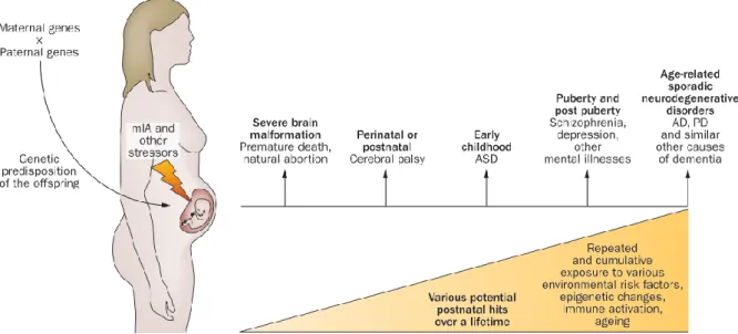
Early-life exposure to environmental risk is thought to contribute to the development of ASD. Illustration 6 describes the different stages of early-life development and gives a good indication of the developmental windows of environmental vulnerability. In utero exposure to environmental pollutants (pesticides, toxins, aerosol particles) or drugs consumed by the mother (e.g. valproate), as well as in utero exposure to maternal obesity are acknowledged risk factors for ASD (Hertz-Picciotto, Schmidt, and Krakowiak 2018). Also, maternal immune activation (MIA) caused by infection or auto-immune disorder is an important environmental factor associated with subsequent ASD diagnosis in the offspring (Careaga, Murai, and Bauman 2017; Hsiao 2013).
Illustration 6. Timeline of developmental stages involving the brain, immune system and the gut development with microbiota acquisition. Adapted from Estes and McAllister 2016.
3.3. Human studies: the case of ASD
Maternal immune activation as a risk factor for ASD: One important environmental factor associated with an increased risk of neurodevelopmental disorders is maternal infection during pregnancy. Many studies have suggested that maternal exposure to various immune stimuli, such as viral and bacterial infections, is associated with abnormal brain development and mental illness in the offspring, particularly schizophrenia and schizophrenia-like disorders (Illustration 7).
There is evidence of other classes of pathogens being able to affect offspring neurodevelopment upon gestational exposure, namely rubella, toxoplasma and maternal genital or reproductive infections (Brown et al. 2001; Brown and Susser 2002; H. J. Sørensen et al. 2009; Penner and Brown 2007; Hyman, Arndt, and Rodier 2005). Moreover, maternal infection was also found to be associated with autism diagnosis in the offspring. This was first described in a group of children with congenital rubella in the wake of the 1964 US rubella pandemic (Chess 1971). Another study on the impact of congenital infections with
Cytomegalovirus and autism diagnosis confirmed the initial association (Stubbs, Ash, and Williams 1984). This effect is now known to be due, not to the exact pathogen, but rather to the intensity of the maternal immune activation (MIA), which occurred during pregnancy (Shi 2003; Patterson 2009; Myka L.Estes 2016). Indeed, prenatal fever or hospitalization following infection, rather than the type of the infection per se, was associated with increased ASD risk (Hornig et al. 2018; Atladóttir et al. 2010).
Illustration 7. The maternal immune activation chain of events, leading to the onset of neurodevelopmental disorders. Adapted from Knuesel et al. 2014.
Maternal microbiota risk factors for ASD: Human microbiota has been shown to play an important role in health and disease, potentially acting as a “hit” to pathology onset (Codagnone et al. 2019). Recent studies have provided more insight into the compositional changes undergone by the microbiota during pre- and post-natal development (Torrazza and Neu 2011; Palmer et al. 2007). Interestingly, maternal microbiota is vertically transmitted to the offspring (Funkhouser and Bordenstein 2013). If dysbiotic or unappropriately transmitted to the offspring, microbiota could contribute to trigger developmental abnormalities in the offspring. Moreover, intestinal microbiota can modulate the postnatal development of the immune system and CNS. Its effects are driven by the mode of delivery, vaginal or C-section (Björkstén 2004; E. A. Mayer et al. 2015). Vaginally-born infants adopt a maternal vaginal and faecal flora, while infants born via caesarean display a microbiota similar to that of maternal skin, which is considered to be transmitted postnatally via
newborn handling at the time of birth (Funkhouser and Bordenstein 2013). Interestingly, children born via C-section have been shown to have significantly higher odds of being later diagnosed with ASD diagnosis (Yip et al. 2017). As pregnancy progresses, so does the diversity of intestinal and vaginal flora: intestinal microbiota diversity increasing as pregnancy progresses (Dominguez-Bello et al. 2010), while vaginal microbiota diversity decreases with pregnancy progression (Aagaard et al. 2012). While most studies describing the maternal-foetal microbiome interaction were performed in humans, recent evidence has also been found in rodents, where the maternal microbiome was found to promote the development of autistic-like behaviour in a mouse model of ASD, which will be described in more detail in the next sections.
Association studies between cytokines and ASD in ASD patients: In ASD patients, anomalies in immune function, both in the CNS and in periphery may be responsible for changes in brain connectivity associated with ASD (Illustration 8). In support of this, analyses of the brain transcriptome of individuals with ASD highlighted the enrichment in both synaptic and immune network modules (Voineagu et al. 2011). Moreover, autism has also been linked to neuroinflammation in the brain with microglial and astroglial activation, and increased proinflammatory cytokines in the cerebrospinal fluid of autistic patients (Pardo, Vargas, and Zimmerman 2005; Morgan et al. 2010; Müller and Schwarz 2006; Vargas et al. 2005; Chez et al. 2007). In addition, cytokine dysregulation has also been associated with the pathogenesis of ASD. One study found elevated levels of CCL2 and TGF-β1 in brain tissue from patients with autism (Vargas et al. 2005). The same study also showed a proinflammatory profile of elevated levels of both cytokines and chemokines (amongst which IL-6, IL-8, IFN-γ, CCL, CCL4, CXCL10) in the CSF from autistic patients. Studies on several cohorts reported elevated levels of TNF in the serum and cerebrospinal fluid in children diagnosed with ASD, as compared with healthy children (Ashwood et al. 2011b; Molloy et al. 2006; Vargas et al. 2005; Abdallah et al. 2013; Chez et al. 2007), as well as increased levels in post-mortem brain tissue of ASD patients (Li et al. 2009). Furthermore, the levels of TNF were found to be associated with the severity of autistic symptoms (Chez et al. 2007). Last, increased TNF has also been positively associated with gastrointestinal (GI) dysfunction in LPS-stimulated PBMCs from children with autism (Rose et al. 2018).
Illustration 8. Overview of the factors leading to maternal immune disruption during gestation. The potential outcome of these events is abnormal faetal brain development leading to behavioural and cognitive impairment. Adapted from Meltzer and Van De Water 2017.
3.4. Mouse models of neurodevelopmental defects triggered by immune activation
The MIA model: Animal studies have further demonstrated that maternal immune activation is a risk factor for neurodevelopmental disorders, such as ASD (Deverman and Patterson 2009). Originally, the MIA model was induced using infection with live human influenza virus into pregnant mice around mid-gestation. The resulting offspring showed abnormal behaviour in tests checking for prepulse inhibition (PPI) of acoustic startle response, exploratory behaviour and social interactions. The authors also observed similar behavioural impairments when they injected Poly(I:C), a viral mimic, described in previous chapters (Shi 2003). This proved that the maternal immune response, rather than the virus itself, was responsible for the behavioural alterations found in offspring. This effect was further characterized in many studies and it was concluded that both bacterial and viral mimics – LPS and Poly(I:C), respectively – can be used as MIA induction models to study ASD pathological processes in mice (Malkova et al. 2012; Hava et al. 2006).