DISCRETE CHANNEL APODIZATION METHOD
FOR THE ANALYSIS OF HIGH-ENERGY X-RAY DATA.
by
JAIME GUILLERMO CARBONELL
Submitted in Partial Fulfillment of the Requirements for the Degree of Bachelor of Science
at the
MASSACHUSETTS INSTITUTE OF TECHNOLOGY
June, 1975
Signature of
Certified by....,.
a rtment rtment of Physi
r e
... oCs
r~~~~~~~~~~~~~~~f''~p`
Ma May 5 1755,V j * - - WThes. e Supervisor -Thesis Supervisor
Accepted by ... ... ... ...
Chairman, Departmental Committed/on Theses
ARCHIVES.
DISCRETE CHANNEL APODIZATION METHOD FOR THE ANALYSIS OF HIGH-ENERGY X-RAY DATA
by
JAIME GUILLERMO CARBONELL
Submitted to the Department of Physics on May, 1975 in partial fulfillment of the requirements for the degree of Bachelor of Science.
ABSTRACT
The discrete channel apodization method to unfold detected x-ray energy spectra is derived for a detector with a Gaussian response function. Other processes re-quired to determine the true source spectrum at the top of the atomosphere are described. A successful computer implementation, with sample results of the spectral
determination process, including the discrete channel apodization method, is presented.
Dr. Anton Scheepmaker
ACKNOWLEDGEMENTS
The author wishes to especially thank Dr. Anton Scheepmaker for his help and encouragement as supervisor of this thesis. Thanks are extended to the whole M.I.T. x-ray balloon group for making this work possible. The
author is also grateful to Stan Ryckman for his help in programming and to Alan Wadja for helping make the text
of this thesis more readable.
TABLE OF CONTENTS
Page
INTRODUCTION ... METHODS OF DETERMINING THE ENERGY SPECTRUM OF
X-RAY SOURCES ... STUDY.OF THE ABSORPTION, FOLDING AND EFFICIENCY
EFFECTS OF THE X-RAY DETECTION PROCESS ... DISCRETE CHANNEL APODIZATION METHOD ... IMPLEMENTATIONS AND RESULTS OF THE APODIZATION
ALGORITHM...
CONCLUSIONS .. . ... ... ...
APPENDIX - FULL LISTING OF SPECTRA AND ITS
SUBROUTINES ... BIBLIOGRAPHY .. . . ... . 3 5 10 17 28 36 38 51 1
LIST OF FIGURES Page Figure 1.1 Figure 2.1 Figure 2.2 Figure 2.3 Figure ]3.1 Figure 3.2 Figure 3.3 Figure 4.1 Figure 4.,2 Figure L4.3
Figure
4.4
Figure 4.5Count rate plot for Crab Nebula
drift scans
X-ray escape probability function Gaussian response function folding Pulse shape discriminator efficiency function
Folding of discrete channel x-ray spectrum
Functions that represent Gaussian folding convolutions
Extrapolation of detected spectrum Tabulation of results printed by SPECTRA
Tabulation of discrete channel
apodization results printed by GAUS Graphical representation of spectral determination processes
Determination of Crab Nebula x-ray energy spectrum
Crab Nebula spectrum
2 b 13 15 18 23 25 30 31 32 34 35
INTRODUCTION
X-ray astronomy is a new and rapidly developing branch of Astronomy. X-ray telescopes, which must be
lifted to stratospheric heights or beyond because of the opacity of the atmosphere to x-rays, provide the means of' observing celestial x-ray sources. The
analy-sis of the x-ray observation data frequently culminates
in the determination of the x-ray energy spectrum for
the observed celestial source. The determination of the spectrum is essential to the theoretical study and modeling of the natural phenomena which produce the x-rays.
In order to determine the x-ray spectrum, the obser-vation mechanism must be well understood, and all
dis-torting effects must be fully accounted for. X-ray detectors have a response function which is convoluted
(i.e., folded ) with the energy spectrum in the detection process. The logical way to determine the spectrum im-pingent on the detector is to unfold the detected spectrum; unfortunately, this is no simple process. The general
problem of reversing the effects of a response function is known as apodization. The theme of this dissertation is the development of an apodization algorithm which may be applied to the phoswich x-ray detector system used by the M.I.T. x-ray balloon group. The discrete channel apodization
method is such an algorithm, generally applicable to the class of' detector systems which have Gaussian response functions and discrete channels.
The high energy x-ray telescope system cited and de-scribed in this investigation was flown to 130,000 feet on a stratospheric balloon by the x-ray balloon group of the M.I.T. Center for Space Research on June, 1974. The telescope system consists primarily of two detector banks of phoswich type x-ray detectors, and associated electronics.
The observed x-ray sources were the Crab Nebula and the Coma and Perseus clusters of galaxies. The sources were observed by the drift-scan method.
METHODS OF DETERMINING THE ENERGY SPECTRUM OF X-RAY SOURCES
The determination of the true spectrum of a celestial x-ray source involves several areas of investigation. For a balloon borne x-ray telescope these areas may be cate-gorized as follows: The measured source spectrum must be determined; i.e., the source and background x-ray fluxes as functions of energy must be separated. Secondly, non-linear efficiency effects of the electronic pulse height analysis must be accounted for in order to determine the spectrumn at the detector level. Thirdly, the convolution of the response function with the spectrum impingent on the detector, known as the folding process, must be con-sidered. Finally, there is energy dependent atmospheric attenuation and some attenuating effects in the telescope
system which must be taken into account.
The drift scan method of observing an x-ray source facilitates the separation of source and background fluxes. Since the diurnal motion of the Earth causes the celestial sphere to rotate at a constant rate with re-spect to Earth based coordinates, the telescope can be aimed just ahead of the x-ray source which will drift through the field of view. This observation method is known as a drift scan. If the aspect of the balloon borne system is known, the increase in x-ray count rates,
i0o.1
o.t
0. -0.oir
TTFI
TITI
T_ TTTI1
I
In
FI6URE 1.1 - ;. CRAB NEBULA DRIFT SCANS
TOP: FRACTIlo OF DETEToR AREA EXPOSED TO TE CKAB
BOTTOM: DETECTED CouvT ATE FRP, 330 FEW.W.M. ETECToR
II I I I I
6
lin 36 . 32 . 28 b N(n 2 . I2'-<D LO-20 . cr) 4-+ =16 c- 4 Q3 LO X * In X 8 -Y a0)
o
N0 -1· 1
ItI.
34;00 35000 35600 36200 36800 37100 38000 CDT SEC 'i . T Iportional to the increase in detector area exposed to the source, can be used to calculate the source intensity
(Fig. 1.1). A straight line least squares fit to the x-ray count rate as a function of the detector area may be applied to different energy ranges. Extrapolating the lines, if necessary, to full exposure and to zero exposure yields the full source plus background, and the background count rates respectively (Ryckman, 1974). The background can be independently determined by extending the scan to include a section where no part of the detector is ex-posed to the source.
The other areas of investigation constitute the de-termination of the unperturbed source spectrum from the detected spectrum. If all of the attenuating and perturb-ing effects of the atmospheric absorbtion of the x-rays, folding in the phoswich detectors, and electronic pulse height analysis could be sequentially reversed, then the true spectrum at the top of the atmosphere could be easily determined. This is not the case because the spectrum im-pingent on the NaI crystal of the phowsich detectors is
folded with the detector response function, a process which is not directly reversible.
Most of the X-ray Astronomy research groups use a repeated trial method to converge on a function which closely approximates the true source spectrum.2 This pro-cess requires the critical assumption that the yet unknown
source spectrum is best approximated by the theoretically predicted spectral functions. The three types of functions most often postulated by theoretical models of x-ray
emit-ting mechanisms are:3
dN -a
a) - = E a power law - synchrotron radiation
dN CeE/KT
(1.1) b) dE E exponential -bremsstrahlung
dN CE2
c) dN _E/K T black body radiation
e -1
In the repeated trial method one function is selected
and the free parameters (e.g., and P for the power
law spectrum) are estimated to generate a trial spectrum. The attenuating and folding effects are applied to the trial spectrum in order to compare it to the detected
spectrum. The closeness of the match is usually
evalu-2
ated under a x criterion. Next, the free parameters are altered and the spectrum generation process is
re-2
iterated in order to minimize 2X Often, after many
iterations to find the best parameters to fit one function, the entire process is repeated for other theoretically
feasible functions.
The repeated trial method has two clear disadvantages over the direct determination of the spectrum at the top
of the atmosphere by apodization of the response function and reversing the attenuating processes. Repeated trials are computationally inefficient, and the number of trials required to find an optimal fit to a given function ex-plodes combinatorically as the number of free parameters
increases. There are algorithms to generate reasonable guesses for the new values of the free parameters for sub-sequent trials given the results of previous trials, but these algorithms are computationally costly and dependent on the form of the spectral function being approximated. The other major disadvantage of the repeated trial method is that the choice of approximating function is constrained to simple, theoretically predicted, trial spectra. It is conceivable that more than one mechanism, including the possibility of some absorbtion mechanism, may be operating simultaneously to generate the observed spectrum.
An apodization method that can be implemented and used efficiently avoids the aforementioned difficulties; it avoids the problem of guessing trial spectra and the combinatorial inefficiency of repeated trials with a moder-ate number of free parameters.4 The apodization method for discrete channels will be discussed in detail after the
different attenuation and pertubation effects are presented. 9
STUDY OF THE ABSORPTION, FOLDING AND EFFICIENCY EFFECTS OF THE X-RAY DTECTION PROCESS
This section will investigate each process in the
sequence of events which a primary x-ray undergoes on its
way to the detector, in the detection process, and in the subsequent pulse height analysis. A brief description of the x-ray telescope system for the June 1974 flight should put these processes in their proper perspective.
A) Description of the Detector.
The high-energy x-ray telescope system consists of two detector banks. Each bank consists of four phoswich type x-ray counters behind a slat collimator. For the June flight, one collimator had a 6°x 6° full width at half maximum (FWHM) field of view, and the other a 30 x30 FWTHM field of view, The phoswich detectors have a
pri-mary 3mni thick NaI crystal coupled to a 1.6" thick CsI
secondary crystal. A plastic scintilliation veto counter surrounds the detector banks to reject charged particles. There is an on-board pulse height analysis and telemetry
system.
B) Atmospheric Absorption.
The detector system was lifted above 99% of the Earth's atmosphere by a stratospheric balloon, since the opacity of the atmosphere to x-rays prevents them from penetrating substancially deeper. Even in the tenuous stratosphere, x-rays are absorbed as a function of x-ray
energy and air thickness traversed. The probability that an x-ray will not be absorbed, called the transmission
probability, is given by:
[5.3 0(10 2.90+ .16] A
(2.1) PTRAIR(wAE) = e
where E = x-ray energy in KeV and P-A = thickness of air traversed measured in gm/cm . Air thickness in the
zenith direction is a tabulated function of altitude. To calculate the air thickness in the observation direc-tion, the zenith air thickness is mutliplied by the
co-secant of the zenith angle of the collimator x-ray axis. C) Styrofoam Absorption.
There is a protective styrofoam layer above the de-tectors which abosrbs a small fraction of the x-ray flux as a function of energy. The transmission probability function is simpler than the one for air because of the macroscopically homogeneous nature of styrofoam. The transmission probability is:
- (8.6)2.69 (2.2) PIR-INS(E) = e
D) Detection Efficiency.
Since the NaI crystal has finite thickness, there is a probability that some x-rays will penetrate the full thickness of the crystal without being detected. In this
case the probability that the x-ray is not lost (i.e., detected) is the absorption probability given by
- A(E) NaI
(2.3) PAB-NaI(P'NaI'= E) 1 - e
where ,NaI = thickness of the NaI crystal ( = 1.17 gm/cm2 for a 3nm NaI crystal), and
5.8 if E < 33 KeV
A(E) =
{
28.0 if E > 33 KeVThe difference in values for A(E) occurs because of the K absorption edge of Iodine at 33 KeV.
E) Escape Probability.
An impingent x-ray whose energy is greater than 33 KeV may excite a K electron in Iodine, giving up 33 TCV' s of' energy. X-rays are re-emitted when an electron, usually an L state electron, falls into the empty K state. Since x-rays are re-emitted isotropically, there is a theoretical probability that some may escape through the front surface of the NaI crystal. The vast majority of x-rays are detected near the front surface of the 3mm crystal; hence, the probability of escape through the back surface is negligible. The average energy of the re-emitted
x-rays is 29.2 KeV. The escape probability as a function of x-ray energy is given in Figure 2.1.
,la 40 60 80 Ioo ENIgERGy (KeV'
(Fig 2.1) Theoretical probability that an x-ray impingent of a 3 mm NaI crystal will produce an Iodine K-flourescent escape x-ray.
F) Detector Response Function Folding.
In the process of detection in a phoswhich type detector,
the impingent x-ray energy spectrum is folded with the re-sponse function of the detector to create the detected pulse height spectrum. Let S(E) and S'(E) represent the x-ray energy spectrum, and the pulse height spectrum respectively. S'(E) = S'(h(H)) where h(H) is the calibration function that assigns to each pulse height the corresponding energy it represents. The general folding process takes the form:
(2.4)
S(E) = .[r X S(E) G(E)dEwhere G(E) is the response function. Since
15
1-0
LD
S(E) =dN E) S (E) NdNI E)
equation (2.5) can be expressed as:
(2.6) N'(E) = X E(E) dN(E)
where N(E) is the number of x-ray counts of energy E per cm sec KeV (i.e., the x-ray flux as a function of energy). The response function for a phoswich type de-tector is a Gaussian, therefore the probability that an x-ray, whose impingent energy is Eo, is detected as having energy between E1 and E2 is given by:
(2.7) Pd(E1<E< E2)
where E is the mean
o tion (Figure 22). 1 and o(Eo)
and
a(E
0)
s~o IEElky cr-Om
~
6 G E. Et V E2 E F1_(E-Eo
)
2d
/2(Eo)
e 00 dEis the standard
devia-(Fig 2.2) Folding effect. X-ray with impingent energy
'P (6 function) has a
prob-0
ability of being detected
between F1 and E2 = shaded
area under Gaussian response function. 14 'EVERjG
(2.5)
dN dEG) Pulse Shape Diserminator Efficiency.
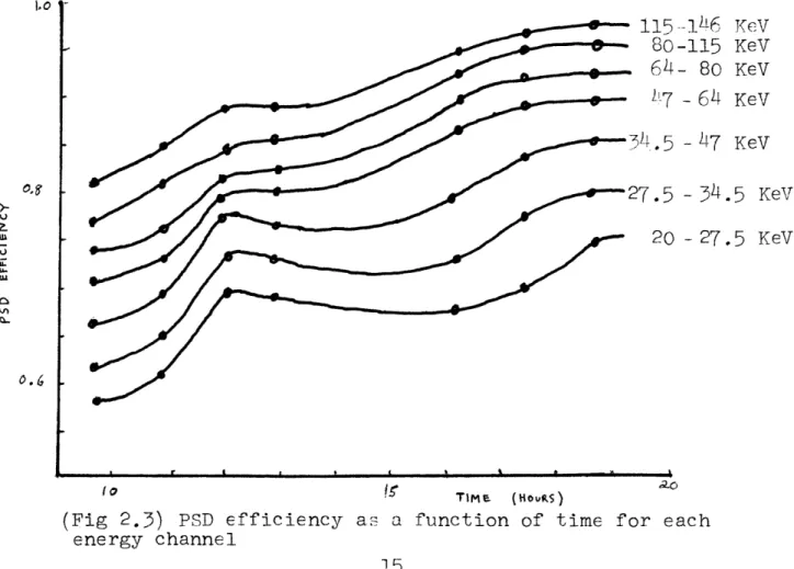
The pulse shape discriminator (PSD) is an electronic system which selects pulses according to the rise time and pulse height. Its efficiency in admitting the appropriate pulses varied (June 1974) as a function of temperature and pulse height during the flight. Therefore, the ef-ficiency had to be determined by an analysis of source
calibrations taken during the flight (Scheepmaker, 1974). A source calibration consists of exposing the detectors
to an x-ray source of known intensity for a few seconds. Figure 2.3 is the best determination of the efficiency
function of the PSD for the June, 1974 flight.
-14d6 KeV -115 KeV - 80 KeV
- 64
KeV
- 47 KeV- 34.5
KeV
- 27.5 KeV 1 0 fr TlMe (HouVS) ao(Fig 2.3) PSD efficiency as a function of time for each energy channel 15 a U .-6 I -I.o
H) Pulse Height Analyzer.
The pulse height analyzer (PHA) bins the detected x-rays according to energy into discrete pulse height channels. Hence, the spectral data consists of x-ray count rates per pulse height channel. If some of the
channel boundaries in the PHA are not well defined, dN'(E)/dE is folded with the boundary resolution
func-tions.
From the mathematical models presented in this sec-tion it is evident that all the processes, except for folding, are easily reversible. The following section will analyze an approximation method to reverse the
fold-ing process.
DISCRETE CHANNEL APODIZATION METHOD
Discrete channel apodization is the process of
unfolding the effects of a response function on a finite number of discrete channels whose energy width is great-er than the minimum resolution of the detection system. In this section a mathematical formulation of the fold-ing process is examined and an algorithm for invertfold-ing the process is derived. This algorithm, implemented in a spectrum determination program, has proven successful in unfolding detected continuous spectra.
The uncertainty factor in the analysis of discrete channel spectral representations is that no direct deter-mination can be made of the x-ray energy distribution within a single channel. Therefore, for an arbitrary im-pingent spectrum, a uniform distribution within each
channel is assumed, giving the spectrum a characteristic step function. For a system with N energy channels
(Fig. 3.1) let (Ei,Ei+l) represent the ith channel,
i.e., the energy range E EiEi+l i < E 1 . Ei is the
energy at the center of the ith channel; Ei= (Ei+Ei+)/2 . The indices i and j are assumed to run from 1 to N
(N=7 for the June 1974 balloon flight system). S(Ei) and S'(i ) represent, respectively, the values of the impingent and detected (unfolded and folded) spectra in
of the ith channel.
(Fig. 3.1) Folding of
discrete channel x-ray spectrum. X-ray flux represented by shaded area in (a) is folded into shaded area in (b) by Gaussian response
function.
E4 ;,, FAERCY (Ke j
The response function of the phoswich x-ray detector is a typical Gaussian distribution:
-(E-E)2 /2(Ej
)2
G(E-j,E) = e 18 > -1 ta -t Q..'I1
(3.1)
where(3.2)
00G(EjE)dE
=
,
2wr
( )
.
E - oEj is the mean of the distribution and a(Ej) the stan-dard deviation. a(Ej) is calculated empirically from calibration data taken from x-ray line emission sources before the balloon flight (Scheepmaker, 1974). The form
of o(Ej) may be estimated to within the limits of ex-perimental accuracy by:
(3.3) a(Eo ) = A j + B · Bj + C
where A, B and C are constants.
Normalizing (3.1) gives a probability distribution function similar to equation (2.7):
E
i+l
(3.4) Pd(Ei< < Ei+l) = A G(faE)dE
2P7r (C) E
This formulation allows the detected spectrum to be ex-pressed as a step function of the impingent spectrum and the response function for each channel:
N Ei+l
(3.5) s'(Ti) = X s(i) 1 -
J
G(EjE)dEj=l a(2) E E
i = 12,,N
The goal of this section is to derive S(Ei) from
S '(Ei ) , hence reversing the folding process of the re-sponse function G(Ej,E) . The apodization method is usually an approximating process converging to a best
6
approximation of the impingent spectrum. In the present apodization scheme the algorithm simply involves solving a set of N linear equations in N unknowns. Since G(E.,E) is directly computable, (3.5) yields a system of linear equations with unknowns S(Ej) for a given set of S'(Ei) . Letting
Ei+l
Ei
(3.6) [aij] =
r
_ 1 G(EE)dE7; o(Ej) i
be the coefficient matrix, (3.5) takes the form:
(3.7) [aij] [S(Ej)] = [S'(YEi)]
Since [ai] is nonsingular and diagonally dominant, there exists a straightforward solution procedure for the S(Ej).
The aforementioned apodization algorithm makes four simplifying assumptions to the general apodization problem. Two of the assumptions are imposed by the detector system:
the finite number of discrete channels and the Gaussian response function, The other assumptions are approxi-mations to simplify the mathematical analysis. The uni-form distribution within each channel is an assumption which does not introduce significant inaccuracies. The development of an apodization method which does not
re-quire this assumption will be discussed at the end of
this section. The fourth assumption is implicit in (3.4) where the probability of detection is calculated to be a
single Gaussian distribution centered about the mean Ej . This assumes that the uniform distribution inside the
.th
jth channel may be considered a delta function at the mean, an assumption which is good only if the channel
width is small with respect to the standard deviation,
c(Ej) > Ej+jl - Ej
For the June 1974 detector system the channels are wide with respect to the standard deviation. Given a
.th
uniform x-ray flux for the i channel, the exact form of the convolution with the Gaussian response function for the jtL channel is:
Ej+l
Pj(E)= j[ U(EjEjl) G (E,E')dE'
Ej
(3.8) Ej+l 2
=[_ e dE
(Eel E)
FrE)
E
Therefore, the probability that an x-ray impingent on the
.th th
3 channel is detected at the i channel is the definite
integral of (.8) over the it h channel:
Ei+l
P.(E
i<
Ei+l)--
P(E)dE
=
Ei (Ej+-Ej) 2w (EFj)
(3.9)
E B 2 2
E i+
EJ!
-E) /2a(E
-(E
.)
x
r
e dE dEx ~ j
E.
The exact expression for the total flux detected in
th
the i channel is:
=
S(Ej)
jL
(EJ+ -Ej )
a (J)
Ei+l
Ej+l
x I: *, t~~5j e
-(E '-E)2/Co(E )2
i dE 'dE
which is a system of linear equations. (3.10) may be re-presented in the same form as (3.7) and solved by inver-ting the coefficient matrix. In the limit as (Ej+l-Ej) -0
(3.9) is equivalent to (3.4); hence, as previously stated, the first apodization algorithm is valid for very narrow energy channels.7 22 (3.10) i = 12,.*.N Ei
E
i ESolving the system of equations (3.10) proved to be impractical in terms of computer time required to gener-ate the coefficient matrix. The numerical evaluation of
Ei+l Ej+l
(3.11) Di = J e
E
i E.3- (E
'E) /2 (j)
dE 'dE
by iterative application of numerical integration tech-niques is somewhat costly, and there are N2 (N2 = 49 for the present system) Dij to evaluate. A more ef-ficient method of calculating the Dij has recently been found, after n approximating system of equations equivalent to (3.10) was programed and used in the data analysis.
E; El;, . Ew [I(k >V)
(Fbig 3.2) Folded channel function for a) exact form of Gaussian response convoluted with uniform dis-tribution, b) close numerical approximation to (a),
c) single Gaussian, considering x-ray flux to be 8 function at center of channel.
Equation (3.9) may be approximated by minimizing the
L1 norm of the concatentation of two half Gaussians and
a constant function (Fig. 3.2). The probability that an x-ray impingent in the jth channel is detected at the ith channel becomes:
1
2ra
(Ej)+(Ei+l-Ei)
Pai
Ei+1
F
e
E.(3.12)
Pdj
(i<E<Ei+])
- (E-Ej) /2F(E )23
dE
,
if
Paj
1 -- ~-_ __--~- 1-" ~2r (E) +(Ei+ -Ei)Pai
E
1
(B
2)2
Eil -(E-E ) 2/2 (j)
2
x J e j+l dE, if j>i
E
I
iFor i = 1,2,. ,N
where Dcj 1 ji Pdj((E i < E <il) is the probability
channel is detected that an x-ray impingent on the jth
th
in the j channel. In this approximation the respective means of the Gaussians are at the channel boundaries, mak-ing the derivative of the approximatmak-ing function well
de-24
j<i
fined and everywhere continuous.
The detected spectrum on the ith
channel can be
ex-pressed as a sum of the impingent spectrum multiplied by the respective detection probabilities, as in the previousmet hods::
N
S'(Ei)
S(Ej)Pd(E < E <
Ei+l)
ij=
i = 1,2,... ,N
The system of linear equations may then be solved for
S
(j)=
S(Ej)P
, which yields
S(Ej)
directly.
S
(E.) is called the alpha spectrum; it is the fractiona
of the spectrum in each channel not carried to a different
channel by the response function.
A/ .1 -0 I I I I
I<
tI C ti EER cy ( kEV )(Fig 3.15) Extrapolation of detected spectrum to estimate the fraction of the flux folded into the extreme channels from x rays whose impingent energy lies beyond the threshold of
the extreme channel boundaries.
25
(3.
0)
d HE -____ { f Z-XrCL/I/ 4,There is a non-negligible probability that x-rays impingent with slightly less energy than the lowest en-ergy channel boundary, or with enen-ergy slightly higher than the highest channel boundary, will be folded into the respective extreme channel by the response function. Likewise, a fraction of the impingent flux at the extreme channels is never detected; it is carried out of detection range by the Gaussian response (Fig. 3.3). In order to minimize errors caused by ignoring this effect, the de-tected spectrum is extrapolated beyond the energy range of the extreme channels. A reasonable extrapolation will yield correction estimates for the x-ray fluxes carried across the extreme boundaries.
At the time of this writing the development of a
more accurate, but also more complicated, method is being investigated This method determines a smooth, best
L2 -approximate to the detected spectrum. The process assigns to each ES'(E)! a tentative d and a
Emewhat more tentative d2
somewhat more tentative d- S(E)IE in order to give
a reasonable approximation to the x-ray distribution with-in each channel. The approximatwith-ing functions under with- investi-gation are interpolating cubic splines, where the number of splined sections is a function of the number of energy
channels. The splining method will give a more accurate
determination of the impingent spectrum in systems incor-porating a somewhat larger number of energy channels.
IMPLEMENTATION AND RESULTS OF THE APODIZATION ALGORITHM
SPECTRA is a Fortran 1 computer program which lifts an x-ray spectrum from the detected count rates in each channel to the true x-ray source spectrum at the top of the atmosphere. An implementation of the discrete channel apodization algorithm lies at the heart of SPECTRA in a subprogram called GAUS.
SPECTRA applies the inverse process of each attenua-tion or pertubaattenua-tion, previously described, in reverse
order from the detection process. Since the apodization algorithm inverts the folding process, a single program run will yield the best values of the source spectrum at a small computational cost. SPECTRA takes as input the set of detector system parameters and the detected count rates per energy channel for each detector bank. The de-tector parameters are: air thickness, NaI crystal thick-ness, detector area at full source exposure for each
de-tector bank, time in the flight when the detected count rates were accumulated, PSD efficiency table with effic-iency values for different x-ray energies and different times during the flight, and PHA channel boundaries in terms of energy. (Pulse height is directly porportional
to energy.)
The count rate in each channel is converted to units of counts/cm2sec KeV. The PSD efficiency table is
terpolated to calculate the closest value for each energy channel at the given time in the flight, and the efficiency correction is applied to the spectrum. SPECTRA calls GAUS with the efficiency corrected spectrum to apply the dis-crete channel apodization algorithm. GAUS, in turn, calls
several functions and subroutines, including RSIMQ to solve the linear matrix equation (3.13). (RSIMQ was developed by the Information Processing Center at M.I.T.). After the escape correction is applied to the unfolded spectrum, there follows a sequential application of the corrections for NaI crystal transmission, styrofoam layer absorption, and atmospheric absorption. The resultant step function is the best discrete determination of the continuous x-ray source spectrum at the top of the atmosphere. The spectral
2
step function can be easily 2 fitted to a theoretically predicted spectrum (e.g., power law).
SPECTRA tabulates the results at each step in lift-ing a detected spectrum to the top of the atmosphere
(Figure 4.1). GAUS prints out the probability coefficient matrix,
(4.1) [aij] = Pdj(Ei< E< Ei)/Pai
and the unfolding of the energy spectrum (Figure 4.2). In order to test the accuracy of SPECTRA, a close approximation to the Crab Nebula power law spectrum,8
Lu Ln r-IU -0 It- t '-0 L-10 In 0 I', N a-Ni .1 I I II 0 I ccl N I I CH N II +-) 4-)-d r t
I
O
I I WI I a ) HH N~~~~~cI - -I: t() PCH 1 Cou] "C --'= ' .0 ¢ L OJ :R co ¢) ,. C a C) ' ,-o 4O b3 CO hi uj -o · e r r - 2 .~( I I' ,(u l :I ; cQ. D oD I >*rl 31 - O P- r I c C) .- L. (D o 0 NI jr Hc&W--: N I . c W HI + O q- -C I 0 a ] 0o I ZC E C d \C) CIr I , o r 1 - O cc l o rl I Tc 1 a 4) Z o wI a .*1 0 > NI UJ .O IIL
0rl
CeO
° 4 ' E w -' cr t I * ,- I I I I I.'.. 0101 -101 I I I I J - -_- _ _ - - -__ -I r-I 30 N I z 4 t I-,cc z 04 I-0. 0 tnccCz zct U 'LI 0U tI 0 U z 0 -J I_11 M I \ IIX co en 0 0 r-11 l cI N IW 0W w IA'0 In l O I t-10 '3t I O 1 10 LL I I IL IN _ I I N, c Z I 01 l I 01 1 t I t. I st 0 I -? 1/' I 0 C. I < 0 ,4 '4 4 Q II 0 W '3 N co 0 I) -, 0 O0 co -. 0 WJ ,0 ,0 -o '0 .4 OU ,1I ,--, t1 IW I I tn 0 '1' I N 1 I0 I, N In W N I c U I_ Oq IW cc IC A0 a0 IG 0 1 N CUJ L"i 01 i: I 0' C, N 0 LL u-I en 0 N I1 I0 -N"I r3-uI~ O In 0 u-I 3-N 0 I ! ' '3 LI W-. In '0 CLi'1 I * I WLI It I I I I_ I Un I I LI I t0 I INWt I ILul 0L LI I * II 0. I In I t- I _ I LII 'J) I U) I 0t I 0k I U, I W/ I I_ I I, I I I C I I >I t I I 1 I') I I O 0 II , - III, 3. U I 0 0 I I I I I - I .U I O I r I a, 1 6 1U 11 1 I I I I1 | I 0 I U I WU UI t c c I 0' I N I N I-) 4 lt 1. I t ) n I I I Ii I I 0 I , I 0 0- I 0 I o' *? I I CI.3I0 .3.I'OI0II t In I o I 0 . I * I '3 I tI s ft I I I I s I I 0 U 0 U u' cli A. U In0'i
II I-0 L, -1 uj I .1 IN I r t I I II I I II l II I I II II II II I II II I i II II II I I , . . II _ vUNFCLDING cr SPECTRUP NUMeER 2
AIR THICKNESS = 3.350 G;S/CM SC
RESPONSE FUNCTICN = 0.C30) * ENERGY
NAI THICKNESS = 1.171 GRAMS/C SC
u.420 * SQRT(ENERGY) + 0.230
PRCHABILITY CChVCLUTION VATRIX
1. 00) 0.189 0.0 C.C ... 0.207 1.000 0.124 9 . C0 O. 0 0.0 O.n ENERGY BIN 0. 00 0.261 1.0o 0.108 . 900 0.0 0.3 o .0UC 0 .000 0. 1 78 1.000 0. 139 0.000 0 . 0 .0 0 .000 0 .000.OOJ 0.155 1.000 0 .075 0.0 0.0 0.0 0 .000 3 .CC .2C2 1.0CO 0. 1 04
FCLCEC SPECT ALPFA SPECT UNFOLCED SP
20.0) 27.50 27.50 34.50 34. 5 47.00 471.C 64.00 64.90 8.CO 80.30 115.00 115.00 146.00 2.2293E-03 2.6318E-03 I .8642E-33 1.2547E-03 7.6734E-04 3 .6748E--04 1. 2J5 5E-04 (Fig 4.2) Discrete re sult s.
Channel Apodization computational GAUS prints the [aij] coefficient probability matrix and the unfolded spectrum.
Results tabulated in this run are from the recovery of the power law mentioned in the previous figure. -
of
0.0 C . C.u 0 .09 0 .113 I .CCC 1.8346E-03 1.9066E-03 1.4476E-C3 1.0 1 14E-03 5.631 E-C4 3.1407E-C4 9 .6763E-05 2.4826E-C3 2.7511E-C3 1.E71SE-C3 1 .2762E-03 7.4EUlE-C4 3.72 14F-C4 1.215CE-04lo o30 io t 0 tio 6l eTER&Y (KeV)
(Fig 4.53) Comparison of original power law spectrum,
folded, attenuated spectrum, and recovered spectrum by SPECTRA. 32 w 0) u 4i N E I U To -A0w 81 k I
(4.2) dN = 22E 2.25 (photons/cm2sec KeV) dE
was folded with the detector response function, and all
other absorption and efficiency effects were applied (Laros, 1973). The resultant spectrum, generated to simulate a
detected spectrum, was given to SPECTRA, which recovered the original spectrum at the top of the atmosphere with small computational errors (Fig. 4.1 and 4.3). The high-est and lowhigh-est energy channels have somewhat larger errors
than the central channels due to the extrapolation of the
"detected" spectrum necessary to calculate the effects of the response function at the boundaries of the spectrum.
SPECTRA was applied to detected spectra from Crab Nebula drift scans. The result of one computer run, for
the second Crab scan in the June, 1974 flight, is pre-sented in Figures 4.4 and 4.5. The second and fourth energy channels are respectively too low and too high with respect to the Laros spectrum. This was found to
be the case for other methods of determining the spectrum
and for other Crab scans.
SPECTRA was tested for different detector system parameters. For instance, the unfolding of a test spec-trum gave more accurate results when 12 energy channels were used to span the 20-146 KeV energy range instead of the 7 channels in the June flight.
O)
0 ) U) (]) U U O -@ _r a) P, r.D · o - CtZ rl co 0 U) U-0 V E S Zj $ %Q ) Z E1 i hO H i a) ; aH) R 4 r Q I- EE I IIw I-10CIt Nl I * I p-I I II c 1010 I-W I 1mEIf 1010 111 I 0l N * i I I I I O I I I f Il I 0 I 0 1111u I .-,-4II 0 u-NJ C' 3t w C, LuI C-0 3 W :L 0 1t .0W '0 r.-0 C', t-0 0co ui w In (P 0 1 LU rfl 0 U '0 N en Ci LU m ,er 'Wim I w II , r'0 I i , I lw (n 0 i 0 i . 0 Io r-Lr) .I -4 I Q III I I r-I u IUr I II 'T I -0 LU 0 '0 '4 (r !¢ I Lu I C I co I 0 Ig Ir I 0 I N _ 01 ZI U--10Cl I u 013U. X I I J I Wj rN 41 UL I ZI I I 1 I r-4u Z I CL I WI N OI t" V: I r C;I OLl L I 0 LLI'7 tu-W I 0. I 'W 4/ I IIC) J I _U U)z I N IN 1 LU I -n :IU zJ I rs IU I e I C I ow 'I r4 l o -1 - I I I I I i l I I C I I o I I o 0 1 I 0I III I 0 co 0 II u I C-I In o ItI IJ 0 0 1 OI 'I I LIUJ ILUI LI N N I I '0 0'| I 0 I 0'v I I I I I I I v ,..- I ,.-4 I I(ff § O I ' Qt I I I I " I W I '7 I -T 0 I 0 I 0 .-1 1111111 I U I 0I 0 I 0 N I LU I I I 0 - I I LU0 -;I · I ; I I I i I 0 10 I0 I0 0 I O I 0) I O I .ru I I ,t I I o I u0 I U, I I I_ 0101010 U, I 0 I 0 I 0 I * I . I JTICI~Tl0 I 4 I '0 I 0 I 't I 0 1 I I 10 I , I o I I I I IC 10 I -I N uj y 0 ui ,/ -r I-0 II 'A I--C 3 I 0 V ) rJ CN 0 0 Nu a C, LU Lu tJ 34 II II II II II II II II II II II II I I II II II I II II II I I II II II I I I 7O 3o 40 lo a00 EnVERY (KeV)
(Fig 4.5)
Graphical representation of thedeter-mined x-ray energy spectrum for the Crab Nebula. The power law, given as a reference, is the Laros spectrum.
35 ,-V ID qJ E 'Q r 1-0 Z Q_ -4 t0
CONCLUS IONS
The discrete channel apodization technique yields the closest obtainable values to the true x-ray source energy spectrum at the top of the atmosphere in a single, efficient pass. These values may be matched to a.
theor-etically predicted spectrum (e.g., under a X2 minimi-zation criterien) if one wishes to study the x-ray pro-duction mechanism. The calculated values for the source
spectrum are independent of any fitting performed after the determination of the spectrum, unlike the repeated trial method, where nothing is known about the source
spectrum until a reasonable fit is found. The determina-tion of the source spectral parameters may be greatly facilitated by graphing the source spectrum and perform-ing preliminary visual fittperform-ing, or, at least, settperform-ing severe constraints on the type of function and values for the free parameters chosen for the '2 fit.
The discrete channel apodization algorithm may be applied to any x-ray detector with discrete channels and a Gaussian response function. It may be possible to generalize the method to include other types of response functions, but the necessity for discrete channels lies at the very heart of the algorithm.
There are some limitations to the apodization method. Only continuous spectra can be unfolded; line spectra
cannot be resolved by discrete channel apodization. In order to get accurate results for continuous spectra, the detector system needs to have at least five energy channels. On the whole, the discrete channel apodization algorithm is an efficient and fruitful process applied to the determination of the x-ray source spectrum at the top of the atmosphere. The author hopes that this method may be used by x-ray astronomers to facilitate and improve their spectrum determinations.
APPENDIX
FULL LISTING OF SPECTRA AND ITS SUBROUTINES
SPECTRA is a Fortram program which should be compatible with most Fortran IV implementations. The data is read in from a set of cards of specified format, fully explained
in the first page of the listing. SPECTRA can process several detected spectra measured during a single balloon flight in one program run.
The discrete channel aodization algorithm is im-plemented in the subroutine GAUS, which may be used in--dependently of SPECTRA for detector systems that are not balloon borne (e.g. satellite detectors), but which have a Gaussian response function and discrete channels.
DATE = 75126 21/55/37
C …....AIN PRCGRAM TO LIFT BALLOON DETECTED X-RAY SPECTRA 10 THE TP CF
C rHE ATPCSPhERE. ASSUMES DISCRETE CHANNELS AND GAUSSIAN RESPCNSE
C FUNCTICh FCR TIE DETECTOR C
C
C---WRITTEN AT MIT JANUARY ThRU APRIL 1975 BY JAIME G. CARBONELL
C C
C---RECUIRES 4 SUBROUTINES,
C 1) GUS = APOCIZATICN ALGORITHM FOR DISCRETE CHANNELS AND GAUSSIAN RESP.
C Z) RSIPC SCLVES SYSTEM OF N LINEAR EQUATIONS IN N LNKNCOINS
C 3) ESCtPE INVERTS ESCAPE EFFECT OF IODINE ABSCRP[lON K EDGE IN AI X-TAL.
C 4) EPSE CALCULATES EFFICIENCY OF PSC BY INIERPOLATICN ON TIME AND ENERGY.
C C
C---RECUIRES 9 FUNCTIONS
C GAUSP XTRPOL ESCR TRAIR [RNAI TRINS SIGF EFFEL FINTRP
C C
C---FIRST SET OF DATA CARCS IS THE EFFICIENCY TABLE PRECEEDED BY 1 CARD C WITH T- (12) NUMBER OF ENTRIES = NLMBER OF lIMES IN FLIGHT = NUMBER
C OF CARCS IN EFFICIENCY TABLE.) EACH CARD CONSISTS CF A TIME IN CCT SEC. C FCLLCWEE BY 7 EFFICIENCY VALUESt ONE FOR EACH CHANNEL.
C C
C---SECOND SET CF DATA CARCS CONSISTS OF DETECICR PARAMETERS
C 1) NUMEER CF ENERGY CHANNELS (12)
C 2) TICKNESS (IF AI CRYSTAL I GM/CM**2 (F7.3)
C 3) A,B8C VALUES FOR SIGF RESPOCiNSE FLNCTION (3F7.3)
C ''t) NINS+1 ENERGY OUNEARY VALUES (1CF7.2)
C !I) NUMBER CF SPECTRA C BE PROCESSEC (12)
C---THE FOLLOWING CARDS APPLY TO EACH SPECTRUM - MUST li. REPEATED
C b) DETECTOR AREA IN C**2 (FIC.I)
C 7) CT TIME (FIO.1)
C B) AIR THICKNESS IN GM/CM**2 (F7.3)
C 9) CCUN7 RATES PER CANN.'L (CETECTED) IN CTS/SEC (10F7.3)
C C C
REAL EKV(51),EKVA(50),TSPECT(5C),SPECT(SC,8)
REAL S ICVAL(!O),S IPARf3) EFF=1.O CALL EFSD REAC(5,10) NBINS 10 FCRMAT(I12) READ5,11) TNAI 11 FCRMAT(F7.3) REAC(5,12) (SGPAR(I),I=1p3) 12 FCFMAT(3F7.3) KK=NE INS+1
REAC(5,13) (EKE.V( I I=1,KK) 13 FCPMAT(10F7.2) ISPEC=O REPD(5,14) NSPEC 14 FCRPAT(12) 100 IF(NSPEC.LE.ISPEC) CC TO 1C1 ISPEC= ISPEC+I READC (,18) AREA
39
0o01 C002 CO03 0004 C005 C006 C007 0'08 C009 COlO 0011 0012 0013 C014 0015 C016 0017 C018 0019DATE = 75126 21/55/37
READ(5,18) CT IME
18 FORMAT(F10.1)
READ(5,11) TAIR
REAC(5,15) (SPECT( I 1)I= INeINS)
15 FCPMAT(10F7.3)
C ---- PRINT CUT INPUT PARAMETERS READ
WRITE(6,16) ISPECTHAIRTI-NAI,(SIGPAR(lI),I==3)
16 FCRMATI'l't//////' UNFOLDING CF SPECTRUM NUMB-R ',12,//
1' AIR T-ICKNESS =',F7.3,' GRAMS/CM SQ NAI T-ICKNESS =',
2F7.3,' GRAMS/CM SQ,/ RESPUNSE FLNCTICN =',F7.3,' * ENERGY
I ',F7.3,' * SQRT(EKERCY) * ',F7.3,////) CC 29 J=1,hBINS 29 EKEVA(J)=(EKEV(J)+EKEYVJ+I))/2 U 3 J=lrNBINS K=J+1 SFECT(J,1)=SPECT(JI)/(AREA*IEKEV(K)-EKEViJ))) SPECT(J,2)=SPECT(J,1)/EFFEL(EKEVA(J)) CALL EFFPSC(EFF,COTIE, EKEVA J )EKEVA)
SPECT(J,3)=SPECT(J,2)EFF TSFECT(J)=SPEC(J, 3)
30 SICVAL(J)=SIGF(EKEV( J )SIGPAR(1) r SIGPAR(2) SIGPAR 3 )/2.0 CALL GAUS(NBINS,EKEVEKEVASIGVALTSPECTtSICPAR)
WRITEI6,17) (SIGVAL( I),I=1,NBINS) 17 FCRMAT('OSIGVAL= ',1OF7.2)
C- ---- 'Il CRYSTAL UNFOLDING JUST COMPLETED -- NOW DC FIRST ORCER
C ESCAPE CORRECTION ANC RING SPECTRUM TO TOP OF ACSPHERE
rFLUX=O CC 41 J=leNEINS 41 SPECT(J,4)=TSPECT(J) CALL ESCAPE(EKEV,TSPECT,NBINS) CC 31 J=1,NBINS K=J+I SPECT(J,5 )=TSPECT( J ) SPECTJ,6)PEcPECT(J,5)/(1I.O-TRNAI(THNAI,EKEVAIJ))) SPECT(J,7)=SPCT(Jt6)/TRINS(KEVA(J) ) SFECT(J,8)=SPECT(Jt7)/TRAIR(fHAIREKEVA(J)) TFLUX=TFLUX+SPECT(JE)*(lKEV(K)-EKEV(J)) 31 CChTINUE
C---PRINT SPECTRUM AT EACI- STEP IN PROCESSING AD UNFULDING
,RITE (6,37)
37 FORMATI'O UNITS = CTS/ICMi**2*SEC*KEV)'/) ARITEI6,32)
32 FCPMAT{'O ENERGY BIN MEASURED SP EFFEL CORR EFF{ IR NAI UNFCLCING ESCAPE CORR NAI ASORPTICN TRINS
2INAL)' ) 4 'SC COR [RAIR(F CC 34 J=ItNBINS K=J+1 RITE(6,38) 38 FORPAT(' I---2---I' ) nRITEC6,33) EKEVIJ),EKEV(K)l(SPECT(JrI),=1r8)
33 FORMATI' I '2F7.2,1P8EI4.4, I')
34 CCATINUE
RITF(6,38
WPITE(6,39) EKEV( 1 tEKEV(KK)rTFLLX WRITE(6,35! ISPEC
39 FCRMATi'O',//' TOTAL INTGRATED FLUX ETWEhN,F7.29' AND'rF7.29
40 C020 C021 0022 C023 C024 0025 CO26 C027 C028 0029 C030 ;0 31 C 32 0033 C034 ¶0 35 C036 0037 0038 CO 39 C040 CO41 t:042 0043 C044 C045 C046 ,347004 !8048 C049 0 50 0351 C .)52 gJ53 054 C055 C056 C057 C058 0059 0360 0061 C062 C063 C064 0065 C066
FCRTRAN IV G LEVEL 19 , AIN DATE = 75126 21/55/37
1'KEV IN UNITS OF XRAYS/(CM**2*SEC) IS ',1PE14.5//)
0067 35 FCRPAT('O',///' ENE CF PROCESSING FOR SPECTRUv ',12///)
C068 ;;C TO 100
C069 101 CCNTINUE
0070 ,RITE6,36)
0071 36 FCRPAT('O',//////' CCMPLETION OF UNFOLDING ANC LIFTING OF SPECTRA
I '/////)
C072 STCP
DATE = 75126 21/55/37
SUERCUTINE GAUSINBINS,XKEV,XKEVA,LMDA,XSPEC1,SIGPAR)
C---CAUSSIPA UNFOLDING CF XRAY SPECTRUM.
REAL XKEV(51),A1(50,5 ),LMDA( 5l)XSPECI(5C),XSPEC2(50) REAL XSPEC3(50),XKEVA(5 ),SIGPAR(3)
CP I= 1 72454 DO 14 J=1,NBINS K=J+1 CC 14 I=1,kEINS L=I+1 XHIGH=XKEV(K) XLCW=XKEV ( J) CE=Xf IC---XLUW IF (I-J) 41,43,42 41 XPEA\= XKEV(L) GC TC 15 42 XFEAN=XEV ( I ) 15 XLfO= LCA( I ) A1(JI1 )=SCP I*XLMDA*GAUSP(XL0h,X ,C TC 14 43 AI(JI)=1.O 14 CONTINUE IGH,XMEANXLMDA)/CE
C---LIPIT EXTRPPOLATION CCRRECTION
C---(CRE PECISICON REC FOR SMALLER E BINING)
CE=XKEV (2)-XKEV( 1)
X4=XKEVA( 1)-DE
SZERC=XTRPOL(XKEVA(3),XKEVA(2),)XKEVA(1),X4tSPECI(3),
1XSFEC(2) ,XSPEC1( 1) )
ALPD=SIGF(X4,SICP R( ),SIGPAR(2),SIGPAR(3))/2.O
S ~LPHA=SZEROCG*E/( E+SQP *ALMDA)
XP EAN=XKEV( 1 ) l LCW=XI<EV( 1 )
XHIGF= XKEV( 2)
SCCRR=SALPFA*SQPI*XLMCA*CALSP(XLChXHIGH,XMEAN·)LVOA)/DE :RITE(6,51) SZERCtSALPI-ASCORR
51 C;AT ( 'OSZERCSALP-/, SCURR= ',3E14.4) (SPECI(1)=XSPEC1(1)-SCCRR NK2=KBINS-2 NK1=NB INS-1 .1=N1e I\S + lC E=XKEV ( NN 1 -XKEV( N IS ) X4=XKEVA(NBINS)+CE SZERC=XTRPOL(XKEVA(NK2),XKEVA(NK1),XKEVA(NBINS),X4,XSPECI(NK2),
lXSFECl(NK1)t.XSPECL{NeINS
)
XLFVA=SIGF(X4,SIGPtR)1)S[ SC-PAR(2)tSIGPAR(3))/2.0SL PHA=SZERO:E/ ( CE+SCP I*XLMOA ) XEAN=XEV ( N8 INS ) XLOW=XKEV( B INS) XF IGH=XKEV (NN1 ) SCCRR=SALP-A*SQPI*XLMCA*CALSP(XLOW,XHIlGH,XMEAN,XLMDA)/DE wRITE(6,51) SZtRO,SALPfA,SCORR XSPECl(IBINS)=XSPEC1(NfEINS)-SCORR
C- ---- Ok SOLVE MATRIX OF CCNVCLUTION COEFF. FOR S ALPHA. ARITE(6,30)
30 FCRMAT('O',///' PROBAeILITY CCNVOLUTION MATRIX'/)
[C 16 I=1,NBINS
WRITEl6,31) (Al I,J),J=l,NBINS)
31 FCRVAT(1OF7.3) 16 XSPEC2(1)=XSPEC1(I) 42 Cool C002 C003 C004 0005 C006 COO7 C008 C009 C010 COll C012 0013 COl4 C015 C016 0017 COb 8 C019 C020 C021 0022 C024 002 5 025 0026 C027 C028 0029 ro- klu, 5 u C 0 3 1 C031 032 C033 C034 C035 0036 C037 C038 0039 004 0 C041 `042 0043 0044 C045 C046 C04 7 C048 C049 C050 C051 C052
OATE = 75126 21/55/37IC 0053 C054 C055 0056 :" 0057 C058 C059 0060 C061 C062 C063 0064 C065 C066 0067 0068 C069 C070
CALL RSIMQ ( 50, NINS, A ,XSPEC2, 0 ) ;CNTINUE DO 17 I=1,NBINS K=I+l (SPEC3(I)=XSPEC2(1)*(I.O+ SQPI*LMCA(I)/(XKE(KI-XKF-V(I))) 17 CCNTINUE WRITE(6, 18)
18 FORPAT('OENERGY BIN FOLCED SPECT ALPHA SPFCT UNFOLDED SPI/)
L)C 19 1=1,tBINS K =I +l
19 WRITE(6,20) XKEV(I),XKEV(K),X<PEC(I lXSPEC2(I),XSPEC3I),
20 FORiAT(2F7.2, IP3E14.4/)
hR ITE(6,21 )
21 FCPMAT('O',//////' END OF GAUSSIAN NFOLDING '////)
DO 22 I=1,NBINS
22 XSPEC1({ )=XSPEC3(I)
RETURN ENC
43
DATE = 75126 21/55/37
SUeROUTINE RSIMQ(NCIM, NORCER, COEFF, RHS, IERR)
C
REAL CCEFF RHS, I8GC, SAVE, TOL, ABS
INTEGER NORCtR, CIM I J K, IMAX, JPI, JJ, NMI
C
!)IMENSICN COEFF(NDIM, NCRCER), RHS(NCRCER)
C
C CHECK FCR ARGUMENT RRCRS.
IF (:DMl .GE. NORCER .ANC. NORDER .GT. C) GC 10 10 IERR = 2
WRITE (, 1001) CIM, NOROER
RETURN C
10 TOL = G.OEO IERR = 0 C
C CC ORWARC ELIMINATION, WITH PARTIAL PIVCTING.
CC 70 J = 1, NORCER
C
C CHCCSE LARCEST ELEMENT. REMAINING IN THIS COLUMN.
eIGC = O.OEO
CC 2 I = J NCRCER
IF (AeS(BICC) .CE. ABS(COEFF(I, J)) CO T 20
EIGC = CEFF(I J) IFAX = I
20 CCNTINUE
C
C IF LL ELEMENTS AVE MAGNITUCES LESS THAN C PMAIIX IS SINGULAR. IF (PBSIEIGC) .GT. TOL) GO TC 30 IERR = 1 WRITE (6, 1002) RETUFN C
C INf-RC-ANCE RCWS IF NECESSARY, AND DIVIDE
C PIVOT ELEMENT.
30 CC 4) K = J, NORCER
SAVE = C(EFFIIMAX, K)
CCEFF(IMAX, K) = COEFF(J, K)
CCEFF(J, K) = SAVE / BIGC 40 CCNTINUE
C
C
CR ECLAL TC TCL, THEN
NEW CURRENT RCW 8Y
CC rI-E SAVE FCR TE RIG-T-HANC SIDE.
SAVE = RHS(IMAX) RHS(IMAX) = RHS(J) RI-S(J) = SAVE / 8IGC
C
C SUeTRACT MULTIPLES OF THIS ROW C LEgCING CCEFFICIENTS VANISI-.
IF (J .GE. NORDER) GO TO
C
FROM ANY REMAINING RCWS TO MAKE
80 JF1 = J + 1
CC 60 I = JPI, NCRCER StVE = COEFF(I, J)
CC 50 K = JP1, NCRCER
50 COEFF(I, K) = COEFF(I, K) - SAVE * CCEFF(J, K)
;ES(I) = RHS(I) - SAVE * RHS(J) 60 CONTINUE 44 COOl 0002 C003 0004 C005 C006 C007 CC008 C309 :010 I01 1 C012 C013 C014 C11015 0016 01 7 C018 e C019 S020 C0021 0022 C023 C324 0025 :0?6 2027 ¢028 C029 C030 C031 C032 C033 0034 0035 C036 0037 FCRTRAN IV G LEVEL 19 RS MQ
DATE = 75126 21/55/37
70 CCNTINU_ C
C NCW FINC ELEMENTS OF SCLUTION VECTOR [N REVERSE CRDER BY CIRECT
C SLESTITUTICN. 8O Nh = CROER -Pl = CRCER + 1 '3C 130 JJ = It NMI J = NORCER - JJ JP1 = J + I DO 9 KK = 1 JJ K = NP1 - KK PI-S(J) = RHS(J) - COEFF(J, K) * RFS(K) 90 CCNTINUE 100 CONTINUE RETURN C 1001 FORVT(23H RSIMQ 1'302 FCRPAT(32k RSIPC F NC ARGUMENT ERRORt 2111) ECUATIONS ARE SINGULAR.)
45 C038 C039 C040 C041 C042 C043 t044 C045 046 C047 C048 2049 '050 0051 C052 FCRTRAN IV LEVEL 19 RS[MQ
DATE = 75126 21/55/37
COO1 SUeRCUTINE ESCAPE(XKEV,TSPECT ,Ne INS )
C ---ESCAPE CORRECTION ON ENTIRE EXPANDED SPECTRLM C RETURNS NEW SPECTRUP Y CLOBBERING TSPECT
C002 PEAL XKEV(51),TSPECT(50),FSPECT 3CC) C003 INTECEP IKEV(51) C034 KK=Ne IS+l C005 H10 1 I=1,KK C006 1) IKEV( I)=IFIX(XKEV( [) ) CC07 LC=IKEV(KK)-IKEV(1) C008 [CCUNT=hINKS CU09 l)0 20 I=1,LD CO10 h=LC- +IKEV(1
COi1 IF (N.LT.IKEV(ICCUNT)) ICOUNT=ICOUNT-1
L012 ICI=ICCUNT+1
^013 FSPECT(N)=TSPECT([COUNT)/(IKEV(IC1)-IKEV(ICOUNT))
C ... RITE(6,101) ICCUNT,IKEV(ICOUNT),TSPCT( COLNT),NFSPECI(l)
C014 20 CCNTINUE Ct)15 CC 3 I=LLC ;01 6 ?1= LC- I KEV + ( 017 N29=N-29 C018 [SC=ESCR(FLCAT(N)) t019 IF N2$.GE.IKEV(1)) FSPECT(N29)=FSPECT(N29)/(1+ESC*FSPECT(N)/FSPEC 1T(K29))
S'20 FSFECT ()=FSPECT( N)/( 1-ESC)
C021 30 CCKTINUE Ci22 UC 40 I-lI,NBINS 023 43 rSPECT(I)=0.2) C024 ICCUNf=B INS C025 EC 50 !=!,LC C026 N=LD-I IKEV ( ) '927 IF (N.LT.IKEV(ICOUN[)) ICCUNT=ICCUNT-1 C028 TSF-CT(ICOCUN-)=TSPECT(ICtiUNT)+FSPECT(N)
C ... RITE(6,101) ICOUT T,IKEV( ICOUNT),ISPECT(ICOLNI) ,N,FSPECT(N)
C329 50 CCNTINU._ '"30 191 FCRMAT(' ICOLNrIKEV(ICOUT),TSPECT(ICOUJ),N,FSPECT(N) = ', 21 4, 14.4, . E 14 4) C031 rETURN .)32 DEPUC SUBCkK 6J33 L hC 46
DATE = 75126 21/55/37
CO01 SUEROCUTINE EPSD
C---EFFICIENCY OF PULSE HEICFT DISCRIPINATOR C THIS ENTRY REACS THE EFFICIENCY TABLE
CC002 IPENSICN PSEC5097),CDTX(5C),CDTY(50),EX(7),EY(7) 00C03 P'EAD(5,10) NTIMES 0004 1) FCRPAT(I2) C005 CC 20 I=1,NTIMES C306 20 READ(5,30) CCTX(I),(PSC(lJ),J=1.7) 007 30 FORT ( F 10.0,7F10.3) G008 'ETURN C009 --NTRY EFFPSO(zFF,TT,EEX)
C---.NTERPCLATES EFFICIENCY ABLE FOR A GIVEN TIME AND ENERGY
C WRITE(6,8L) EFF-T, E,(EX(I)pI=1,'7)
CO10 81 FCRPAT('OEFFT,E,EX= ',10F10.2) COil CC 50 J=1,7 C'J12 DO 40 I=1,NTIMES 0013 4n CTY(I)=PSC(I,J) C014 5J FY(J)=FINTRP(T,NTIMES,CCTX,CDTY) C015 EFF=FINTRP(E,7,EXEY)
C iRITE(6,82) EFF,(EY(I),=17),(CDTX ,CX(,CTY(II=1,TIPES) 0016 R2 rCRYPT('OEFF,EYCOTXC£TY= ',F7.2,/7F7.2,/10FFI.2,/1JF10.2,/
t CF. 10F.210lF1.2/)
C017 RETURN
C318 ENC
47
DATE = 75126 21/55/37 FUhCTICh GAUSP(X1,X2,XMEAN,SIGMA)
C---CCPUTES TI-E INTEGRAL UNDER A NORMALIZED GALSSIAN DISTRIBUTIGK
C AFPRCX. ACCURATE TO SIX CIGITS. X1=LOWER AND X2=UPPER INTEGRATION
C LIMITS IN TERMS OF X CCCRDINATE, NOT SIGMA LNITS
REAL Be(5)/.31S38153, -. 35656378, 1.7814779, -1.8212560, 1.3302744
1/
REAL XX(2 ),RR( 2 ),PP(2),EE(21)
XX (11= (X1-XMEAN I/S IGMA
XX (2 )= X2-XMEAN )/S IGtA CC 30 1=1,2 FP( I )=1./(1.+.2316419*AESXX I ) ) 1=1.0 RR( I )=C.O I;C 10 J,5 T=T*PP( I) 10 RR(II=RR( I +r*BB(J) IF(XX(I).LT.O.0) RR(II=1.0-RR(Il EE(I 1=0.0
30 IF(XX( I ).LT.10.AND.XX( I).GT.-10.C EE( I ) =EXP(-XX(I)*)X(I )/2)
CAUSP=ES((RR(2)*EE(2)-RR( 1)*EE( 1 )2.5C6628
C WRITE (6,20) AUSP,X1,X2,XMEAN,SIGA
20 FCRMAT(' GAUSP,X1,X2,MEAN,SIGIA= ',5F10.4)
C WRITE(6,21) (XX(I),RR( I),PP( I ),EE(I 1=1,2)
21 FORPOT(10E12.3) RETURN '.C FCRTRAN IV G LEVEL 19 _001 CC02 C003 C304 C0J5 CC06 C007 u038 C009 COlO COll C012 C013 _SCR DATE = 75126 21155/37 FUNCTICK ESCR(E)
C---APPRCXIUATE ESCAPE PRCBABILITY FOR X-RAYS FROM ICDINE FLOURESCENCE C VIA FRCNT FACE CF 3 MM NAI CRYSTAL
IF(E.Lt.33.) GU TU 1 IF(E.G[.33..AND.E.LE.5C.) GO 10 2 IF(E.G.50..AND.ELELE.80.) GO TO 3 IF(E.GT.80.1 ESCR=.C55 RETURN 1 ESCR=O. RETURN 2 cSCR=.27- (E-33.)/25.)*.21 RETURN 3 ESCR=.14-((E-50.)/25.)*.C8 RETURN 48 C001 0002 C003 C004 C005 0006 C007 C008 C009 COl C013 C014 C015 C016 C017 -018 C319 C020
CATE = 75126 21/55/37 FUNCTICN XTRPOL( XItX2,X3,X4tSltS2,S3!
C---AFFRCXIPATE EXTRAPOLATION FOR DETECTED SPECTRUM FNCTION. SLCPEI=(S2-Sl)/(X2-XII
SLOPE2=(S3-S2)/(X3-X2) SLCPE3=O
!F(SLOP l.EC..O ) CU TO 5
IF (SLCPEl.GT.0. .aNC. SLCPE2.CT.C.C! GO TC I IF (SLCFE1.LT.O.0 .hAC. SLOPE2.LT.C.C) GO C 1
SLCPE3=SLOPE2-SLCPE1 GC TO 5 1 SLCPE3=SLCPE2*SLOPE2/SLOPEI IF(AeS!SLOPEJ).GT.AeS(SLOPE2)) SLOPE3=SLOPE2 5 XTRPCL=SLOPE3*(X4-X3).S3 \4RITE(6,10) SLPE1,SLOPE2,SLUPE3pXTRPOL 10) FiRMAT('OSLCPEl,SLOPE2,SLOPE3pXRPOL = ',4E14.6/) RETURN ENC FLRTRAN IV G LEVEL 19 C301 --O02 C003 034 CC05 C'006 CO07 C038 C009 2 10 LOll C012 01 3 CO14 CO 5 C016 0017 COl 8 C019 C020 r INTRP DATE = 15126 FChNCT IC FINIRP(XNVALSXVALS,YP6LS)
C---IKTERPCLATES TAEULATEC FUNCTICN, XVALS AND
C FC CALCULArE F(X). LINEAR EXTRAPOLATION IF
DIFENSICN XVALS(50),YVALS(5C0
[F(X.LE.XVALS(1)) GC TO 20 IF(X.GE.XVALS(NVALS)) CO TO 30
21/55/37
F( VALS) = YVALS
X ELISIDE TIAULATED RANGE.
N=1 10 N1=N ~=1+1 IFIN.GT.NVALS) G TC 30 IF(X.Gr.xvALS(NJII O TO 1J r-INTRP=YVALS(Nl+i(X-XVALS(Nl))*(YVALS(N)-YVALS(hl))/(XVALS(N)-I XVALS(N1)) GCC TE 40
20 FINTRP=YVALS( 1)-(XVALS(I)-X)*(YVALS(2)-YVALS(l)
)/(XYALS(21-1 XVALS(1))
GO TC 4;
33 F ITRP=YVALSINVALS )+ (X-XVALS(NVAL S) )* (YVALS(NVALS)-YVALS(NVALS-1))
I /(XVALS(NVALS)-XVALS(NVALS-1))
43 CCNT INUE
C WRITE(6,81) FIN[RP,X,NVALS
81i FCRMAT('O INTERP,XtNVALS= '2F1C.213)
C WRITE (6982) XVALS(I),YVALS(I),I=1tNVALS) 82 FORATi' XVALS,YVALS= 'CF10.2) RETURN CEeUG SUBC-K ENC
49
COO C002 ;303 CO034 C005 COO6 C007 C008 CC009 C'010 COll C013 C914 ::016DATE = 75126 21/55/37 FUNCTILN TRAIR(T-AIR E )
C---PRCAeeLITY OF TRANSIISSION TIROLGH ATMOSPHERE
IRAIR=EXP(-(5.30*( 1C ./E)**2.C+.16)*THAIR) RETURN ENC FCRTRAN IV G LEVEL 19 C001 C002 C033 Q004 [R INS DATE = 5126 21/55/37 FUNCTILN TRINS(EE)
C---PRCeABILITY OF TRANSMISSION THRU STYROFOAM LAVER (JLNE, 1974)
rRINS=EXP(?-1.0*(8.6/EE **2.65) RETURN ENC FCRTRAN IV G LEVEL 19 CfOO C002 CC03 C004 :J 5 006 C007 *008 FRNAI DATE = 75126 FUNCTICN TRNAI(TNA ,E )
C---PRCeABILTY LF TRANSMISSION TRCOGH NAI CRYSTAL
IF(E-33.0) 1,1,2
1 c=5.8
GO TC 3
2 4a=28.0
3 fRhNAI= XP(-T-NAI*( A* 3.Oi/E )* 2.65 )
RETURN ENC FCRTRAN IV G LEVEL 19 COOl C002 C003 C004 SIGF CATE = 75126 21 /55/37
FUNCI ICN SICGF(E,A,e,C )
C---;GIVES SIGMA FOR GAUSSIAN RESPCNSE FiCTION -- EMPIRICAL FORPULA
SICF = i(A*E + B*SQRT(E) + C)
RETURN C'C FCRTRAN IV G LEVEL 19 CCO1 0002 C033 CC04 EFFEL DATE = 75126 FUNCTICh EFFEL( E)
C---EFFICIENCY CF ELECTRONICS ON SCALE 1 TO 0
EFFEL=C.94 RETURN ENC 50 cool C002 C003 C004 21/55/37 21/55/37
BIBLIOGRAPHY
1) Ricker, G. R., Scheepmaker, A., et al, Astrophysical J.
V 197, pp. L83-87, 1975.
2) Clark, G. W., Lewin, W. H. G., Smith, W. B.,
Astro-physical J. V 151, pp. 21-32, 1968.
3) Rose, W. K., Astrophysics. Holt, Rinehart and Winston, 1973.
4) Lloyd, K. H., American J. Physics, V 37, pp. 329-330,
1969.
5) Proposal to the National Science Foundation for x-ray balloon observations. Center for Space Research, M.I.T.,
1974.
6) Hoyng, P., Stevens, G. A., Astrophysics Sp. Sci., V 27,
pp. 07-321, 1974.
7) Handbook of Mathematical Functions, Abramowitz and Stegun (eds.) U.S. Department of Commerce, 1964.
8) Laros, J. G., Matteson, J. L., Pelling, R. M., Nature
Physical Sci., V 246, pp. 109-111, 1973.