HAL Id: hal-03152330
https://hal-univ-tlse3.archives-ouvertes.fr/hal-03152330
Submitted on 25 Feb 2021
HAL is a multi-disciplinary open access
archive for the deposit and dissemination of
sci-entific research documents, whether they are
pub-lished or not. The documents may come from
teaching and research institutions in France or
abroad, or from public or private research centers.
L’archive ouverte pluridisciplinaire HAL, est
destinée au dépôt et à la diffusion de documents
scientifiques de niveau recherche, publiés ou non,
émanant des établissements d’enseignement et de
recherche français ou étrangers, des laboratoires
publics ou privés.
Phenolic Compounds Isolated from Caesalpinia coriaria
Induce S and G2/M Phase Cell Cycle Arrest
Differentially and Trigger Cell Death by Interfering with
Microtubule Dynamics in Cancer Cell Lines
Jessica Nayelli Sánchez-Carranza, Laura Alvarez, Silvia Marquina-Bahena,
Enrique Salas-Vidal, Verónica Cuevas, Elizabeth Jiménez, Rafael Veloz,
Maëlle Carraz, Laeticia Gonzalez-Maya
To cite this version:
Jessica Nayelli Sánchez-Carranza, Laura Alvarez, Silvia Marquina-Bahena, Enrique Salas-Vidal,
Verónica Cuevas, et al.. Phenolic Compounds Isolated from Caesalpinia coriaria Induce S and G2/M
Phase Cell Cycle Arrest Differentially and Trigger Cell Death by Interfering with Microtubule
Dy-namics in Cancer Cell Lines. Molecules, MDPI, 2017, 22 (4), pp.666. �10.3390/molecules22040666�.
�hal-03152330�
See discussions, stats, and author profiles for this publication at: https://www.researchgate.net/publication/324748615
Phenolic Compounds Isolated from Caesalpinia coriaria Induce S and G2/M
Phase Cell Cycle Arrest Differentially and Trigger Cell Death by Interfering
with Microtubule Dynamics in Ca...
Article in Molecules · April 2017
DOI: 10.3390/molecules22040666 CITATIONS 23 READS 162 9 authors, including:
Some of the authors of this publication are also working on these related projects: Salamander OmicsView project
SINANOTOXView project Jessica Nayelli Sanchez Carranza
Universidad Autónoma del Estado de Morelos
12PUBLICATIONS 52CITATIONS
SEE PROFILE
Laura Alvarez
Universidad Autónoma del Estado de Morelos
116PUBLICATIONS 1,749CITATIONS
SEE PROFILE
Silvia Marquina
Universidad Autónoma del Estado de Morelos
38PUBLICATIONS 400CITATIONS
SEE PROFILE
Enrique Salas-Vidal
Universidad Nacional Autónoma de México
37PUBLICATIONS 869CITATIONS
SEE PROFILE
All content following this page was uploaded by Leticia Gonzalez on 29 May 2017. The user has requested enhancement of the downloaded file.
molecules
Article
Phenolic Compounds Isolated from
Caesalpinia coriaria Induce S and G2/M Phase Cell
Cycle Arrest Differentially and Trigger Cell Death by
Interfering with Microtubule Dynamics in Cancer
Cell Lines
Jessica Nayelli Sánchez-Carranza1, Laura Alvarez2, Silvia Marquina-Bahena2,
Enrique Salas-Vidal3, Verónica Cuevas1, Elizabeth W. Jiménez1, Rafael A. Veloz G.4,
Maelle Carraz5,* and Leticia González-Maya1,*
1 Facultad de Farmacia, Universidad Autónoma del Estado de Morelos, Av. Universidad 1001,
Col. Chamilpa, C.P. 62209 Cuernavaca, Mexico; jes_chazaq@hotmail.com (J.N.S.-C.); vero_7cd89@hotmail.com (V.C.); sumasr@hotmail.com (E.W.J.)
2 Centro de Investigaciones Químicas-IICBA, Universidad Autónoma del Estado de Morelos,
Av. Universidad 1001, Col. Chamilpa, C.P. 62209 Cuernavaca, Mexico; lalvarez@uaem.mx (L.A.); smarquina@uaem.mx (S.M.-B.)
3 Departamento de Genética del Desarrollo y Fisiología Molecular, Instituto de Biotecnología,
Universidad Nacional Autónoma de México, Cuernavaca, C.P. 62209 Morelos, Mexico; esalas@ibt.unam.mx
4 Departamento de Ingenieria Agroindustrial, Universidad de Guanajuato, Salvatierra,
C.P. 38000 Guanajuato, Mexico; alejandroveloz@hotmail.com
5 UMR152 Pharma Dev, Université de Toulouse, IRD, UPS, 31062 Toulouse, France
* Correspondence: maelle.carraz@ird.fr (M.C.); letymaya@uaem.mx (L.G.-M.); Tel.: +52-777-3297000 (ext. 2325) (L.G.-M.)
Academic Editor: Nancy D. Turner
Received: 8 March 2017; Accepted: 18 April 2017; Published: 22 April 2017
Abstract: Caesalpinia coriaria(C. coriaria), also named cascalote, has been known traditionally in
México for having cicatrizing and inflammatory properties. Phytochemical reports on Caesalpinia species have identified a high content of phenolic compounds and shown antineoplastic effects against cancer cells. The aim of this study was to isolate and identify the active compounds of a water:acetone:ethanol (WAE) extract of C. coriaria pods and characterize their cytotoxic effect and cell death induction in different cancer cell lines. The compounds isolated and identified by chromatography and spectroscopic analysis were stigmasterol, ethyl gallate and gallic acid. Cytotoxic assays on cancer cells showed different ranges of activities. A differential effect on cell cycle progression was observed by flow cytometry. In particular, ethyl gallate and tannic acid induced G2/M phase cell cycle arrest and showed interesting effect on microtubule stabilization in Hep3B cells observed by immunofluorescence. The induction of apoptosis was characterized by morphological characteristic changes, and was supported by increases in the ratio of Bax/Bcl-2 expression and activation of caspase 3/7. This work constitutes the first phytochemical and cytotoxic study of C. coriaria and showed the action of its phenolic constituents on cell cycle, cell death and microtubules organization.
Keywords:phenolic compounds; Caesalpinia coriaria; ethyl gallate; gallic acid; tannic acid; cell cycle; cancer; cell death; apoptosis; microtubules
Molecules 2017, 22, 666 2 of 14
1. Introduction
Cancer can be defined as the abnormal uncontrolled proliferation of cells in the body with the ability to invade and damage other surrounding tissues. Worldwide it is one of the main causes of death [1]. It is well known that polyphenols are one of the greatest groups of phytochemicals present in plants, and one of their principal functions is to protect them from stress induced by reactive oxygen species. Polyphenols are also an essential part of the human diet, particularly flavonoids and phenolic acids which are the most common in food. There is a growing awareness that the lower incidence of cancer in certain populations may be due to the consumption of certain nutrients, especially diets rich in polyphenols. Consequently, it has been demonstrated that dietary polyphenols possess cancer preventive and therapeutic activities [2,3]. Polyphenolic compounds may promote apoptosis in cancer cells, modulating key elements in signal transduction pathways linked to apoptosis [4].
C. coriaria (Jack) Willd, belongs to the Caesalpinaceae family, and it is known in México with the vernacular name “cascalote” (in Náhuatl the pod-like fruit of the tree is named nacascolotl, which means twisted ear). Due to its astringent, antiseptic and anti-inflammatory properties the infusion made with the C. coriaria pods is traditionally used for infectious skin problems [5].
Phytochemical reports on Caesalpinia species have identified a high content of phenolic compounds and many studies have shown a chemopreventive or antineoplastic effect of these compounds against cancer. For example, an extract rich in polyphenols from Caesalpinia mimosoides Lamk showed inhibition of proliferation and induction of apoptosis in HeLa, SiHa, and C33A human cervical carcinoma cells [6]. Phenolic compounds isolated from this species have shown antiproliferative activities in cancer cells. For instance, gallic acid (GA) induced apoptosis in cholangiocarcinoma cell lines [7]) while ethyl gallate potently inhibited proliferation and induced apoptosis in MDA-MB-231 (ER-) breast cancer [8]. Another phenolic compound from Caesalpinia species, tannic acid (TA), was studied for its radical scavenging properties and its antitumoral effect. Recently, TA had been proven to inhibit intracellular FAS activity, down-regulating fatty acid synthase (FAS) expression in human breast cancer MDA-MB-231 and MCF-7 cells, and to induce cancer cell apoptosis [9].
Microtubules, essential constituents of the cytoskeleton in eukaryotic cells, are involved in a number of important structural and regulatory functions, including the maintenance of cell shape, intracellular transport machinery, as well as cell-growth and division [10,11]. Microtubules are in dynamic equilibrium with tubulin dimers as tubulin is polymerized into microtubules and depolymerized as free tubulin. This dynamic equilibrium is targeted by microtubule disrupting agents; often promote G2/M phase arrest of cell cycle [12]. There are several natural products like paclitaxel, podophyllotoxin, vinca alkaloids (vincristine and vinblastine), combretastatins, dolastatins, epothilones, etc. targeting microtubule dynamics. These agents either stabilize or destabilize the polymerization process of tubulins into microtubules. In both cases the equilibrium of this process is disturbed which ultimately induce cell death, therefore these natural products are used in the therapy against cancer [13].
In the present study, we have in on the one hand investigated the chemical composition of C. coriaria species pods, and in the other hand, we have conducted biological evaluations of the WAE extract of C. coriaria pods and characterized compounds on cancer cell lines with high cancer incidence and mortality as PC3 (prostate), HeLa and Ca Ski (cervical), Hep3B and HepG2 (hepatocellular) carcinoma cells. In order to explore the possible mechanism of action of these compounds, experiments were performed to determine their effects on cell cycle progression, microtubule polymerization, and cell death in the different tumor cell lines.
2. Results
2.1. Antiproliferative Activity of WAE Extract of C. coriaria
The WAE extract of the pods of C. coriaria was assayed to determine its antiproliferative activity against PC3 (prostate), Hep3B, and HepG2 (hepatocellular), Ca Ski and HeLa (cervical) human cancer cell lines, as well as the immortalized human hepatocytes cell line IHH as control. The corresponding
Molecules 2017, 22, 666 3 of 14
IC50were calculated, and the results showed that hepatocellular carcinoma HepG2 (16.5 µg/mL) and
Hep3B (20 µg/mL) cells were the most sensitive to the WAE extract, while IHH cells, which are not tumor cells (202 µg/mL), were the less sensitive (Table1). The WAE extract of C. coriaria showed also antiproliferative activities on the PC3, Ca Ski and HeLa cells with IC50values of 24, 25.3 and 40 µg/mL,
respectively. These results were consistent enough to envisage the fractionation and isolation of the active compounds.
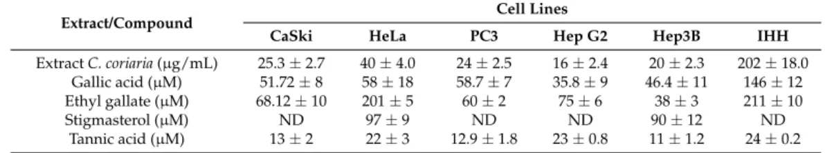
Table 1.IC50values in µg/mL for the C. coriaria extract and µM for isolated compounds.
Extract/Compound Cell Lines
CaSki HeLa PC3 Hep G2 Hep3B IHH
Extract C. coriaria (µg/mL) 25.3 ± 2.7 40 ± 4.0 24 ± 2.5 16 ± 2.4 20 ± 2.3 202 ± 18.0 Gallic acid (µM) 51.72 ± 8 58 ± 18 58.7 ± 7 35.8 ± 9 46.4 ± 11 146 ± 12 Ethyl gallate (µM) 68.12 ± 10 201 ± 5 60 ± 2 75 ± 6 38 ± 3 211 ± 10 Stigmasterol (µM) ND 97 ± 9 ND ND 90 ± 12 ND
Tannic acid (µM) 13 ± 2 22 ± 3 12.9 ± 1.8 23 ± 0.8 11 ± 1.2 24 ± 0.2 IC50values represent the concentration causing 50% growth inhibition. They were determined by linear regression
analysis. Each sample is the mean of three independent experiments.±standard deviation (SD).
2.2. Isolation of Active Compounds
Next the WAE extract of C. coriaria was fractionated by repeated column chromatography to obtain stigmasterol, ethyl gallate (EG), and GA. Stigmasterol was identified by direct comparison with the spectroscopic data of an authentic sample, while EG and GA were identified and characterized by spectroscopic data analysis and comparison with data described in the literature.
The1H-NMR spectrum of GA showed only two single signals at δ 13.5 (1H, s), and δ 7.14 (2H, s) indicating the presence of benzoic acid (Figure S2). The13C-NMR spectrum shows a signal at δ 168.25 characteristic of the carbon atom of a carboxylic acid group, as well as four aromatic carbon signals at δ 146.0 (2 C), 138.8 (1 C), 122.0 (1 C), and 110.2 (2 CH), indicating the presence of a symmetrical tetrasubstituted ring (Figure S3). Comparison of these spectral data, together with the mass spectrum (Figure S1) with those described for the same compound in the literature confirmed its structure [14]. The1H-NMR spectrum of EG displayed signals for a phenol at δ 8.2 (1H, sbr), one tetrasubstituted aromatic ring at δ 7.12 (2H, s), and one ethoxy group at δ 4.24 (2H, q, J = 7 Hz) and δ 1.30 (3H, t, J = 7 Hz) (Figure S5). The13C-NMR showed the same signals as GA, except for the presence of the signals for the ethoxy group at δ 60.96 and 14.69 (Figure S6). The above spectral features are in close agreement to those observed for EG (Figure1) [14]. The most polar extract was constituted mainly of tannic acid, which was previously identified in this species [15]. For further biological studies, we didn’t purify this substance from the WAE extract but rather used a commercial sample.
Molecules 2017, 22, 666 3 of 14
Table 1. IC50 values in μg/mL for the C. coriaria extract and µM for isolated compounds.
Extract/Compound Cell Lines
CaSki HeLa PC3 Hep G2 Hep3B IHH
Extract C. coriaria (µg/mL) 25.3 ± 2.7 40 ± 4.0 24 ± 2.5 16 ± 2.4 20 ± 2.3 202 ± 18.0 Gallic acid (µM) 51.72 ± 8 58 ± 18 58.7 ± 7 35.8 ± 9 46.4 ± 11 146 ± 12 Ethyl gallate (µM) 68.12 ± 10 201 ± 5 60 ± 2 75 ± 6 38 ± 3 211 ± 10 Stigmasterol (µM) ND 97 ± 9 ND ND 90 ± 12 ND
Tannic acid (µM) 13 ± 2 22 ± 3 12.9 ± 1.8 23 ± 0.8 11 ± 1.2 24 ± 0.2 IC50 values represent the concentration causing 50% growth inhibition. They were determined by linear regression analysis. Each sample is the mean of three independent experiments. ± standard deviation (SD).
2.2. Isolation of Active Compounds
Next the WAE extract of C. coriaria was fractionated by repeated column chromatography to obtain stigmasterol, ethyl gallate (EG), and GA. Stigmasterol was identified by direct comparison with the spectroscopic data of an authentic sample, while EG and GA were identified and characterized by spectroscopic data analysis and comparison with data described in the literature.
The 1H-NMR spectrum of GA showed only two single signals at δ 13.5 (1H, s), and δ 7.14 (2H, s)
indicating the presence of benzoic acid (Figure S2). The 13C-NMR spectrum shows a signal at δ 168.25
characteristic of the carbon atom of a carboxylic acid group, as well as four aromatic carbon signals at δ 146.0 (2 C), 138.8 (1 C), 122.0 (1 C), and 110.2 (2 CH), indicating the presence of a symmetrical tetrasubstituted ring (Figure S3). Comparison of these spectral data, together with the mass spectrum (Figure S1) with those described for the same compound in the literature confirmed its structure [14]. The 1H-NMR spectrum of EG displayed signals for a phenol at δ 8.2 (1H, sbr), one tetrasubstituted
aromatic ring at δ 7.12 (2H, s), and one ethoxy group at δ 4.24 (2H, q, J = 7 Hz) and δ 1.30 (3H, t, J = 7 Hz) (Figure S5). The 13C-NMR showed the same signals as GA, except for the presence of the signals for
the ethoxy group at δ 60.96 and 14.69 (Figure S6). The above spectral features are in close agreement to those observed for EG (Figure 1) [14]. The most polar extract was constituted mainly of tannic acid, which was previously identified in this species [15]. For further biological studies, we didn’t purify this substance from the WAE extract but rather used a commercial sample.
Figure 1. Compounds isolated from the WAE extract of C. coriaria.
2.3. Antiproliferative Activity of Pure Compounds
Antiproliferative activities evaluation of the isolated compounds, GA, EG, stigmasterol, and TA, showed interesting effects on cell proliferation (Table 1). GA inhibited cell proliferation in all cancer cell lines analyzed, but was most potent on hepatocellular carcinoma cells with IC50 of 35.8 µM
and 46.4 µM on HepG2 and Hep3B cells, respectively. Interestingly the IC50 of GA on IHH
immortalized human hepatocytes cells was much higher (146 µM). Similar results were observed
Figure 1.Compounds isolated from the WAE extract of C. coriaria. 2.3. Antiproliferative Activity of Pure Compounds
Antiproliferative activities evaluation of the isolated compounds, GA, EG, stigmasterol, and TA, showed interesting effects on cell proliferation (Table1). GA inhibited cell proliferation in all cancer
Molecules 2017, 22, 666 4 of 14
cell lines analyzed, but was most potent on hepatocellular carcinoma cells with IC50of 35.8 µM and
46.4 µM on HepG2 and Hep3B cells, respectively. Interestingly the IC50of GA on IHH immortalized
human hepatocytes cells was much higher (146 µM). Similar results were observed with EG exhibiting the best antiproliferative activity on Hep3B cells (IC50of 38 µM) and the worse on IHH cells (IC50of
211µM). With respect to TA, the most sensitive cells were Hep3B (IC50of 11 µM), followed by PC3
(IC50of 12.9 µM) and Ca Ski cells (IC50of 13 µM). Finally, we found that on all the cancer cell lines
tested, stigmasterol was not active, with an IC50higher than 90 µM.
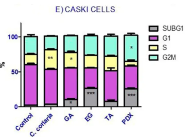
2.4. Cell Cycle Analysis
The effect of the WAE extract and the isolated compounds of C. coriaria on cell cycle progression of cancer cells was determined at the corresponding IC50. Podophyllotoxin (PDX) was included as a
positive control of the G2/M phase arrest. Histograms acquired are shown in Supplementary Materials in the following order, PC3 (Figure S7); Hep3B (Figure S8); HepG2 (Figure S9); Hela (Figure S10) and CaS ki (Figure S11) cells. Interestingly, the different compounds showed differential effects on the cell cycle progression of the different cell lines (Figure2). More specifically, we observed that the WAE extract induced an S phase arrest in PC3 (Figure2A), Hep3B (Figure2B) and Ca Ski cells (Figure2E). Moreover GA induced an S phase arrest too in PC3 (Figure2A), Hep3B (Figure2B) and HepG2 cells (Figure2C). TA induced an S phase arrest only in Hep3B cells (Figure2B). On the other hand, the characterized compounds, but not the extract of C. coriaria, induced a G2/M phase cell cycle arrest depending on the cells observed. GA induced a G2/M phase arrest in HeLa cells (Figure2D), while TA increased the population of G2/M phase of PC3 cells (Figure2A). EG increased meaningfully the G2/M population in Hep3B cells (Figure2B) and in HepG2 cells (Figure2C). After treatment with the WAE extract, EG, GA, and TA, most of cancer cells showed a significant increase in the subG1 population (Figure2A–E). This suggests DNA damage, cell death or cellular senescence. PC3 and Hep3B cells were the most affected (Figure2A,B), followed by HeLa cells (Figure2D) and Ca Ski cells (Figure2E). That was also the case in HepG2 cells treated with EG or TA (Figure2C).
Molecules 2017, 22, 666 4 of 14
with EG exhibiting the best antiproliferative activity on Hep3B cells (IC50 of 38 µM) and the worse on
IHH cells (IC50 of 211µM). With respect to TA, the most sensitive cells were Hep3B (IC50 of 11 µM),
followed by PC3 (IC50 of 12.9 µM) and Ca Ski cells (IC50 of 13 µM). Finally, we found that on all the
cancer cell lines tested, stigmasterol was not active, with an IC50 higher than 90 µM.
2.4. Cell Cycle Analysis
The effect of the WAE extract and the isolated compounds of C. coriaria on cell cycle progression of
cancer cells was determined at the corresponding IC50. Podophyllotoxin (PDX) was included as a
positive control of the G2/M phase arrest. Histograms acquired are shown in Supplementary Materials in the following order, PC3 (Figure S7); Hep3B (Figure S8); HepG2 (Figure S9); Hela (Figure S10) and CaS ki (Figure S11) cells. Interestingly, the different compounds showed differential effects on the cell cycle progression of the different cell lines (Figure 2). More specifically, we observed that the WAE extract induced an S phase arrest in PC3 (Figure 2A), Hep3B (Figure 2B) and Ca Ski cells (Figure 2E). Moreover GA induced an S phase arrest too in PC3 (Figure 2A), Hep3B (Figure 2B) and HepG2 cells (Figure 2C). TA induced an S phase arrest only in Hep3B cells (Figure 2B). On the other hand, the characterized compounds, but not the extract of C. coriaria, induced a G2/M phase cell cycle arrest depending on the cells observed. GA induced a G2/M phase arrest in HeLa cells (Figure 2D), while TA increased the population of G2/M phase of PC3 cells (Figure 2A). EG increased meaningfully the G2/M population in Hep3B cells (Figure 2B) and in HepG2 cells (Figure 2C). After treatment with the WAE extract, EG, GA, and TA, most of cancer cells showed a significant increase in the subG1 population (Figure 2A–E). This suggests DNA damage, cell death or cellular senescence. PC3 and Hep3B cells were the most affected (Figure 2A,B), followed by HeLa cells (Figure 2D) and Ca Ski cells (Figure 2E). That was also the case in HepG2 cells treated with EG or TA (Figure 2C).
Molecules 2017, 22, 666 5 of 14
Molecules 2017, 22, 666 5 of 14
Figure 2. Effect of the WAE extract of C. coriaria, GA, EG and TA on cell cycle progression in cancer cells
lines. (A) PC3 (prostate); (B) Hep3B and (C) HepG2 (hepatocellular carcinoma); (D) HeLa and 2E) Caski (cervical cancer). Podophillotoxin (PDX) was used as a positive control (0.005 µM). * p < 0.05, ** p < 0.01, *** p < 0.001 compared with the control group.
2.5. Effect of Characterized Compounds on Microtubules Dynamics
With the purpose of exploring the effect on microtubule dynamics, immunofluorescence of α-tubulin in Hep3B cells in the presence of pure compounds EG, GA, and TA was monitored by confocal microscopy. Hep3B cells had already been reported as a good cellular model to analyze microtubule perturbations because of their phenotypic characteristics [16]. Taxol and podophyllotoxin, which are known to target microtubules, were used as positive controls. As expected, we obtained a complete disassembly of microtubules after podophyllotoxin treatment and a stabilization of microtubules by an inhibition of their depolymerization after taxol treatment (Figure 3). Our results showed that in Hep3B cells treated with EG the microtubules appeared rigid and formed large bundles in the cytoplasm. EG induced a complete rearrangement of microtubules structure. It is clear that polymerized microtubules formed bulky rigid structures, localized in the cell periphery, in a manner parallel to the plasma membrane. We observed that they are typically arranged perpendicular to the cell edge and the cells showed microtubules stabilized around the nucleus, similar but to a lesser extent than observed in taxol treated cells. These structures were also observed in the cells treated with tannic acid, despite the low fluorescence intensity detected. These results are in accordance with those of the cell cycle analysis suggesting that EG could induce cell cycle G2/M phase arrest in hepatocellular carcinoma cells by stabilizing microtubule polymerization. This is the first report describing the effect of EG on microtubule stabilization in cancer cells. Moreover GA treated cells showed a disperse tubulin signal although some microtubules appear normal, indicating a mild effect of GA on microtubule stability (Figure 3). Moreover, we evaluated the effect of EG on PC3 cells and although some polymerized microtubules localized in the cell periphery were observed, the changes observed were not significant respect to taxol treatment in this cell line (Figure S12). Further studies with these compounds would be interesting to further determine their action mechanism on microtubules and a possible interaction with tubulins.
Figure 2.Effect of the WAE extract of C. coriaria, GA, EG and TA on cell cycle progression in cancer cells lines. (A) PC3 (prostate); (B) Hep3B and (C) HepG2 (hepatocellular carcinoma); (D) HeLa and 2E) Caski (cervical cancer). Podophillotoxin (PDX) was used as a positive control (0.005 µM). * p < 0.05, ** p < 0.01, *** p < 0.001 compared with the control group.
2.5. Effect of Characterized Compounds on Microtubules Dynamics
With the purpose of exploring the effect on microtubule dynamics, immunofluorescence of α-tubulin in Hep3B cells in the presence of pure compounds EG, GA, and TA was monitored by confocal microscopy. Hep3B cells had already been reported as a good cellular model to analyze microtubule perturbations because of their phenotypic characteristics [16]. Taxol and podophyllotoxin, which are known to target microtubules, were used as positive controls. As expected, we obtained a complete disassembly of microtubules after podophyllotoxin treatment and a stabilization of microtubules by an inhibition of their depolymerization after taxol treatment (Figure3). Our results showed that in Hep3B cells treated with EG the microtubules appeared rigid and formed large bundles in the cytoplasm. EG induced a complete rearrangement of microtubules structure. It is clear that polymerized microtubules formed bulky rigid structures, localized in the cell periphery, in a manner parallel to the plasma membrane. We observed that they are typically arranged perpendicular to the cell edge and the cells showed microtubules stabilized around the nucleus, similar but to a lesser extent than observed in taxol treated cells. These structures were also observed in the cells treated with tannic acid, despite the low fluorescence intensity detected. These results are in accordance with those of the cell cycle analysis suggesting that EG could induce cell cycle G2/M phase arrest in hepatocellular carcinoma cells by stabilizing microtubule polymerization. This is the first report describing the effect of EG on microtubule stabilization in cancer cells. Moreover GA treated cells showed a disperse tubulin signal although some microtubules appear normal, indicating a mild effect of GA on microtubule stability (Figure3). Moreover, we evaluated the effect of EG on PC3 cells and although some polymerized microtubules localized in the cell periphery were observed, the changes observed were not significant respect to taxol treatment in this cell line (Figure S12). Further studies with these compounds would be interesting to further determine their action mechanism on microtubules and a possible interaction with tubulins.
Molecules 2017, 22, 666 6 of 14
Molecules 2017, 22, 666 6 of 14
Figure 3. Effect of isolated compounds on stabilization of microtubules in Hep3B cells by immunofluorescence of α-Tubulin and confocal microscopy. EG; GA; TA; Podophillotoxin (Microtubules destabilizing agent) and Taxol (Microtubules stabilizing agent).
2.6. Cellular Death
Apoptosis is a central mechanism to eliminate unwanted cells that may accumulate during physiological processes and pathologic conditions such as cancer and autoimmune diseases. The induction of apoptosis promotes morphological characteristic changes: cell shrinkage (cells get smaller in size, the cytoplasm is dense and the organelles are more tightly packed), pyknosis which is the result of chromatin condensation and this is the most characteristic feature of apoptosis, extensive Figure 3. Effect of isolated compounds on stabilization of microtubules in Hep3B cells by immunofluorescence of α-Tubulin and confocal microscopy. EG; GA; TA; Podophillotoxin (Microtubules destabilizing agent) and Taxol (Microtubules stabilizing agent).
2.6. Cellular Death
Apoptosis is a central mechanism to eliminate unwanted cells that may accumulate during physiological processes and pathologic conditions such as cancer and autoimmune diseases. The induction of apoptosis promotes morphological characteristic changes: cell shrinkage (cells get smaller in size, the cytoplasm is dense and the organelles are more tightly packed), pyknosis which is the result of chromatin condensation and this is the most characteristic feature of apoptosis, extensive plasma membrane blebbing karyorrhexis and separation of cell fragments into apoptotic bodies during a process called “budding”. Apoptotic bodies consist of cytoplasm with tightly packed
Molecules 2017, 22, 666 7 of 14
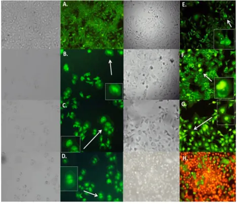
organelles with or without a nuclear fragment [17]. In the present study, the effect of the C. coriaria extract and pure compounds (AG, AG and TA) on cell death was characterized by morphological changes observed by acridine orange-ethidium bromide fluorescence microscopy. Hep3B (Figure4), PC3 (Figure S13), and HepG2 (Figure S14) cells showed morphological characteristic changes similar to the pro-apoptotic controls podophyllotoxin and H2O2. For instance, Hep3B cells treated with the
C. coriaria extract, AG and EG showed clear chromatin condensation and plasma membrane blebbing (magnification, white arrows Figure4B–D); while the formation of apoptotic bodies and chromatin condensation was observed after the TA treatment (magnification, white arrows 4E); the morphological changes were similar to podophyllotoxin (Figure4G). Major effects on cell death in PC3, HepG2 and CaSki cells were observed after TA treatment with a clear chromatin condensation, plasma membrane blebbing and formation of apoptotic bodies (white arrows Figure S13E, S14E and S15E, respectively). Similar results were observed in PC3 cells after the C. coriaria extract treatment (Figure S13B). On HeLa cells, chromatin condensation and green yellow cores (white arrows) were observed only with GA treatment (Figure S16D). It is noteworthy that necrosis was not observed in none of the cells treated with the compounds. These results showed that the WAE extract of C. coriaria as well as the isolated molecules EG, GA and TA induced cell death, with characteristic apoptotic morphological changes, that are in accordance with the increase of subG1 population previously observed in the cell cycle histograms (Figures S7–S11) and bar charts (Figure2).
Molecules 2017, 22, 666 7 of 14
plasma membrane blebbing karyorrhexis and separation of cell fragments into apoptotic bodies during a process called “budding”. Apoptotic bodies consist of cytoplasm with tightly packed organelles with or without a nuclear fragment [17]. In the present study, the effect of the C. coriaria extract and pure compounds (AG, AG and TA) on cell death was characterized by morphological changes observed by acridine orange-ethidium bromide fluorescence microscopy. Hep3B (Figure 4), PC3 (Figure S13), and HepG2 (Figure S14) cells showed morphological characteristic changes similar to the pro-apoptotic controls podophyllotoxin and H2O2. For instance, Hep3B cells treated with the
C. coriaria extract, AG and EG showed clear chromatin condensation and plasma membrane
blebbing (magnification, white arrows Figure 4B–D); while the formation of apoptotic bodies and chromatin condensation was observed after the TA treatment (magnification, white arrows 4E); the morphological changes were similar to podophyllotoxin (Figure 4G). Major effects on cell death in PC3, HepG2 and CaSki cells were observed after TA treatment with a clear chromatin condensation, plasma membrane blebbing and formation of apoptotic bodies (white arrows Figure S13E, S14E and S15E, respectively). Similar results were observed in PC3 cells after the C. coriaria extract treatment (Figure S13B). On HeLa cells, chromatin condensation and green yellow cores (white arrows) were observed only with GA treatment (Figure S16D). It is noteworthy that necrosis was not observed in none of the cells treated with the compounds. These results showed that the WAE extract of C. coriaria as well as the isolated molecules EG, GA and TA induced cell death, with characteristic apoptotic morphological changes, that are in accordance with the increase of subG1 population previously observed in the cell cycle histograms (Figures S7–S11) and bar charts (Figure 2).
Figure 4. Effect of WAE extract of C. coriaria extract and pure compounds on cell death in Hep3B cells by epifluorescence microscopy. (A) Negative control; (B) C. coriaria extract; (C) GA; (D) EG; (E) TA;
(F) Podophillotoxin 0.005 µM (positive control); (G) H2O2 apoptosis positive control; (H) Necrosis control.
2.7. Bax and Bcl2 Expression
In order to determine whether the pure compounds induce cell death by apoptosis by regulating anti-apoptotic (Bcl-2) and pro-apoptotic (Bax) proteins in the hepatocellular carcinoma Hep3B cells, expression of Bax and Bcl-2 was analyzed by RT-PCR. Ours results showed a clear inhibition of Bcl-2 and an increase of Bax transcripts after the treatment with TA, GA and EG (Figure 5). The Bax/Bcl-2 ratio was evidently increased when compared to the control after TA, GA and EG treatments.
Figure 4.Effect of WAE extract of C. coriaria extract and pure compounds on cell death in Hep3B cells by epifluorescence microscopy. (A) Negative control; (B) C. coriaria extract; (C) GA; (D) EG; (E) TA; (F) Podophillotoxin 0.005 µM (positive control); (G) H2O2apoptosis positive control; (H) Necrosis control.
2.7. Bax and Bcl2 Expression
In order to determine whether the pure compounds induce cell death by apoptosis by regulating anti-apoptotic (Bcl-2) and pro-apoptotic (Bax) proteins in the hepatocellular carcinoma Hep3B cells, expression of Bax and Bcl-2 was analyzed by RT-PCR. Ours results showed a clear inhibition of Bcl-2 and an increase of Bax transcripts after the treatment with TA, GA and EG (Figure5). The Bax/Bcl-2 ratio was evidently increased when compared to the control after TA, GA and EG treatments.
Molecules 2017, 22, 666 8 of 14 Molecules 2017, 22, 666 8 of 14
BAX
BCL2
Control
TA
GA
EG
GAPDH
Figure 5. Effect of TA, GA and EG on mRNA expression levels of Bcl-2 and Bax in Hep3B cells after 72 h treatment. GAPDH was used as an internal control.
2.8. Caspases 3/7 Activity
Caspases play a key role in the initiation and execution of apoptosis. Therefore to support the effect previously observed on Bax/Bcl-2 expression; the caspase 3/7 activity was evaluated on Hep3B cells after treatment by GA, EG and TA (Figure 6). All pure compounds showed a higher caspase 3/7 activity when compared to the non-treated control. TA showed the most significant effect, just below taxol (positive control).
Caspase 3/7 activity
CON TROL GA EG TA TX 0.0 200000.0 400000.0 600000.0 800000.0 1000000.0 *** ** * * Compounds Lum in escence ( R L U )Figure 6. Caspase 3/7 activity after treatment with GA; EG; TA and taxol (TX) in Hep3B cells. * p < 0.05, ** p < 0.01, *** p < 0.001 were obtained when compared to the negative control.
3. Discussion
A number of studies have demonstrated the antitumoral and antimutagenic properties of polyphenols and phenolic compounds isolated from plants [15]. Our results with the WAE extract of
C. coriaria and isolated phenolic compounds on cell proliferation, cell cycle arrest and cell death
confirm such antitumoral potential. In this study, the hepatocellular carcinoma cells were the most sensitive to the antiproliferative effect of the WAE extract of C. coriaria, although they are known to be chemoresistant. Oppositely, the immortalized cells from normal human hepatocytes (IHH cells),
were the less sensitive to the WAE extract of C. coriaria extract and compounds with an IC50 10 fold
higher than the one seen for Hep3B cancer cells. This effect was expected, as it seems that this extract
showed selectivity for cancer cells. Moreover, the WAE extract of C. coriaria induced S phase cell
Figure 5.Effect of TA, GA and EG on mRNA expression levels of Bcl-2 and Bax in Hep3B cells after 72 h treatment. GAPDH was used as an internal control.
2.8. Caspases 3/7 Activity
Caspases play a key role in the initiation and execution of apoptosis. Therefore to support the effect previously observed on Bax/Bcl-2 expression; the caspase 3/7 activity was evaluated on Hep3B cells after treatment by GA, EG and TA (Figure6). All pure compounds showed a higher caspase 3/7 activity when compared to the non-treated control. TA showed the most significant effect, just below taxol (positive control).
Molecules 2017, 22, 666 8 of 14
BAX
BCL2
Control
TA
GA
EG
GAPDH
Figure 5. Effect of TA, GA and EG on mRNA expression levels of Bcl-2 and Bax in Hep3B cells after 72 h treatment. GAPDH was used as an internal control.
2.8. Caspases 3/7 Activity
Caspases play a key role in the initiation and execution of apoptosis. Therefore to support the effect previously observed on Bax/Bcl-2 expression; the caspase 3/7 activity was evaluated on Hep3B cells after treatment by GA, EG and TA (Figure 6). All pure compounds showed a higher caspase 3/7 activity when compared to the non-treated control. TA showed the most significant effect, just below taxol (positive control).
Caspase 3/7 activity
CON TROL GA EG TA TX 0.0 200000.0 400000.0 600000.0 800000.0 1000000.0 *** ** * * Compounds Lum in escence ( R L U )Figure 6. Caspase 3/7 activity after treatment with GA; EG; TA and taxol (TX) in Hep3B cells. * p < 0.05, ** p < 0.01, *** p < 0.001 were obtained when compared to the negative control.
3. Discussion
A number of studies have demonstrated the antitumoral and antimutagenic properties of polyphenols and phenolic compounds isolated from plants [15]. Our results with the WAE extract of
C. coriaria and isolated phenolic compounds on cell proliferation, cell cycle arrest and cell death
confirm such antitumoral potential. In this study, the hepatocellular carcinoma cells were the most sensitive to the antiproliferative effect of the WAE extract of C. coriaria, although they are known to be chemoresistant. Oppositely, the immortalized cells from normal human hepatocytes (IHH cells),
were the less sensitive to the WAE extract of C. coriaria extract and compounds with an IC50 10 fold
higher than the one seen for Hep3B cancer cells. This effect was expected, as it seems that this extract
showed selectivity for cancer cells. Moreover, the WAE extract of C. coriaria induced S phase cell
Figure 6.Caspase 3/7 activity after treatment with GA; EG; TA and taxol (TX) in Hep3B cells. * p < 0.05, ** p < 0.01, *** p < 0.001 were obtained when compared to the negative control.
3. Discussion
A number of studies have demonstrated the antitumoral and antimutagenic properties of polyphenols and phenolic compounds isolated from plants [15]. Our results with the WAE extract of C. coriaria and isolated phenolic compounds on cell proliferation, cell cycle arrest and cell death confirm such antitumoral potential. In this study, the hepatocellular carcinoma cells were the most sensitive to the antiproliferative effect of the WAE extract of C. coriaria, although they are known to be chemoresistant. Oppositely, the immortalized cells from normal human hepatocytes (IHH cells), were
Molecules 2017, 22, 666 9 of 14
the less sensitive to the WAE extract of C. coriaria extract and compounds with an IC5010 fold higher
than the one seen for Hep3B cancer cells. This effect was expected, as it seems that this extract showed selectivity for cancer cells. Moreover, the WAE extract of C. coriaria induced S phase cell cycle arrest in PC3, Hep3B and CaSki cells but no changes were observed in G2/M population cells. Previous studies with extracts rich in phenolic compounds have shown S phase cell cycle arrest as mechanism of inhibition of cancer cell proliferation [18]. In addition, we observed a significant increase in the subG1 population (in the cell cycle analysis) by the WAE extract of C. coriaria treatment in PC3 and Hep3B cells that correlated with the observed morphological characteristic changes similar to apoptosis.
Further chromatographic fractionation of the WAE extract of C. coriaria allowed the isolation or identification of stigmasterol, EG, GA and TA. GA and TA had been previously identified as components of the WAE extract of C. coriaria but the presence of stigmasterol and EG had not been reported before in this species [15]. We found that GA, a metabolic intermediate of plants seemed to act selectively on cancer cells, particularity against hepatocellular carcinoma cell lines with IC50
values 4- and 3-fold lower in HepG2 and Hep3B cells, respectively, compared to the one in normal IHH cells. This selectivity had been also observed in dRLh-84 mouse hepatoma cells that showed cell death after treatment with GA at a concentration of 20 µg/mL, while in primary cultures of rat hepatocytes and macrophages it was not cytotoxic, showing again some selectivity for cancer cells [19]. EG is an ester derivative of GA that we also found more anti-proliferative on Hep3B and HepG2 cells than on IHH cells (with a 5- and 3-fold lower IC50, respectively). Finally, TA, known as a potent anti-oxidant,
showed lower IC50values than EG and GA in the majority of the cancer cells.
Interestingly, GA induced cell cycle arrest differentially in PC3, Hep3B and HepG2 cells, where we observed an S phase arrest, than in HeLa cells where we observed a G2/M phase arrest. Previously, it was demonstrated that GA treatment of human prostate carcinoma DU145 cells resulted in an S phase cell cycle arrest [20]. Recently, it was elucidated that GA induced G2/M phase arrest in HeLa cells accompanied by mitotic catastrophe, and formation of cells with multiple nuclei, followed by impaired centrosomal clustering [21]. Similarly, TA induced S phase arrest in Hep3B whereas it induced a G2/M phase arrest in HepG2 and PC3 cells. A preceding study in malignant human cholangiocytes indicated for TA an S phase cell cycle arrest [22]. Finally EG induced a clear G2/M phase cell cycle arrest in the hepatocellular carcinoma Hep3B and HepG2 cells. One explanation of a cell cycle G2/M phase arrest is the inhibition by stabilization or destabilization of microtubules polymerization as that can be observed with taxol or podophillotoxin. Our results showed by confocal microscopy a microtubules stabilization of Hep3B cells treated with EG. Similar results but to a lesser extent were observed with TA. Interestingly we expected to observe an effect on microtubules destabilization rather than stabilization since it has been previously shown that the 3,4,5-trimethoxyphenyl unit of GA, present in the EG structure, is crucial for interactions disturbing the assembly of tubulin [23,24]. Therefore, further studies on the activity of EG on microtubule dynamics in vitro are necessary to better understand the EG mechanism of action.
It was known that GA induces apoptosis by activating PARP, caspase-9, and caspase-3, related to an increase of the pro-apoptotic Bax expression [25]. In addition, TA was known to induce apoptotic death in acute myeloid leukemia (AML) HL-60 cells in a dose- and time-dependent manner as well as an increase of the sub-G1 fraction, chromosome condensation, and DNA fragmentation [26]. EG was reported to induce DNA fragmentation and apoptosis via mitochondrial-mediated pathways by increasing the ratio of Bax/Bcl-2 and activation of Caspases-8, -9, -3 in HL-60 cells [27]. Similar results were given when EG was tested against the human breast cancer cells (MDA-MB-231 (ER-) invasion and showed to modulate the PI3K/Akt pathway and to inhibit their downstream targets such as Bcl-2/Bax at mRNA levels [8]. This motivated us to look at the effect of the GA, TA and EG compounds on apoptosis on Hep3B hepatocarcinoma cells. We found chromatin condensation, plasma membrane blebbing and formation of apoptotic bodies. Moreover, GA, EG and AT clearly inhibited Bcl-2 and increased Bax transcripts in Hep3B cells and showed a clear increase in caspase 3/7 activation. This is in line with a pro-apoptotic activity of these compounds in Hep3B cells.
Molecules 2017, 22, 666 10 of 14
4. Materials and Methods
4.1. Plant Material
The pods of C. coriaria were collected by Jessica Nayelli Sánchez Carranza on March 2013 in Cutzamala the Pinzon Guerrerro, México. A sample was authentified by Alejandro Flores and deposited at the Herbarium of the Universidad Autónoma del Estado de Morelos (HUMO-UAEM), with the voucher number 33400.
4.2. Chemical Procedures
Isolation procedures and purity of compounds were checked by thin layer chromatography (TLC, Merck Millipore, Billerica, MA, USA) visualized by UV light, and sprayed with Ce(SO4)2·2(NH4)2SO4·2H2O. All1H,13C, and 2D-NMR experiments were recorded on acetone-d6,
on a Varian Unity 400 spectrometer (Varian, Palo Alto, CA, USA), at 400 MHz for1H-NMR, and at 100 MHz for13C-NMR. EI-EM spectra were recorded on a JEOL JMX-AX 505 HA mass spectrometer (Jeol Ltd., Tokyo, Japan). All the reagents and solvents used were of analytical grade. Tannic acid (CAS: 1401-55-4) was purchased from Sigma Aldrich (St. Louis, MO, USA).
4.3. Extraction and Isolation.
The collected material (600 g) was dried and powdered and extracted with 2 L of a water:acetone:ethanol solvent mixture (80:10:10, v/v) at room temperature. After 72 h, the extract was filtered and concentrated under reduced pressure at 60◦C, to yield 75 g of extract. This WAE extract of C. coriaria was dissolved in acetone and filtered again. The soluble part (12.4 g) was subjected to a first chromatographic fractionation using Si-gel open column chromatography (60×10 cm) and eluted with a gradient of CH2Cl2–acetone (from CH2Cl2100% to CH2Cl2–acetone 70:30, 200 mL each fraction)
to obtain three main fractions JS33A, 1.3 g (100:0); JS33B, 900 mg (90:10); JS33C, 2.4 g (80:20). Fraction JS33A was subjected to CC purification to yield 72 mg of stigmasterol. Fraction JS33B was purified by column chromatography (30 g Si gel), eluted with a gradient of n-hexane–acetone. Fractions eluted with n-hexane–acetone (75:25) contained pure ethyl gallate, which was crystallized from CH2Cl2–EtOH,
to yield 820 mg of ethyl gallate. Fraction JS33C was purified by column chromatography eluted with a CH2Cl2:acetone gradient system (100:00→80:20). Fractions eluted with CH2Cl2–acetone (62:38)
afforded 920 mg of a yellow solid, which was purified by column chromatography using an isocratic mixture of CH2Cl2–MeOH to yield 501.4 mg of gallic acid. These compounds were characterized by
comparison of their spectroscopic data with those described [14]. 4.4. Cytotoxic Assay
The WAE extract of C. coriaria as well as pure compounds 1 and 2 and tannic acid (3) were subjected to antiproliferative assays in PC3 (prostate), Hep3B, and HepG2 (hepatocellular), and Ca Ski and HeLa (cervical) human cancer cell lines, obtained from ATCC (American Type Culture Collection, Manassas, VA, USA). We also included an immortalized human hepatocytes cell line (IHH) as a control of non-cancerous cells [28]. PC3 and CaSKi cells were grown in RPMI-1640 medium (Sigma Aldrich, St. Louis, MO, USA), while Hep3B, HepG2, IHH and HeLa in DMEM medium (Invitrogen, Thermo Fisher Scientific, Inc., Waltham, MA, USA) and supplemented with fetal bovine serum 10% (SFB, Invitrogen) and with 2 mM glutamine, all cultures were incubated at 37◦C in atmosphere of 5% CO2. 4000 cells per well in 96-well plate were cultured for starting the cytotoxic evaluation. The extract
was solubilized in culture medium and sterilized by 0.2 µm membrane filtration, the concentrations used for the extract were 1000, 100, 10, 1, 0.1, 0.001 µg/mL for a dose/response curve, and incubated at 37◦C in 5% CO2 atmosphere for 72 h. Concentrations of 160, 80, 40, 20, 10, 5, 2.5, 1.25, 0.6,
0.3 µg/mL were used for pure compounds (1–3), and incubated at 37◦C in 5% CO2atmosphere for
72 h. The number of viable cells in proliferation was then determined by using CellTiter 96®AQueous One Solution Cell Proliferation Assay kit (Promega, Madison, WI, USA), following the manufacturer's
Molecules 2017, 22, 666 11 of 14
instruction. Cell viability was determined by absorbance at 450 nm using an automated ELISA reader. The experiments were conducted by triplicate in three independent experiments. Data were analyzed in Prism 5.0 statistical program and the IC50values were determined by regression analysis.
4.5. Cell Cycle Analysis
PC3, Hep3B, HepG2, HeLa and Ca SKi cells (1.25×10⁵) were plated in 6-well plates. Exponential growing cells were exposed to each compound for 72 h in accordance with their IC50 values.
Podophyllotoxin (PDX) at 0.005 µM was included as a positive control of the G2/M phase arrest. Cells from each treatment were trypsinized and collected into single cell suspensions, centrifuged, and fixed in cold ethanol (70%) overnight at−20◦C. The cells were then treated with RNase (0.01 M, Sigma Aldrich) and stained with propidium iodide (PI) (7.5 µg/mL, Invitrogen) for 30 min in the dark, PI has the ability to bind to DNA molecules, and then RNase was added in order to allow PI to bind directly to DNA. The percentage of cells in G1, S, and G2 phases was analyzed with a flow cytometer (Becton Dickinson, FACS Calibur, San Jose, CA, USA); the number of cells analyzed for each sample was 10,000. The experiments were conducted by triplicate in three independent experiments. Data obtained from the flow cytometer were analyzed using the FlowJo Software (Tree Star, Inc., Ashland, OR, USA) to generate DNA content frequency histograms, and to quantify the number of cells in the individual cell cycle phases. The results were summarized in bar charts arranged by cell line, compound and cell cycle phase. Data were analyzed in Prism 5.0 statistical program and statistical differences were evaluated using ANOVA followed by Dunnett test and considered significant at p value < 0.05. 4.6. Immunofluorescence of α-Tubulin
3.5×10⁴Hep3B cells were added in 24-well culture plates containing glass slides and allowed
to attach overnight at 37◦C in 5% CO2. Then they were treated with ethyl gallate (7.7 µg/mL), GA
(7.9 µg/mL), tannic acid (20 µg/mL) and controls podophillotoxin (0.005 µM) and taxol (75 nM) for 72 h. The cells were fixed with paraformaldehyde (PFA, 4% in PEM buffer). After 15 min PFA/NaHCO3
was added and incubated for 45 min at room temperature. The slides were rinsed with PBS and treated with 0.1% Triton X-100 (Sigma Aldrich) and then incubated with the primary antibody mouse anti-α-tubulin (1:300, T9026, Sigma Aldrich) overnight at 4◦C. The secondary antibody (anti-mouse Alexa 647 1:1000, A-21235, Molecular Probes, Thermo Fisher Scientific, Inc., Waltham, MA, USA) was added and incubated for two hours at 37◦C. The cells were stained with Sytox Green (1:5000, S7020, Molecular Probes) for one hour, mounted, and imaged by confocal microscopy. The experiments were conducted by triplicate in three independent experiments.
4.7. Cell Death
PC3, Hep3B, HepG2, HeLa, and CaS Ki cells were cultured 20000 cells per well in 24-well plates, and then were treated for 72 h according to the IC50of each compound in each cell line. For controls,
cells were grown 71 h before being treated 30 min with H2O2 (positive control of apoptosis) or
being boiling in water at 95◦C for 10 sec (positive control of necrosis). After 72 h of treatment cells were exposed to a solution of acridine orange (AO) and ethidium bromide (EB) (100 µg/mL AO, 100 µg/mL EB), according to procedures reported [29]. Cells were observed using a fluorescence microscope. AO/EB are fluorescent intercalating nucleic acids that when bounded to DNA give green and orange fluorescence respectively. It is well known that AO can pass through cell membranes, but EB cannot. Necrotic cells stain red but have a nuclear morphology resembling that of viable cells. Apoptotic cells appear green, and morphological changes such as formation of apoptotic bodies are observed. The criteria for identification are as follows: viable cells appear to have green nucleus with intact structure; early apoptosis cells exhibit a bright green nucleus showing condensation of chromatin; late apoptosis appears as dense orange areas of chromatin condensation; and orange intact nucleus depicts secondary necrosis [30,31]. The experiments were conducted by triplicate in three independent experiments.
Molecules 2017, 22, 666 12 of 14
4.8. RT-PCR
1.25× 10⁵ Hep3B cells were plated and treated with GA, TA and EG for 72 h., then RNA was isolated. The total RNA extraction was performed employing a Quick-RNA MiniPrep Kit (Zymo Research, Irvine, CA, USA), following the manufacturer’s instructions. RNA was quantified using NanoDrop® ND-1000 (Thermo Scientific, Waltham, MA, USA), and the RNA content of the samples was normalized. The RT-PCR was performed using a One-Step RT-PCR Kit with Thermo-Start Taq (Thermo Scientific) following the manufacturer’s instructions.
The primer sequences for Bcl-2 were 50-CCC TCC AGA TAG CTC ATT-30, and 50-CTAGAC AGACAAGGAAAG-30. The Bax primer sequences were 50-ATGGACGGGTCCGGGGAG-30, and 50-TCAGAAAACATGTCAGCTGCC-30 [32]. The GAPDH primers were 50-CAAGGTCATCCA TGACAACTTTG-30and 50-GTCCACCACCCTGTTGCTGTAG-30. All primers were synthesized by IDT-Integrated DNA Technologies (Redwood, CA, USA), the reaction products of the samples were analyzed in 1.5% agarose gel.
4.9. Caspases Activity
Hep3B cells (8000 per well) were plated on 96-well plate and treated with GA, TA and EG for 72 h. After treatment the caspase 3/7 activity was determined using the luminescent Caspase-Glo® 3/7 Assay (Promega, cat. G811C) following the manufacturer’s instructions. The results were represented as relative units of luminescence and represented in graphs, the statistical analysis was performed using the Prism 5.0 statistical program and the test performed was t-student test with significant p values < 0.05.
5. Conclusions
Our results suggest that the extract of C. coriaria and its isolated phenolic compounds exhibit strong antiproliferative activities by inducing cell cycle arrest, apoptosis, microtubules reorganization. Ethyl gallate showed an interesting effect on Hep3B hepatocellular carcinoma cells by microtubule stabilization and promoting cell cycle G2/M phase arrest and cell death. These results show the great antineoplasic potential of these phenolic compounds on cancer cells. Further studies will have to be performed to determine the precise mechanism of action of each of these phenolic compounds on the studied cancer cells. This is the first report about the antiproliferative biological activities of the species C. coriaria.
Supplementary Materials:Supplementary materials are available online.
Acknowledgments: Jessica Nayelli Sánchez Carranza acknowledges fellowship 351450 from CONACYT and the support from CONACYT (CB240801), CONACYT-FOMIX (224038). Partial support from UAEM (Grants SI-DGDI-UAEM/13/289) is acknowledged.
Author Contributions: L.G.-M. formulated the original ideas and working hypothesis, together with L.A. designed the research study; J.N.S.-C. participated to the entire experimental process of the study; L.A., S.M.-B. and R.A.V.G supervised the phytochemical part; V.C. and E.W.J. participated to the flow cytometry analysis; E.S.-V. carried out the confocal microscopy study; M.C. participated to the immunofluorescence study and antiproliferative studies. All authors analyzed and interpreted the data. J.N.S. Carranza and L.G.M. wrote the manuscript and L.A., M.C. and S.V. provided important reviews and considerations. All authors read and approved the final manuscript.
Conflicts of Interest:The authors declare no conflict of interests.
References
1. Saracci, R.; Wild, C.P. International Agency for Research on Cancer: The First 50 Years, 1965–2015; IARC Press: Albany, NY, USA, 2015.
2. Yang, C.S.; Landau, J.M.; Huang, M.T.; Newmark, H.L. Inhibition of carcinogenesis by dietary polyphenolic compounds. Annu. Rev. Nutr. 2001, 21, 381–406. [CrossRef] [PubMed]
Molecules 2017, 22, 666 13 of 14
3. Sporn, M.B.; Suh, N. Chemoprevention: An essential approach to controlling cancer. Nat. Rev. Cancer. 2002, 2, 537–543. [CrossRef] [PubMed]
4. Link, A.; Balaguer, F.; Goel, A. Cancer Chemoprevention by Dietary Polyphenols: Promising Role for Epigenetics. Biochem. Pharmacol. 2010, 12, 1771–1792. [CrossRef] [PubMed]
5. Soto Nunez, J.C.; Sousa, M.S. Plantas Medicinales de la Cuenca del Rio Balsas; Universidad Nacional Autonoma de Mexico: Distrito Federal, Mexico, 1995; No. 25; pp. 7–198.
6. Palasap, A.; Limpaiboon, T.; Boonsiri, P.; Thapphasaraphong, S.; Daduang, S.; Suwannalert, P.; Daduang, J. Cytotoxic effects of phytophenolics from Caesalpinia mimosoides Lamk. on cervical carcinoma cell lines through an apoptotic pathway. Asian Pac. J. Cancer Prev. 2014, 15, 449–454. [CrossRef] [PubMed]
7. Rattanata, N.; Klaynongsruang, S.; Daduang, S.; Tavichakorntrakool, R.; Limpaiboon, T.; Lekphrom, R.; Boonsiri, P.; Daduang, J. Inhibitory effects of gallic acid isolated from Caesalpinia mimosoides Lamk. on cholangiocarcinoma cell lines and foodborne pathogenic bacteria. Asian Pac. J. Cancer Prev. 2016, 17, 1341–1345. [CrossRef] [PubMed]
8. Cui, H.; Yuan, J.; Du, X.; Wang, M.; Yue, L.; Liu, J. Ethyl gallate suppresses proliferation and invasion in human breast cancer cells via Akt-NF-κB signaling. Oncol. Rep. 2015, 33, 1284–1290. [PubMed]
9. Nie, F.; Liang, Y.; Jiang, B.; Li, X.; Xun, H.; He, W.; Lau, H.T.; Ma, X. Apoptotic effect of tannic acid on fatty acid synthase over-expressed human breast cancer cells. Tumor Biol. 2016, 37, 2137–2143. [CrossRef] [PubMed]
10. Mitchison, T.; Kirschner, M. Dynamic instability of microtubule growth. Nature 1984, 312, 237–242. [CrossRef] [PubMed]
11. Desai, A.; Mitchison, T.J. Microtubule polymerization dynamics. Annu. Rev. Cell Dev. Biol. 1997, 13, 83–117. [CrossRef] [PubMed]
12. Ballatore, C.; Brunden, K.R.; Huryn, D.M.; Trojanowski, J.Q.; Lee, V.M.-Y.; Smith, A.B. Microtubule stabilizing agents as potential treatment for Alzheimer’s disease and related neurodegenerative tauopathies. J. Med. Chem. 2012, 55, 8979–8996. [CrossRef] [PubMed]
13. Kamal, A.; SrinivasaReddy, T.; Polepallietal, S. Synthesis and biological evaluation of podophyllotoxin congeners as tubulin polymerization inhibitors. Bioorg. Med. Chem. 2014, 22, 5466–5475. [CrossRef] [PubMed] 14. Kamatham, S.; Kumar, N.; Gudipalli, P. Isolation and characterization of gallic acid and methyl gallate
from the seed coats of Givotia rottleriformis Griff. and their anti-proliferative effect on human epidermoid carcinoma A431 cells. Toxicol. Rep. 2015, 2, 520–529. [CrossRef]
15. Veloz-García, R.A.; Marín-Martínez, R.; Veloz-Rodríguez, R.; Muñoz-Sánchez, C.I.; Guevara-Olvera, L.; Miranda-López, R.; González-Chavira, M.M.; Torres-Pacheco, I.; Guzmán-Maldonado, S.H.; Cardador-Martínez, A.; et al. Antimutagenic and antioxidant activities of cascalote (Caesalpinia cacalaco) phenolics. J. Sci. Food Agric. 2004, 84, 1632–1638. [CrossRef]
16. Carraz, M; Lavergne, C.; Jullian, V.; Wright, M.; Gairin, J.E.; Gonzales de la Cruz, M.; Bourdy, G. Antiproliferative activity and phenotypic modification induced by selected Peruvian medicinal plants on human hepatocellular carcinoma Hep3B cells. J. Ethnopharmacol. 2015, 166, 185–199. [CrossRef] [PubMed] 17. Elmore, S. Apoptosis: A Review of Programmed Cell Death. Toxicol. Pathol. 2007, 35, 495–516. [CrossRef]
[PubMed]
18. Tagne, R.S.; Telefo, B.P.; Nyemb, J.N.; Yemele, D.M.; Njina, S.N.; Goka, S.M.; Lienou, L.L.; Nwabo, A.H.; Moundipa, P.F.; Farooq, A.D. Anticancer and antioxidant activities of methanol extracts and fractions of some Cameroonian medicinal plants. Asian Pac. J. Trop Med. 2014, 7, S442–S447. [CrossRef]
19. Inoue, M.; Suzuki, R.; Sakaguchi, N.; Li, Z.; Takeda, T.; Ogihara, Y.; Jiang, B.Y.; Chen, Y. Selective induction of cell death in cancer cells by gallic acid. Biol. Pharm Bull. 1995, 18, 1526–1530. [CrossRef] [PubMed]
20. Agarwal, C.; Tyagi, A.; Agarwal, R. Gallic acid causes inactivating phosphorylation of cdc25A/cdc25C-cdc2 via ATM-Chk2 activation, leading to cell cycle arrest, and induces apoptosis in human prostate carcinoma DU145 cells. Mol. Cancer Ther. 2006, 5, 3294–3302. [CrossRef] [PubMed]
21. Tan, S.; Guan, X.; Grün, C.; Zhou, Z.; Schepers, U.; Nick, P. Gallic acid induces mitotic catastrophe and inhibits centrosomal clustering in HeLa cells. Toxicol. In Vitro 2015, 30, 506–513. [CrossRef] [PubMed] 22. Saleem, A.; Husheem, M.; Härkönen, P.; Pihlaja, K. Inhibition of cancer cell growth by crude extract and the
Molecules 2017, 22, 666 14 of 14
23. Singh, A.; Fatima, K.; Singh, A.; Behl, A.; Mintoo, M.J.; Hasanain, M.; Luqman, R.A.S.; Shanker, K.; Mondhe, D.M.; Sarkar, J.; et al. Anticancer activity and toxicity profiles of 2-benzylidene indanone lead molecule. Eur. J. Pharm. Sci. 2015, 76, 57–67. [CrossRef] [PubMed]
24. Singh, A.; Fatima, K.; Srivastava, A.; Khwaja, S.; Priya, D.; Singh, A.; Mahajan, G.; Alam, S.; Saxena, A.K.; Mondhe, D.M.; et al. Anticancer activity of gallic acid template-based benzylidene indanone derivative as microtubule estabilizer. Chem. Biol. Drug Des. 2016, 88, 625–634. [CrossRef] [PubMed]
25. Chen, H.M.; Wu, Y.C.; Chia, Y.C.; Chang, F.R.; Hsu, H.K.; Hsieh, Y.C.; Chen, C.C.; Yuan, S.S. Gallic acid, a major component of Toona sinensis leaf extracts, contains a ROS-mediated anti-cancer activity in human prostate cancer cells. Cancer Lett. 2009, 286, 161–171. [CrossRef] [PubMed]
26. Chen, K.S; Hsiao, Y.C.; Kuo, D.Y.; Chou, M.C.; Chu, S.C.; Hsieh, Y.S.; Lin, T.H. Tannic acid-induced apoptosis and -enhanced sensitivity to arsenic trioxide in human leukemia HL-60 cells. Leuk. Res. 2009, 297–307. [CrossRef] [PubMed]
27. Kim, W.-H.; Song, H.-O.; Choi, H.-J.; Bang, H.-I.; Choi, D.-Y.; Park, H. Ethyl gallate induces apoptosis of HL-60 cells by promoting the expression of caspases-8, -9, -3, apoptosis-inducing factor and endonuclease G. Int. J. Mol. Sci. 2012, 9, 11912–11922. [CrossRef] [PubMed]
28. Basu, A.; Saito, K.; Meyer, K.; Ray, R.B. Stellate cell apoptosis by a soluble mediator from immortalized human hepatocytes. Apoptosis 2006, 11, 1391–1400. [CrossRef] [PubMed]
29. Kasibhatla, S.; Amarante-Mendes, G.P.; Finucane, D.; Brunner, T.; Bossy-Wetzel, E.; Green, D.R. Acridine orange/ethidium bromide (AO/EB) staining to detect apoptosis. Cold Spring Harb. Protoc. 2006, 3, pdb-prot4493. [CrossRef] [PubMed]
30. Yang, G.-Y; Liao, J.; Kim, K.; Yurkow, E.J.; Yang, C.S. Inhibition of growth and induction of apoptosis in human cancer cell lines by tea polyphenols. Carcinogenesis 1998, 19, 611–616. [CrossRef] [PubMed]
31. Mohan, S.; Bustamam, A.; Ibrahim, S.; Al-Zubairi, A.S.; Aspollah, M.; Abdullah, R.; Elhassan, M.M. In vitro ultramorphological assessment of apoptosis on CEMss induced by linoleic acid-rich fraction from Typhonium flagelliforme tuber. Evid. Based Complement. Altern Med. 2011, 2011, 421894. [CrossRef] [PubMed] 32. Jurado, R.; Lopez-Flores, A.; Alvarez, A.; García-López, P. Cisplatin cytotoxicity is increased by mifepristone
in cervical carcinoma: An in vitro and in vivo study. Oncol. Rep. 2009, 22, 1237–1245. [PubMed]
Sample Availability:Samples of the compounds ethyl gallate, gallic acid, tannic acid or WAE extract of C. coriaria are not available from the authors.
© 2017 by the authors. Licensee MDPI, Basel, Switzerland. This article is an open access article distributed under the terms and conditions of the Creative Commons Attribution (CC BY) license (http://creativecommons.org/licenses/by/4.0/).
View publication stats View publication stats