HAL Id: hal-01426282
https://hal.archives-ouvertes.fr/hal-01426282
Submitted on 18 Apr 2018HAL is a multi-disciplinary open access archive for the deposit and dissemination of sci-entific research documents, whether they are pub-lished or not. The documents may come from teaching and research institutions in France or abroad, or from public or private research centers.
L’archive ouverte pluridisciplinaire HAL, est destinée au dépôt et à la diffusion de documents scientifiques de niveau recherche, publiés ou non, émanant des établissements d’enseignement et de recherche français ou étrangers, des laboratoires publics ou privés.
Distributed under a Creative Commons Attribution| 4.0 International License
Cerium Dioxide Nanoparticles on Mouse Oocytes
Blandine Courbiere, Melanie Auffan, Raphael Rollais, Virginie Tassistro,
Aurélie Bonnefoy, Alain Botta, Jérôme Rose, Thierry Orsiere, Jeanne Perrin
To cite this version:
Blandine Courbiere, Melanie Auffan, Raphael Rollais, Virginie Tassistro, Aurélie Bonnefoy, et al.. Ultrastructural Interactions and Genotoxicity Assay of Cerium Dioxide Nanoparticles on Mouse Oocytes. International Journal of Molecular Sciences, MDPI, 2013, 14 (11), pp.21613-21628. �10.3390/ijms141121613�. �hal-01426282�
International Journal of
Molecular Sciences
ISSN 1422-0067 www.mdpi.com/journal/ijms
Article
Ultrastructural Interactions and Genotoxicity Assay of Cerium
Dioxide Nanoparticles on Mouse Oocytes
Blandine Courbiere 1,2,*, Mélanie Auffan 3,4, Raphaël Rollais 1, Virginie Tassistro 1, Aurélie Bonnefoy 1, Alain Botta 1, Jérôme Rose 3,4, Thierry Orsière 1 and Jeanne Perrin 1,2
1 Institut Méditerranéen de Biodiversité et d’Ecologie marine et continentale (IMBE),
Biogénotoxicologie–Santé humaine et environnement (UMR CNRS 7263–FR CNRS 3098), Aix-Marseille Université, Faculté de médecine, 27 Bd Jean Moulin, Marseille 13005, France; E-Mails: r.rollais@gmail.com (R.R.); virginie.tassistro@imbe.fr (V.T.);
aurelie.bonnefoy@univ-amu.fr (A.Bon.); alain.botta@imbe.fr (A.Bot.); thierry.orsiere@imbe.fr (T.O.); jeanne.perrin@imbe.fr (J.P.)
2 Department of Gynaecology, Obstetrics and Reproduction, Gynepole, AP-HM La Conception,
Marseille 13005, France
3 Centre Européen de Recherche et d’Enseignement des Géosciences de l’Environnement (CEREGE),
UMR CNRS 7330, Technopôle de l'Arbois-Méditerranée BP80, 13545 Aix en Provence cedex 4, France; E-Mails: auffan@cerege.fr (M.A.); rose@cerege.fr (J.R.)
4 iCEINT, international consortium for the Environmental Implications of Nanotechnology,
Technopôle de l’Environnement Arbois Méditerranée, Avenue Louis Philibert, 13545 Aix-en-Provence, France
* Author to whom correspondence should be addressed; E-Mail: blandine.courbiere@ap-hm.fr or blandine.courbiere@imbe.fr; Tel.: +33-4-91-38-37-13; Fax: +33-4-91-38-39-71.
Received: 23 August 2013; in revised form: 4 October 2013 / Accepted: 17 October 2013 / Published: 31 October 2013
Abstract: Cerium dioxide nanoparticles (CeO2 ENPs) are on the priority list of
nanomaterials requiring evaluation. We performed in vitro assays on mature mouse oocytes incubated with CeO2 ENPs to study (1) physicochemical biotransformation of ENPs in
culture medium; (2) ultrastructural interactions with follicular cells and oocytes using Transmission Electron Microscopy (TEM); (3) genotoxicity of CeO2 ENPs on follicular
cells and oocytes using a comet assay. DNA damage was quantified as Olive Tail Moment. We show that ENPs aggregated, but their crystal structure remained stable in culture medium. TEM showed endocytosis of CeO2 ENP aggregates in follicular cells. In oocytes,
revealed significant DNA damage in follicular cells. In oocytes, the comet assay showed a dose-related increase in DNA damage and a significant increase only at the highest concentrations. DNA damage decreased significantly both in follicular cells and in oocytes when an anti-oxidant agent was added in the culture medium. We hypothesise that at low concentrations of CeO2 ENPs oocytes could be protected against indirect oxidative stress
due to a double defence system composed of follicular cells and ZP.
Keywords: oocyte; cerium dioxide nanoparticles; genotoxicity; comet assay
1. Introduction
Engineered nanoparticles (ENPs) (size between 1 to 100 nm) are widely and extensively used in many industries such as environmental testing [1], electronics [2], textiles [3], cosmetics [4], pharmacology, and medicine (e.g., oncology, radiology) [5,6]. This growing interest is based on their large specific surface area and the novel properties specifically resulting from their small size and surface reactivity [7]. The world production of ENPs is estimated to reach 500,000 metric tons/year in 2015 [8] and raises the question of the potential long-term effects of ENP residue in the environment. Cerium dioxide nanoparticles (CeO2 ENPs) are used in the automotive industry [9], wood care applications [10],
and medicine [11,12]. They are also used as additives in diesel to decrease fuel consumption and CO2
gas emissions [13]. According to the Health Effects Institute (HEI), CeO2 ENPs emissions are expected
to reach up to 22 million pounds annually in the European Union after their introduction as diesel-additives. Consequently, the Organization for Economic Cooperation and Development (OECD) decided to include CeO2 ENPs in the priority list of the ENPs requiring evaluation. Cassee et al.
demonstrated that environmental and human health impacts resulting from exposure to emissions with new diesel mixtures containing CeO2 ENPs were unknown and required further nanotoxicological studies [9].
Nanotoxicology is a new field in toxicology [14]. Several authors have identified insufficient nanotoxicological data [15] to perform relevant risk assessment studies. There is also a lack of standardised toxicological assays available for risk assessment of ENPs [16,17].
The effects of CeO2 ENPs on human cells remain a paradox. CeO2 ENPs have been shown to be
powerful antioxidant agents [18–20], which can be used as anti-cancer treatments [21]. ENPs are also considered a promising therapy against inflammation and oxidative stress due to their free radical scavenger ability [21]. Such antioxidant properties are related to the presence of oxygen vacancies and redox transformations (Ce4+/Ce3+) occurring at the surface of CeO2 ENPs. Conversely, several publications
have demonstrated harmful effects of CeO2 ENPs on somatic cells [22], aquatic organisms [23–26], or
bacteria [27]. CeO2 ENPs are able to penetrate through cell membranes or can be internalised by
endocytosis [22] to induce oxidative stress, inflammation, cytotoxicity and genotoxicity [28,29]. In a previous work, Auffan et al. demonstrated the internalisation of CeO2 ENPs using human dermal fibroblasts
and DNA damage related to oxidative stress [30]. Such stress was related to the biotransformation and surface reactivity of the CeO2 ENPs in the biological medium. These paradoxical results demonstrate
that the evaluation of ENP safety is a public health priority requiring basic research concerning interactions of ENPs with ecosystems [31] and human health [16,32].
Exogenous compounds are able to accumulate in ovaries. For example, mercury can accumulate after application of skin-lightening creams [33]. However, the biodistribution of ENPs in ovaries is unknown [34,35]. Several publications have focused on the effects of ENPs in the reproduction of aquatic organisms [36–41]. Few studies focused on mammalian male germ cells and spermatogonia [42,43], and a limited number of studies have addressed the effects of ENPs on human germ cells [44]. Chaudhury et al. suggested that ovarian exposure to CeO2 ENPs by intraperitoneal injection had no
adverse effect on the rate of mature oocytes. However, this study provided no information regarding genotoxicity of CeO2 ENPs on oocytes [45]. The current work is the first study focusing on the
mechanisms of interactions and biological effects between mouse oocytes and CeO2 ENPs. The novelty
of our approach is a combination of in vitro genotoxicity testing with a thorough physico-chemical characterisation of CeO2 ENPs in the biological media of oocytes.
2. Results and Discussion
2.1. Physico-Chemical Behaviour of the CeO2 ENPs in the Oocyte Culture Medium M16
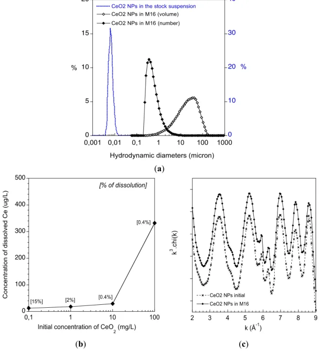
The colloidal stability of the CeO2 ENPs was studied by dynamic light scattering (DLS) after 2 h of
incubation in the abiotic M16 culture medium. While the ENPs are stable in their stock suspension, a significant aggregation occurs in M16 (Figure 1a). Aggregates with hydrodynamic diameters (volume distribution) centred at ~35 μm quickly formed. When expressed as a number distribution most of these aggregates have hydrodynamic diameters of approximately 350 nm. It is noteworthy that this number distribution is based on several assumptions (e.g., shape, density of the aggregates) but it highlights that most of the CeO2 ENPs interacted with the oocytes as small aggregates. The dissolution
of the CeO2 ENPs in the M16 was studied by ICP-MS (Figure 1b). After 2 h, less than 30 μg/L of
dissolved Ce was measured in the abiotic M16 media for an initial CeO2 concentration below 10 mg/L.
For 100 mg/L of initial CeO2 ENP concentration, 331 μg/L of dissolved Ce was measured in solution
(0.4% of the total Ce content). The release of Ce ions was low, typically <0.4% of the initial
concentrations. EXAFS (Extended X-ray Absorption Fine Structure) was used to study the crystal structure of the CeO2 ENPs (i.e., the number, nature and distances of atoms surrounding Ce from 0 to 5 Å)
after incubation in abiotic M16 (Figure 1c). The experimental spectra of CeO2 ENPs before and after
2 h of incubation in M16 perfectly superimpose, indicating that the atomic structure of the CeO2 ENPs
is not affected (Figure 1c). Such local-scale stability suggests that the ENPs surface interaction with macromolecules from the M16 (proteins) is not associated with major surface complexation or reduction of Ce4+ into Ce3+. Notably, EXAFS is not sensitive to minor Ce species (i.e., <10%). Therefore,
the detection of less than 0.4% Ce dissolution is not contradictory with the EXAFS main result concerning
CeO2 structure.
After 2 h of incubation with the abiotic M16 medium the CeO2 ENPs can be considered structurally
stable and have a slow release of dissolved Ce. However, the ENPs have a strong colloidal destabilisation,
Figure 1. (a) Aggregation stage of the CeO2 ENPs in the M16 medium. Distribution of the
hydrodynamic diameters of the CeO2 ENPs in their stock suspension and after 2 h in the
M16 medium (expressed as a volume or number distribution); (b) Chemical stability of the CeO2 ENPs in M16. Dissolution of CeO2 ENPs after 2 h in the abiotic M16 determined by
ICP-MS; (c) Structural stability of the CeO2 ENPs in M16. EXAFS at the Ce L3-edge of the
CeO2 ENPs before and after 2 h of incubation within the abiotic M16 medium.
(a) 2 3 4 5 6 7 8 9 CeO2 NPs initial CeO2 NPs in M16 k 3 .c hi (k ) k (Å-1) (b) (c)
2.2. Transmission Electron Microscopy (TEM) Study of CeO2 ENPs Internalisation
The TEM study showed the internalisation of CeO2 ENP aggregates by endocytosis in follicular
cells (Figure 2b,c). In oocytes surrounded by zona pellucida (ZP+) incubated with CeO2 ENPs, the
not surrounded by zona pellucida (ZP−), we did not observe any CeO2 ENPs in the oocyte
cytoplasm (Figure 2f).
Figure 2. Transmission electron microscopy image of follicular cells and oocytes exposed to CeO2 ENPs. (a) Unexposed follicular cells; (b) Follicular cells exposed to CeO2 NPs
(wide shot); (c) Follicular cells exposed to CeO2 ENPs (close-up); (d) Unexposed oocyte
surrounded by zona pellucida; (e) Oocyte surrounded by zona pellucida (ZP+) exposed to CeO2 ENPs; (f) Oocyte not surrounded by zona pellucida (ZP−) exposed to CeO2 ENPs.
2.3. CeO2 ENPs Induced DNA Damage in Follicular Cells
Although several genetic toxicology tests have been validated for chemicals according to the Organisation for Economic Co-operation and Development (OECD) test guidelines, the relevance of these assays for nanoparticulate materials remains to be determined. To address this issue, the OECD has established current projects designed to evaluate the relevance and reproducibility of safety hazard tests for representative nanomaterials, including genotoxicity assays [17]. No reference gene mutation test has been described for oocytes. The genotoxicity of CeO2 ENPs on mouse oocytes and on follicular
cells was assessed with the protocol previously described in our research laboratory [46]. A comet assay was used in this study because it is very sensitive and allows the detection of DNA double and single-strand breaks in individual eukaryotic cells. The principle underlying the comet assay is that denatured DNA fragments can be measured migrating out of the cell nucleus during electrophoresis [47]. The image obtained is a “comet” with a distinct head consisting of intact DNA and a tail containing relaxed DNA
loops or broken pieces of DNA [48]. The comet assay quantifies DNA damage with the Olive Tail Moment (OTM = % DNA in the tail × length of the comet tail). At least 350 follicular cells were studied for each condition using the comet assay. Significant dose-dependent DNA damage was observed in follicular cells exposed to 2, 5, 10 and 100 mg/L of CeO2 ENPs with OTM, respectively at 5.9 ± 0.4;
8.1 ± 0.6; 10.1 ± 0.6 and 12.9 ± 0.7 vs. 2.3 ± 1.6 for the negative control group and 22.2 ± 1.6 for the positive control group (Figure 3).
Figure 3. Genotoxicity assessment of CeO2 ENPs on follicular cells by comet assay. * p ≤ 0.05.
2.4. DNA Damage Assay in Oocytes
In ZP+ oocytes, we did not observe significant DNA damage at 2 mg/L (OTM = 4.8 ± 0.8) and at 5 mg/L (OTM = 5.1 ± 1) compared to 2.4 ± 0.4 for the negative control group and 27.8 ± 3.8 for the positive control group (Figure 4a). The two highest exposure concentrations induced a significant increase of OTM values: 9.1 ± 0.74 at 10 mg/L and OTM = 12.7 ± 1.1 at 100 mg/L. In ZP− oocytes, we observed a significant increase of DNA damage after CeO2 ENPs exposure, with OTM,
respectively, at 14.4 ± 2.1, 14.5 ± 2.3, 13.3 ± 2.2 and 14.4 ± 2.7 for 2, 5, 10 and 100 mg/L vs. 6.7 ± 1.3 for the negative control group and 22.8 ± 2.2 for the positive control group (Figure 4b).
Figure 4. Genotoxicity assessment of CeO2 ENPs on mouse oocytes by comet assay
(a) Oocytes surrounded by zona pellucida (ZP+); (b) Oocytes not surrounded by zona pellucida (ZP−). * p ≤ 0.05.
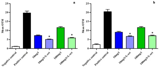
2.5. CeO2 ENPs and Anti-Oxidant
When L-ergothioneine was added in culture medium before incubation of ZP+ oocytes and
follicular cells with CeO2 ENPs, DNA damage decreased significantly in both cell types. At 10 mg/L
and 100 mg/L, OTM in the groups treated with L-ergothioneine were 5.1 ± 0.2 vs. 7.2 ± 0.3,
respectively, in follicular cells with L-ergothioneine and 5.9 ± 0.3 vs. 11.6 ± 0.5 without L-ergothioneine (Figure 5a). In ZP+ oocytes, OTM were respectively 2.8 ± 0.5 vs. 6.7 ± 0.3 with L-ergothioneine (p < 0.05) and 7.1 ± 0.3 vs. 11.6 ± 0.5 without L-ergothioneine (p < 0.05) (Figure 5b). The
OTM in negative and positive control groups were respectively 1.1 ± 0.1 and 19.7 ± 1.1 in follicular cells and 2.2 ± 0.3 and 20.4 ± 1.3 in ZP+ oocytes.
a
Figure 5. Reduction of DNA damage induced by CeO2 ENPs using the comet assay after
adding an anti-oxidant agent (L-ert = L-ergothioneine) in culture media. (a) Follicular cells; (b) Mouse ZP+ oocytes. * p ≤ 0.05.
2.6. Discussion
To our knowledge, this is the first study focused on the genotoxicity of CeO2 ENPs in oocytes. The
genotoxicity of CeO2 ENPs depends on the physico-chemical properties of the cell’s environment
because it determines aggregation, redox modifications, and surface adsorption [49]. To safely use ENPs future research on the properties of ENPs must focus on biochemical and physical interactions between ENPs and the environment. In the conclusions of a working group from the International Life Sciences Institute Research Foundation/Risk Science Institute Nanomaterial Toxicity Screening Working Group, Oberdorster et al. suggested a toxicity assay of ENPs evaluating their potential effects on human health should involve a multidisciplinary approach including physico-chemical characterisation as well as in vitro and in vivo assays [16]. For the in vitro genotoxicity assay, we used the comet assay, which is a widely used test in toxicology and well-adapted to mouse oocytes [46]. Given the new and unique physico-chemical properties of ENPs, standardised genotoxicity assays must be adapted to respond to risk assessment questions. The dose-response relationship is a function of the physico-chemical behaviour and surface reactivity of different classes of ENPs [16].
2.6.1. Are Oocytes Protected from CeO2 ENP Induced Oxidative Stress and DNA Damage by
Follicular Cell Endocytosis and Zona Pellucida Trapping?
Our study demonstrated that the intracellular delivery of CeO2 ENPs into follicular cells was
possible by endosomal trapping. Endosomal trapping is a mechanism developed by cells to protect themselves from foreign organisms [50]. A combination of DLS and TEM analyses showed that despite aggregation in the exposure media, CeO2 ENPs were internalised by follicular cells. The
kinetics of intracellular uptake most likely depends on the size of ENP aggregates, as shown by Rejman et al. [51]. While the internalisation of ENPs can be useful in biomedical applications [52], we hypothesised it could induce the oxidative stress observed in follicular cells in our experimental conditions. This oxidative stress was indirectly demonstrated by the significant decrease of DNA damage in follicular cells after the addition of L-ergothioneine in the exposure medium.
Regarding the oocytes, TEM images did not show any intracellular delivery into oocytes. However, CeO2 ENPs were observed trapped on the ZP surface outside the oocyte membrane. The ZP could act
as a mechanical barrier excluding CeO2 ENPs, which decreases their direct harmful effects or decreases
the indirect effects of Ce3+ ions released from the ENPs in the M16 culture medium. Following oocyte
exposure to 2 and 5 mg/L, we did not observe significant DNA damage in ZP+ oocytes. Conversely, DNA damage was significantly increased in ZP− oocytes. At high ENP concentrations, DNA damage was observed in oocytes both with and without zona pellucida. This result suggests that the defence system preventing DNA damage and oxidative stress was overwhelmed. This hypothesis requires further evaluation by in vivo studies.
The DNA damage was dose-dependent in follicular cells and in oocytes surrounded by zona pellucida. However, no dose-response relationship was observed in oocytes lacking ZP. The OTM values obtained at the lowest CeO2 ENP concentrations tested in ZP− oocytes were similar to the
highest dose-related effect of CeO2 ENPs tested in ZP+ oocytes. Thus, the dose-related effect of CeO2
ENPs observed with follicular cells and ZP+ oocytes was not present in ZP− oocytes. This result may be because DNA damage quantified by the comet assay was maximal at the lowest tested concentration in ZP− oocytes.
2.6.2. Mechanisms of Oxidative Stress Induced by CeO2 ENPs
In ZP+ oocytes, exposure to CeO2 ENPs statistically increased DNA damage only at high
concentrations (10 and 100 mg/L), while in ZP− oocytes DNA damage was observed at all the tested concentrations. We observed that the DNA damage significantly decreased in ZP+ oocytes after addition of an anti-oxidant agent. Consequently, we hypothesised that CeO2 ENPs induced oxidative
stress in oocytes and that ZP may protects oocytes against oxidative stress at low concentrations of CeO2 ENPs.
Several biophysico-chemical reactions occurring at the ENP/biological interface are able to influence the surface properties of ENPs and consequently their biological effects [53]. Changes of the redox state of the ENPs, their dissolution, the adsorption of organic matter, and the aggregation state (size and density) can influence the exposure and toxicological impacts of ENPs. Dynamic interactions between ENPs and cells, membranes, DNA, and intracellular organelles can lead to favourable or adverse biological effects according to ENP properties and biotransformation [54]. For instance, Asati et al. showed that the internalisation profile in normal and cancer cell lines and the cytotoxicity potential depended on the CeO2 ENPs’ surface charge [22]. Zeyons et al., (2009) have shown that CeO2
ENPs induced cytotoxicity on Escherichia coli via a direct mechanism of bio-reduction requiring a close contact between ENPs and the cell membranes [55]. Additionally, the authors found that CeO2
ENPs induced indirect cytotoxicity on Synechocystis by extracellular polymeric substances preventing direct cellular contacts with the ENPs in addition to the acidity of the ENP stabilising agent.
Indirect biological effects were observed in our experimental conditions. Our results suggest that when mature oocytes are exposed to low concentrations of CeO2 ENPs, follicular cell endocytosis and
zona pellucida trapping protected oocytes by counteracting oxidative stress that prevents DNA damage in oocytes. However, we observed DNA damage at high concentrations of CeO2 ENPs. The DNA
ions released from the ENPs in the M16 culture medium. Based on our results, it is likely that the follicular cells and zona pellucida may prevent direct contact between the CeO2 ENPs and the oocytes.
The Ce3+ ions could diffuse through the zona pellucida to indirectly stress the cells. These findings
demonstrate the importance of studying the physico-chemical behaviour and biotransformation of ENPs to thoroughly understand the mechanisms of genotoxicity towards germinal cell lines.
3. Experimental Section
3.1. Chemical Agents
All chemicals were from Sigma (St. Quentin–Fallavier, France) unless stated otherwise.
3.2. CeO2 ENPs Physico-Chemical Characterisation in Culture Medium
The CeO2 ENPs (Rhodia®, Courbevoie, France) used in this study are pseudo-spherical crystallites
of cerianite with an average size of 3 nm (total number of clusters measured: 70). These ENPs are uncoated and are dispersed in pure water with an average hydrodynamic diameter of ~8 nm (Figure 1a). The size, shape and mineralogy were characterised by Transmission Electron Microscopy (TEM) (using a JEOL 2010F at 200 kV). Dynamic light scattering (DLS) (using a nano ZS and a Mastersizer S, Malvern Instruments SA, Orsay, France) was used to determine their aggregation states before and after 2 h of incubation in the M16 media. The dissolution of the CeO2 ENPs in the M16 medium was
assessed using ICP-MS. Briefly, the CeO2 ENPs were incubated for 2 h in the abiotic M16. After
incubation, the suspension was ultra-centrifuged (200.000 g for 1 h) and the supernatant was analysed by ICP-MS. The solid phase of the centrifugation was freeze-dried and analysed by X-ray absorption spectroscopy (XAS) for structural characterisation. XAS at the Ce L3-edge (5723 eV) was performed on the XAFS 11.1 beamline [56] at the ELETTRA synchrotron (Trieste, Italy). Samples were diluted in boron nitride, pressed to thin pellets, and analysed in transmission mode. The spectra were compiled from the merge of three scans, and the energy was calibrated using a CeO2 standard reference. EXAFS
(Extended X-ray Absorption Fine Structure) data were obtained after performing standard procedures for pre-edge subtraction, normalisation, polynomial removal, and wave vector conversion using the IFEFFIT software package [57].
3.3. Animals
Prepubescent 4-week old female mice CD1 (Charles River Laboratory, L’Arbresle, France) were housed in a temperature and light controlled room with free access to food and water. Institutional Review Board approval n° 12-18042012 was obtained after submission of the experimental protocol and animal handling procedures to the National Ethics Committee on Animal Experimentation.
3.4. Oocytes Isolation
Mice were injected intraperitoneally with 0.1 mL of 10 IU Pregnant Mare Serum Gonadotropin. Three days later, they received an additional injection with 0.1 mL of 5 IU of Human Chorionic Gonadotropin. Sixteen hours later, mice were sacrificed by cervical dislocation [58] and oviducts were
collected. Intact cumulus masses were released from excised oviducts, and decoronisation was performed after incubation with hyaluronidase (10 mg/mL). Digestion of zona pellucida needed for TEM evaluation and genotoxicity assays was performed with acidic Tyrode’s solution. The oocytes were then washed three times in M2 medium. Approximately 25–30 mature oocytes were released for each mouse. Each experimental condition included at least 40 mature oocytes.
3.5. Transmission Electron Microscopy (TEM)
Follicular cells and oocytes with or without the zona pellucida (ZP) were incubated for 2 h in vitro with CeO2 ENPs (100 mg/L) and the potential internalisation was studied by TEM (transmission
electronic microscopy). After incubation, follicular cells and oocytes were washed twice in cacodylate buffer (0.1 M, pH = 7.4) fixed in 2.5% glutaraldehyde and post-fixed in osmium tetroxide 2% in the same buffer. After centrifugation, the pellets were dehydrated in graded alcohol solutions and embedded in Embed-812 kit using a standard procedure. Ultrathin sections (60–70 nm) were counterstained with uranyl acetate and lead citrate before observation with a JEOL/JEM 1400 electron microscope at 80 kV.
3.6. Exposure Conditions for Genotoxicity Assay
We used the comet assay on mature mouse oocytes as described in a previous work [46]. Oocytes (n = 40 for each group) and follicular cells (at least 100 for each group) were exposed to 4 concentrations of CeO2 ENPs (2, 5, 10 and 100 mg/L) in the M16 medium at 37 °C and 5% CO2 during 2 h. Two
groups of oocytes were studied with (ZP+) or without the zona pellucida (ZP−). Negative control oocytes and follicular cell groups were incubated for 2 h in the M16 medium. For the positive control group, oocytes were incubated at 37 °C with 5% CO2 in M16 medium for 2 h. The cells were placed at
the end of incubation in a 250 µM hydrogen peroxide (H2O2) solution for 5 min at 4 °C in the dark.
Each condition was replicated three times.
3.7. Incubation of Oocytes with an Anti-Oxidant
For the conditions that induced DNA damage for the comet assay each experiment was repeated with an anti-oxidant added in the M16 medium. For the conditions with 10 mg/L and 100 mg/L of CeO2
ENPs, L-ergothioneine (5 mM) was added to the culture medium with follicular cells and ZP+ oocytes.
L-ergothioneine has a well-known anti-oxidant activity that can scavenge hydroxyl radicals and inhibit
the generation of hydroxyl radicals from hydrogen peroxide [59]. These two incubation conditions were compared with 2 groups (10 mg/L and 100 mg/L of CeO2 ENPs) without L-ergothioneine and to
the same negative and positive control groups previously described. Each condition was repeated three times with at least 100 follicular cells and 40 matures oocytes were analysed by the comet assay.
3.8. Main Outcome Measures and Statistical Analysis of the Comet Assay
For each tested condition, all the oocyte and follicular cell images were analysed by the validated Komet software (version 6.0; Andor. Bioimaging, Nottingham, UK). DNA damage was expressed as Olive Tail Moment (OTM, arbitrary units), which is the association of tail length and DNA% contained in tail [48]. The results were expressed as the mean values ± SEM and analysed by ANOVA followed
by Fisher LSD post-hoc test using Statview® 5.1 for Windows (Abacus Concepts, Berkeley, CA, USA). The statistical significance was set at p < 0.05.
4. Conclusions
Our study was a first examining the mechanisms of interactions between CeO2 ENPs and germ
cells. Our results cannot be extrapolated to in vivo function of CeO2 ENPs by inhalation, but offer a
tool to understand the mechanisms of possible interactions between CeO2 ENPs and female germ cells.
Our results suggested that when mature oocytes are exposed to low concentrations of CeO2 ENPs,
follicular cell endocytosis and zona pellucida trapping could protect oocytes by decreasing oxidative stress and DNA damage. At low concentrations (more expected in environmental exposure), these preliminary data seem to us reassuring as for the possible impact of an environmental exposure of CeO2 ENPs on mature oocytes. However, at high concentrations this defence system may be insufficient
to prevent induction of oxidative stress leading to DNA damage. Moreover, oocytes exposed at earlier stages of maturation, with no or immature ZP and fewer follicular cells, could be more vulnerable to DNA damage induced by CeO2 ENPs. Given the impact demonstrated by the CeO2 ENPs on the
reproduction of the aquatic organisms [23,24] and the hypothesis of a possible bioaccumulation in ovary [33], we plan to study interactions between CeO2 ENPs and oocytes. In vitro studies will help us to
determine the mechanisms of ENP interactions under controlled conditions that represent in vivo situations. Acknowledgments
We thank Joel Courageot for expert technical assistance in the preparation of TEM samples and Erika Lopez, DMV, for care and counselling concerning laboratory mice (Centre de Formation et de Recherches Experimentales Medico-Chirurgicales, Aix-Marseille University).
Conflicts of Interest
The authors declare no conflict of interest. References
1. Bottero, J.Y.; Rose, J.; Wiesner, M.R. Nanotechnologies: Tools for sustainability in a new wave of water treatment processes. Integr. Environ. Assess. Manag. 2006, 2, 391–395.
2. Ko, S.H.; Park, I.; Pan, H.; Grigoropoulos, C.P.; Pisano, A.P.; Luscombe, C.K.; Frechet, J.M.J. Direct nanoimprinting of metal nanoparticles for nanoscale electronics fabrication. Nano Lett. 2007, 7, 1869–1877.
3. Som, C.; Wick, P.; Krug, H.; Nowack, B. Environmental and health effects of nanomaterials in nanotextiles and facade coatings. Environ. Int. 2011, 37, 1131–1142.
4. Botta, C.; Labille, J.; Auffan, M.; Borschneck, D.; Miche, H.; Cabie, M.; Masion, A.; Rose, J.; Bottero, J.Y. TiO2-based nanoparticles released in water from commercialized sunscreens in a
life-cycle perspective: structures and quantities. Environ. Pollut. 2011, 159, 1543–1550.
5. Gupta, A.K.; Gupta, M. Synthesis and surface engineering of iron oxide nanoparticles for biomedical applications. Biomaterials 2005, 26, 3995–4021.
6. Lammers, T.; Hennink, W.E.; Storm, G. Tumour-targeted nanomedicines: Principles and practice.
Br. J. Cancer 2008, 99, 392–397.
7. Auffan, M.; Bottero, J.Y.; Chaneac, C.; Rose, J. Inorganic manufactured nanoparticles: How their physicochemical properties influence their biological effects in aqueous environments.
Nanomedicine 2010, 5, 999–1007.
8. Avis de l’Afsset; Rapport d’Expertise Collective. Évaluation des risques liés aux nanomatériaux pour la population générale et pour l’environnement. La Doc. Fr. 2010, 3, 1–207. Available online: http://www.ladocumentationfrancaise.fr/var/storage/rapports-publics/104000168/0000.pdf (accessed on 25 October 2013).
9. Cassee, F.R.; van Balen, E.C.; Singh, C.; Green, D.; Muijser, H.; Weinstein, J.; Dreher, K. Exposure, health and ecological effects review of engineered nanoscale cerium and cerium oxide associated with its use as a fuel additive. Crit. Rev. Toxicol. 2011, 41, 213–229.
10. Blanchard, V.; Blanchet, P. Color stability for wood products during use: Effects of inorganic nanoparticles. BioResources 2011, 6, 1219–1229.
11. Niu, J.; Azfer, A.; Rogers, L.M.; Wang, X.; Kolattukudy, P.E. Cardioprotective effects of cerium oxide nanoparticles in a transgenic murine model of cardiomyopathy. Cardiovasc. Res. 2007, 73, 549–559.
12. Tarnuzzer, R.W.; Colon, J.; Patil, S.; Seal, S. Vacancy engineered ceria nanostructures for protection from radiation-induced cellular damage. Nano Lett. 2005, 5, 2573–2577.
13. Park, B.; Donaldson, K.; Duffin, R.; Tran, L.; Kelly, F.; Mudway, I.; Morin, J.P.; Guest, R.; Jenkinson, P.; Samaras, Z.; et al. Hazard and risk assessment of a nanoparticulate cerium oxide-based diesel fuel additive—A case study. Inhal. Toxicol. 2008, 20, 547–566.
14. Oberdorster, G. Safety assessment for nanotechnology and nanomedicine: Concepts of nanotoxicology. J. Intern. Med. 2010, 267, 89–105.
15. Hansen, S.F.; Baun, A. European regulation affecting nanomaterials—Review of limitations and future recommendations. Dose Response 2012, 10, 364–383.
16. Oberdorster, G.; Maynard, A.; Donaldson, K.; Castranova, V.; Fitzpatrick, J.; Ausman, K.; Carter, J.; Karn, B.; Kreyling, W.; Lai, D.; et al. Principles for characterizing the potential human health effects from exposure to nanomaterials: Elements of a screening strategy. Part. Fibre Toxicol. 2005, 2, 8.
17. Warheit, D.B.; Donner, E.M. Rationale of genotoxicity testing of nanomaterials: Regulatory requirements and appropriateness of available OECD test guidelines. Nanotoxicology 2010, 4, 409–413.
18. Celardo, I.; Pedersen, J.Z.; Traversa, E.; Ghibelli, L. Pharmacological potential of cerium oxide nanoparticles. Nanoscale 2011, 3, 1411–1420.
19. Heckert, E.G.; Karakoti, A.S.; Seal, S.; Self, W.T. The role of cerium redox state in the SOD mimetic activity of nanoceria. Biomaterials 2008, 29, 2705–2709.
20. Hirst, S.M.; Karakoti, A.; Singh, S.; Self, W.; Tyler, R.; Seal, S.; Reilly, C.M. Bio-distribution and in vivo antioxidant effects of cerium oxide nanoparticles in mice. Environ. Toxicol. 2013, 28, 107–118.
21. Celardo, I.; Traversa, E.; Ghibelli, L. Cerium oxide nanoparticles: A promise for applications in therapy. J. Exp. Ther. Oncol. 2011, 9, 47–51.
22. Asati, A.; Santra, S.; Kaittanis, C.; Perez, J.M. Surface-charge-dependent cell localization and cytotoxicity of cerium oxide nanoparticles. ACS Nano 2010, 4, 5321–5331.
23. Rodea-Palomares, I.; Boltes, K.; Fernandez-Pinas, F.; Leganes, F.; Garcia-Calvo, E.; Santiago, J.; Rosal, R. Physicochemical characterization and ecotoxicological assessment of CeO2 nanoparticles
using two aquatic microorganisms. Toxicol. Sci. 2011, 119, 135–145.
24. Van Hoecke, K.; de Schamphelaere, K.A.C.; van der Meeren, P.; Smagghe, G.; Janssen, C.R. Aggregation and ecotoxicity of CeO2 nanoparticles in synthetic and natural waters with variable
pH, organic matter concentration and ionic strength. Environ. Pollut. 2011, 159, 970–976.
25. Shaw, B.J.; Handy, R.D. Physiological effects of nanoparticles on fish: A comparison of nanometals vs. metal ions. Environ. Int. 2011, 37, 1083–1097.
26. Zhang, X.; Sun, H.; Zhang, Z.; Niu, Q.; Chen, Y.; Crittenden, J.C. Enhanced bioaccumulation of cadmium in carp in the presence of titanium dioxide nanoparticles. Chemosphere 2007, 67, 160–166.
27. Pelletier, D.A.; Suresh, A.K.; Holton, G.A.; McKeown, C.K.; Wang, W.; Gu, B.; Mortensen, N.P.; Allison, D.P.; Joy, D.C.; Allison, M.R.; et al. Effects of engineered cerium oxide nanoparticles on bacterial growth and viability. Appl. Environ. Microbiol. 2010, 76, 7981–7989.
28. Li, J.J.; Muralikrishnan, S.; Ng, C.T.; Yung, L.Y.; Bay, B.H. Nanoparticle-induced pulmonary toxicity. Exp. Biol. Med. 2010, 235, 1025–1033.
29. Smita, S.; Gupta, S.K.; Bartonova, A.; Dusinska, M.; Gutleb, A.C.; Rahman, Q. Nanoparticles in the environment: Assessment using the causal diagram approach. Environ. Heal. 2012, 11, S13. 30. Auffan, M.; Rose, J.; Orsiere, T.; Demeo, M.; Thill, A.; Zeyons, O.; Proux, O.; Masion, A.;
Chaurand, P.; Spalla, O.; et al. CeO2 nanoparticles induce DNA damage towards human dermal
fibroblasts in vitro. Nanotoxicology 2009, 3, 161–171.
31. Priester, J.H.; Ge, Y.; Mielke, R.E.; Horst, A.M.; Moritz, S.C.; Espinosa, K.; Gelb, J.; Walker, S.L.; Nisbet, R.M.; An, Y.J.; et al. Soybean susceptibility to manufactured nanomaterials with evidence for food quality and soil fertility interruption. Proc. Natl. Acad. Sci. USA 2012, 109, E2451–E2456.
32. Becker, H.; Herzberg, F.; Schulte, A.; Kolossa-Gehring, M. The carcinogenic potential of nanomaterials, their release from products and options for regulating them. Int. J. Hyg. Environ.
Health 2011, 214, 231–238.
33. Al-Saleh, I.; Shinwari, N.; Al-Amodi, M. Accumulation of mercury in ovaries of mice after the application of skin-lightening creams. Biol. Trace Elem. Res. 2009, 131, 43–54.
34. Morishita, Y.; Yoshioka, Y.; Satoh, H.; Nojiri, N.; Nagano, K.; Abe, Y.; Kamada, H.; Tsunoda, S.; Nabeshi, H.; Yoshikawa, T.; et al. Distribution and histologic effects of intravenously administered amorphous nanosilica particles in the testes of mice. Biochem. Biophys. Res. Commun. 2012, 420, 297–301.
35. Klein, J.P.; Boudard, D.; Cadusseau, J.; Palle, S.; Forest, V.; Pourchez, J.; Cottier, M. Testicular biodistribution of 450 nm fluorescent latex particles after intramuscular injection in mice. Biomed. Microdevices 2013, 15, 427–436.
36. Lim, D.; Roh, J.; Eom, H.; Choi, J.Y.; Hyun, J.; Choi, J. Oxidative stress-related PMK-1 P38 MAPK activation as a mechanism for toxicity of silver nanoparticles to reproduction in the nematode Caenorhabditis elegans. Environ. Toxicol. Chem. 2012, 31, 585–592.
37. Musee, N.; Oberholster, P.J.; Sikhwivhilu, L.; Botha, A.M. The effects of engineered nanoparticles on survival, reproduction, and behaviour of freshwater snail, Physa acuta (Draparnaud, 1805).
Chemosphere 2010, 81, 1196–1203.
38. Philbrook, N.A.; Winn, L.M.; Afrooz, A.R.; Saleh, N.B.; Walker, V.K. The effect of TiO2 and Ag
nanoparticles on reproduction and development of Drosophila melanogaster and CD-1 mice.
Toxicol. Appl. Pharmacol. 2011, 257, 429–436.
39. Ramsden, C.S.; Henry, T.B.; Handy, R.D. Sub-lethal effects of titanium dioxide nanoparticles on the physiology and reproduction of zebrafish. Aquat. Toxicol. 2013, 126, 404–413.
40. Schlich, K.; Klawonn, T.; Terytze, K.; Hund-Rinke, K. Effects of silver nanoparticles and silver nitrate in the earthworm reproduction test. Environ. Toxicol. Chem. 2013, 32, 181–188.
41. Wang, J.; Zhu, X.; Zhang, X.; Zhao, Z.; Liu, H.; George, R.; Wilson-Rawls, J.; Chang, Y.; Chen, Y. Disruption of zebrafish (Danio rerio) reproduction upon chronic exposure to TiO2
nanoparticles. Chemosphere 2011, 83, 461–467.
42. Braydich-Stolle, L.; Hussain, S.; Schlager, J.J.; Hofmann, M.C. In vitro cytotoxicity of nanoparticles in mammalian germline stem cells. Toxicol. Sci. 2005, 88, 412–419.
43. Braydich-Stolle, L.K.; Lucas, B.; Schrand, A.; Murdock, R.C.; Lee, T.; Schlager, J.J.; Hussain, S.M.; Hofmann, M.C. Silver nanoparticles disrupt GDNF/Fyn kinase signaling in spermatogonial stem cells. Toxicol. Sci. 2010, 116, 577–589.
44. Wiwanitkit, V.; Sereemaspun, A.; Rojanathanes, R. Effect of gold nanoparticles on spermatozoa: The first world report. Fertil. Steril. 2009, 91, e7–e8.
45. Chaudhury, K.; Babu, N.K.; Singh, A.K.; Das, S.; Kumar, A.; Seal, S. Mitigation of endometriosis using regenerative cerium oxide nanoparticles. Nanomed. Nanotechnol. Biol. Med. 2013, 9, 439–448.
46. Berthelot-Ricou, A.; Perrin, J.; di Giorgio, C.; de Meo, M.; Botta, A.; Courbiere, B. Comet assay on mouse oocytes: An improved technique to evaluate genotoxic risk on female germ cells.
Fertil. Steril. 2011, 95, 1452–1457.
47. Tice, R.R.; Agurell, E.; Anderson, D.; Burlinson, B.; Hartmann, A.; Kobayashi, H.; Miyamae, Y.; Rojas, E.; Ryu, J.C.; Sasaki, Y.F. Single cell gel/comet assay: Guidelines for in vitro and in vivo genetic toxicology testing. Environ. Mol. Mutagen. 2000, 35, 206–221.
48. Singh, N.P.; McCoy, M.T.; Tice, R.R.; Schneider, E.L. A simple technique for quantitation of low levels of DNA damage in individual cells. Exp. Cell Res. 1988, 175, 184–191.
49. Browning, L.M.; Lee, K.J.; Huang, T.; Nallathamby, P.D.; Lowman, J.E.; Xu, X.-H.N. Random walk of single gold nanoparticles in zebrafish embryos leading to stochastic toxic effects on embryonic developments. Nanoscale 2009, 1, 138–152.
50. Levy, R.; Shaheen, U.; Cesbron, Y.; See, V. Gold nanoparticles delivery in mammalian live cells: A critical review. Nano Rev. 2010, 1, doi:10.3402/nano.v1i0.4889.
51. Rejman, J.; Oberle, V.; Zuhorn, I.S.; Hoekstra, D. Size-dependent internalization of particles via the pathways of clathrin- and caveolae-mediated endocytosis. Biochem. J. 2004, 377, 159–169. 52. Faraji, A.H.; Wipf, P. Nanoparticles in cellular drug delivery. Bioorg. Med. Chem. 2009, 17,
53. Yoshida, T.; Yoshioka, Y.; Matsuyama, K.; Nakazato, Y.; Tochigi, S.; Hirai, T.; Kondoh, S.; Nagano, K.; Abe, Y.; Kamada, H.; et al. Surface modification of amorphous nanosilica particles suppresses nanosilica-induced cytotoxicity, ROS generation, and DNA damage in various mammalian cells. Biochem. Biophys. Res. Commun. 2012, 427, 748–752.
54. Nel, A.E.; Madler, L.; Velegol, D.; Xia, T.; Hoek, E.M.V.; Somasundaran, P.; Klaessig, F.; Castranova, V.; Thompson, M. Understanding biophysicochemical interactions at the nano-bio interface. Nat. Mater. 2009, 8, 543–557.
55. Zeyons, O.; Thill, A.; Chauvat, F.; Menguy, N.; Cassier-Chauvat, C.; Orear, C.; Daraspe, J.; Auffan, M.; Rose, J.; Spalla, O. Direct and Indirect CeO2 Nanoparticles Toxicity for Escherichia
coli and Synechocystis. Available online: http://www.informahealthcare.com/doi/abs/10.3109/ 17435390903305260 (accessed on 21 August 2013).
56. Cicco, A.D.; Aquilanti, G.; Minicucci, M.; Principi, E.; Novello, N.; Cognigni, A.; Olivi, L. Novel XAFS capabilities at ELETTRA synchrotron light source. J. Phys. 2009, 190, 012043. 57. Ravel, B.; Newville, M. ATHENA, ARTEMIS, HEPHAESTUS: Data analysis for X-ray absorption
spectroscopy using IFEFFIT. J. Synchrotron Radiat. 2005, 12, 537–541.
58. Roustan, A.; Perrin, J.; Berthelot-Ricou, A.; Lopez, E.; Botta, A.; Courbiere, B. Evaluating methods of mouse euthanasia on the oocyte quality: Cervical dislocation vs. isoflurane inhalation.
Lab. Anim. 2012, 46, 167–169.
59. Akanmu, D.; Cecchini, R.; Aruoma, O.I.; Halliwell, B. The antioxidant action of ergothioneine.
Arch. Biochem. Biophys. 1991, 288, 10–16.
© 2013 by the authors; licensee MDPI, Basel, Switzerland. This article is an open access article distributed under the terms and conditions of the Creative Commons Attribution license (http://creativecommons.org/Licenses/by/3.0/).