OCIMUM
Research Article
Anti-inflammatory and Antioxidant Effect of a
ᴅ-galactose-rich Polysaccharide Extracted from
Aloe vera Leaves
Khalef Lefsih1, Liticia Iboukhoulef2, Emmanuel
Petit3, Hana Benouatas2, Guillaume Pierre4* and
Cédric Delattre4
1Laboratoire de Biomathématiques, Université de
Bejaia, Algeria
2Laboratoire National de Contrôle des Produits
Pharmaceutiques, Institut Pasteur, Alger, Algeria
3EA3900 BIOPI Université de Picardie Jules Verne,
France
4Université Clermont Auvergne, CNRS, SIGMA
Clermont, Institut Pascal, F-63000 Clermont-Ferrand, France
Received Date: 11 October, 2018 Accepted Date: 19 November, 2018 Published Date: 28 November, 2018
Citation
Lefsih K, Iboukhoulef L, Petit E, Benouatas H, Pierre G, et al. (2018) Anti-inflammatory and Antioxidant Effect of a ᴅ-galactose-rich Polysaccharide Extracted from Aloe vera Leaves. Adv Appl Chem Biochem 2018(1): 18-26.
Correspondence should be addressed to Guillaume Pierre, France
E-mail: guillaume.pierre@uca.fr
Copyright
Copyright © 2018 Guillaume Pierre et al. This is an open access article distributed under the Creative Commons Attribution License which permits unrestricted use, distribution, and reproduction in any medium, provided the original author and work is properly cited.
Abstract
This work deals with studying the structure of a galactan extracted from Aloe vera Barbadensis Miller leaves harvested in Algeria (AVP). AVP was characterized by FTIR spectroscopy to identify functional groups confirming its polysaccharidic nature and analyzed by High-Pressure Anion Exchange Chromatography (HPAEC) to determine its composition as constituent monosaccharides. AVP was essentially composed of ᴅ-galactose (75.25%), ᴅ-glucuronic acid (10.82%), ʟ-rhamnose (6.52%), ʟ-arabinose (4.95%), ᴅ-galacturonic acid (2.16%) and ᴅ-mannose (0.28%). The total carbohydrate content (96.84%) was determined by a phenol-sulfuric acid colorimetric assay and the protein content (2.85%) was measured by the Bradford method. The analysis of anti-inflammatory activity is based on the model of xylene-induced ear edema. The percentage reduction of ear edema obtained during the analysis of the anti-inflammatory activity of the AVP at doses 1%, 5% and 10% (m/v) are respectively 7.92%; 43.30% and 71.49%. The anti-inflammatory activity of AVP was found very close to that of pure Aloe vera Gel (AVG) used in Algerian medicine.
Keywords
Aloe vera; Anti-Inflammatory Activity; Antioxidant Activity; Galactan; Pectin
Introduction
Through the ages, A. vera has been venerated by many civilizations
and cultures. So much so that it acquired the name of divine plant and
symbolized “beauty, health and well-being”. A. vera was already largely
part of the Chinese pharmacopoeia 4000 years BC and nicknamed
“the plant of harmony” thanks to its effects, e.g. wound dressing,
treatment of burns, skin condition controller, on all the body. Later,
around 700-800, Chinese people used it to treat sinusitis, fever and
convulsions in children, stomach aches but also for the treatment of
urticaria and other skin diseases. The current Chinese medicine uses
its pulp for the treatment of arteriosclerosis [1]. Phyto-polysaccharides
are a class of biomacromolecules having some biological properties
that can be valued at different scales. Extracted mainly from plants, the
water-soluble polymers establish specific interactions with water and
can thicken, stabilize or gel a solution, even at low concentrations. The
interest of polysaccharides is not limited to their rheological properties
since they often show interesting pharmaceutical and dermo-cosmetic
10.33513/ACBC/1801-03
activity [2]. Aloe leaf can be separated into two parts containing the vascular bundles and the aloe gel. Gel or mucilage correspond to the solution within the parenchyma cells whereas pulp or parenchyma tissue are associated to parenchyma tissue [3]. In general, glucomannans have been reported as the main polysaccharides in Aloe pulp [4]. Other polysaccharides have also been highlighted such as galactan, arabinan, pectin or pectin-like, arabinogalactan or arabinorhamonogalactan for the last two decades [5-7]. Galactan and arabinogalactan structures are often described as the neutral part of pectins and can be extracted from cell wall and degenerated cellular organelle [8]. Pectins are a complex class of polysaccharides involved in the structure of plant cell walls. Their main component is a central linear backbone chain made of α-ᴅ-(1,4)-galacturonic acid residues. Usually, some neutrals sugars are often reported such as ᴅ-galactose, ᴅ-xylose, ʟ-arabinose, ʟ-rhamnose of ᴅ-glucose, up to 10% (wt). The degree of esterification as well as the molecular weight distribution and length of GalA blocks greatly contribute to the multiple functional properties of the different classes of pectins [9]. Pectins are widely used in Food (E440) and authors evaluate that an average daily intake of 5 grams of pectin is consumed in a western diet [10]. The annual world consumption of pectins is estimated at 45 Mkg with a market around 400 M€. The benefits of natural pectin are also more and more appreciated by scientists and consumer due to its biodegradability, biocompatibility and bioavailability [11]. Note that pectins are widely used in other fields, from drug delivery system to membrane devices and tissue engineering [12]. This work focused on the extraction, chemical characterization and assessing bioactivity (antioxidant and anti-inflammatory) of pectin from A. vera Barbadensis Miller leaves harvested in Algeria.
Materials and Methods
Vegetal material
The aerial part (leaves) of A. vera Barbadensis Miller plant, belonging to the Liliaceae family, was used. The leaves were harvested in December 15, 2016 in Tizi-Ouzou, Northern Algeria, an area with a Mediterranean climate characterized by a cold and rainy winter, and a hot and dry summer with temperatures oscillating between 0°C and 40°C. Four kg of fresh A. vera were cut into small pieces (1 cm), dried in an ventillated oven at 60°C for 48 h, then reduced to powder using a grinder. A step of automatic sieving was performed to obtain a fine granulometry powder with a particle of 250 µm. The final powder was stored in a hermetically sealed bottle at 4°C.
Polysaccharide extraction
The polysaccharide was extracted with conventional solid-liquid method from 134 g of the fine powder [13]. Hexane washing
was necessary to eliminate lipids. After delipidation, the fine powder was reconstituted in distilled water at pH=3 (with 5N HCl) and then heated at 80°C under magnetic stirring for 1h. The solution was then centrifuged for 20 min at 4000 g and 4°C. The supernatant was neutralized with NaOH (5M). Ethanol precipitation was carried out with cold ethanol (96%) (3/1 v/v) overnight at 4°C. The precipitate was subjected to a Sevag deproteination process and a second precipitation (96% cold ethanol, 3/1 v/v) was repeated as well as an acetone wash. Finally, the precipitate was centrifuged for 20 min at 4000 g and 20°C, then freeze-dried (AVP fraction).
Total sugars and proteins content
The amount of total carbohydrate in AVP was determined as glucose equivalents using the phenol-sulfuric acid assay [14]. The protein content was measured by the Braford method using bovine serum albumin as standard [15].
FTIR spectroscopy
AVP was analyzed using a Fourier transform infrared spectrometer. The fraction was ground with a spectroscopically pure Potassium Bromide powder (KBr). Solid-FTIR is used because liquid FTIR preparations involve dispersion of samples in water (for polysaccharides). The presence of water greatly interferes with the analysis of specific regions to due overabsorption of O-H peak for example [16]. The mixture was pressed to make pellets (150 mg of dried KBr and 1 mg of freeze-dried AVP) and the spectra were recorded at room temperature in transmission mode (medium infrared region, from 4000 to 600 cm−1) using a Nicolet spectrometer. A total
of 40 scans were measured with a resolution of 4 cm−1 and the
data were analyzed by using the OMNIC software.
Monosaccharide composition analysis with
HPAEC-PAD
HPAEC analysis coupled with Pulsed Amperometry Detection (HPAEC-PAD) is based on the ionization of monosaccharides in a strongly alkaline medium, which makes it possible to separate them on an anion exchange column, then to detect them and to be quantified by pulsed amperometry. The hydroxyl groups of the monosaccharides (OH) can ionize oxyanions or alkolates (O-) at pH higher than the pKa of the monosaccharides. In
this form they can be separated according to their affinity with a stationary phase consisting of quaternary ammoniums which act as anion exchangers. The experiments were based on the method described by Nadour et al. [17]. The preliminary hydrolysis was carried out by dissolving 5 mg of AVP in 1 ml of 2M Trifluoroacetic Acid (TFA). This mixture was vortexed for 5 s and incubated in a dry water bath at 120°C for 90 min. The solution was then evaporated under a stream of nitrogen.
Citation: Lefsih K, Iboukhoulef L, Petit E, Benouatas H, Pierre G, et al. (2018) Anti-inflammatory and Antioxidant Effect of a ᴅ-galactose-rich
The hydrolysate was dissolved in 1 mL of Milli-Q water and then filtered through 0.22 μm before injection. The analysis of the monosaccharides was carried out using a CarboPac PA1 pre-column (Dionex 4×50 mm) coupled to a CarboPac PA1 column (Dionex 4×250 mm). The stationary phase was consisted of 10 μm diameter polystyrene and divinylbenzene beads on which particles functionalized with NR4+ groups were
attached. The technique also provided a specific quantitative analysis of neutral and acid monosaccharides. The elution was carried out in isocratic mode by a solution of decarbonated NaOH at 16 mM for 20 min at a flow rate of 0.5 mL/min. After each elution, a flush of 30 min with a solution of 100 mM NaOH was carried out to elute any contaminants still in interaction with the stationary phase. Before each analysis, the column was equilibrated for 10 min with a solution of 16 mM NaOH. Calibration was performed with standard solutions of ʟ-rhamnose, ʟ-arabinose, ᴅ-glucose, ᴅ-xylose, ᴅ-mannose, ᴅ-galactose, ᴅ-glucuronic acid and ᴅ-galacturonic acid to identify the respective elution times of these compounds. The injections (samples and standards) were performed in triplicate and the data acquisition and processing were performed by Chromeleon software (version 6.8).
SEC-MALS experiments
Determining the molecular weight of AVP was carried out with Size Exclusion Chromatography (SEC) coupled to a multi-angle light scattering detector and a refractometer differential, following the method described by Benaoun et al. [18]. The Multi-Angle Light Scattering measurements (MALS) were performed using a mini-DAWN spectrophotometer. This system, equipped with a laser source and a 50 μL K5 cell (Wyatt Technology Corp., Santa Barbara, CA) allowed simultaneous measurements of the intensity scattered at 3 different angles, with photodiodes installed at fixed angles (45°,90°,180°). The solutions were injected through columns, an OHPAK SB-G pre-column (6 mm x 50 mm) and two SHODEX columns OHPAK SB806 HQ and SB804 HQ (stationary phase, polyhydroxymethyl-methacrylate gel, dimensions: 8
mm x 300 mm). The columns were eluted with 0.1M NaNO3
at 0.7 mL/min. AVP was solubilized at 0.2 g/L in NaNO3
(0.1M) for 24 h under stirring (250 rpm) at room temperature, then the solution was filtered through a 0.45 μm filter before injection onto a full loop of 100 μL. The triple detection made it possible to determine continuously for each elution volume, the molar mass, the viscosity and the concentration of the separated polysaccharide fractions. All data were analyzed using the Astra 4.50 software using a dn/dc of 0.15 mL/g.
Anti-oxidant activity
Anti-DPPH radical: The measurement of the anti-radical
activity of AVP was carried out by the
2,2'-Diphenyl-1-Picrylhydrazyl (DPPH) assay [19]. The fraction was dissolved at different concentrations (from 0 to 4 g/L) in ultrapure water. A volume of 1 mL of each solution was mixed with 1mL of a solution of DPPH (0.1mM in ethanol). After vortex homogenization, the mixtures were incubated at room temperature (25°C) in the dark. After 30 min of incubation, the absorbance was read at 517 nm. Vitamin C was used as a positive control. The assays were carried out in triplicate. The DPPH inhibition (%) was calculated using eq. (1):
DPPH inhibition(%) = 1 −
Where Asample is the absorbance at 517 nm of the mixture: 1
mL of polysaccharidic solution + 1 mL de DPPH (0.1 mM in ethanol) and Acontrol is the absorbance at 517 nm of the mixture: 1 mL of ultrapure water + 1 mL de DPPH (0.1 mM in ethanol).
Anti-hydroxyl radical: The measurement of the anti-radical
activity (hydroxyl radical) of AVP was performed according to a protocol adapted by Delattre et al. [19]. The polysaccharide fraction was dissolved at different concentrations (from 0 to 4 g/L) in ultrapure water. A volume of 0.2 mL of each solution
was mixed with 0.2 mL of a solution of FeSO4 (5 mM in
ultrapure water). After vortex homogenization, 0.2 mL of H2O2 (1% in ultrapure water) was added before mixing the solution. The mixtures were incubated at room temperature (25°C). After 60 min of incubation, 1mL of distilled water was added before reading the absorbance at 510 nm. Vitamin C was used as a positive control. The assays were carried out in triplicate. The hydroxyl radical inhibition (%) was calculated using eq. (2).
Hydroxyl radical inhibition(%) = ×100 (2) ×100 (1)
( )
( )
Ac − As AcWhere As is the sample absorbance at 510 nm and Ac corresponds to the control absorbance at 510 nm (mixture where the sample to be assayed is replaced by 0.2 mL of ultrapure water).
Anti-inflammatory activity
The study of the anti-inflammatory activity of AVP and gel from A. vera leaves on the model of xylene-induced ear edema in mice was performed according to the method adapted by Rotelli et al., [20] adapted by Gloaguen and Krausz [21].
Gel and AVP preparation: Extraction of pure gel from A. vera
leaves (AVG) was made without any treatment nor addictive; the AVG consist of the inner mucilagineous, colorless and viscuous substance of leaves [22]. The preparation of AVP by reconstitution in paraffin oil was in the presence of tween 20 as a solubilizing agent.
Asample Acontrol
Animals experimentation: The mice were weighed, labeled and then divided into 7 lots of 10 where each group was located in a labeled cage and treated as follow: Diclofenac 1% ointment (Lot 1); xylene for control edema (Lot 2); AVP 1% (Lot 3); AVP 5% (Lot 4); AVP 10% (Lot 5); AVG (Lot 6); paraffin oil (Lot 7). At time T0, Lot 1: each mouse received on the inner face of its right ear 100 μl of Diclofenac ointment 1% applied locally; Lot 2: received no application; Lots 3, 4 and 5: each mouse received on the inner face of the right ear 100 μl of the AVP extract at concentrations of 1%, 5% and 10% respectively, applied locally; Lot 6: each mouse received on the inner face of right ear 100 μL of AVG applied locally; Lot 7: each mouse received on the inner right ear 100 μL of paraffin oil applied locally. At time T0+ 1h, all groups simultaneously
received 30 μL of xylene (irritant solvent) applied locally on the inner face of the right ear. At time T0 + 4h, the mice were sacrificed by asphyxiation in the presence of petroleum ether, then with a steel hole punch, discs 8 mm in diameter were cut at the level of the upper part of the inner side of the Right Ear (RE) and the Left Ear (LE). The cutted pieces were immediately weighed down with an analytical balance, then a comparison was made between the weight of the ears that received the treatment compared to those that received nothing (right compared to the left). The mean edema weight of the ear was calculated according to eq. (3):
The mean edema weight = ∑ ×100 (3)
W
TE−W
NEN
( )
Where, WTE is the weight of treated ear; WNE is the weight of non-treated ear; N is the number of mice in a lot.
Statistical analysis
The analyses were performed using the OriginPro 8.1 statistical program (OriginLab Corp, Northampton, MA, USA). Difference is considered statistically significant according to the Student’s t test at a 95% confidence rate and they were deduced under the Analysis of Variance test (ANOVA).
Results and Discussions
Extraction and solid FTIR
The mass yield of AVP polysaccharides in A. vera leaves was estimated at 2.02% of dry weight. This yield is noticeably higher than many Lamiaceae species. It was slightly higher than those reported from the leaves of Opuntia ficus indica (1.33%) [23] and much higher than for Asphodelus tenuifolius (0.65%) [24]. Regarding the literature, it is reported that the mass yield of water-soluble polysaccharides extracted from leaves depends on the eco-physiological state of the plant, the stage of ripening and the extraction procedures [25]. The FTIR spectrum of AVP is shown in figure 1. The results were analysed in three characteristic regions: O-H stretching range (3200-3600 cm˗1); stretching range of C-H in methyl
group 2800-3000 cm-1 and spectral fingerprint 700-1800 cm˗1
domain. A broad band at 3434 cm-1 was due to the O-H group
Figure 1: Solid FTIR spectrum of AVP.
SubmitManuscript
Advances in Applied Chemistry and Biochemistry [ISSN: 2652-3175]
Citation: Lefsih K, Iboukhoulef L, Petit E, Benouatas H, Pierre G, et al. (2018) Anti-inflammatory and Antioxidant Effect of a ᴅ-galactose-rich
stretching frequency in water molecule [26]. The fingerprint region of the FTIR spectrum showed three characteristic bands at 1034, 1085 and 1154 cm-1, assigned to the C-OH, C-C
and C-O stretching vibrations of polysaccharides, respectively [27]. AVP belong to the class of carboxypolysaccharides, which differs from neutral polysaccharides, with an intense band in the 1750-35 cm-1 region relative to the esterified carboxyl group
vibration at 1400-1450 cm-1, but also at 1600-1650 cm-1 with
the unesterified free carboxyl group [28]. Vibrations around
400-900 cm-1 were mainly due to free monosaccharide
molecules, except for the peak at 690 cm-1 which was attributed
to the C-Br stretching vibration formed during the preparation of pellets with KBr.
Monosaccharide composition and molecular
weight
The total carbohydrate content of AVP was estimated at 96.84 wt%, which was higher than previous reports, e.g. 82 wt% in a study carried out on A. vera leaves [29]. A. vera pulp consists mainly of water (> 98% by weight) and polysaccharides (pectin, acetylated galactoglucomannan called acemannan, galactan, etc.) which account for more than 60% of the dry matter [30]. From table 1, AVP was mainly composed of 75.3% ᴅ-galactose, 6.52% ʟ-rhamnose, 4.95% ʟ-arabinose, 0.28% ᴅ-mannose, 10.82% ᴅ-glucuronic acid and 2.16% of ᴅ-galacturonic acid. It has been reported that acemannan is the main functional component of the A. vera gel and constituted by a long chain of acetylated mannose intercepted with glucose residues and where the mannose backbone contains galactose branches [29,31]. The low mannose content and the absence of glucose should indicate that AVP could be a neutral part of acemannan. According to Jin M et al., [32], the presence of ᴅ-glucuronic and ᴅ-galacturonic acid, ʟ-arabinose and ʟ-rhamnose could be associated to pectin structures, which are polysaccharides consisting of α-(1,4) linked polygalacturonic acid with intra-chain rhamnose intercession, with different degree of side chains and ramifications of neutral sugars, i.e., mainly composed by galactose, arabinose and mannose [33]. According to a study carried out on water-soluble polysaccharides of A. vera barbadensis leaves, the presence of ᴅ-mannose, ʟ-rhamnose and ʟ-arabinose as well as a wide range of uronic acids, suggest the prevalence of pectins [29]. There is more chance that AVP should be the neutral ramification of a pectin structures, due to the extraction procedure. Finally, SEC-MALS experiments were carried to determine the molecular mass distribution of AVP (Table 1). This analysis clearly showed that AVP corresponded to low molecular weight polymer with a relative homogeneous and low distribution of molecular
weight as shown by a PDI close to 1.46 (Table 1). The analysis revealed that the average molecular weight (Mw) and the average molar mass number of AVP were about 1.02×10⁴ g/
mol and 7.02×10³ g/mol, respectively, which was consistent
Antioxidant and chelating activities
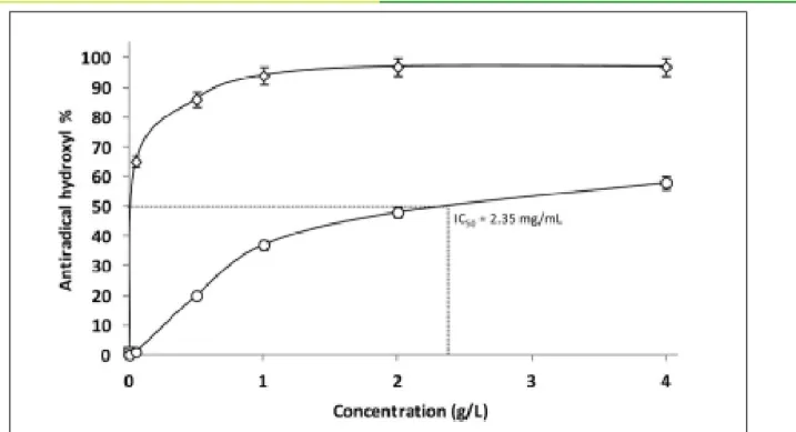
The scavenging abilities of AVP against hydroxyl radical increased with increasing concentration reaching a plateau of 50% at concentrations of AVP higher than 2 g/L and an IC50 close to 920 μg/mL (Figure 2). AVP showed a lower antioxidant activity as it scavenged DPPH radical and IC50 was around 2.35 mg/mL (Figure 3). It is well described that the reducing capacities of polysaccharides is related to their sulfation rate, molecular weight, glycosidic linkages, hydroxyl groups but also carboxylic groups of uronic acids [34]. It was previously reported that a direct correlation between the antioxidant activity and the reducing power of polysaccharides [35]. Indeed, this antioxidant activity is explained by the presence of many free hydroxyl groups in the structure of polysaccharides. In addition, some papers reported that the presence of arabinose in the structure of polysaccharides could reduce the production of hydroxyl radicals by chelation of pro-oxidant ions [36]. Overall, many studied have highlighted the antioxidant properties of polysaccharides extracted from Aloe leaves [3,6-8]. Even if extensive in vitro antioxidant studies for polysaccharides are sorting out [37], many conflicting results are detailed in the literature and yet no real mechanism are given. Note that authors claim that the antioxidant properties of polysaccharides are not determined by a single factor but a combination of several related factors, from the purity to the structural features and the physicochemical properties.
Concentration (mol%)a Monosaccharide 75.3 ± 0.51 ᴅ-galactose 10.8 ± 0.32 ᴅ-glucuronic acid 6.52 ± 0.67 ʟ-rhamnose 4.95 ± 1.22 ʟ-arabinose 2.16 ± 1.04 ᴅ-galacturonic acid 0.28 ± 0.12 ᴅ-mannose
Table 1: Monosaccharide composition and molecular weight of AVP.
aMonosaccharide composition was estimated by HPAEC-PAD; bMn: number average molecular weight estimated by SEC-MALS; cMw: molecular weight estimated by SEC-MALS;
dPDI: polydispersity index estimated by SEC-MALS;
All analyses were run in triplicate and the relative standard deviations are less than 5%.
Mw (g/mol)c PDId
7.02 x 10³
Mn (g/mol)b
1.02 x 10⁴ 1.46
Advances in Applied Chemistry and Biochemistry [ISSN: 2652-3175] SubmitManuscript
.05
.
Figure 2: Determination of the anti-radical (anti-radical hydroxyl) activities of the polysaccharide extracted from A. vera. (-O-): AVP, (- -): Ascorbic acid.
Figure 3: Determination of the anti-radical activities (anti-DPPH) of the polysaccharide extracted from A. vera. (-O-):
AVP, (- -): ascorbic acid.
SubmitManuscript
Advances in Applied Chemistry and Biochemistry [ISSN: 2652-3175]
.06
.
Citation: Lefsih K, Iboukhoulef L, Petit E, Benouatas H, Pierre G, et al. (2018) Anti-inflammatory and Antioxidant Effect of a ᴅ-galactose-rich
Anti-inflammatory activity
The percent reduction in ear edema obtained during the analysis of the anti-inflammatory activity of AVPs at doses of 1%, 5% and 10% were respectively 7.92%, 43.30% and 71.49% (Table 2). The percentage of reduction obtained with the use of the A. vera Pure Gel (AVG) was 73.23%. It has been found that the percentage of inhibition of edema obtained with a concentration of 10% of AVP was greater than that obtained with a concentration of 5 %, the latter was also greater than the percentage of reduction obtained with a concentration of 1 % in AVP. This result showed that AVP and AVG were endowed with anti-inflammatory activity, which were dose dependent. As shown in table 2, a very slight
t Student Edema Reduction (%) Edema weight (mg) Lot / / 5.68 ± 0.010 Xylene1 (edema control)
0.018* 56.33 2.48 ± 0.010 Diclofenac 1% 0.743 7.92 5.23 ± 0.015 AVP 1% 0.058 43.3 3.22 ± 0.020 AVP 5% 0.003* 71.49 1.62 ± 0.017 AVP 10% 0.0001* 73.23 1.52 ± 0.010 AVG
Table 2: Edema reduction after treatment with pure AVG and different concentrations of AVP.
1Both xylene and 12-O-Tetradecanoyl Phorbol 13-Acetate (TPA) can be used in topical induction of inflammation, few hours after
application. Xylene was used here because of its availability.
decrease in edema weight was found between the mean weight of the control edema and the mean weight of edema treated (5.68 mg to 5.23 mg, i.e., 7.92% reduction) with the 1% AVP extract and no significant difference was detected by a Student’s test (p > 0.05). Regarding the lot treated with AVP extract at 5%, a slight decrease in the average weight of edema was detected compared to the average weight of the edema control; it was reduced by 43.3%. This difference was not significant (p > 0.05). For lots treated with 10% AVP and AVG, a very significant decrease in edema weight was observed, the mean edema weight decreased by 71.49% reduction for AVP at 10% and by 73.23% for AVG, compared to a reduction percentage of 56.33% for the reference treatment (Diclofenac 1%). Thus, the edema reduction by treatment with AVP (10 %) and AVG seemed to be better compared to that of Diclofenac 1%. According to the same test, no significant difference (p > 0.05) was reported between the treatment with AVP extract (10 %) and that of AVG, and finally these two treatments seemed to have the same effect (p > 0.05). All these results demonstrated that AVP has a significant anti-inflammatory activity. According to the last result, it can be supposed that the anti-inflammatory activity of A. vera gel was mainly stemming from the presence of polysaccharides which represented than 60 % of its dry matter [29,30]. The results obtained were in
accordance to those obtained in different studies conducted on pectic and galactan extracts [25,38]. The authors demonstrated the presence of bioactive polysaccharides in C. deserticola responsible for the anti-inflammatory and immunomodulatory activities of two pectic fractions including homogalacturonan and rhamnogalacturonan. The anti-inflammatory activities of the pectic polysaccharide obtained from Adansonia digitata (Malvaceae) at different concentrations were evaluated by the inhibition test of cyclooxygenases type-1 (COX-1) and 2 (COX-2), enzymes that catalyse the oxygenation of polyunsaturated fatty acids to form prostanoids. Both enzymes were inhibited by this polysaccharide which was more effective in inhibiting COX-2 than COX-1 [39]. A similar study also demonstrated that water-soluble polysaccharides significantly
inhibited the production of Nitric Oxide (NO), prostaglandin E2, TNF-α and IL-6 in response to LPS [40]. Modern phytochemistry and pharmacological experiments have also shown that polysaccharides are major active ingredients of
Astragalus monspessulanus root with various biological
activities such as anti-inflammatory activity [32].
Generally, water-soluble polysaccharides participate in the mobility of leukocytes on the endothelial surface of inflammation sites, the regulation of chemokines, the transendothelial migration of leukocytes, the inhibition of cyclooxygenase or lipooxygenase, and thus by the inhibition of leukocyte adhesion, which is one of the first steps in the initiation of the inflammatory response and for the accumulation of active immune cells at inflammatory sites [41]. Because of these and other functions, water-soluble polysaccharides of different structures and origins can be used to positively regulate inflammation processes [42].
Among the different biological activities of natural plant products that have been published until now, anti-inflammation is one of the most reported effects [43]. When a whole extract is used, there is a good chance for synergism between active components that might be lost when each of these components is isolated, this case being usually reported with essential oils
and polyphenols. Such synergism was discovered in several medicinal tests, including those for anti-inflammatory activity [44]. Solvent selection for extraction of plant materials is one of the most important factors in determining the potential activity of the extract, since the solvent polarity determines which compounds will be extracted or not [43].
The anti-inflammatory activity of the plant extract can be expressed depending on the chemical structure of the constituting molecules, their size and polarity. Thus, carotenoïd, flavonoïd, phenolic acid, monoterpene or sulfide showed reduction in C-Reactive Protein (CRP) and IL-6 levels but also inhibition of NFκB [45]. Polyphenol, capsaicin, curcumin, ascorbic acid, indol-3-carbinol, geraniol, sulphoraphane, gingerol, lycopene, deoxyelephantophin demonstrated significant reduction in cytokines levels and inhibition of COX-2, Inductible Nitric Oxide synthase (iNOS), NFκB and Signal Transducers and Activators of Transcription (STAT) activities [46]. Finally, sesquiterpenoid, diterpenoid, steroid, ceramide, cerebroside showed reduction in cytokines, NO and ProstaGlandins (PGs) levels and inhibition of COX and iNOS activities [47].
Conclusion
A galactose-rich polysaccharide from A. vera leaves have been extracted and its monosaccharide composition was determined. With a homogeneous and low molecular weight, the fraction showed decent antioxidant (IC50 of 920 μg/mL) and anti-inflammatory activities. In fact, ear edema reduction showed that the pure A. vera gel and the AVP fraction have very similar anti-inflammatory capacities. This provided a basis and direction for further study concerning the bioactivity of important traditional plant polysaccharides, and specific branches (neutral/carboxy) of very well-known glycosidic structures such as pectin; which can be recovered from specific extraction patterns.
References
1. Benzie IF, Wachtel-Galor S (2011) Herbal medicine: biomolecular and clinical aspects. 2ndedn. CRC Press, Boca Raton, USA. Pg no: 500.
2. Perez S (1997) Les vertus cachées des sucres. Biofutur 171: 21-23.
3. Ni Y, Tizard IR (2004) Analytical methodology: the gel-analysis of aloe pulp and its derivatives. In: Reynolds T (ed.). Aloes the genus aloe. CRC Press, Boca Raton, USA. Pg no: 111-126. 4. Chow JTN, Williamson DA, Yates KM, Goux WJ (2005)
Chemical characterization of the immunomodulating polysaccharide of Aloe vera L. Carbohydr Res 340: 1131-1142. 5. Mandal G, Das A (1980) Structure of the D-galactan isolated
from Aloe barbadensis Miller. Carbohydr Res 86: 247-257. 6. Mabusela WT, Stephen AM, Botha MC (1990) Carbohydrates
polymers from Aloe ferox leaves. Phytochem 29: 3555-3558. 7. Wozniewski T, Blaschek W, Franz G (1990) Isolation and
structural analysis of a glucomannan from the leaves of Aloe
arborescens var. Miller. Carbohydr Res 198: 387-391.
8. Ni Y, Turner D, Yates KM, Tizard I (2004) Isolation and characterisation of structural components of Aloe vera L. leaf pulp. Int Immuno Pharmacol 4: 1745-1755.
9. Lefsih K, Delattre C, Pierre G, Michaud P, Aminabhavi TM, et al. (2016) Extraction, characterization and gelling behavior enhancement of pectins from the cladodes of Opuntia ficus
indica. Int J Biol Macromol 82: 645-652.
10. Renard C, Voragen A, Thibault J, Pilnik W (1990) Studies on apple protopectin: I. Extraction of insoluble pectin by chemical means. Carbohydr Polym 12: 9-25.
11. Ciesielski W, Lii CY, Yen MT, Tomasik P (2003) Interactions of starch with salts of metals from the transition groups. Carbohydr Polym 51: 47-56.
12. Rinaudo M (2010) New way to crosslink chitosan in aqueous solution. Eur Polym J 46: 1537-1544.
13. Kulkarni SG, Vijayanand P (2010) Effect of extraction conditions on the quality characteristics of pectin from passion fruit peel (Passiflora edulis f. flavicarpa L.). LWT-Food Sci Technol 43: 1026-1031.
14. Dubois M, Gilles KA, Hamilton JK, Rebers PT, Smith F (1956) Colorimetric method for determination of sugars and related substances. Anal Chem 28: 350-356.
15. Bradford MM (1976) A rapid and sensitive method for the quantitation of microgram quantities of protein utilizing the principle of protein-dye binding. Anal Biochem 72: 248-254. 16. Casu B, Scovenna G, Cifonelli AJ, Perlin AS (1978) Infrared
spectra of glycosaminoglycans in deuterium oxide and deuterium chloride solution: quantitative evaluation of uronic acid and acetamidodeoxyhexose moieties. Carbohydr Res 63: 13-27.
17. Nadour M, Laroche C, Pierre G, Delattre C, Moulti-Mati F, et al. (2015) Structural characterization and biological activities of polysaccharides from olive mill wastewater. Appl Biochem Biotechnol 177: 431-445.
18. Benaoun F, Delattre C, Boual Z, Ursu AV, Vial C, et al. (2017) Structural characterization and rheological behavior of a heteroxylan extracted from Plantago notata Lagasca (Plantaginaceae) seeds. Carbohydr Polym 175: 96-104. 19. Delattre C, Pierre G, Gardarin C, Traikia M, Elboutachfaiti
R, et al. (2015) Antioxidant activities of a polyglucuronic acid sodium salt obtained from TEMPO-mediated oxidation of xanthan. Carbohydr Polym 116: 34-41.
20. Rotelli AE, Guardia T, Juárez AO, De la Rocha NE, Pelzer LE (2003) Comparative study of flavonoids in experimental
SubmitManuscript
Advances in Applied Chemistry and Biochemistry [ISSN: 2652-3175]
Citation: Lefsih K, Iboukhoulef L, Petit E, Benouatas H, Pierre G, et al. (2018) Anti-inflammatory and Antioxidant Effect of a ᴅ-galactose-rich
models of inflammation. Pharmacol Res 48: 601-606. 21. Gloaguen V, Krausz P (2008) Propriétés anti-inflammatoires
du polysaccharide capsulaire produit par la cyanobactérie thermophile mastigocladus laminosus. La Presse thermale et Climatique 145: 135-141.
22. Tambe R, Kulkarni M, Joice A, Gilani I (2009) Formulation an evaluation of Aloe vera gels. J Pharm Res 2: 1588-1590. 23. Sepúlveda E, Sáenz C, Aliaga E, Aceituno C (2007) Extraction
and characterization of mucilage in Opuntia spp. J Arid Environ 68: 534-545.
24. Boual Z (2009) Contribution à l'étude des polysaccharides de quelques plantes spontanées à caractère médicinal de la région de Ghardaïa (Sahara septentrional Est Algérien). Mémoire de magister. Université Kasdi Merbah Ouargla, Algeria. Pg no: 80.
25. Ebringerová A, Kardošová A, Hromádková Z, Hřı́balová V (2003) Mitogenic and comitogenic activities of polysaccharides from some European herbaceous plants. Fitoterapia 74: 52-61. 26. Sawut A, Yimit M, Sun W, Nurulla I (2014) Photopolymerisation and characterization of maleylated cellulose-g-poly(acrylic acid) superabsorbent polymer. Carbohydr Polym 101: 231-239. 27. Cárdenas A, Goycoolea FM, Rinaudo M (2008) On the gelling
behaviour of ‘nopal’ (Opuntia ficus indica) low methoxyl pectin. Carbohydr Polym 73: 212-222.
28. Nejatzadeh-Barandozi F, Enferadi ST (2012) FT-IR study of the polysaccharides isolated from the skin juice, gel juice, and flower of Aloe vera tissues affected by fertilizer treatment. Org Med Chem Lett 2: 33.
29. Femenia A, Sánchez ES, Simal S, Rosselló C (1999) Compositional features of polysaccharides from Aloe vera (Aloe barbadensis Miller) plant tissues. Carbohydr Polym 39: 109-117.
30. Delatorre-Herrera J, Delfino I, Salinas C, Silva H, Cardemil L (2010) Irrigation restriction effects on water use efficiency and osmotic adjustment in Aloe vera plants (Aloe barbadensis Miller). Agric Water Manag 97: 1564-1570.
31. Lee SC, Prosky L (1995) International survey on dietary fiber: definition, analysis, and reference materials. J AOAC Int 78: 22-36.
32. Jin M, Zhao K, Huang Q, Shang P (2014) Structural features and biological activities of the polysaccharides from Astragalus
membranaceus. Int J Biol Macromol 64: 257-266.
33. Hamman JH (2008) Composition and applications of Aloe vera leaf gel. Molecules 13: 1599-1616.
34. Hentati F, Delattre C, Ursu AV, Desbrières J, Le Cerf D, et al. (2018) Structural characterization and antioxidant activity of water-soluble polysaccharides from the Tunisian brown seaweed Cystoseira compressa. Carbohydr Polym 198: 589-600.
35. El-Shourbagy GA, El-Zahar KM (2014) Oxidative stability of ghee as affected by natural antioxidants extracted from food processing wastes. Ann of Agric Sci 59: 213-220.
36. Wu H, Zhu J, Diao W, Wang C (2014) Ultrasound-assisted enzymatic extraction and antioxidant activity of polysaccharides from pumpkin (Cucurbita moschata). Carbohydr Polym 113: 314-324.
37. Wang J, Hu S, Nie S, Yu Q, Xie M (2016) Reviews on mechanisms of in vitro antioxidant activity of polysaccharides. Oxid Med Cell Longev 2016: 5692852.
38. Lin LW, Hsieh MT, Tsai FH, Wang WH, Wu CR (2002) Anti-nociceptive and anti-inflammatory activity caused by
Cistanche deserticola in rodents. J Ethnopharmacol 83:
177-182.
39. Ibrahim AY, Mahmoud M, Asker MM (2014) Anti-inflammatory and antioxidant activities of polysaccharide from Adansonia
digitata: An in vitro study. Int J Pharm Sci Rev Res 25: 174-182.
40. Du Z, Liu H, Zhang Z, Li P (2013) Antioxidant and anti-inflammatory activities of Radix Isatidis polysaccharide in murine alveolar macrophages. Int J Biol Macromol 58: 329-335. 41. Wu Y, Cui SW, Tang J, Wang Q, Gu X (2007) Preparation,
partial characterization and bioactivity of water-soluble polysaccharides from boat-fruited sterculia seeds. Carbohydr Polym 70: 437-443.
42. Pomin VH (2015) Sulfated glycans in inflammation. Eur J Med Chem 92: 353-369.
43. Azab A, Nassar A, Azab AN (2016) Anti-inflammatory activity of natural products. Molecules 21: 1321.
44. Deharo E, Ginsburg H (2011) Analysis of additivity and synergism in the anti-plasmodial effect of purified compounds from plant extracts. Malar J 10: 01-05.
45. Watzl B (2008) Anti-inflammatory effects of plant-based foods and of their constituents. Int J Vitam Nutr Res 78: 293-298. 46. Aravindaram K, Yang NS (2010) Anti-inflammatory plant natural
products for cancer therapy. Planta Med 76: 1103-1117. 47. Wei WC, Sung PJ, Duh CY, Chen BW, Sheu JH, et al. (2013)
Anti-inflammatory activities of natural products isolated from soft corals of Taiwan between 2008 and 2012. Mar Drugs 11: 4083-4126.