HAL Id: tel-02514804
https://tel.archives-ouvertes.fr/tel-02514804
Submitted on 23 Mar 2020HAL is a multi-disciplinary open access archive for the deposit and dissemination of sci-entific research documents, whether they are pub-lished or not. The documents may come from teaching and research institutions in France or abroad, or from public or private research centers.
L’archive ouverte pluridisciplinaire HAL, est destinée au dépôt et à la diffusion de documents scientifiques de niveau recherche, publiés ou non, émanant des établissements d’enseignement et de recherche français ou étrangers, des laboratoires publics ou privés.
Marine fungi from sponges : biodiversity, chemodiversity
and biotechnological applications
Elena Bovio
To cite this version:
Elena Bovio. Marine fungi from sponges : biodiversity, chemodiversity and biotechnological applica-tions. Other. COMUE Université Côte d’Azur (2015 - 2019); Università degli studi (Torino, Italia), 2019. English. �NNT : 2019AZUR4009�. �tel-02514804�
University of Turin
Department of Life Sciences and Systems Biology
University of Côte d’Azur
Institute of Chemistry of Nice, UMR 7272 CNRS
Ph.D. Program in Biology and Applied Biotechnologies
Doctoral School of Sciences Fondamentales et Appliquées
Doctoral School of Sciences Fondamentales et Appliquées
Elena Bovio
Ph.D. Coordinators: Prof. Silvia Perotto
Prof. Elisabeth Taffin de Givenchy
Academic years: 2015-2018
Academic disciplines: BIO/02 - Chemistry
Marine fungi from sponges: biodiversity, chemodiversity and
biotechnological applications
University of Turin
Ph.D. Programme in Biology and Applied Biotechnologies
University of Côte d’Azur
Doctoral School of Sciences Fondamentales et Appliquées
Marine fungi from sponges: biodiversity,
chemodiversity and biotechnological applications
Elena Bovio
Tutors: Prof. Giovanna Cristina Varese
Prof. Mohamed Mehiri
The sea, once it casts its spell,
holds one in its net of wonder forever
Table of contents
List of Abbreviations i
List of Figures and Tables iv
1. INTRODUCTION 1
1.1 Marine environment 1
1.2 Marine fungi 2
1.2.1 Origin 2
1.2.2 Diversity and occurrence of fungi in the marine environment 5
1.2.3 Ecology 8
1.3 Marine organisms and microorganisms: an unlimited source of novel
compounds 12
1.3.1 The Mycotechnology 13
1.4 The sponge holobiont 16
1.4.1 Sponges 166
1.4.1.1 Origins, anatomy and physiology 166
1.4.1.2 Sponges, as producers of secondary metabolites 18
1.4.2 Fungi associated with sponges 19
1.4.3 Secondary metabolites from marine fungi associated with sponges 23 1.5 Classical and epigenetic approach to diversify the production of secondary
metabolites in marine fungi 25
2. AIM OF THE WORK 30
2.1 Project outline 32
3. The culturable mycobiota associated with 4 Atlantic sponges 33
3.1 MATERIAL AND METHODS 34
3.1.1 Sampling sites and axenic isolation 34
3.1.2 Fungal identification 36
3.1.3 Statistical analyses 38
3.2 RESULTS AND DISCUSSION 38
3.2.1 Influence of isolation techniques on the fungal community 38
3.2.3 Fungal diversity among the sponges 58
3.3 CONCLUSION 63
4. Thelebolus balaustiformis and Thelebolus spongiae: two new fungal species
from the Atlantic sponge D. fragilis 65
4.1 MATERIAL AND METHODS 65
4.1.1 Thelebolus spp. growth conditions and molecular study 65
4.2 RESULTS AND DISCUSSION 65
4.2.1 Thelebolus balaustiformis and Thelebolus spongiae sp. nov. 65
4.2.2 Taxonomy 67
4.2.2.1 Description of Thelebolus balaustiformis 67
4.2.2.2 Description of Thelebolus spongiae 72
4.2.2.3 Ecology and peculiar features of Thelebolus spp. 77 5. The culturable fungal communities inhabiting the Mediterranean sponges
Aplysina cavernicola, Crambe crambe and Phorbas tenacior 82
5.1 MATERIAL AND METHODS 83
5.1.1 Sampling sites and fungal isolation 83
5.1.2 Fungal identification 84
5.1.3 Statistical analyses 85
5.2 RESULTS AND DISCUSSION 85
5.2.1 Influence of isolation techniques on the fungal community 85
5.2.2 Fungal diversity 86
5.2.3 Fungal culturable diversity among the sponges 96
5.2.4 Mycobiotas vs metabolome 99
5.2.5 Fungi from A. cavernicola isolated by direct plating 100
5.3 CONCLUSION 105
6. Chemical diversity of the fungal community associated with the Atlantic
sponge G. compressa 106
6.1 MATERIALS AND METHODS 106
6.1.1 Small scale fermentation of the fungal community and OSMAC
approach 106
6.1.1.1 Multi-well culture and co-culture conditions 106
6.1.2 Scale-up of E. chevalieri MUT 2316 and molecules purification 108 6.1.2.1 Solid media culture condition and secondary metabolites extraction
and purification from E. chevalieri 108
6.1.2.2 Liquid media culture condition and secondary metabolites
extraction from E. chevalieri 110
6.1.2.3 High Resolution Mass Spectrometry analysis and molecular
networking 111
6.2 RESULTS AND DISCUSSION 112
6.2.1 OSMAC approach 112
6.2.1.1 Effect of media on fungal development and chemical fingerprint 112
6.2.1.2 Effect of salts on the fungal development and the chemical
fingerprint 117 6.2.1.3 Effect of co-culture on the fungal development and the chemical
fingerprint 119
6.2.1.4 General remarks on the OSMAC approach 120
6.2.2 Scale up of Eurotium chevalieri MUT 2316 121
6.2.2.1 E. chevalieri MUT 2316 metabolites from solid culture
condition 122 6.2.2.2 E. chevalieri MUT 2316 metabolites from liquid culture
condition 127 6.2.2.3 Molecular networking of E. chevalieri MUT 2316 in solid and
liquid culture condition 130
6.3 CONCLUSION 134
7. The antibacterial activity of the molecules produced by E. chevalieri MUT
2316 135
7.1 INTRODUCTION 135
7.1.1 The Golden Age of antibiotics, the antibiotic crisis and the need of new
medicines 135
7.1.2 Developing of AMR 139
7.1.3 What determines a bacterium to become MDR? 141
7.2 MATERIAL AND METHODS 142
7.2.1 Bacterial growth conditions and inoculum preparation 142 7.2.2 Stock solutions of the molecules isolated from E. chevalieri
7.2.3 Determination of the MIC 144 7.2.4 Determination of the Minimal Bactericidal Concentration (MBC) 146
7.3 RESULTS AND DISCUSSION 146
7.3.1 E. chevalieri MUT 2316 derived compounds show antibacterial
activity 146
7.3.2 Structure-activity relationship 151
7.4 CONCLUSION AND FUTURE PERSPECTIVES 151
8. The antiviral activity of the molecules produced by E. chevalieri MUT
2316 153
8.1 INTRODUCTION 153
8.1.1 Viruses and antivirals 153
8.1.2 Influenza A virus (IAV) 155
8.1.3 Herpes Simplex virus 1 (HSV-1) 156
8.2 MATERIAL AND METHODS 158
8.2.1 Cell culture conditions and viruses titre determination 158
8.2.2 Cytotoxic assays 159
8.2.3 Antiviral assay 159
8.3 RESULTS AND DISCUSSION 161
8.3.1 Cytotoxic activity of E. chevalieri MUT 2316 compounds 161 8.3.2 E. chevalieri MUT 2316-derived compounds show antiviral activity
against HSV-1 and IAV 162
8.3.3 Structure-activity relationship 164
8.4 CONCLUSION AND FUTURE PERSPECTIVES 164
9. The antifouling activity of the molecules produced by E. chevalieri MUT
2316 166
9.1 INTRODUCTION 166
9.1.1 The biofouling: definition and impact 166
9.1.2 Biofouling formation 166
9.1.3 Antifouling methods 168
9.1.4 Why sponges and their associated microorganisms as candidates for the
production of new antifouling? 171
9.2.1 Marine Antibacterial Assays 173 9.2.1.1 Growth inhibition 174 9.2.1.2 Adhesion inhibition 175 9.2.2 Microalgal assay 176 9.2.2.1 Growth inhibition 176 9.2.2.2 Adhesion inhibition 177
9.2.3 Inhibition of blue mussel M. edulis settlement – tyrosinase assay 177
9.3 RESULTS AND DISCUSSION 178
9.3.1 The molecules produced by E. chevalieri MUT 2316 display
antifouling activity 178
9.4 CONCLUSION AND FUTURE PERSPECTIVES 184
10. GENERAL CONCLUSION AND FUTURE PERSPECTIVES 186
References 190 Annexe 226 Annexe 2 242 Ph.D. activities 245 PUBLICATIONS 247 ACKNOWLEDGEMENTS 250
i
List of Abbreviations
AC Algobank of Caen
ACT Actin gene
AIC Akaike Information Criterion
AMR Antimicrobial resistance
ANOVA Analysis of variance
ATCC American type culture collection
BHI Brain-Heart Infusion
BPP Bayesian posterior probabilities
CA Carrot Agar
CAL Calmodulin gene
CAP Canonical analysis of principal coordinates
CFU Colony forming unit
CLSI Clinical and laboratory standards institute CMASW Corn meal agar seawater
cRNP Ribonucleoprotein
DAAs Directly acting antivirals
DBIOS Department of life sciences and system biology DDD 1,1-dichloro-2,2-bis-(4-chlorophenyl)ethane DMEM Dulbecco’s Modified Eagle Medium
DMSO Dimethyl sulfoxide
DNA Deoxyribonucleic acid
DNMT DNA methyltransferase
dNTPs Deoxynucleotides
EPS Extracellular polymeric substances
FA Formic acid
FBS Fetal bovine serum
FDA Food and Drug Administration
GA Gelatin agar
GAPDH Glyceraldehyde-3-phosphate dehydrogenase gene
GAS Gelatin agar added with 3% NaCl
ii
GNPS Global natural products social molecular networking HAAs Host-acting antivirals
HBV Hepatitis B virus
HCV Hepatitis C virus
HDAC Histone deacethylase
HIV Human immunodeficiency virus
HPLC High Performance Liquid Chromatography HR-MS/MS High-resolution tandem mass spectrometry HSV1 Herpes simplex virus 1
HSV2 Herpes simplex virus 2
IAV Influenza A virus
IBV Influenza virus B
IC Inhibitor concentration
ITS Internal transcribed spacer regions
LBCM Laboratoire de Biotechnologie et Chimie Marines LC-MS Liquid chromatography - mass spectrometry LOEC Low observable effect concentration
LSU Large subunit
MBC Minimal bactericidal concentration
MBM Marine bacterial medium
MCMC Markov chains monte carlo MDCK Madin darby canine kidney cells
MDR Multi drug resistant
MEA Malt extract agar
MEASW Malt extract agar seawater
MeOH Methanol
MHBCA Mueller-hinton broth cation adjusted MHF Mueller hinton fastidius agar
MIC Minimal inhibitory concentration
mRNA Messenger RNA
MRSA Methicillin-resistant Staphylococcus aureus MUT Mycotheca Universitatis Taurinensis
NGS Next Generations Sequencing
iii
nrRNA Nuclear ribosomal RNA
OD Optical density
OSMAC One strain - many compounds
PC Positive control
PCR Polymerase chain reaction
PDA Potato dextrose agar
PDAS Potato dextrose agar added with 3% NaCl
PDB Potato dextrose broth
PERMANOVA Permutational multivariate analysis of variance
PFU Plaque forming unit
PRA Plaque reduction assays
rDNA Ribosomal DNA
RNA RiboNucleic Acid
SPE Solid phase extraction
SW Seawater
SWA Seawater agar
TBT Tributyltin
TSA Tryptic soy agar
TUB Beta-tubulin gene
VERO African green monkey kidney cells (verda reno) vRNAs Viral ribonucleoproteins
WH Wickerham’s medium
WHO World health organisation
iv
List of Figures
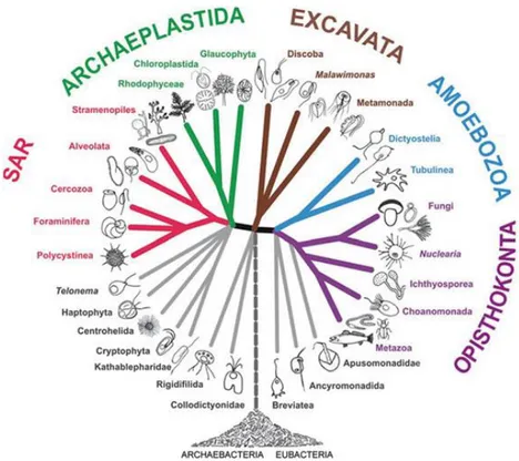
Figure 1.1 Division of the marine environment (Raghukumar, 2017). 1 Figure 1.2 General eukaryote phylogenetic tree. The clade Fungi and Metazoa have a common ancestor and are grouped under the Opisthokonta (Adl et al., 2012).
3
Figure 1.3 Divergent evolution of Kingdom fungi from their ancestors (Microsporidia, Cryptomycota and Aphelida) (Karpov et al., 2014). 4
Figure 1.4 Global distribution of marine sponges (Van Soest et al., 2012). 17
Figure 1.5 General anatomy of a marine sponge (Jackson et al., 2015). 18
Figure 1.6 The coloured arrows report the key functions carried out by the microbiome that directly influence the sponge holobiont and indirectly play ecosystems services influencing nutrient cycles and the web food chain (Pita
et al., 2018).
22
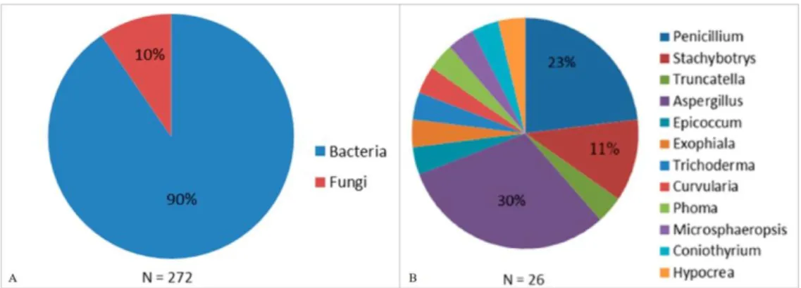
Figure 1.7 Percentage of microorganisms associated with sponges found able to produce antimicrobial molecules (antibacterial, antiviral, antifungal, antiprotozoal). B. Sponge derived fungal genera from which new molecules with antimicrobial activity were isolated (Indraningrat et al., 2016).
24
Figure 3.1 A. D. fragilis (Ph. B. Picton). B. G. compressa (Ph. B. Picton). C. P. johnstonia (Ph. B. Picton). D. S. ciliatum (Ph. M. De Kluijver). 33
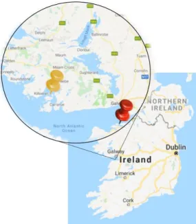
Figure 3.2 Sampling site of D. fragilis and P. johnstonia marked with the yellow pin; G. compressa and S. ciliatum sampling site is shown by the red pin.
35
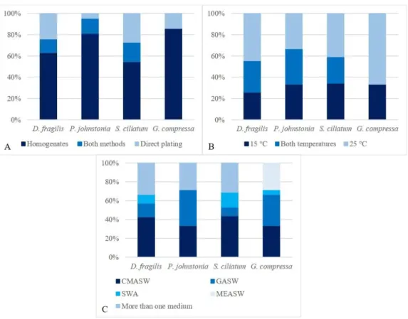
Figure 3.3 Influence of A. isolation methods; B. incubation temperatures; C. growth media, on the fungal community associated with D. fragilis, G.
compressa, P. johnstonia and S. ciliatum.
v
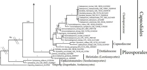
Figure 3.4 Bayesian phylogram of Capnodiales (Dothideomycetes) based on rDNA large subunit (LSU). One fungal isolate (MUT 2352) is included and identified as Pseudocercosporella sp.. Branch numbers indicate BPP values.
48
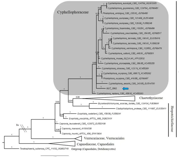
Figure 3.5 Bayesian phylogram of Chaetothyriales (Eurotiomycetes) based on rDNA large subunit (LSU). MUT 2862 is included and clusters within the genus Cyphellophora. Branch numbers indicate BPP values.
49
Figure 3.6 Bayesian phylogram of Pleosporales (Dothideomycetes) based on rDNA large subunit (LSU). Six and four fungal isolates clustered within the Phaeosphaeriaceae and the Pleosporaceae, respectively. Six fungal taxa clustered individually within the Didymellaceae, Cucurbitariaceae,
Montagnulaceae, Periconiaceae, Sporormiaceae and
Roussoellaceae/Thyridariaceae. One fungus was included in the Pleosporales order. Branch numbers indicate BPP values.
52
Figure 3.7 Bayesian phylogram of Leotiomycetes based on rDNA large subunit (LSU). Two fungal isolates were identified as Mollisia sp. and
Gremmenia infestans within the Dermateaceae and Phacidiaceae, respectively.
Branch numbers indicate BPP values.
54
Figure 3.8 Bayesian phylogram of Sordariomycetes based on rDNA large subunit (LSU). Three fungal taxa clustered individually within the Nectriaceae, Hypocreaceae and Microascaceae. Five fungal isolates clustered within the Bionectriaceae. Branch numbers indicate BPP values.
56
Figure 3.9 Bayesian phylogram of the genus Emericellopsis based on a combined dataset of ITS and beta-tubulin partial sequences. MUT 2273 and MUT 2274 were identified as Emericellopsis pallida. Branch numbers indicate BPP values.
vi
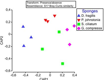
Figure 3.10 CAP of the fungal communities of the 4 Atlantic sponges D. fragilis (DF), G. compressa (GC), P. johnstonia (PJ) and S. ciliatum (SC). 59
Figure 4.1 Bayesian phylogram of the genus Thelebolus based on a combined dataset of ITS and beta-tubulin partial sequences. MUT 2357 and MUT 2359 were identified as new species, T. balaustiformis and T. spongiae, respectively. Branch numbers indicate BPP values.
66
Figure 4.2 T. balaustiformis MUT 2357. A, B. Closed subglobosus ascoma in the first stage of development. C. Ascoma becoming apothecial with mature asci. D. Apothecial ascoma with cortical excipulum dehiscent; E, F. Mature asci with 48–64 ascospores. G. Ascospores. Scale bars: A–D, F = 10 µm; E, G = 5 µm.
68
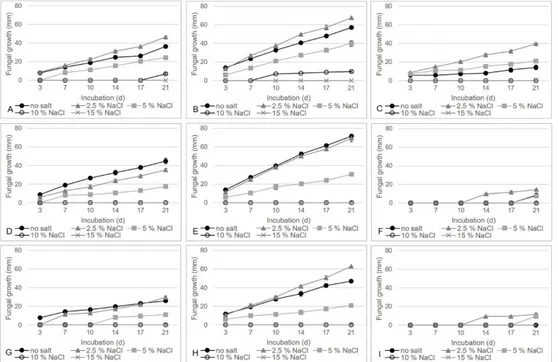
Figure 4.3 T. balaustiformis MUT 2357 growth curve with no and different NaCl concentrations on CA at A. 4 °C; B. 15 °C; C. 25 °C; on PDA at D. 4 °C; E. 15 °C; F. 25 °C; on MEA at G. 4 °C; H. 15 °C; I. 25 °C.
69
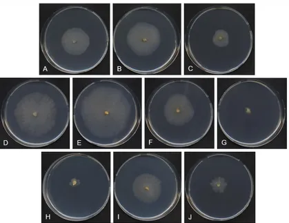
Figure 4.4 T. balaustiformis MUT 2357: 21-d-old colonies on CA at 4 °C with A. 0% NaCl; B. 2.5% NaCl; C. 5% NaCl; at 15 °C with D. 0% NaCl; E. 2.5% NaCl; F. 5% NaCl; G. 10% NaCl; at 25 °C with H. 0% NaCl; I. 2.5% NaCl; J. 5% NaCl.
70
Figure 4.5 T. balaustiformis MUT 2357: 21-d-old colonies on PDA at 4 °C with A. 0% NaCl; B. 2.5% NaCl; C. 5% NaCl; at 15 °C with D. 0% NaCl; E. 2.5% NaCl; F. 5% NaCl; G. 10% NaCl; at 25 °C with H. 0% NaCl; I. 2.5% NaCl; J. 5% NaCl.
70
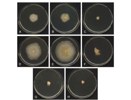
Figure 4.6 T. balaustiformis MUT 2357: 21-d-old colonies on MEA at 4 °C with A. 0% NaCl; B. 2.5% NaCl; C. 5% NaCl; at 15 °C with D. 0% NaCl; E. 2.5% NaCl; F. 5% NaCl; at 25 °C with G. 2.5% NaCl; H. 5% NaCl.
vii
Figure 4.7 T. spongiae MUT 2359: A. Initial ascoma; B. Ascomata with two globular asci; C. Mature ascoma opening with four asci; D. Ascoma with two sacciform asci; E. Ascospores. — Scale bars: A, E = 10 µm; B–D = 30 µm.
73
Figure 4.8 T. spongiae MUT 2359 growth curve with no and different NaCl concentrations on CA at A. 4 °C; B. 15 °C; C. 25 °C; on PDA at D. 4 °C; E. 15 °C; F. 25 °C; on MEA at G. 4 °C; H. 15 °C; I. 25 °C.
74
Figure 4.9 T. spongiae MUT 2359: 21-d-old colonies on CA at 4 °C with A. 0% NaCl; B. 2.5% NaCl; C. 5% NaCl; at 15 °C with D. 0% NaCl; E. 2.5% NaCl; F. 5% NaCl; G. 10% NaCl; at 25 °C with H. 0% NaCl; I. 2.5% NaCl; J. 5% NaCl.
75
Figure 4.10 T. spongiae MUT 2359: 21-d-old colonies on PDA at 4 °C with A. 0% NaCl; B. 2.5% NaCl; C. 5% NaCl; at 15 °C with D. 0% NaCl E. 2.5% NaCl; F. 5% NaCl; at 25 °C with G. 0% NaCl; H. 2.5% NaCl; I. 5% NaCl.
75
Figure 4.11 T. spongiae MUT 2359: 21-d-old colonies on MEA at 4 °C with A. 0% NaCl; B. 2.5% NaCl; C. 5% NaCl; at 15 °C with D. 0% NaCl; E. 2.5% NaCl; F. 5% NaCl; G. 10% NaCl; at 25 °C with H. 0% NaCl; I. 2.5% NaCl; J. 5% NaCl.
76
Figure 5.1 A. A. cavernicola (Ph. J. de Vaugelas). B. C. crambe (Ph P.
Amade). C. P. tenacior (Ph. J. de Vaugelas). 82
Figure 5.2 Sampling site of A. cavernicola, C. crambe and P. tenacior along
the coast of Villefranche sur Mer, France. 83
Figure 5.3 A. Influence of incubation temperatures and B. growth media on the fungal community isolated from A. cavernicola, C. crambe and P. tenacior. 86
viii
Figure 5.4 Bayesian phylogram of Capnodiales (Dothideomycetes) based on rDNA large subunit (LSU). MUT 3036 is identified as U. dekkeri. Branch numbers indicate BPP values.
90
Figure 5.5 Bayesian phylogram of Pleosporales (Dothideomycetes) based on rDNA large subunit (LSU). Two taxa clustered within the Phaeosphaeriaceae. Of the two remaining taxa, one belongs to the Sporormiaceae and one to the Torulaceae. Numbers indicate BPP values.
92
Figure 5.6 Bayesian phylogram of Sordariomycetes based on rDNA large subunit (LSU). Of 6 taxa, each one individually cluster in the families Cordycipitacaeae, Microascaceae, Sporormiaceae, Apiosporaceae and Sordariaceae; while one is in the incerta sedis group. Three taxa were within the Xylariaceae. Branch numbers indicate BPP values.
95
Figure 5.7 Number of exclusive and common fungal taxa in the three Mediterranean sponges’ A. cavernicola, C. crambe and P. tenacior. 97
Figure 5.8 PCO on the fungal communities of the Atlantic and
Mediterranean sponges. 104
Figure 6.1 Secondary metabolites extraction from E. chevalieri MUT 2316 in
solid culture condition. 110
Figure 6.2 Secondary metabolites extraction from E. chevalieri MUT 2316 in
liquid culture condition. 111
Figure 6.3 A. Absence of sporulation on WH and B. High sporulation (h) on PDAS for P. paneum MUT 2327. C. Production of exudates (e) on WH by E.
chevalieri MUT 2316. D. Production of soluble pigments (s) on PDA for F. solani MUT 2317. E. Contrast (c) with the bacterium and the fungus C. allicinum MUT 2313 on PDA. F. Predominance (p) of C. cladosporioides
ix
MUT 2314 on the bacterium on PDAS. G. E. chevalieri MUT 2316 on PDAS completely inhibit the bacterial growth (n).
Figure 6.4 A. Fungal growth on three media, classified as development of the fungus on the inoculum (class 1), occupation of half the area of the well (class
2), complete growth of the fungus on all the surface of the well (class 3); B.
Changing in the chemical diversity of fungal extracts due to the different conditions assessed. Classification based on the number of HPLC-UV 280 nm peaks: 0-3 peaks (class 1), 4-7 peaks (class 2), 8-11 peaks (class 3), ≥ 12 peaks
(class 4).
114
Figure 6.5 Percentage of fungi in which the presence of salts increased, decreased or did not influence the metabolic diversity (same metabolic classes), related to the different conditions tested (three media in pure and co-culture).
117
Figure 6.6 Percentage of fungi interacting in different ways with the bacterium. Interactions are classified as contrast (c), predominance (p) and total inhibition of the bacterial growth (n), on the six tested media.
120
Figure 6.7 Chemical fingerprints ( = 280 nm) of the crude organic extract of
E. chevallieri MUT 2316 on six different media in A. pure culture and B.
Co-culture.
122
Figure 6.8 Extraction scheme of E. chevalieri MUT 2316 in solid culture condition. The amount of the extracts, the solvents and the techniques used are reported. In light blue the pure molecules obtained and their names, in grey the molecules for which no signals were recorded by extensive NMR analysis.
123
Figure 6.9 Chemical fingerprints (Macherey-Nagel NUCLEODUR® Sphinx column - 250 x 4.6 mm, 5 μm. H2O:ACN + 0.1% FA - 90:10 to 0:100 in 30
min, 0:100 for 5 min, 0:100 to 90:10 in 15 min. Flow rate 1 mL/min. = 280 124
x
nm). A. MeOH:CH2Cl2 crude organic extract. B. EtOAc:CH2Cl2 crude organic
extract. C. F3 eluted with H2O:MeOH. D. F4 eluted with MeOH.
Figure 6.10 Echinulin (1), Neoechinulin A (2), Neoechinulin D (3), Dihydroauroglaucin (4), Flavoglaucin (5), Isodihydroauroglaucin (6) and Physcion (7) isolated from E. chevalieri MUT 2316 in solid culture condition.
126
Figure 6.11 Extraction scheme of E. chevalieri MUT 2316 in liquid culture condition. The amount of the extracts, the solvents and the techniques used are reported. In light blue the pure molecules obtained and their names, in grey the molecules for which no signals were recorded by extensive NMR analysis.
127
Figure 6.12 Chemical fingerprints (Macherey-Nagel NUCLEODUR® Sphinx column - 250 x 4.6 mm, 5 μm. H2O:ACN + 0.1% FA - 90:10 to 0:100 in 30
min, 0:100 for 5 min, 0:100 to 90:10 in 15 min. Flow rate 1 mL/min. = 280 nm). A. EtOAc crude organic extract from broth. B. F2 MeOH fraction from broth C. EtOAc:CH2Cl2 crude organic extract from the mycelium. D.
MeOH:CH2Cl2 crude organic extract from the mycelium.
128
Figure 6.13 Asperflavin (8), Cinnalutein (9) and Cyclo-L-Trp- L-Ala (10), isolated from E. chevalieri MUT 2316 in liquid culture condition. 129
Figure 6.14 Molecular network of the crude extract of E. chevalieri MUT 2316 in solid (green), liquid (azure) and both culture conditions (fuchsia). 131
Figure 6.15 Molecular clusters of Echinulin (1), Neoechinulin A (2), Neoechinulin D (3), Dihydroauroglaucin (4), Flavoglaucin (5), Physcion (7), Asperflavin (8), Cinnalutein (9) and Cyclo-L-Trp- L-Ala (10). Colours highlight the molecules only detected in solid culture condition (green), in liquid culture condition (azure) and on both (fuchsia).
133
Figure 7.1 Timeline of the developing of new antibiotics and the occurrence
xi
Figure 7.2 Evolution of resistance through A. vertical evolution and B. horizontal evolution with phage conjugation, transduction or natural transformation. Blue cells are susceptible to bacteria, red cells are resistant bacteria (Sommer et al., 2017).
140
Figure 7.3 Broth microdilution for antibacterial assay as recommended by the
CLSI guidelines. 145
Figure 7.4 Red circles indicate the MIC values for (6), numbers are referred to the different concentrations tested, from one (128 µg/mL) to 12 (0.06 µg/mL), making two-fold serial dilution. A. S. aureus ATCC 29213 (MIC 64 µg/mL). B. S. aureus Monza-FD1 (MIC 32 µg/mL). C. S. aureus Monza-PFI (MIC 64 µg/mL). D. E. fecalis ATCC 29212 (MIC 64 µg/mL). S. pneumoniae Monza-82 is not reported because the colour of the medium did not allow to obtain significant pictures.
148
Figure 8.1 Antiviral approved by the FDA in the period 1987-2017 (Chaudhuri
et al., 2017, modified). 154
Figure 8.2 Influenza viral cycle. The influenza virus enters into the cell thanks to endosomal uptake; once inside it releases the genetic material as viral ribonucleoproteins (vRNAs) that enter in the nucleus for the transcription of messenger RNA (mRNA) and the replication thanks to the intermediation of a positive-sense complementary ribonucleoprotein (cRNP). In the cytoplasm, the mRNA is translated into the viral proteins that will constitute new virions once assembled together with the vRNPs (Krammer et al., 2018).
155
Figure 8.3 HSV-1 cycle. The glycoproteins present on the surface of HSV-1 are fundamental for the virus-receptor interaction; the virus can enter into the cell by membrane fusion or endocytosis. The genetic material is then transferred in the nucleus. Three genes groups mediate the transcription of HSV-1 and the expression of the proteins: α coding for proteins mainly
xii
responsible of the first steps related with the infection, β with proteins involved in the viral replication and, γ responsible for the synthesis of structural proteins. These three groups are subsequently activated.
For the replication of the HSV-1 DNA, 7 viral proteins are involved, including the DNA polymerase; first, the replication proceeds via the theta form to change in the rolling cycle replication that after the further cut, represent the final genome ready for encapsidation.
Figure 8.4 Schema of the treatment of the cells with the molecules for the
PRA, before, during and after the infection. 160
Figure 8.5 A. the arrow indicates the viral plaque generated by HSV-1, seen at the microscope; B. the arrow indicates the plaques produced by IAV, visible without magnification in the multiwell plates; C. control without plaques.
162
Figure 8.6 Effect of the molecules derived from E. chevalieri on A. IAV and B. HSV-1 replication. The mean plaque counts for each molecule was expressed as percent of inhibition of viral replication compared to the positive control (PC). The data shown are the means ± standard deviations from two independent experiments performed in duplicate. * p<0.05, ** p<0.01.
163
Figure 9.1 Fouling formation on a surface over time (Amara et al., 2018). 167
Figure 9.2 Scheme of a multiwall plate prepared with different concentrations
of the tested compounds and the controls. 174
Figure 9.3 Number of bacterial and algal species inhibited in the growth and adhesion by the seven tested (1, 2, 4, 5, 7, 9, 10) molecules produced by E.
chevalieri MUT 2316.
xiii
List of Tables
Table 3.1 Gene loci sequenced, primers for molecular analysis and PCR
programs. 37
Table 3.2 Fungal taxa isolated from D. fragilis (DF), G. compressa (GC), P. johnstonia (PJ) and S. ciliatum (SC) and their relative abundance in percentage
(RA%). The species already found in the marine environment (MA) and associated with sponges (SP) are reported, as well as the first record (FR).
42
Table 4.1 Thelebolus species and main morphological features (ascomata,
asci and ascospores). 79
Table 5.1 Fungal taxa isolated from A. cavernicola (AC), C. crambe (CC) and P. tenacior (PT) and their relative abundance in percentage (RA%). First
record (FR) and species already found in the marine environment (MA) and associated with sponges (SP) are reported.
87
Table 5.2 Fungal taxa isolated from A. cavernicola by direct plating and their relative abundance in percentage (RA%). First record (FR) and species already found in the marine environment (MA) and associated with sponges (SP) are reported.
101
Table 6.1 Effect of different growth conditions on the development of the fungus (class 0, 1, 2, 3); production of exudates (e), soluble pigments (s) and high sporulation (h); fungal-bacteria interaction as contrast (c), predominance of the fungus (p) or complete inhibition of the bacterial growth (n) are reported in red. The changing in colours from dark to light blue and white, underlined the increasing of growth classes.
113
Table 6.2 Effect of different growth conditions on the HPLC-UV 280 nm chemical fingerprint of fungi, expressed as (class 1, 2, 3, 4) at the increasing of the metabolic diversity. Exclusive peaks of each condition indicated in
xiv
parenthesis. The changing in colours from dark to light blue and white indicated the increasing of the metabolic diversity.
Table 7.1 Marine pharmaceutical in clinical pipelines (http://marinepharmacology.midwestern.edu/clinPipeline.htm accessed on 25th May 2018).
137
Table 7.2 Bacterial strains used for the antibacterial tests with their code,
resistance and classification (Gram +/-). 143
Table 7.3 Molecules isolated from E. chevalieri MUT 2316, their weights and concentrations in 100% DMSO (stock 1) and further diluted in the bacterial media for the bioassays (stock 2).
144
Table 7.4 Minimum inhibitory concentration (MIC µg/mL) and minimum bactericidal concentration (MBC µg/mL) values of compounds (1–10) and reference antibiotics against reference strains and multidrug-resistant isolates. Positive values are highlighted in light blue.
147
Table 8.1 Concentration of the molecules (1–10) that guaranteed at least 70%
of viability of MDCK and VERO cells. 161
Table 9.1 Marine bacteria used for the antifouling test. 173
Table 9.2 Molecules isolated from E. chevalieri MUT 2316 tested for the antifouling activity, their concentrations in 100% DMSO (stock 1) and further diluted for the bioassays (stock 2).
174
Table 9.3 Amsterdam table indicating the volume of bacterial suspension to add to the medium (final volume 10 mL) to obtain an inoculum concentration of 2 × 10⁸ CFU/mL, based on the OD value at 630 nm.
175
xv
Table 9.5 Low Observable Effect Concentration (LOEC µg/mL) values of compounds (1, 2, 4, 5, 7, 9, 10) against the growth (Gr) and adhesion (Ad) of marine bacteria and algae, representatives of fouling organisms. Positive values are highlighted in light blue.
180
Table 9.6 Low Observable Effect Concentration (LOEC µg/mL) values of compounds (1, 2, 4, 5, 7, 9, 10) in the tyrosinase assay. Positive values are highlighted in light blue.
1
1. INTRODUCTION 1.1 Marine environment
The Oceans cover almost three quarter of the Earth’s surface and host 50% – 80% of life forms (Choudhary et al., 2017). Marine ecosystems are the result of complex interactions between the physiochemical environment and the inhabiting organisms, and have a dramatic impact on climate and human life (Raghukumar, 2017). Marine species are perfectly adapted to harsh conditions, represented by an average salinity of 3.5%, reduced light penetration and high hydrostatic pressure (Nicoletti and Andolfi, 2018; Raghukumar, 2017). On the contrary, others parameters like pH ranging from 7.5 to 8.5 and temperature (-2 to 30 °C, on average) are more stable than in other terrestrial ecosystems, with some exceptions, i.e. the hydrothermal vents (Raghukumar, 2017). These different physical and chemical conditions define the zones in which the marine environment is divided (Figure 1.1).
2
Horizontally, the coastal habitat (neritic zone) from the littoral (high tide level) to the edge of the continental shelf (about 200 m depth) is markedly influenced by land. The remaining part, less conditioned by land, is named oceanic habitat (Matthiessen et
al., 2018; Raghukumar, 2017). The vertical division highlights the pelagic and the
benthic zone, each with several subdivisions. The pelagic zone is the largest habitat on Earth counting for 99.5% of inhabitable space from the sea-surface, to the sea-bottom (Matthiessen et al., 2018). The upper waters are influenced by the atmosphere conditions (i.e. temperature, seasonal variability, organic and inorganic material transported); while, light penetration delimits the euphotic zone, where the photosynthesis occurs. Thus, all the inhabitants of the deeper zones (except chemoautotrophic organisms) depend on oxygen coming from the sea-surface (Matthiessen et al., 2018). As regards the benthic zone, it extends from the shore to the unexplored deep sea floor and represents a sink for the organic matter; here most of invertebrates, algae and plants live (Matthiessen et al., 2018).
The vastness of the oceans makes that the biodiversity and the ecological role of several life forms are still largely unknown even in the more accessible habitats for humans (Nicoletti and Andolfi, 2018). The classified eukaryotes represent about 200,000 species, while the 90% is awaiting to be discovered, among them the majority of marine fungi (Mora et al., 2011).
1.2 Marine fungi 1.2.1 Origin
The first report of a fungus in the marine environment is dated back in 1849 when the Ascomycota Phaeosphaeria typharum was isolated from Thypha sp.; while, the first record of a yeast in marine environment occurred only in 1922 (Raghukumar, 2017). Today, the origin, distribution and occurrence of marine fungi remain largely unexplored.
Based on a limited number of fossil records, fungi were already present on earth in the Proterozoic, 900-750 millions of years ago (Taylor et al., 2014); however, whether fungi evolved in the sea or on land remains an open question. Most of the
3
life forms evolved in the sea and the Kingdom Fungi and Metazoa (Animalia) both grouped in the supergroup Opisthokonta (Figure 1.2) have a common ancestor (Adl et
al., 2012).
Figure 1.2 General eukaryote phylogenetic tree. The clade Fungi and Metazoa have a common
ancestor and are grouped under the Opisthokonta (Adl et al., 2012).
The early divergent branches of the Kingdom Fungi is represented by Microsporidia, Cryptomycota and Aphelida (Figure 1.3) mainly freshwater organisms with some representatives of marine origin, usually parasites of algae and animals (Raghukumar, 2017). In the last years, several sequences of Cryptomycota were retrieved in samples coming from hydrothermal vents and anoxic regions that most probably present environmental conditions closer to the ancestral Earth (Manohar and Raghukumar 2013; Le Calvez et al., 2009). However, despite these findings, the fresh water origin seems to be the most correct (Raghukumar, 2017). Indeed, divergent of
4
extant fungi probably occurred from ancestral Chytridiomycota that represent the basal clade of Kingdom fungi (Figure 1.3). Chytridiomycota are aquatic organisms, with flagellated spores, that were more likely replaced by the peculiar microscopic structures of “higher fungi”, including terrestrial Ascomycota and Basidiomycota (James et al., 2013).
Figure 1.3 Divergent evolution of Kingdom fungi from their ancestors (Microsporidia,
Cryptomycota and Aphelida) (Karpov et al., 2014).
Overall, the hypothesis on the terrestrial origin of fungi is both based on the phylogenetic analysis, which sees Chytidriomycota closer to the “higher fungi”, and the recovery of the same mainly on land. To this concern, Chytidriomycota early believed to be terrestrial, are more and more found in the marine environment; surprisingly, several studies point out the dominance of Chytidriomycota sequences above all the others groups of fungi, with putative new lineages (Comeau et al., 2016; Hassett and Gradinger, 2016; Richards et al., 2015).
In conclusion, only new samples and data could be able to clarify the origin of fungi. In this context, SCUBA (Self-Contained Underwater Breathing Apparatus) diving
5
and ROV (Remotely Operated underwater vehicles) are making Oceans more and more accessible to collect samples in extreme and unexplored environments.
1.2.2 Diversity and occurrence of fungi in the marine environment Historically, the fungal identification was based on the morphology of the reproductive structures and on the physiology of fungi. However, the morphological variability of some groups and the increasing number of cryptic isolates, indistinguishable for their morpho-physiology, created confusion within the fungal systematic (Wang et al., 2012a). The advent of molecular techniques significantly increased the knowledge on marine fungal diversity; over the last two decades, thanks to the availability of nucleotide sequences and to the discovery of new marine lineages, the systematics of marine fungi has been rewritten. Today, the most updated review of marine fungi by Jones et al. (2015) listed 1,112 known species of marine fungi represented by Ascomycota (943 species), Basidiomycota (96 species), Chytridiomycota and related phyla (26 species), Zygomycota (three species), Blastocladiomycota (one species) and asexual morphs of filamentous fungi (43 species). The phylum most represented in terms of species (Ascomycota followed by Basidiomycota) is also the most frequently retrieved, using cultural dependent techniques (Jones et al., 2015). New species and genera are continuously described in the marine environment (Bovio et al., 2018; Devadatha et al., 2018; Abdel-Wahab et al., 2017), up-to-date the website http://marinefungi.org/ reports 1,254 marine fungal species described. Moreover, well known terrestrial taxa are repeatedly reported for the first time in marine ecosystems (Garzoli et al., 2018; Bovio et al., 2017; Garzoli et al., 2015a). The narrow definition of a marine fungus, proposed by Kohlmeyer and Kohlmeyer (1979): “obligate marine fungi” are those growing and reproducing only in the marine environment; “facultative marine fungi” are terrestrial species actively growing also in the marine environment, was overcome by Jones et al. (2015).
Despite the increasing effort of marine mycologist to contribute to the discovery of new species, thus mapping the worldwide biodiversity, marine fungi are still an understudied group compared to others marine microorganisms (Tisthammer et al.,
6
2016). Jones and collaborators (2015) estimated that about 10,000 marine fungi are still waiting to be described. The only seawater gives an idea of the fungal occurrence in the marine environment: few drops of water (one milliliter) contain over 103 fungal
propagules (Wang et al., 2012a). The distribution of fungi in the water column follow the abundance of the organic matter and its seasonal variability, occurring more frequently in the first meters and close to the coastline, compared to the deep seawaters (Wang et al., 2012a). On the contrary, deep-sea sediments represent a sink for the organic matter creating a habitat where fungi are the dominant eukaryotic microbes (Nicoletti and Andolfi, 2018). Also in the deep-sea hydrothermal vents, 120 fungal species (including new ones) have been retrieved (Xu et al., 2018). Among these fungi, there are pathogens of animals and saprobe that can contribute to nutrient recycling (Nicoletti and Andolfi, 2018; Xu et al., 2018). The challenge for these studies is to demonstrate if the isolated fungi were also active and capable to grow in the environment from which they were recovered. The research of Burgaud and colleagues (2015) highlighted that yeasts frequently isolated from hydrothermal vents tolerate, under laboratory conditions, the pressure at which they were isolated (20 MPa), by studying their vital sign as biomass production, ribosomal and protein activity.
Several others substrates have been investigated for the isolation of marine fungi, however, due to the increasing interest in natural products, the most studied fungal communities are those associated with invertebrates, algae and plants (Raghukumar, 2017). The role of these fungi and the interactions with their hosts will be described in the next sections.
Despite all the efforts made to describe the marine fungal community, the studies available are not enough to cover the vastness of the oceans. Another important factor that influenced the knowledge of marine fungi is our capability to cultivate these organisms under standard laboratory conditions. Nowadays, the implementation of different media and isolation techniques increased the possibility of success in the cultivation of a higher number of fungi. However, the uncultivable fungi still represent the major component of the marine fungal community, hence, to describe the real biodiversity (cultivable and uncultivable species) Next Generation Sequencing (NGS)
7
techniques started to be largely employed. Up to date, several studies reported the fungal community of marine environment based on NGS analysis, in some case confirming at least the phyla abundance reported in cultural dependent studies. This was the case of deep-sea sediments of a hydrothermal vents system in India, were Ascomycota counted for 96.96% of the fungal community, followed by Basidiomycota (3.00%) and unidentified fungi (Xu et al., 2018). A recent review of the Arctic marine fungi, reported the dominance of Ascomycota in driftwood, sediments, seawater and ice (Rämä et al., 2017). On the contrary, sediments under Arctic land highlighted the dominance of Chytidriomycota; from an ecological point of view, this environment is characterised by abundant phytoplankton biomasses that provide the substrate and the niche for parasitic fungi such as Chytridiomycota (Rämä et al., 2017). Chytidriomycota were also the dominant group in the water samples of Arctic and temperate zones, representing more than three-quarter of fungi in the samples from the Atlantic Ocean, while the others phyla were in the order of abundance Ascomycota, Cryptomycota and Basidiomycota (Comeau et al., 2016).
Certainly, also the microbial community described by the mean of NGS analysis is strongly influenced by environmental factors; moreover, the sampling methods, the DNA extraction procedure and the sequencing techniques can condition the results. To overcome this problem, thanks to the International Census of Marine Microbes (ICoMM) project (Amaral-Zettler et al., 2010), water and sediments samples were collected across the world and standard protocols were used for the identification of marine fungi. In parallel, in correspondence of the sampling sites, the environmental parameters (e.g. temperature, salinity, concentrations of phosphate, nitrate, dissolved oxygen and silicate) were registered. By analysing these data, Tisthammer and colleagues (2016) observed the dominance of Ascomycota and Basidiomycota in the sediments and water column, together with a scarce overlap (15.4%) between water and sediments OTUs. Environmental factors played the major role in shaping the fungal community (73% of the total explainable variance) compared to the geographic location (18%). Depth, dissolved oxygen and nitrate were the three main factors that explained the fungal variability. Other studies underlined the influence of temperature and salinity
8
in determining the distribution of marine fungi (Shearer et al. 2007). Overall, several physicals and chemicals factors modulate marine fungal distribution, this without taking into account the availability of abiotic and biotic substrates, where fungi are abundantly recorded.
In conclusion, although very useful to outline the real biodiversity, NGS usually do not allow low taxonomic rank classification, description of new species and biotechnological exploitation of fungi. Therefore, the improvement of specific isolation techniques and cultural media, mimicking the fungal natural environment, could be the key factor to increase the number of cultivable fungi (Raghukumar, 2017).
1.2.3 Ecology
Marine fungi are widespread in the oceans and colonise different ecological niches; they were found associated with organisms of all trophic levels and can act as saprobes, symbionts and parasites (Raghukumar, 2017). This wide ecological diversity is a consequence of the evolution of fungal cell biology and feeding strategies.
Saprotrophic
Saprotrophic fungi use dead organic matter as a food source by secretion of enzymes; compared to their prokaryotic counterpart (bacteria), they are able to penetrate organic and inorganic materials, thanks to the development of hyphae, rhizoids, or ectoplasmic nets (Raghukumar, 2017). These features make saprobe terrestrial fungi the major contributors to nutrient recycling of organic matter. Several facts indicate that marine fungi too are critical and abundant components of nutrient cycling in the oceans (Tisthammer et al., 2016).
Among the most recalcitrant natural substrates, there are cellulose and chitin, the first and second most abundant biomass in nature (Alamgir, 2017). Lignocellulose materials are extremely abundant in seawater for the presence of plants and due to the wood coming from lands (Raghukumar, 2017). The recovery of fungi on these substrates is influenced by the type of wood and by climate, which determines slow colonisation rate in temperate regions (Panebianco et al., 2002) compared to tropical waters
9
(Vishwakiran et al., 2001). Moreover, it has been observed that marine fungi isolated from algae and seaweeds produce oxidative enzymes and tannases involved in the degradation of lignocellulose materials (Balabanova et al., 2018). Ascomycota are the main producers of these key enzymes, replacing the role of Basidiomycota in terrestrial environment as degraders of recalcitrant organic matter (Balabanova et al., 2018; Pointing and Hyde, 2000). In this context fungi offer peculiar ecosystems services: by degrading organic matter, new space becomes available for the development of seaweeds such as P. oceanica meadows and, by decomposing recalcitrant organic matter, nutrients make the return in the web food chain (Panno et al., 2013).
Likewise the most persistent polymers of plants, the most complex matrixes of animals too are degraded by fungi, while soft tissues remain of bacteria competence: fungi degrade carbonatic shells, chitinous exoskeleton and bones. The retrieval of a fungus on an oyster shell goes back to 1891 when Ostracoblabe implexa was first described (Golubic et al., 2005; Bornet, 1891).Shells colonised by fungi present peculiar uniform tunnels, made up by the mineralisation process of developing hyphae (Raghukumar, 2017). Similarly, Bongiorni and colleagues (2005) demonstrated that fungi recorded on the exoskeleton of dead crabs’ were able to produce several enzymes, including chitinase (N-acetyl-β-glucosaminidase), involved in the exoskeleton decomposition. Interestingly, prawn wastes increased chitinase production in the marine fungus Beauveria bassiana (Suresh and Chandrasekaran, 1998).
Parasites and pathogens
Fungal parasites and pathogens are associated with organisms of all trophic levels and play an essential role in the ecosystem functioning (Raghukumar, 2017). Indeed, they maintain a balance in the ecosystems diversity and only under exceptional conditions, they can devastate a population; hence, the organic matter becomes soon available to saprotrophs to return in the nutrient cycles (Raghukumar, 2017).
Environmental factors, such as the increase of water temperature or oceans acidification have been addressed as one of the causes that determine commensal or mutualistic fungi to become pathogens. This was the case of Aspergillus sidowii, the
10
causal agent of “sea fans diseases”, detected both on healthy and diseased Gorgonia spp. (Toledo-Hernandez et al., 2008); experimental studies highlighted that A. sidowii grows better as temperature rise, becoming able to extensively parasitize corals (Alker et al., 2001). Generally, climate changes and others stress factors, as livestock overpopulation may induce physiological stresses in organisms, making them susceptible to diseases; therefore, weak parasitic fungi may become highly virulent (Harvell et al., 1999). Indeed, what we know about fungal pathogens of marine animals mainly comes from aquacultures studies (Raghukumar, 2017). Several species of commercial prawns, shrimps, lobsters, crabs and fish have been affected by mycosis. To cite the most virulent diseases, the mortality rate of Penaeus japonicas raised up to 100% when injected with a conidial suspension of the pathogen Acremonium sp. (Raghukumar, 2017). The same mortality, caused by Atkinsiella dubia is observed in the larvae of the Japanese mitten crab Eriocheir japonicus (Raghukumar, 2017). As for fish, Ochroconis humicola determine the death of several striped jack Pseudocaranx dentex in a fish farm in Japan; the same fish was infected by Exophiala xenobiotica in another fish farm in Japan (Raghukumar, 2017). Probably, the same pathogens of aquaculture are found in wild animals, with repercussions on the global food chain.
Parasitic fungi can be also among the major contributors in the nutrient cycling; this is the case of Chytridiomycota, (pathogens of phytoplankton, zooplankton, animals, fungi and plants) the main actors of the “mycoloop”, firstly described in fresh water (Kagami et al., 2014). The “mycoloop” concept provides that large inedible phytoplankton species are infected by Chytridiomycota; the nutrients within host cells are consumed by Chytrids, and transferred, thanks to the zoospores, to zooplankton (Kagami et al., 2014). Therefore, Chytridiomycota transfer nutrients that otherwise would be lost by sinking from the euphotic zone (Kagami et al., 2014). Noteworthy, zooplankton is able to control the population of Chytridiomycota with a positive impact for example on the reduction of chytridiomycosis in fresh water amphibians (Kagami et
al., 2014).
Despite the “mycoloop” have been mainly studied in fresh water ecosystems, also marine Chytridiomycota are recognised as actors of the “mycoloop” (Reich et al.,
11
2017; Hassett and Gradinger, 2016). Probably several others “mycoloop” exist and are still waiting to be discovered; recently a new one has been reported in the lakes, where Chytridiomycota play a key role transferring the nutrients from the inedible pollens to the zooplankton (Kagami et al., 2017).
Symbionts
The two major mutualistic symbioses known in terrestrial environment are represented by mycorrizhae (plant-fungus association) and lichens (alga/cyanobacterium-fungus association). Lichens count about 700 species in the marine ecosystem, with Ascomycota mainly involved in the symbiosis. Marine lichens are frequently reported in the supralittoral and intertidal zone (Raghukumar, 2017), where they are exposed to high solar irradiance and, with the high tide, they are covered by seawater (Lipnicki, 2015). These extreme conditions induced phenotypic variability (morphologically and anatomically) in lichens and influenced their distribution (Lipnicki, 2015). Interestingly, few lichens have been reported also below the tide level, on algae and shells (Raghukumar, 2017).
As for the other most common symbiosis in the terrestrial environment, mycorrhizae have been reported in marine ecosystems only in the salt-marsh plant (Hyde
et al., 1998). However, preliminary data hypothesised mycorrhizal relationship also in
the roots of the seagrasses P. oceanica (Vohník et al., 2017) and Enhalus acoroides (Sakayaroj et al., 2010).
Several other symbiotic interactions have been hypothesised in the marine environment (sponges, corals, echinoderms, arthropods, etc.). For instance, fungi were retrieved in the gut and digestive tract of sea urchins and marine arthropods, probably contributing to break down complex polymers (Raghukumar, 2017). In the three species of the marine sponge Chondrilla sp., yeast cells were found to be maternally-transmitted, transferred from the soma through the oocytes to the fertilized eggs (Maldonado et al., 2005). Noteworthy, this symbiosis was found in different geographical locations (Mediterranean area, Caribbean and Australia).
12
Within corals, fungi may be involved in the nitrogen cycle, making this element available to corals that usually live in low-nitrogen waters; in parallel, fungi may protect their host from UV radiations. All these findings are supported by metabolomic studies of fungi inhabiting corals (Raghukumar, 2017).
However, few studies clearly state the role of fungi within their hosts; because of this, the term symbiosis should refer only to different organisms that live together for a long period of time (Taylor et al., 2007).
1.3 Marine organisms and microorganisms: an unlimited source of novel compounds
Bioactive marine natural products, deriving from both primary and secondary metabolisms, count for 28,609 new molecules with 1,277 new compounds only described in 2016 (Blunt et al., 2018). This research field has important economic implications: the global market of marine-derived natural products is around 5 billion dollars (Thompson et al., 2017).
Up to date, the main problem concerning the use of marine organisms is due to the difficulties to grow them under controlled conditions and the impossibility to collect large amounts of samples from the environment. As an example, a huge amount (1 tonnes - wet weight) of the tunicate Ecteinascidia turbinata or of the sponge
Halichondria okadai would be necessary to obtain few milligrams of two promising
anticancer compounds without resorting to the chemical synthesis (Jackson et al., 2015; Proksch et al., 2003).
In order to overcome the supply problem, natural products research has started focusing on marine microorganisms, including fungi that can be easily cultivated and manipulate (Silber et al., 2016). In addition, it is more than likely that bioactive molecules known produced by marine organisms are of bacterial or fungal origin (Jackson et al., 2015; Henriquez et al., 2014).
13
1.3.1 The Mycotechnology
The term mycotechnology was introduced to indicate the wide range of applications of fungi in biotechnology, from industrial processes to bioremediation and development of new drugs (Nicoletti and Andolfi, 2018). In this context marine fungi, in light also of the putative ecological role that cover in the sea, represent a promising source of new secondary metabolites, extremophilic enzymes and bioremediation agents.
Secondary metabolites
Marine fungi are the candidates to supply the huge amount of pharmaceutical required worldwide, i.e. 100,000 tons per year only for antibiotics (Silber et al., 2016). In addition, their biotechnological potential is incontestable: among the 1,277 new natural products described in 2016, marine fungi account for 36% of these newly described molecules (Blunt et al., 2018). In five years (2010-2015), 285 antibacterial and antifungal compounds were isolated from marine fungi (Nicoletti and Andolfi, 2018). Several molecules isolated from marine fungi proved to be active against the high priority strains of Staphylococcus aureus methicillin resistant (MRSA). For instance, two compounds produced by the fungi Diaporthaceae sp. PSU-SP2/4 and Nigrospora sp. MA75 displayed a Minimal Inhibitory Concentration (MIC) of 2 μg/mL and 8 μg/mL against S. aureus MRSA (Xu et al., 2015). As for the most interesting antifungal compounds, Didymellamide A produced by the marine fungus Stagonosporopsis
cucurbitacearum was active against azole-resistant and -sensitive Candida albicans, Candida glabrata, and Candida neoformans strains with MIC values ranging from 1.6
to 3.1 µg/mL (El-Hossary et al., 2017).
New anticancer with a better pharmaco-toxicological profile are also needed, malignant tumours are responsible for about 8.8 million victims every year (1/6 of deaths) (Calcabrini et al., 2017). Up to date, several compounds produced by marine fungi have been described for the anticancer activity; however, molecules with cytotoxic activity are often wrongly named anticancer compounds. An anticancer should be selective for cancer cells and preferably be active against cancer cells resistant to already
14
known drugs and with a mechanism different from the induction of apoptosis (Deshmukh et al., 2018; Gomes et al., 2015). There are some promising compounds produced by marine fungi. For instance, Pinophilin A produced by the marine fungus
Penicillium pinophilum is selective against cancer cells line, with a non-apoptotic
mechanism (Gomes et al., 2015; Myobatake et al., 2012). An unidentified species of
Penicillium sp. produced several gliotoxin analogous with an innovative anticancer
mechanism, targeting enzymes commonly involved in the malignant transformation of cells (Gomes et al., 2015; Sun et al., 2011). Ophiobolin O originally isolated from
Aspergillus ustus and its synthetic analogous present several mechanisms of actions and
was found able to kill glioblastoma cells that are apoptosis-resistant (Lv et al., 2015). Another strain of A. ustus produced a molecule named phenylahistin, whose synthetic derivate (plinabulin) after 20 years is still in the clinical stage (phase II) despite the promising anti-cancer activity, underlining the long time required before obtain a new drug ready to enter into the market (Mohanlal et al., 2018; Nicoletti and Andolfi, 2018; Kanoh et al., 1997).
As for the antiviral, there are promising compounds produced by marine fungi targeting several viruses with different replication mechanisms (Moghadamtousi et al., 2015). For instance, two fungi, Aspergillus terreus and an unidentified deep sea fungus were able to inhibit Influenza strains resistant to the currently available drugs (Abdelmohsen et al., 2017). The fungus Dichotomomyces cejpii F31-1 produced the scequinadoline A, significantly able to inhibit the dengue virus (Wu et al., 2018). Two fungi Fusarium heterosporum and Phoma sp. produced Equisetin and its enantiomeric homologous, phomasetin, respectively, targeting an important enzyme for the replication of the Human Immunodeficiency Virus (HIV) (Moghadamtousi et al., 2015).
Enzymes
Marine fungi are an appealing source of extremophilic enzymes that preserve their structure and activity at low temperatures, high salt concentrations and pH (Nicoletti and Andolfi, 2018). Several applications can be found for these enzymes. For instance, in the degradation of recalcitrant plants residues as cellulose and inulin, to
15
obtain ethanol suitable for the biofuel (Nicoletti and Andolfi, 2018; Rawat et al., 2017). Also, the recalcitrant waste of the paper industries can be treated with fungal enzymes or using the fungus alive, as demonstrated by Chen and collaborators (2014) with
Penicillium janthinellum and Pestalopsis sp.. Other environmental applications are
based on the use of marine fungal enzymes such as azoreductases, peroxidases and laccases for the degradation of textile effluents (Nicoletti and Andolfi, 2018; Theerachat
et al., 2018). Overall, the advantages of using marine fungi lie in their ability to grow at
high concentrations of salt or with high osmotic pressure, as it is common in industrial wastewaters (Nicoletti and Andolfi, 2018).
Others recalcitrant polymers as lignin and chitin can be degraded by marine fungi, as already mentioned in the section of saprotrophic fungi with, as result, the valorisation of waste materials.
Bioremediation agents
Bio-based systems to treat crude oil polluted sites, offer interesting alternatives to the classic methods, is an economic and environmentally friendly alternative (Zhang
et al., 2011). Due to their robustness and adaptation skill, marine fungi can be directly
employed as bioremediation agents and proved to be highly successful in crude oil degradation, facing one of the biggest environmental issues, oil spill. Several fungi can use aliphatic compounds as a sole carbon source, while a lower percentage can also use aromatic compounds. This is the case of several Aspergillus spp. capable of degrading benzo[a]pyrene (Nicoletti and Andolfi, 2018; Passarini et al., 2011). The main limit to the degradation process is the availability of the compounds to microorganisms, in this context biosurfactants and emulsificants can play a key role. This is even better if they are produced by marine fungi, for instance, hydrophobins are the most powerful surface-proteins known so far (Artini et al., 2017; Cicatiello et al., 2017; Piscitelli et al., 2017; Cicatiello et al., 2016).
Another environmental issue is represented by heavy metals, such as Cd(II), Hg(II), and Pb(II) that are abundant in the environment due to industrialisation and are responsible for several human pathologies (Mahmoud et al., 2017). Scientific studies
16
underlined a correlation between halophilism and tolerance to heavy metals, making of marine fungi the best candidates in bioremediation of heavy metals (Nicoletti and Andolfi, 2018). For instance, several deep-sea fungi accumulate heavy metals in different cells districts, including cell wall, vacuoles and cytoplasm (Nicoletti and Andolfi, 2018). Certainly, the selective pressure played by the accumulation of heavy metals in the ocean floor might have been the driving force for the outliving of these highly efficient fungi. Finally, even dead fungal biomass is extremely useful for the biosorption of heavy metals, as in the case of A. ustus (Mahmoud et al., 2017).
1.4 The sponge holobiont
Marine sponges (phylum Porifera) are the oldest metazoans on Earth; over their 660–635 M years evolution formed a close association with a wide variety of microorganisms including bacteria, archaea, fungi, and algae (Zumberge et al., 2018; Taylor et al., 2007). This close association was described for the first time by Vacelet and Donadey (1977) who observed bacteria within the sponges’ tissues. Today, it is well recognised that microorganisms represent up to 40–60% of the sponge biomass (Yarden 2014). Therefore, the term “sponge holobiont” is more appropriate when sponges and the associated microbial communities are considered as a whole (Webster and Thomas 2016; He et al., 2014). Holobionts are complex systems whose function is determined by both the processes carried out by the single members and those resulting from their interactions (Pita et al., 2018). The stability and health of the holobiont are the results of a dynamic equilibrium characterised by the resistance and the resilience to external pressure (Pita et al., 2018). Therefore, improving our knowledge of the sponge holobiont, will help us to understand their ability to survive to climate changes, maintaining their key role in the ecosystems.
1.4.1 Sponges
1.4.1.1 Origins, anatomy and physiology
Porifera counts more than 8,600 described species and about 15,000 estimated, not yet discovered (Webster and Thomas 2016). Sponges are globally distributed (Figure