Cardiovascular responses to sugary drinks in humans: galactose
presents milder cardiac effects than glucose or fructose
Nathalie Charrière1 · Cathriona Loonam1 · Jean-Pierre Montani1 · Abdul G. Dulloo1 · Erik K. Grasser1
Conclusions Galactose thus presents the interesting
char-acteristics of a low-glycemic sugar with mild cardiovas-cular effects. Further studies are warranted to confirm the clinical relevance of the milder cardiovascular effects of galactose than other sugars for insulin resistant obese and/ or diabetic patients with cardiac insufficiency.
Keywords Sucrose · Beat-to-beat · Randomized controlled
trial · Cardiac effects · Clinical implication
Introduction
A rise in the consumption of refined sugars in food and bev-erages has often been implicated in the epidemic of obesity, type 2 diabetes and cardiovascular diseases. While most added sugars are consumed as the disaccharide sucrose or in the form of its two constituent monosaccharides (glu-cose and fructose), it is its fructose moiety which, despite its classification as a low-glycemic sugar, is viewed as the more harmful sugar component [1].
Indeed, chronic studies comparing diets containing fruc-tose in substitution for glucose have demonstrated a more adverse lipid profile and greater cardiometabolic risks [1,
2].
During the past decade, there has been increasing inter-est in the potential beneficial effects of galactose [3–10], a low-glycemic sugar monosaccharide which in combination with glucose constitutes the disaccharide lactose. Although galactose is absorbed at the same rate as glucose, most of it is taken up by the liver where it is metabolized to other substrates (glucose, lactate or fatty acids) that are readily metabolized by extrahepatic cells [11]. Consequently, as the release of galactose in the form of glucose into the blood is delayed, it could provide an energy source of choice with
Abstract
Purpose There is increasing interest into the potentially
beneficial effects of galactose for obesity and type 2 dia-betes management as it is a low-glycemic sugar reported to increase satiety and fat mobilization. However, fructose is also a low-glycemic sugar but with greater blood pressure elevation effects than after glucose ingestion. Therefore, we investigated here the extent to which the ingestion of galactose, compared to glucose and fructose, impacts upon haemodynamics and blood pressure.
Methods In a randomized cross-over study design, 9
overnight-fasted young men attended 3 separate morning sessions during which continuous cardiovascular monitor-ing was performed at rest for at least 30 min before and 120 min after ingestion of 500 mL of water containing 60 g of either glucose, fructose or galactose. These meas-urements included beat-to-beat systolic and diastolic blood pressure, heart rate deduced by electrocardiography, and stroke volume derived by impedance cardiography; these measurements were used to calculate cardiac output and total peripheral resistance.
Results Ingestion of galactose, like glucose, led to
signifi-cantly lesser increases in systolic, diastolic and mean blood pressure than fructose ingestion (p < 0.05). Furthermore, the increase in cardiac output and reduction in total periph-eral resistance observed after ingestion of glucose were markedly lower after galactose ingestion (p < 0.01).
* Erik K. Grasser
erikkonrad.grasser@unifr.ch
1
Laboratory of Integrative Cardiovascular and Metabolic Physiology, Division of Physiology, Department of Medicine, University of Fribourg, 1700 Fribourg, Switzerland
http://doc.rero.ch
Published in "European Journal of Nutrition 56(6): 2105–2113, 2017"
which should be cited to refer to this work.
a low-glycemic index and a low-insulinemic response [12–
15]. Indeed, several studies in healthy humans have shown that the ingestion of galactose elicits only small increases in plasma glucose and insulin, which are not different from those in response to fructose ingestion [12–15].
This specific metabolic fate of galactose has formed the basis of interest in this low-glycemic sugar with respect to its potential impact on exercise performance [5, 8, 10] and more recently for weight control [3, 4, 6, 7, 9, 10]. In the latter context, several acute studies have shown that inges-tion of galactose leads to increased satiety [3, 4, 10]. Further-more, a chronic study involving intermittent intake of sug-ary drinks in women has shown that intake of galactose in substitution for glucose resulted in increased fat mobilization and oxidation [6], which would be consistent with the much lesser impact of galactose than glucose ingestion on circulat-ing insulin [12–15]. These studies have led to the proposal that diets with galactose as a source of carbohydrates could be useful in the management of obesity and type 2 diabetes.
Little is known, however, about the impact of galactose ingestion on the cardiovascular system. By contrast, it has repeatedly been demonstrated that the ingestion of glucose and fructose drinks is characterized by specific haemody-namic responses in healthy young [16, 17] and older people [18]. Unlike drinking glucose, which decreases total periph-eral resistance by an insulin-mediated release of nitric oxide accompanied by a metabolic vasodilation leading to an increase in cardiac output (CO), thereby resulting in little or no change in blood pressure (BP), the ingestion of fructose elicits a significant increase in BP that results from a more modest increase in CO but without a decrease in TPR [16,
17]. These differential BP responses to glucose and fructose ingestion have been attributed to their differential effects on insulin secretion [17], and to the well-known effect of insu-lin not only in enhancing sympathetic activation of cardiac contractility (hence contributing to the increased CO) [19], but also in stimulating peripheral vasodilatation and hence decreasing TPR [20]. In this context, the question arises as to whether ingestion of galactose, which like fructose has only a mild stimulatory effect on insulin release [12–15], would also lead to higher BP. In testing this hypothesis, the aim of our study here was to investigate, using comprehensive and continuous cardiovascular monitoring, potential differences in BP and haemodynamics between galactose, glucose and fructose in a cross-over study design in young men.
Methods Subjects
Nine healthy young men aged 24 ± 1 year (mean ± SD) were recruited from local students and their friends. The
mean height of the participants was 182 ± 7 cm, body weight was 79 ± 13 kg and BMI was 23.8 ± 2.8 kg/ m2. Before their first test, subjects had to fill out a ques-tionnaire about their health and lifestyle and under-went BP and anthropometric measurements to ensure a healthy condition. We defined healthy on the basis that all of our test subjects were non-obese with normal rest-ing BP (brachial systolic/diastolic BP < 140/90 mmHg and >100 mmHg systolic BP) and none of our subjects had any diseases or were taking any medication affect-ing cardiovascular regulation. All participants fasted for ≥12 h and abstained from alcohol, smoking and caffeine as well as from vigorous exercise for 24 h prior to each test and were advised not to change their dietary habits between the tests. This study was conducted according to the guidelines laid down in the Declaration of Helsinki, all procedures involving human subjects were approved by the Cantonal Ethics Committee, and written informed consent was obtained from all subjects prior to their inclu-sion in the study.
Experimental design
All studies started between 08:00 and 09:00 in a quiet, air-conditioned laboratory (21 °C), with the subjects at thermal comfort. Every test subject attended three sepa-rate experimental sessions (sepasepa-rated by at least 2 days) according to a randomized cross-over design. Randomi-zation was performed using a random sequence gen-erator (https://www.randomizer.org/) where the sessions order was determined for 9 test subjects before the study started. At each experimental session, the cardiovascu-lar responses to one of three drinks were monitored. The drinks tested were as follows: (1) water containing 60 g of D(+)-glucose, (2) water containing 60 g of D(+)-galactose, and (3) water containing 60 g D(−)-fructose. The purity of all sugars was 99 % (Argos Organics, Che-mie Brunschwig SA, Basel, Switzerland). Each drink also contained 10 mL lemon juice (to mask the difference in sweetness) and was made up to a total of 500 mL by addi-tion of distilled water (21 °C). The subjects were not told the order of the drinks.
On arrival at the laboratory, subjects were asked to empty their bladders if necessary and to sit in a com-fortable armchair. After waiting for a 20–30 min period to attain cardiovascular stability, continuous beat-by-beat recordings were initiated starting with at least a 30-min baseline period after which the subject ingested the respective test drink over a period of 4 min, which was followed by post-drink cardiovascular recordings over the next 120 min. The subjects were allowed to watch documentaries and calm movies in order to reduce boredom.
Haemodynamics
A Task Force Monitor (CNSystems, Medizintechnik, Graz, Austria) was used to perform cardiovascular recordings, and data were sampled at a rate of 1000 Hz [21]. Con-tinuous BP was monitored using the Penaz principle from either the index or middle finger (automatically finger switch every 30 min) of the right hand and was calibrated to oscillometric brachial BP measurements on the con-tralateral arm. The right hand with the continuous BP cuff rested on a ductile pillow which was positioned at heart level on a height adjustable table. Impedance cardiogra-phy measurements [22], in which the changes in thoracic impedance are converted to reflect changes in thoracic fluid content/volume over time, were taken based on the original Kubicek [23] approach, but using an improved estimate of thoracic volume [24], which allows calculation of cardiac stroke volume (SV). ECG/impedance electrodes were posi-tioned together with upper arm and finger BP cuffs. Elec-trode strips were placed at the neck and thoracic regions, the latter specifically at the midclavicular at the xiphoid process level (CNSystems standard electrode kits).
Statistical analysis
Values of cardiac interval, systolic BP (SBP), diastolic BP (DBP) and SV were averaged every 20 min during the baseline period and the 120 min post-drink period. CO was derived as the product of SV and heart rate (HR), where HR was calculated from the appropriate cardiac interval. Total peripheral resistance (TPR) was calculated as mean BP (MBP) divided by CO, where MBP was calculated as the result of DBP + 1/3 (SBP–DBP). All values are reported as mean ± SD. Statistical analysis was performed by two-way
ANOVA for repeated measures with time and treatment of
sugar-type (glucose, galactose, fructose) as within-subject factors using statistical software (Statistix version 8.0; Ana-lytical Software, St. Paul, MN). Where significant differ-ences were found, the effects of each drink over time were analysed by comparing values at each time point over the post-drink period with the basal values recorded before
drinking. One-way ANOVA with Dunnett’s multiple com-parison test and repeated-measures ANOVA with
Newman-Keuls post hoc testing were used to test for changes over
time from baseline level and to compare mean changes between the drink types. All reported p values are two-sided. For all tests, significance was set at p ≤ 0.05.
Sample size
Our sample size is based on the results of our previous study [16], comparing BP responses to glucose and fruc-tose. Assumptions are (1) a clinically relevant difference of 5 mmHg between galactose and fructose in healthy subjects (averaged response over 120 min post-drink) and (2) a standard deviation of the difference in post-drink BP response of 5 mmHg. Therefore, using a type I error (α) of 0.05 and a desired power (1−β) of 0.80, such an investi-gation of repeated measures (on the same subject) would require a total number of 8 subjects.
Results Blood pressure
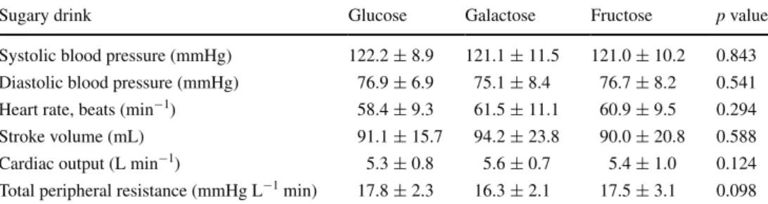
For all cardiovascular parameters assessed, there were no significant differences in the baseline values across days (Table 1).
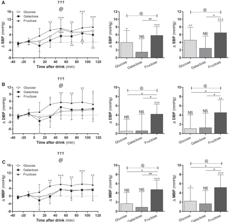
The BP responses to the sugary drinks are shown in Fig. 1 (panels a–c). In response to all three sugary drinks, SBP increased gradually to reach a plateau within an hour, and the significantly higher SBP values were maintained until the end of the study, i.e. 120 min post-drink. ANOVA indicates a significant effect of sugar-type (p < 0.05), with the increase in SBP in response to fructose (7–8 mmHg) being greater than after glucose (4–5 mmHg) or galac-tose (2–3 mmHg). A significant effect of sugar-type has also been found in the DBP response (p < 0.05). As shown in Fig. 1 (panel b), DBP was also found to be increased to a greater extent with fructose, reaching a plateau of ~5 mmHg relative to baseline (p < 0.05), compared to
Table 1 Baseline
haemodynamic data before ingestion of the sugary drinks, each ingested as 500 mL of distilled water containing 60 g glucose, galactose or fructose
All data are presented as mean ± SD. Baseline haemodynamic parameters for glucose, galactose and fruc-tose were compared using one-way ANOVA
Sugary drink Glucose Galactose Fructose p value
Systolic blood pressure (mmHg) 122.2 ± 8.9 121.1 ± 11.5 121.0 ± 10.2 0.843 Diastolic blood pressure (mmHg) 76.9 ± 6.9 75.1 ± 8.4 76.7 ± 8.2 0.541 Heart rate, beats (min−1) 58.4 ± 9.3 61.5 ± 11.1 60.9 ± 9.5 0.294 Stroke volume (mL) 91.1 ± 15.7 94.2 ± 23.8 90.0 ± 20.8 0.588 Cardiac output (L min−1) 5.3 ± 0.8 5.6 ± 0.7 5.4 ± 1.0 0.124 Total peripheral resistance (mmHg L−1 min) 17.8 ± 2.3 16.3 ± 2.1 17.5 ± 3.1 0.098
non-significant increases of only 2–3 mmHg in response to glucose or galactose. These more pronounced increases in SBP and DBP in response to fructose than to glucose or galactose are reflected in MBP (Fig. 1, panel c), which
showed, in the plateau phase, increases of about 6 mmHg for fructose compared to only a 3 mmHg increase for glu-cose or galactose (ANOVA: p < 0.05). Pair-wise compari-sons across the sugars for the average increases in BP over
Fig. 1 Time course of changes in systolic blood pressure (SBP) a,
diastolic blood pressure (DBP) b and mean blood pressure (MBP) (C) before and after ingestion of glucose (open circle), galactose (closed square) and fructose (closed triangle). Repeated-measures
ANOVA assessed statistical differences as follows: effect of time
(symbol dagger), effect of type (symbol @) and the Sugar-type × time interaction (symbol double dagger); one, two and three
symbols denoting p < 0.05, p < 0.01 and p < 0.001, respectively. For data on post-drink changes averaged over 60 and 120 min (shown as bars), post hoc pair-wise comparison for statistical differences across sugar-types is assessed by Dunnett’s multiple comparison tests and indicated as follows: *p < 0.05, **p < 0.01 and ***p < 0.001. Signifi-cant difference between post-drink and baseline values are indicated as follows: NS non-significant; °p < 0.05, °°p < 0.01 and °°°p < 0.001
the 120 min (or first 60 min) post-drink indicate that SBP, DBP and MBP are all significantly different between fruc-tose versus glucose or between frucfruc-tose versus galacfruc-tose, but not between glucose versus galactose.
Heart rate and stroke volume
While all sugars increased HR reaching plateau values between 60 and 90 min post-drink (Fig. 2, panel a), a sig-nificant interaction effect of sugar-type and time (ANOVA,
p < 0.001) is observed, with fructose ingestion resulting in
the greatest elevation in HR (+7 bpm) in the second hour post-drink compared to glucose (+4–5 bpm) or galactose (+2–3 bpm). In contrast, fructose ingestion, like galac-tose ingestion, did not significantly increase SV (Fig. 2, panel b), while glucose increased SV with peak values (+8–12 mL) between 15 and 45 min post-drink; the effect of sugar-type for SV is statistically significant, particularly during the first hour post-drink (p < 0.01).
Cardiac output and total peripheral resistance
The increase in BP in response to the fructose drink was accompanied by a modest increase in CO (Fig. 2, panel c) which reached a plateau after 45 min (Δ = 0.5 L/min), but with no change in TPR (Fig. 2, panel d). This contrasts with the response to glucose, showing a significantly greater increase in CO, particularly during the first 60 min post-drink (Δ = 0.80–1.0 L/min), but also a significant decrease in TPR over that same time period before returning gradu-ally towards baseline levels. In response to galactose, CO also increased, albeit to a much lesser extent than after glu-cose, reaching a plateau which was maintained between 30 and 90 min post-drink (Δ ~ 0.35 L/min), and this plateau value after galactose tended to be lower and less sustained than that for fructose. This was accompanied by a small decline in TPR during 15–30 min post-drink before a return to baseline levels; this decline in TPR being much less pro-nounced than with glucose.
Discussion
This study compared acute cardiovascular responses of three dietary monosaccharides, galactose, glucose and fruc-tose, in a randomized cross-over design. Our results confirm the greater impact of fructose than glucose on BP and also demonstrate that the BP-elevating effect of fructose is not mimicked by galactose ingestion. Furthermore, we show here that the increase in CO, as well as the decrease in TPR
in response to glucose, is much less pronounced if com-pared to galactose ingestion. Galactose therefore combines the advantages of a low-glycemic index sugar with mild car-diovascular effects compared to fructose or glucose.
To our knowledge, only one study has previously exam-ined acute BP responses to galactose in comparison with glucose and fructose. It was reported that SBP was not altered after galactose or fructose, but transiently increased (1–2 h) after glucose and that DBP was not altered by any of these three sugars [13]. The interpretation of this study’s data is, however, limited for several reasons: (1) a higher baseline value was observed for fructose (118–121 mmHg) than for glucose and galactose (111–114 mm Hg), which could hence contribute to the inability to demonstrate an increase in BP after fructose ingestion, (2) all of their sub-jects experienced watery diarrhoea after ingestion of fruc-tose (1 g/kg), most of them within 40–60 min post-drink, (3) the consumption of a large distilled water preload (>1 L) 2 h before the sugary drinks may also be an inter-fering factor in the cardiovascular responses after the sug-ary drinks and (4) BP was monitored by discontinuous oscillometric method and measured only once every hour. In the current study, in which we performed continuous beat-by-beat measurement of BP in response to the sugary drinks, we are able to confirm the BP-elevating effects of fructose observed in two previous studies in our laboratory using a similar experimental protocol and sugar load (60 g in 0.5 L water) [16, 17], namely a greater and sustained increase over at least 90 min in both SBP and DBP after fructose than after glucose ingestion. In these latter stud-ies, glucose led to marginal or no increase in BP similar to that observed in the present studies after glucose and galactose. Moreover, based upon our previous observations [16, 21] that SBP tended to increase by a few mmHg over 1–2 h after a 0.5 L water load, our findings here of a small gradual increase in BP after glucose or galactose ingestion could be largely attributed to the water-load component of the sugar drinks rather than an effect of these sugars per
se. Overall, in previous studies from our laboratory [16, 17] and in the present study, the BP response to fructose was found to be significantly greater than that after glucose by 3–4 mmHg. Furthermore, no differences are observed here between galactose and glucose in BP, whether SBP, DBP or MBP, and unexpectedly the ingestion of galactose, com-pared to glucose ingestion, resulted in a lesser effect with marginal impact on HR, SV and CO.
According to classic theory linking diet to BP regulation [19], the presence of macronutrients (particularly carbohy-drates) in the diet would lead to increased circulating insu-lin levels, which can act centrally to stimulate sympathetic
neural activity to the heart resulting in increased HR and CO. However, as insulin also possesses potent vasodilatory properties in skeletal muscle micro- and macro-vasculature
[20], which are believed to contribute importantly to glu-cose clearance by this tissue, the resulting insulin-induced decrease in TPR would largely compensate for its central sympathetic stimulatory effects on the heart, thereby result-ing in marginal or no increase in BP. The contrastresult-ing vas-cular responses to fructose and glucose ingestion might therefore be explained, at least in part, by the haemody-namic actions of insulin. Whether, in our present study, the decrease in TPR observed after galactose, albeit much less pronounced than after glucose, involves peripheral vasodila-tors distinct from insulin remains an intriguing question.
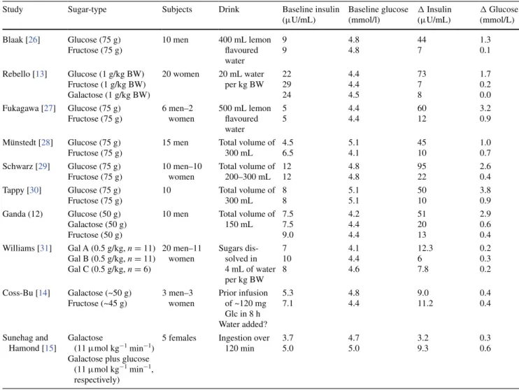
Our study has a number of limitations. First, we did not take venous blood samples in order to assess insulin and glucose values because the cannulation process could influence the subtle haemodynamic changes that are being monitored [25]. However, as summarized in Table 2, sev-eral past studies comparing postprandial glucose and
Fig. 2 Time course for changes of heart rate (HR) (A), stroke
vol-ume (SV) (B), cardiac output (CO) (C) and total peripheral resist-ance (TPR) (D), before and after ingestion of glucose (open circle), galactose (closed square) and fructose (closed triangle). Repeated-measures ANOVA assessed statistical differences as follows: effect of time (symbol dagger), effect of type (symbol @) and the sugar-type × time interaction (symbol double dagger); one, two and three symbols denoting p < 0.05, p < 0.01 and p < 0.001, respectively. For data on post-drink changes averaged over 60 and 120 min (shown as bars), post hoc pair-wise comparison for statistical differences across sugar-types is assessed by Dunnett’s multiple comparison tests and indicated as follows: *p < 0.05, **p < 0.01 and ***p < 0.001. Sig-nificant difference between post-drink and baseline values are indi-cated as follows: NS non-significant; °p < 0.05; °°p < 0.01; and °°°p < 0.001)
Table 2 Summary of studies reporting plasma insulin and glucose before and after ingestion of a sugary drink
Glc glucose. Values for plasma insulin and glucose levels were either obtained from tables or estimated from figures. A conversion factor of
0.555 was used to convert mg/dL to mmol/L in the study by Williams et al. [31]. Data from the study by Ganda et al. [12] have been reported for not more than 30 min post-drink
Study Sugar-type Subjects Drink Baseline insulin (μU/mL) Baseline glucose (mmol/l) Δ Insulin (μU/mL) Δ Glucose (mmol/L) Blaak [26] Glucose (75 g) Fructose (75 g) 10 men 400 mL lemon flavoured water 9 9 4.8 4.8 44 7 1.3 0.1 Rebello [13] Glucose (1 g/kg BW) Fructose (1 g/kg BW) Galactose (1 g/kg BW) 20 women 20 mL water per kg BW 22 29 24 4.4 4.4 4.5 73 7 8 1.7 0.2 0.0 Fukagawa [27] Glucose (75 g) Fructose (75 g) 6 men–2 women 500 mL lemon flavoured water 5 5 4.4 4.4 60 12 3.2 0.9 Münstedt [28] Glucose (75 g) Fructose (75 g)
15 men Total volume of 300 mL 4.5 6.5 5.1 4.1 45 10 1.0 0.7 Schwarz [29] Glucose (75 g) Fructose (75 g) 10 men–10 women Total volume of 200–300 mL 12 12 4.8 4.8 95 22 2.6 0.4 Tappy [30] Glucose (75 g) Fructose (75 g) 10 Total volume of 300 mL 8 8 5.1 5.1 50 10 3.8 0.9 Ganda (12) Glucose (50 g) Galactose (50 g) Fructose (50 g)
10 men Total volume of 150 mL 7.5 7.5 9.0 4.2 4.4 4.4 51 20 13 2.9 0.6 0.4 Williams [31] Gal A (0.5 g/kg, n = 11) Gal B (0.5 g/kg, n = 11) Gal C (0.5 g/kg, n = 6) 20 men–11 women Sugars dis-solved in 4 mL of water per kg BW 7 10 8 4.1 4.4 4.6 12.3 6 7.8 0.2 0.3 0.2 Coss-Bu [14] Galactose (~50 g) Fructose (~45 g) 3 men–3 women Prior infusion of ~120 mg Glc in 8 h Water added? 5.3 7.1 4.8 4.4 9.0 11.2 0.4 0.4 Sunehag and Hamond [15] Galactose (11 μmol kg−1 min−1)
Galactose plus glucose (11 μmol kg−1 min−1, respectively)
5 females Ingestion over 120 min 3.7 5.0 4.7 5.0 3.2 9.3 0.3 0.6 ◂
http://doc.rero.ch
insulin responses to these monosaccharides (in amounts similar to those used in our study here) have consistently shown markedly lower rise in circulating glucose and insu-lin following fructose and galactose than after glucose [12–15, 26–31]. Second, our study was conducted only in men. In this first “proof-of-concept” study about the cardi-ovascular impact of galactose, we focused on men because including women would have increased substantially the number of subjects and testing days (cross-over design) in order to observe a potential gender effect with sufficient statistical power. Third, based on our selective study pop-ulation, which included only young and healthy men of normal body weight, our findings cannot be extrapolated to the general population. Further studies are warranted to explore the proof-of-concept presented here of a favour-able haemodynamic profile of galactose (besides its low-glycemic index) in patients with cardiac insufficiency and impaired glucose metabolism (i.e. impaired glucose toler-ant patients and/or type 2 diabetic patients).
In conclusion, this study demonstrates that galactose resembles glucose in its marginal impact on BP, which contrasts with the significant BP-elevating effect of fruc-tose ingestion. Furthermore, it has a much less pronounced effect on cardiac workload than glucose. Galactose thus presents the interesting characteristics of a low-glycemic sugar with mild cardiovascular effects. Further studies are warranted to confirm the clinical relevance of the milder cardiovascular effects of galactose compared to other sug-ars for insulin resistant obese and/or diabetic patients with cardiac insufficiency and also to investigate whether this beneficial cardiac effect of galactose would persist when consumed together with other monosaccharides (i.e. glu-cose, fructose or sucrose), as well as when integrated in meals in more chronic studies.
Acknowledgments Research related to this paper was funded in part
by the Swiss National Science Foundation and in part by intramural funding.
Compliance with ethical standards
Conflict of interest The authors declare that they have no conflict of
interest.
References
1. Bray GA, Nielsen SJ, Popkin BM (2004) Consumption of high-fructose corn syrup in beverages may play a role in the epidemic of obesity. Am J Clin Nutr 79(4):537–543
2. Tappy L, Le KA, Tran C et al (2010) Fructose and meta-bolic diseases: new findings, new questions. Nutrition 26(11–12):1044–1049
3. Adam TC, Westerterp-Plantenga MS (2005) Nutrient-stimulated GLP-1 release in normal-weight men and women. Horm Metab Res 37(2):111–117
4. Adam TC, Westerterp-Plantenga MS (2005) Glucagon-like pep-tide-1 release and satiety after a nutrient challenge in normal-weight and obese subjects. Br J Nutr 93(6):845–851
5. Burelle Y, Lamoureux MC, Peronnet F et al (2006) Comparison of exogenous glucose, fructose and galactose oxidation during exercise using 13C-labelling. Br J Nutr 96(1):56–61
6. Mohammad MA, Sunehag AL, Rodriguez LA et al (2011) Galactose promotes fat mobilization in obese lactating and non-lactating women. Am J Clin Nutr 93(2):374–381
7. Sclafani A, Ackroff K (2012) Flavor preferences conditioned by intragastric glucose but not fructose or galactose in C57BL/6J mice. Physiol Behav 106(4):457–461
8. O’Hara JP, Carroll S, Cooke CB et al (2012) Preexercise galac-tose and glucose ingestion on fuel use during exercise. Med Sci Sports Exerc 44(10):1958–1967
9. Duckworth LC, Backhouse SH, Stevenson EJ (2013) The effect of galactose ingestion on affect and perceived exertion in recrea-tionally active females. Appetite 71:252–258
10. Duckworth LC, Backhouse SH, O’Hara JP et al (2016) Effect of galactose ingestion before and during exercise on substrate oxidation, postexercise satiety, and subsequent energy intake in females. J Am Coll Nutr 35(1):1–12
11. Mayes PA (1993) Intermediary metabolism of fructose. Am J Clin Nutr 58:754S–765S
12. Ganda OP, Soeldner JS, Gleason RE et al (1979) Metabolic effects of glucose, mannose, galactose, and fructose in man. J Clin Endocrinol Metab 49(4):616–622
13. Rebello T, Hodges RE, Smith JL (1983) Short-term effects of various sugars on antinatriuresis and blood pressure changes in normotensive young men. Am J Clin Nutr 38(1):84–94
14. Coss-Bu JA, Sunehag AL, Haymond MW (2009) Contribution of galactose and fructose to glucose homeostasis. Metabolism 58(8):1050–1058
15. Sunehag AL, Haymond MW (2002) Splanchnic galactose extrac-tion is regulated by coningesextrac-tion of glucose in humans. Metabo-lism 51:827–832
16. Brown CM, Dulloo AG, Yepuri G et al (2008) Fructose ingestion acutely elevates blood pressure in healthy young humans. Am J Physiol Regul Integr Comp Physiol 294(3):R730–R737
17. Grasser EK, Dulloo A, Montani JP (2014) Cardiovascular responses to the ingestion of sugary drinks using a randomised cross-over study design: does glucose attenuate the blood pres-sure-elevating effect of fructose? Br J Nutr 112(2):183–192 18. Visvanathan R, Chen R, Garcia M et al (2005) The effects of
drinks made from simple sugars on blood pressure in healthy older people. Br J Nutr 93(5):575–579
19. Landsberg L, Young JB (1985) Insulin-mediated glucose metab-olism in the relationship between dietary intake and sympathetic nervous system activity. Int J Obes 9:63–68
20. de Jongh RT, Clark AD, IJzerman RG et al (2004) Physiological hyperinsulinaemia increases intramuscular microvascular reac-tive hyperaemia and vasomotion in healthy volunteers. Diabeto-logia 47(6):978–986
21. Girona M, Grasser EK, Dulloo AG et al (2014) Cardiovas-cular and metabolic responses to tap water ingestion in young humans: does the water temperature matter? Acta Physiol (Oxf) 211(2):358–370
22. Grasser EK, Goswami N, Hinghofer-Szalkay H (2009) Presyn-copal cardiac contractility and autonomic activity in young healthy males. Physiol Res 58(6):817–826
23. Kubicek WG, Patterson RP, Witsoe DA (1970) Impedance car-diography as a non-invasive method of monitoring cardiac func-tion and other parameters of the cardiovascular system. Ann NY Acad Sci 170:724–732
24. Fortin J, Habenbacher W, Heller A et al (2006) Non-invasive beat-to-beat cardiac output monitoring by an improved method
of transthoracic bioimpedance measurement. Comput Biol Med 36(11):1185–1203
25. Langham BT, Harrison DA (1993) The pressor response to venous cannulation: attenuation by prior infiltration with local anaesthetic. Br J Anaesth 70(5):519–521
26. Blaak EE, Saris WH (1996) Postprandial thermogenesis and substrate utilization after ingestion of different dietary carbohy-drates. Metabolism 10:1235–1242
27. Fukagawa NK, Veirs H, Langeloh G (1995) Acute effects of fructose and glucose ingestion with and without caffeine in young and old humans. Metabolism 44:630–638
28. Münstedt K, Böhme M, Hauenschild A et al (2011) Consump-tion of rapeseed honey leads to higher serum fructose levels
compared with analogue glucose/fructose solutions. Eur J Clin Nutr 65:77–80
29. Schwarz JM, Schutz Y, Froidevaux F et al (1989) Thermogenesis in men and women induced by fructose vs glucose added to a meal. Am J Clin Nutr 49:667–674
30. Tappy L, Randin JP, Felber JP et al (1986) Comparison of ther-mogenic effect of fructose and glucose in normal humans. Am J Physiol Endocrinol Metab 13:718–724
31. Williams CA, Phillips T, Macdonald I (1983) The influence of glucose on serum galactose levels in man. Metabolism 32(3):250–256